Home Blog Page 4

SEATTLE II trial shows Ekos system beneficial in treatment of acute pulmonary embolism

0
SEATTLE II trial shows Ekos system beneficial in treatment of acute pulmonary embolism

The trial’s results were published in JACC: Cardiovascular Interventions and show no intracranial haemorrhage and no death among massive pulmonary embolism patients.

EKOS Corporation, a BTG International group company, announced the publication of results of the SEATTLE II trial in the JACC: Cardiovascular Interventions. The study concluded that treatment with ultrasound-facilitated catheter-directed low-dose thrombolysis for acute pulmonary embolism improves right heart function, reduces blood clot size, and decreases pulmonary hypertension, in patients with intermediate to high risk pulmonary embolism. Importantly, no patient experienced intracranial haemorrhage, a serious adverse experience related to full-dose thrombolysis treatment.

The SEATTLE II study was a prospective, single-arm, multicentre trial designed to evaluate the safety and effectiveness of ultrasound-facilitated catheter-directed low-dose thrombolysis, using the Ekos EkoSonic Endovascular System. One hundred and fifty patients diagnosed as acute massive (n=31) or submassive (n=119) pulmonary embolism were enrolled. Patients received low dose (24mg) of tPA (thrombolytic) for 24 hours with a unilateral catheter or for 12 hours with bilateral catheters. The size of the right heart measured as RV/LV ratio significantly decreased from 1.55 to 1.13 (p<0.0001) by 48 hours after start of treatment.

For patients with severe pulmonary embolism, the mortality rate is nearly one third. In the SEATTLE II study, 31 patients presented to the emergency room with massive pulmonary embolism, syncope and hypotension. All 31 survived the 30-day follow up period. Of 150 patients in the study, one death was directly attributed to pulmonary embolism.

There were no intracranial haemorrhages and no fatal bleeding events. Major bleeds occurred in 15 patients and were comprised of one severe bleed and 16 moderate bleeds. Six of the major bleeds occurred in patients with co-morbidities known to be associated with an increased risk of bleeding during thrombolytic therapy.

“The SEATTLE II findings establish a new rationale for considering ultrasound-facilitated catheter-directed low-dose thrombolysis in both massive and submassive PE,” said Gregory Piazza, assistant professor of Medicine, Harvard Medical School, staff physician, Cardiovascular Division, Brigham and Woman’s Hospital, and principal investigator for SEATTLE II. “Without any intracranial haemorrhage and using a much reduced lytic dose, a substantial and clinically meaningful reduction of the RV/LV ratio was achieved.”

“The SEATTLE II and ULTIMA trials add to the body of evidence showing treatment with the EkoSonic Endovascular System improves the standard of care for patients with acute pulmonary embolism,” said Matt Stupfel, general manager of EKOS Corporation. “The only endovascular device cleared by the FDA for the treatment of pulmonary embolism, EkoSonic represents a potential game-changer in the treatment of high-risk pulmonary embolism patients. EKOS ultrasonic waves greatly accelerate lytic dispersion by driving the drug deeper into the clot and unwinding fibrin to expose plasminogen receptor sites.”

The ULTIMA trial, published January 2014 in Circulation, demonstrated that EKOS treatment was clinically superior to anticoagulation with heparin alone in reversing right ventricular enlargement at 24 hours, without an increase in bleeding complications. EKOS is launching the next study, OPTALYSE PE, to better understand the optimal dose of thrombolytic and duration of its treatment.

SIR calls for abstracts to be submitted for 2016 annual meeting

0
SIR calls for abstracts to be submitted for 2016 annual meeting

SIR_Logo_TAG_main_Main

The Society of Interventional Radiology (SIR) is accepting scientific abstracts covering all areas of vascular and nonvascular interventional radiology for its 41st Annual Scientific Meeting, which will be held 2–7 April, 2016, in Vancouver, British Columbia, Canada. Online scientific abstract submission is open until 6 October.

Practicing experts, early career professionals, research scientists, clinical associates, trainees and medical students are invited to submit scientific abstracts for oral or poster presentation by 6 October. Researchers will have the opportunity to advance the field of image-guided interventions by sharing their basic science results, device and procedure innovations and clinical outcomes research at SIR 2016.

All selected scientific abstracts will be published in SIR’s flagship publication, the Journal of Vascular and Interventional Radiology, and on its website. Selected abstracts may be designated as “distinguished”, “featured” or “abstract of the year”.


Research awards to support travel are available through SIR Foundation for medical students, residents and fellows whose abstracts are accepted for oral presentation at SIR 2016. SIR’s International Scholarship program enables physicians (who are within 10 years of completion of training and who are practicing outside North America) an opportunity to train and learn at the meeting. SIR also offers two SIR 2016 scholarships for interventional radiologists-in-training: the SIR Medical Student Scholarship and SIR Resident-in-Training Scholarship.

For more information, please visit www.SIRmeeting.org.

Patient selection is key for SIRT with first-line chemotherapy

0
Patient selection is key for SIRT with first-line chemotherapy
Ricky Sharma
Ricky Sharma
Ricky Sharma

Speaking to Interventional News on the SIRFLOX trial results at the Global Embolization Symposium and Technologies (GEST EU, 24–27 June, Seville, Spain) meeting, Ricky Sharma, professor and honorary consultant of Clinical Oncology at the University of Oxford and Oxford University Hospitals Trust said: “We need to know more about patient selection for radioembolization.

Speaking to Interventional News on the SIRFLOX trial results at the Global Embolization Symposium and Technologies (GEST EU, 24–27 June, Seville, Spain) meeting, Ricky Sharma, professor and honorary consultant of Clinical Oncology at the University of Oxford and Oxford University Hospitals Trust said: “We need to know more about patient selection for radioembolization. Treatment with radioembolization using SIR-spheres (Sirtex) plus chemotherapy in the first-line setting is particularly suited to patients with liver-limited disease. However, at the moment we treat patients with extra-hepatic disease and we are getting more of an opinion about how little extra hepatic disease patients can have in order to derive optimal benefit from the treatment, which we did not know before.”

Commenting on the importance of the SIRFLOX trial’s key findings, Sharma noted, “The SIRFLOX trial has shown a significant improvement in liver control in the first-line setting for metastatic colorectal cancer. The improvement, compared to chemotherapy alone, was 7.9 months, median statistic.The study was really designed to test radioembolization with SIR-spheres along with radiosensitising chemotherapy. At the time the trial began, oxaliplatin-based chemotherapy was the most common first-line chemotherapy and both oxaliplatin and 5-fluorouracil are known radiosensitisers. The significant finding from SIRFLOX is confirmation that selective internal radiotherapy (SIRT) can really improve local control in the liver. We knew that from largely retrospective data, but this is the first randomised, prospective trial that shows, in 530 selected patients, you can improve liver control by combining a liver-directed therapy with chemotherapy.”

Sharma also emphasised the importance of liver control. “Patients with metastatic colorectal cancer, more often than not, die from progression of their liver metastases. Once we have run out of chemotherapeutic options and systemic biological options, then unfortunately such patients almost invariably die of progression of liver metastases. So improving local control in the liver is a very important goal. There are several ways this can be achieved and this trial importantly shows that SIRT can be integrated into the standard regimen either in the first-line setting or, indeed in any setting in which FOLFOX is being used as the chemotherapy backbone.”

Sharma clarifies that in England, Wales and Scotland, there is a commissioning programme to treat patients with SIRT in the third and subsequent lines of treatment for metastatic colorectal cancer, mainly to look at overall survival and quality of life data.

“Increasingly, I think that, as a result of SIRFLOX, we will see patients asking for radioembolization in earlier lines of therapy, although it is not commissioned in that setting. So if the Commissioning through Evaluation programme goes well, I think in the future, we will see the procedure moving forward from third and subsequent lines of treatment, although we might need better patient selection for first-line treatment.”

Sharma also adds that “so far, there is not much published evidence on quality of life data, but there will be from 2017 onwards. Other studies that are being performed, such as FOXFIRE, have rigorous quality of life data collection built into them, and we will get those results in 2017. However, from our experience, we have observed that we can improve chemotherapy-free intervals by using SIRT, and that patients, after the initial phase of fatigue (that can last from two to four weeks), in general have an excellent quality of life.”

Pointers on consenting patients

“We now have very reliable data on the safety and toxicity profile of SIRT plus chemotherapy in the setting of metastatic colorectal cancer, so it is really important that when patients agree to this therapeutic combination that the slight increase in the incidence of blood count complications, such as neutropenia, is noted; the likelihood of common side-effects like fatigue is noted; and the rare increase in more serious complications (less than five per cent) is noted,” Sharma said.

“The other thing to consent patients for is the risk that they might not proceed to receive SIRT as planned. In the SIRFLOX trial, 7% of patients who were randomised to the SIRT arm, did not go on to receive the therapy. This was due to technical reasons that were related to their vasculature, antiangiogenic drugs they might have taken prior to the procedure, dissections that occurred during work-up or any factors that made the risk with SIR-spheres too great to be able to treat the patient safely.”


First low dose CT lung cancer screening gets US FDA clearance

0
First low dose CT lung cancer screening gets US FDA clearance

GE Healthcare’s low dose computed tomography lung cancer screening option is the first to be cleared by the US FDA. Early detection with low dose CT can improve prognosis and treatment and reduce mortality rates for patients with a high risk of lung cancer.

Both the USA Preventative Services Task Force (USPSTF) and the Centers for Medicare and Medicaid Services (CMS) recommend the use of low dose CT lung cancer screening for high-risk individuals. Additionally, Medicare has approved insurance reimbursement for its beneficiaries who are eligible for the use of low dose CT lung cancer screening in high-risk patients.Early detection from an annual lung cancer screening with low dose CT in high-risk persons can prevent a substantial number of lung cancer related deaths, a press release from the company states.

“Bringing low dose CT screening to patients at a high risk for lung cancer will reduce death from the most deadly cancer worldwide,” said Ella Kazerooni, chair of committee on lung cancer screening, American College of Radiology. “Performing low dose CT with attention to high image quality at the lowest radiation exposure to detect early cancer is at the core of a successful screening programme, and requires collaboration with imaging partners to bring technology to bear for this purpose. The FDA’s clearance of these CT scanners for this purpose illustrates that commitment.”

Physicians using low dose CT for lung cancer screening will now have access to the benefits from a screening indication, including GE Healthcare’s new low-dose screening reference protocols. These new protocols are tailored to the CT system, patient size, and the most current recommendations from a wide range of professional medical and governmental organisations. Now, qualified GE Healthcare CT scanners can be confidently used by physicians within their FDA cleared indications for use, delivering low dose, short scan times, and clear and sharp images for the detection of small lung nodules. These nodules are critical in identifying lung cancer at its earliest stages when it is the most treatable and curable.


The landmark National Lung Screening Trial (NLST) was conducted at 33 medical institutions with over 53,000 older, otherwise healthy patients at high-risk due to smoking history. Half of the participants were screened using low dose CT, the other half were screened using chest X-ray. The trial demonstrated that the use of low dose CT screening decisively reduced the mortality rate from lung cancer by 20% compared to use of chest X-ray. The NLST also revealed a significant 6.7% reduction in the rate of death from any cause using low dose CT screening.

All new 64-slice and greater CT scanners, and virtually all of the 16-slice CT scanners that GE Healthcare sells are qualified systems and will include the screening option. The solution that GE Healthcare developed is also available to thousands of qualified GE scanners currently in use, increasing access to the quality scanners that satisfy both patient and physician needs. The new protocols are able to utilise GE Healthcare’s industry-leading technologies such as ASiRTM, ASiR-VTM and VeoTM that are designed to reduce image noise, which is undesirable for physicians looking for small nodules.

In the USA, lung cancer is the third most common cancer and the leading cause of cancer deaths; it kills almost twice as many women as breast cancer and three times as many men as prostate cancer. Survival rates from lung cancer are highly correlated to its initial stage at the time of first diagnosis. The overall five year survival rate for lung cancer is among the lowest (17%) of all types of cancer but is significantly higher when the cancer is diagnosed at an early stage (54%). However, currently only 155 of lung cancer cases are diagnosed at such an early stage. The use of low dose CT lung cancer screening increases this percentage of early stage detection, providing patients a much better prognosis.


Hansen announces growing series of Magellan robotic prostatic artery embolization procedures

0
Hansen announces growing series of Magellan robotic prostatic artery embolization procedures

The procedures have been performed by Sandeep Bagla, interventional radiologist, using the Magellan Robotic System. Bagla is one of the world leaders in the development of prostate artery embolization and is at the forefront of applying the Magellan Robotic System to this expanding procedure.

“We are excited about our clinical experiences with the Magellan Robotic System for prostatic artery embolization,” said Bagla, “Prostate artery embolization is a highly complex procedure that demands precision and control, key elements which are enhanced through robotics. We have completed 30 procedures thus far with the Magellan Robotic System and are delighted with the results we are seeing. We are highly confident in our ability to navigate through tortuous anatomy, our patients are very open to the approach and personally, I am drawn to the ability to reduce my procedural radiation exposure.”

Prostate artery embolization is a novel therapy in which the size of the prostate is reduced by a less invasive catheter-based approach that works to reduce blood flow to the main arteries that feed the prostate. Successfully performed prostatic artery embolization can reduce the prostate gland size and eliminate the symptoms associated with benign prostatic hyperplasia. Currently a patient must choose between long-term medical therapy or highly invasive surgical options that may often result in complications such as pain, bleeding, incontinence or impotence.

The Magellan Robotic System is designed to offer procedural predictability, control, and catheter stability to physicians as they remotely navigate the robotic catheter through the vasculature. Magellan’s remote workstation allows physicians to navigate through the vasculature while seated away from the radiation field, potentially reducing physicians’ radiation exposure and procedural fatigue.

Penumbra to distribute InspireMD’s CGuard embolic prevention system

0
Penumbra to distribute InspireMD’s CGuard embolic prevention system
CGuard EPS
CGuard EPS
CGuard EPS

InspireMD has announced that it has entered into a partnership with Penumbra to distribute its carotid CGuard EPS through their direct commercialisation team.

InspireMD has aggressively been pursuing a strategy in treating carotid artery disease and other neurovascular applications for its proprietary MicroNet technology.  Clinical trials of the CGuard system, including the recent CARENET and PARADIGM studies, have continued to support the benefits of the MicroNet enhanced CGuard system resulting in increased commercial adoption throughout the European market.

Penumbra has a direct and distributor sales organisation in all markets where the CGuard system is commercially available. Penumbra’s neurovascular product portfolio includes medical devices for ischemic stroke and brain aneurysms, as well as peripheral vascular products that include products for embolization and thrombectomy.  Penumbra has approximately 1,000 global employees with direct sales operations in North America, Europe, and Australia.  Penumbra sells through distributors in Asia and select other international markets.

CGuard EPS is CE mark approved. CGuard EPS, however, is not approved for sales in the US by the US FDA at this time.

Sorry, I don’t speak IR

0
Sorry, I don’t speak IR
Online patient material is not easy for patients to understand

Two recently published papers in the Journal of Vascular and Interventional Radiology (JVIR) reveal that patient materials in interventional radiology available online are way above the understanding of most patients. Both studies were carried out from the perspective of American patients and the investigators are urging hospitals and interventional radiologists to pay more attention to health literacy.

Referring to these studies, Ziv Haskal, editor-in-chief of JVIR, Charlottesville, USA, wrote in his editorial, “Consider this a call to arms as over 100 million users navigate the Web for healthcare information.”

One of the studies (Sadigh et al) set out to assess the readability of online education materials offered by hospitals describing commonly performed interventional radiology procedures. The investigators assessed online patient education materials from over 400 hospitals selected from the Medicare Hospital Compare database. They found that 76% of the hospitals presumed to offer interventional radiology services offered online patient education material for one or more of the eight service lines.

They assessed patient online education materials about uterine artery embolization for fibroid tumours, liver cancer embolization, varicose vein treatment, central venous access, inferior vena cava filter placement, nephrostomy tube insertion, gastrostomy tube placement, and vertebral augmentation using six validated readability scoring systems.

The authors reported that the average readability scores corresponding to grade varied between the ninth- and 12th-grade levels. All were higher than the recommended seventh-grade level (p<0.05) except for nephrostomy and gastrostomy tube placement. Average Flesch-Kincaid Reading Ease (that are readability tests designed to indicate how difficult a reading passage in English is to understand) scores corresponded with “fairly difficult” to “difficult” readability for all service lines except inferior vena cava filter and gastrostomy tube placement, which corresponded with standard readability.

A majority of hospitals offering interventional radiology services provide at least some online patient education material. Most, however, are written significantly above the reading comprehension level of most Americans, the authors conclude.

Another study (McEnteggart et al) set out to assess the readability of online patient education materials related to common diseases treated by and procedures performed by interventional radiology.

The investigators selected online patient education materials from the following websites due to their average Google search return: Society of Interventional Radiology (SIR), Cardiovascular and Interventional Radiological Society of Europe (CIRSE), National Library of Medicine, RadiologyInfo, Mayo Clinic, WebMD, and Wikipedia.

The interventional radiology content was assessed for the following procedures: peripheral arterial disease, central venous catheter, varicocoele, uterine artery embolization, vertebroplasty, transjugular intrahepatic portosystemic shunt, and deep vein thrombosis. The investigators analysed data using general mixed modelling and used a variety of common algorithms to estimate and compare readability levels.

They found that on average, online sources that required beyond high school grade-level readability were Wikipedia, SIR and RadiologyInfo; sources that required high school grade-level readability were CIRSE, Mayo Clinic, WebMD, and National Library of Medicine.

“On average, online patient education materials pertaining to uterine artery embolization, vertebroplasty, varicocele, and peripheral arterial disease required the highest level of readability,” the authors reported in JVIR.

The authors concluded that the materials assessed were “written above the recommended sixth-grade reading level and the health literacy level of the average American adult” and that “many patients may not have the ability to read and understand health information related to interventional radiology procedures.”


New CPT code established for Gynesonics‰Ûª Sonata system

0
New CPT code established for Gynesonics‰Ûª Sonata system

The American Medical Association (AMA) Current Procedure Terminology (CPT) Editorial Panel has established a new AMA CPT code specifically for transcervical uterine fibroid ablation with ultrasound guidance. Gynesonics applied for the new code, which was presented to the Editorial Panel in February.

CPT codes are used by medical practitioners including physicians, hospitals, and other healthcare providers, to report healthcare services to insurers for the purpose of reimbursement. This standardised nationwide system of identification provides a uniform language for reporting medical services.

The code has the following descriptor: Transcervical uterine fibroid(s) ablation with ultrasound guidance, radiofrequency. The announcement was posted and made available on the AMA website. The final code number will be released at a future date and will become effective on 1 January 2016.

The Sonata system from Gynesonics provides a uterus preserving, incision-free treatment option to women suffering from symptomatic uterine fibroids, including those associated with heavy menstrual bleeding. Sonata combines and integrates a high resolution, compact ultrasound probe at the tip of a radio frequency ablation device to provide a single handheld transcervical treatment delivery system under integrated sonographic guidance.

Gynesonics president and chief executive officer Christopher M Owens noted that the company pursued the Category III designation in conjunction with beginning its pivotal investigational device exemption (IDE) Sonata trial.

“This initiative provides us the opportunity to establish a dialogue with the AMA CPT Editorial Panel, the gynecology societies and their advisors to ensure proper procedure coding, while completing our Food and Drug Administration (FDA) Sonata IDE trial,” Owens, said. “Widespread reimbursement coverage for the Sonata technology is essential to making it available to women with fibroids who can benefit from its application. In addition to the FDA IDE Sonata trial, the company will continue to move forward with additional clinical and health economics and outcomes research trials. Our intention is to pursue reimbursement coverage and a migration to a CPT Category I code for the Sonata system upon FDA clearance.”


Merit’s QuadraSpheres get new indication of Hepatoma in USA

0
Merit’s QuadraSpheres get new indication of Hepatoma in USA

Merit Medical has announced that it has received 510(k) clearance from the US FDA expanding indication for QuadraSphere microspheres to include the embolization of hepatoma.

Malignant hepatoma, also known as hepatocellular carcinoma (HCC), is the sixth most common cancer and the third leading cause of cancer deaths worldwide. QuadraSphere Microspheres are precisely calibrated and designed to offer controlled, targeted embolization, treating HCC by stopping the blood flow to the tumours.

“The FDA’s clearance of QuadraSphere for the embolization of hepatoma provides another treatment option for patients and physicians in their battle against primary liver cancer,” said Fred Lampropoulos, Merit’s chairman and CEO. “This clearance also represents a milestone in our continual efforts to improve patients’ lives through the development of innovative technologies that produce better patient outcomes.”


Health Canada approves Varithena for patients with varicose veins

0
Health Canada approves Varithena for patients with varicose veins

BTG_Varithena_main_Main

BTG has announced that Health Canada has issued a notice of compliance approving Varithena (polidocanol injectable foam) for the treatment of incompetent great saphenous veins, accessory saphenous veins, and visible varicosities of the great saphenous vein system, above and below the knee.

An estimated 3.7 million Canadians have varicose veins, with women twice as likely as men to develop varicosities.

Louise Makin, CEO of BTG plc, said: “We are pleased to receive approval in Canada for Varithena and look forward to the commercial launch during the first half of 2016. The controlled launch continues in the US reimbursed sector, where we consistently receive excellent feedback from physicians in relation to clinical performance and patient acceptability. We are also progressing development of related products for other venous disorders.”

Varithena is a uniform, low-nitrogen, polidocanol microfoam, dispensed from a proprietary canister device. Treatment is minimally invasive, non-surgical and requires neither tumescent anaesthesia nor sedation. It is intended for use in adults with clinically significant venous reflux as diagnosed by duplex ultrasound and is indicated for the treatment of incompetent great saphenous veins, accessory saphenous veins, and visible varicosities of the GSV system, above and below the knee.

Varithena is intended for intravenous injection using ultrasound guidance, administered via a single cannula into the lumen of the target incompetent trunk veins or by direct injection into varicosities. The use of Varithena is contraindicated in patients with known allergy to polidocanol and those with acute thromboembolic disease.


Varithena received US FDA approval in 2013. It is not approved for use in Europe.


Cook Medical issues global recall of Beacon Tip Angiographic Catheter products

0
Cook Medical issues global recall of Beacon Tip Angiographic Catheter products

Cook Medical has initiated a lot-specific voluntary recall of 2,239 lots of Beacon Tip Angiographic Catheters. Globally, 95,167 devices are subject to this recall. The products include specific versions and lot numbers of the Torcon NB Advantage Beacon Tip Catheters, Royal Flush Plus Beacon Tip High-Flow Catheters, and Slip-Cath Beacon Tip Catheters.

The Beacon Tip Angiographic Catheters have been found to exhibit tip splitting or separation, which has resulted in adverse events. Cook has received 26 complaints of catheter-tip splitting and/or separation. There have been 14 Medical Device Reports to date in which a tip split or separation occurred.

Tip splitting has the potential to lead to loss of device function. Tip separation may require medical intervention to retrieve a separated segment or may occlude blood flow to end organs.

The Beacon Tip Angiographic Catheters in this recall were distributed between June 2013 and June 2015. Product can be identified by the part number and lot number provided on the outer package product label. The lot numbers for products that are subject to this recall can be found on the following pages.

Cook Medical has notified its customers and distributors by recall notification letters. The letters requested that all customers and distributors quarantine and discontinue use of all potentially affected units and return the affected product to the company as soon as possible for credit.

Latest update to TASC II published

0
Latest update to TASC II published

The Journal of Endovascular Therapy (JEVT) has announced the publication of the latest update of the Inter-Society Consensus for the Management of Peripheral Arterial Disease (TASC II), an internationally recognised set of guidelines for the management of patients with peripheral arterial disease.

Originally published in 2000, the TASC document represents the collaboration of international vascular specialties involved in the treatment of peripheral arterial disease patients. One highly utilised aspect of the TASC guidelines is the TASC anatomical artery lesion classification (A-D), which characterises the various patterns of disease and provides guidance on treatment decisions regarding the optimal revascularisation strategy (endovascular vs. surgical). The TASC II guidelines published in 2007 included a revision of the original TASC classification for peripheral arterial disease patients, with a focus on the aortoiliac and femoropopliteal territories.


“In this update, the classification of disease is extended to involve the below the knee arteries as well. This [Not addressing this anatomical region] was a shortfall of TASC-I and II. In future updates, the hope is to further refine the classification and add recommendations as more data becomes available,” Mahmood K Razavi, an interventional radiologist from Orange County, USA, and one of the authors on the paper in JEVT told Interventional News.

Since the publication of the TASC II document, a number of scientific publications and observational reports have documented the rapid adoption of endovascular therapy as a primary strategy for the treatment of symptomatic peripheral arterial disease patients. The overall result is that there has been an increase in the adoption of the endovascular-first strategy for even the most complex anatomies. This shift was not clearly reflected in TASC II. In addition, the TASC II lesion classification did not include the infrapopliteal arteries, which was an important omission given today’s expanding technologies and techniques for catheter-based tibial and distal interventions for patients with critical limb ischemia. Thus, the focus of the TASC II supplement published today in the JEVT is to update the endovascular and surgical revascularisation strategies and techniques for PAD and to expand the TASC lesion classification to include below-the-knee arteries. The document has been prepared in relationship with a number of vascular societies worldwide and has been endorsed by the ISES.

According to George A Antoniou of the Royal Liverpool University Hospital, whose commentary accompanies the TASC II supplement, “The treatment of peripheral arterial disease patients has seen dramatic evolutionary changes over the past decade. Updates of literature, techniques, and practices by expert committees are an essential tool to facilitate and guide clinical practice.” Given the worldwide importance of this TASC II supplement, the article is being co-published with permission of the authors and SAGE in the following journals: Vascular Medicine, Catheterization and Cardiovascular Interventions, Annals of Vascular Diseases, Journal of the Japanese College of Angiology (Japanese translation), and Técnicas Endovasculares (Spanish translation).


3D printing helps doctors rehearse complex paediatric brain interventions

0
3D printing helps doctors rehearse complex paediatric brain interventions

Boston Children’s Hospital physicians report the first cases of children benefitting from 3D printing of their anatomy before undergoing high-risk brain procedures. The four children had life-threatening cerebrovascular malformations that posed special treatment challenges.

Reporting online in late July in the Journal of Neurosurgery: Pediatrics, the physicians describe the use of 3D printing and synthetic resins to create custom, high-fidelity models of the children’s vessel malformations along with nearby normal blood vessels. In some cases, the surrounding brain anatomy was also printed.

“These children had unique anatomy with deep vessels that were very tricky to operate on,” says Boston Children’s neurosurgeon Edward Smith, senior author of the paper and co-director of the hospital’s Cerebrovascular Surgery and Interventions Center. “The 3D-printed models allowed us to rehearse the cases beforehand and reduce operative risk as much as we could.”

The children ranged in age from two months to 16 years old.

The two-month-old infant had a rare vein of Galen malformation in which arteries connect directly with veins—bypassing the capillaries—and was treated with an interventional radiology technique to seal off the malformed blood vessels from the inside.

“Even for a radiologist who is comfortable working with and extrapolating from images on the computer to the patient, turning over a 3D model in your hand is transformative,” says Darren Orbach, chief of Interventional and Neurointerventional Radiology at Boston Children’s and co-director of the Cerebrovascular Surgery and Interventions Center. “Our brains work in three dimensions, and treatment planning with a printed model takes on an intuitive feel that it cannot otherwise have.”

Three of the four children had arteriovenous malformations and were treated surgically.

“Arteriovenous malformations are high-risk cases and it is helpful to know the anatomy so we can cut the vessels in the right sequence, as quickly and efficiently as possible,” says Smith. “You can physically hold the 3D models, view them from different angles, practice the operation with real instruments and get tactile feedback.”

All four children’s malformations were successfully removed or eliminated with no complications. When two of the arteriovenous malformations patients were compared with controls who did not have 3D-printed models—matched for age, size and type of AVM, surgeon and operating room—those with 3D models had their surgical time reduced by 12% (30 minutes). (Actual surgical time was 254 and 257 minutes for the cases with 3D models and 285 and 288 minutes for the controls.) Even a 30-minute reduction is significant for children who are especially sensitive to anaesthesia.

Smith and Orbach are continuing to use 3D models for their trickier cases. “3D printing has become a regular part of our process,” says Smith. “It’s also a tool that allows us to educate our junior colleagues and trainees in a way that’s safe, without putting a child at risk.”

The life-sized and enlarged 3D models were created in collaboration with the Boston Children’s Hospital Simulator Program (SIMPeds) using brain magnetic resonance (MR) and MR arteriography data from each child. Measurements of the models showed 98% agreement with the children’s actual anatomy. 


Hourglass over-the-wire peripheral embolization plug obtains CE mark

0
Hourglass over-the-wire peripheral embolization plug obtains CE mark

Emba Medical has announced receiving the European CE mark for the Hourglass peripheral embolization plug making the device the first over-the-wire embolic device cleared for EU commercial release.

Hourglass peripheral embolization plug was awarded a CE certificate on July 10, 2015, enabling the company to pursue commercial activities in EU nations. The Hourglass device represents a breakthrough in peripheral embolization devices in that it is designed to provide precise, secure, over-the-wire delivery and immediate occlusion with a single integrated device, a press release from the company states.

“This is the first integrated, over-the-wire device designed for peripheral embolization procedures,” said George Wallace, CEO of Emba Medical. “The goal with over-the-wire design is to provide physicians with accurate, stent-like delivery of the device in the vessel. We believe that the Hourglass implant will provide a level of confidence, precision, and control for peripheral embolization procedures that physicians have come to enjoy over the years while performing other types of over-the-wire endovascular procedures.”

Andrew Cragg (Minneapolis, USA) interventional radiologist and Hourglass co-developer, noted: “The Hourglass device is designed to provide immediate occlusion in a wide range of vessel sizes with a single device. The device’s unique design was created to take advantage of natural haemodynamic forces to provide immediate, focal, stable occlusions.”

The device is not approved for sale in the United States.

Gastric balloon for obesity treatment gains FDA nod

0
Gastric balloon for obesity treatment gains FDA nod

The US FDA has approved the Reshape Integrated Dual Balloon System (ReShape). The system is a temporary weight-loss system of gastric balloons that are placed in a minimally invasive manner to occupy space in the stomach. They should be removed after six months.

The Reshape Dual Balloon system consists of two attached balloons that are filled and sealed separately. The balloons are placed into the stomach through the mouth using an endoscopic procedure while the patient is under mild sedation. Once in place, the balloons are filled with saline and methylene blue. When it is time to remove the balloons, they are first deflated then removed using another endoscopic procedure.


The device is for use in adult obese patients who have a body mass index (BMI) of 30-40 kg/m2 who have been unable to lose weight through diet and exercise. Patients must also have one or more obesity-related conditions such as diabetes, high blood pressure, or high cholesterol. Reshape Dual Balloon is intended to be used while a patient participates in a diet and exercise plan supervised by a healthcare provider.


In the REDUCE pivotal clinical study, a prospective, randomised multicentre study to evaluate the safety and efficacy of the intragastric balloon system in obese patients as an adjunct to diet and exercise in the treatment of obese patients with one or more obesity-related comorbid conditions, the group of people who used this device lost more weight than those who did not use it. The study included a total of 326 patients at eight investigational sites in the USA. Of the 326 patients, 187 received the device and 139 underwent the endoscopic procedure but did not receive the device. All study participants received diet and exercise counselling.


Patients with the device lost an average of 14.3 pounds (25.1% of their excess weight and 6.8% of their total body weight). The patients who did not receive the device lost an average of 7.2 pounds (11.3% of their excess weight and 3.3% of their total body weight).


The device should not be used in patients who have had previous gastrointestinal or bariatric surgery; have gastrointestinal inflammatory disease; have potential upper gastrointestinal bleeding conditions; have a gastric mass; have a large hiatal hernia; have structural abnormality in the oesophagus or pharynx; have serious oesophageal motility disorders; have severe coagulopathy; have hepatic insufficiency or cirrhosis; have serious alcoholism or drug addition; have serious or uncontrolled psychiatric illness or disorders; are pregnant or are breastfeeding or plan to become pregnant; have ever developed too much serotonin (serotonin syndrome) and are currently taking any drug known to affect serotonin levels; use aspirin, anti-inflammatory agents, anticoagulants, or other gastric irritants daily; are known or suspected to have an allergic reaction to materials in the device; are unwilling or unable to participate in a medically-supervised diet and behaviour modification programme; and take prescribed proton pump inhibitor medication for the duration of the device implant.


Guerbet bids to acquire Mallinckrodt‰Ûªs contrast media and delivery systems business

0
Guerbet bids to acquire Mallinckrodt‰Ûªs contrast media and delivery systems business

Guerbet has announced entering into a definitive agreement under which it will acquire Mallinckrodt’s contrast media and delivery systems business in its entirety for approximately US$270 million. With this acquisition, Guerbet aims to create a new global leader in medical imaging.

The key strengths of the contrast media and delivery systems business as identified by Guerbet are:

  • An efficient industrial and commercial platform with four production sites in Ireland and in North America and a global distribution network covering approximately 65 countries,
  • A comprehensive range of delivery systems and imaging solutions for X-ray scanners, X-ray cardiac and vascular imaging, urological imaging and magnetic resonance imaging,
  • A large portfolio of patents and other intellectual property rights.

For the first six months of fiscal 2015, Mallinckrodt’s CMDS business recorded sales of close to US$210 million.
Under the terms of the agreement, Guerbet is acquiring 100% of the contrast media and delivery systems business including the production sites, the intellectual property rights and the distribution subsidiaries in the various geographical areas, in a transaction valued approximately US$270M, entirely paid in cash and financed through term loan facilities syndicated by BNP Paribas.

The transaction is subject to customary closing conditions and is expected to close in the next few months. Guerbet’s works council has unanimously issued a favourable opinion in respect of the acquisition of the contrast media and delivery systems business.

Yves L’Epine, Guerbet’s CEO comments: “This acquisition transforms and accelerates Guerbet’s future: it doubles our turnover, enables us to extend our geographical footprint and creates complementarities in both contrast media and imaging solutions and services. We will be very pleased to welcome Mallinckrodt’s contrast media and delivery systems business colleagues to successfully and rapidly complete this integration together after the closing and create a new global leader in medical imaging”

Could combretastatin A4 be an alternative to sirolimus and paclitaxel in the inhibition of the smooth muscle cell cycle?

0
Could combretastatin A4 be an alternative to sirolimus and paclitaxel in the inhibition of the smooth muscle cell cycle?

Preclinical research published online ahead of print in the Journal of Vascular and Interventional Radiology (JVIR) in July shows that combretastatin A4 is a stronger inhibitor of the smooth muscle cell cycle than sirolimus or paclitaxel. Thus, it may represent an alternative coating for drug-eluting stents and balloons in atherosclerotic luminal stenosis, the investigators write.

Sirolimus and paclitaxel are drugs that are loaded onto a wide variety of drug-eluting or drug-coated device platforms for use in the coronary arteries and periphery. For example, Ultimaster, is an example of a sirolimus-eluting bioabsorbable coronary stent (Terumo) and Zilver PTX (Cook Medical), a peripheral paclitaxel-eluting stent system that has shown beneficial long-term effects in combating femoropoliteal disease.

Researchers Daniel Spira and colleagues set out to compare the effects of sirolimus, paclitaxel, and combretastatin A4 on the regulatory proteins of the cell cycle in proliferating smooth muscle cells.

Spira and colleagues treated human aortic smooth muscle cells with sirolimus, paclitaxel, and combretastatin A4 at 5×10⁻⁹ mol/L. After one day, half of the cells were harvested (D1 group). The treatment medium of the other half was replaced with culture medium on day four, and those cells were harvested on day five (D5 group). Cyclins D1, D2, E, and A and cyclin-dependent kinase (CDK) inhibitors p16, p21, and p27 were detected by Western blot technique. Quantification was performed by scanning densitometry of the specific bands.

As reported in JVIR, the researchers found that in the D1 group, treatment with sirolimus resulted in decreased intracellular levels of cyclins D2 and A (p0.05). In the D1 group, combretastatin A4 decreased intracellular levels of cyclins D2, E, and A (p<0.05). Despite recovery effects in the D5 group (increase of cyclins D1, D2, and A compared with D1 group; p<0.05), the upregulation of the CDK inhibitor p21, increased D cyclins, and decreased cyclins E and A (p <0.05) are compatible with a G1 arrest.

“The effect of combretastatin A4 on neointima formation should be evaluated further,” the authors recommend.


Factfile

  • Combretastatin belongs to a class of natural phenols (and despite its name, is unrelated to statins). Several natural combretastatin molecules are found in the bark of South African Bush willow trees.
  • Paclitaxel (also called Taxol) is found in the bark of yew trees.
  • Sirolimus is the generic name for the natural product rapamycin and is produced by a strain of Streptomyces hygroscopicus.

Choosing horses for courses: Using liquid agents to embolize endoleaks

0
Choosing horses for courses: Using liquid agents to embolize endoleaks

It is important to use the right embolic agent for the job – an embolic agent that is particularly suitable for one indication may not at all be appropriate for another. Robert Morgan, vascular interventional radiologist, St Georges Hospital, London, UK, tells Interventional News why he uses liquid embolic agents, such as ethylene vinyl oxide copolymer (EVOH-based) liquid agents, of which Onyx (Medtronic) is an example, for endoleak embolization.

Some potential advantages that liquid embolics offer in endoleak embolization are that they enable more rapid and complete filling of the endoleak cavity when compared with coils. They can also represent a less expensive option in situations where otherwise multiple coils would be needed.

Morgan makes the point that liquid embolic agents are particularly suitable for endoleak embolization because they can be used “to fill the cavity”. Other embolic agents such as coils have been used for endoleak embolization (although they are unsuitable for large cavities) and particles are completely unsuitable for endoleak embolization, he notes. He also clarifies that he would use embolization with particles mainly for small vessel embolization, such as uterine fibroid embolization, and embolization with coils mainly for the embolization of haemorrhage.

Commenting on some of the difficulties of using liquid embolic agents, Morgan makes the point that there are mainly two types of liquid embolic agents: “The cyanoacrylate adhesives (or glue-type embolics) and the EVOH-based agents (or Onyx-type embolics).”

While glue is relatively inexpensive in Europe, it polymerises in ionic fluids (such as blood) and is used with a non-ionic solvent such as dextrose to stop the glue polymerising in the catheter. “This is why there is a need to inject glue and withdraw the catheter, to prevent the catheter sticking to the glue, which makes it a more difficult embolic for endoleaks. On the other hand, Onyx is easier to use and fills the cavity without adhering to the catheter, so you can inject it and then leave the catheter there until the end, but is more costly than glue. Onyx is dissolved in dimethyl sulphoxide in order to stop it from polymerising in the catheter. It is a useful embolic for endoleaks. The main difference between glue and Onyx is that the glue has the potential to stick to the catheter in the small vessels that the catheter is located in, and this is a pitfall of glue use,” Morgan clarifies. He also notes that due to these reasons, he does not use glue to embolize endoleaks.

“I would embolize the majority of type I endoleaks that are not suitable for treatment with other more conventional treatments with Onyx, and I would only embolize type II endoleaks in the presence of an increasing sac size,” he said. In some cases, a combination of coils and Onyx may be more effective than Onyx alone, he adds.

In a debate on the suitability of liquid embolics for the embolization of type I endoleaks at the Charing Cross International Symposium held in April in London, UK, Morgan told delegates that embolization with Onyx is effective for some patients with anatomically suitable type I endoleaks that are not suitable for conventional treatment options. He also maintained that liquid embolic agents are ideally suited for type I endoleak embolization, occasionally in combination with coils.

This approach is not universally accepted. While many experts consider Onyx to be a valid tool in peripheral embolization and for type II endoleak management, they believe that its use must be restricted as an embolic agent for type I endoleaks. His opponent on the debate at the Charing Cross International Symposium, Fabrizio Fanelli, Rome, Italy, said: “Liquid embolic agents have a limited spectrum of applications. There is a high risk of non-target embolization in the case of high flow conditions”, and stated that more “standard” techniques must be selected in case of type I endoleaks.

Liquid embolics have a wide range of indications and are commonly used to embolize vascular malformations. At the European Conference on Interventional Oncology (ECIO) meeting in April in Nice, France, there were presentations on the use of liquid embolics for portal vein embolization, particularly glue. “Some interventionists use them for less mainstream indications such as embolization of haemorrhages or embolization of internal iliac artery aneurysms instead of other embolics that are more widely used, but these are niche indications at the present time,” Morgan clarifies.

Recent advances

Commenting on recent technological advances, Morgan said: “There is another type of glue (Glubran, Gem) that takes longer to polymerise, so it is more forgiving than the conventional cyanoacrylate that is available in Europe. There is a new Onyx formulation with a reduced tantalum concentration that is available in larger bottles, potentially making its use in the periphery easier (Onyx was originally developed for use in the central nervous system and was previously available in small bottles). The reduced radiodensity and the difference in formulation potentially makes it easier for use in the periphery for endoleak embolization. Finally, new EVOH-based agents have been developed and are undergoing evaluation, although there are no data on their use in endoleaks at this time. ”


Metactive secures US$224,000 NIH grant for further studies of new embolization device

0
Metactive secures US$224,000 NIH grant for further studies of new embolization device

Metactive announced that it has received a phase I National Institutes of Health (NIH) grant to fund comparative studies of its embolization device, Blockstent microcatheter.

The grant proceeds will be used to continue the development of a novel over-the-wire Blockstent microcatheter embolic device for the occlusion of peripheral arteries and veins. Nonclinical research data presented as a poster at the 2015 Annual Meeting of the Society of Vascular Surgery in June, showed immediate and complete occlusion of axillary and internal thoracic arteries and 100% complete occlusion at four week follow up in a small number of cases (seven).

“This grant will fund larger nonclinical studies comparing the acute and chronic performance of the Blockstent Microcatheter vs. commercially available vascular occlusion devices such as coils and mesh plugs, which can be difficult to use and often result in incomplete treatment,” stated Howard M Loree II, vice president of Research and Development for Metactive and principal investigator for the new grant. “We believe that Blockstent has the potential to enable more precise device placement; faster, more complete, and more durable blood vessel occlusion; and fewer complications relative to currently available devices and look forward to continuing our development programme with the support of this new NHLBI grant funding.”

According to a press release from the company, more than 150,000 peripheral vascular embolization procedures are performed each year worldwide using coils and vascular plugs. Similarly, more than 150,000 cerebral aneurysm embolization procedures are performed worldwide each year using coils and flow-diverting vascular stents. “There is currently a need in these markets for endovascular embolization devices that can be placed quickly and easily, with a high degree of precision, and that result in immediate and lasting occlusion,” the release further adds.

In 2014, the company presented data on one of its investigational devices at the Society of Interventional Radiology’s (SIR) Annual Scientific Meeting. Metactive presented findings from a pilot nonclinical study in which the company’s cerebral aneurysm embolic device demonstrated immediate and complete mechanical occlusion of a large, terminal saccular aneurysm, as well as full endothelialization and sealing of the aneurysm neck at one month. By comparison, in the same aneurysm model, treatment using widely used conventional coils did not provide immediate and complete occlusion. Compared to coiling, the procedure to place Metactive’s device took half as long, and the devices cost one-third as much.

Endovascular repair of abdominal aortic aneurysm is safe and helps patients recover faster

0
Endovascular repair of abdominal aortic aneurysm is safe and helps patients recover faster

Each year, nearly 40,000 Americans undergo elective surgery to repair an abdominal aortic aneurysm (AAA) with the goal of preventing a life-threatening rupture of this potentially dangerous cardiovascular condition. A new study from researchers at Beth Israel Deaconess Medical Center, Boston, USA, compared open surgical repair with a catheter-based procedure and found that the less invasive endovascular aortic repair (EVAR) has clear benefits for most patients, providing both a safer operation and a quicker recovery. The study was published in The New England Journal of Medicine.

“Abdominal aortic aneurysm is common, especially among men over the age of 65 who have ever smoked and among individuals with a family history of the condition,” said lead author Marc Schermerhorn, chief of vascular surgery at Beth Israel Deaconess Medical Center and associate professor of surgery at Harvard Medical School.

In order to evaluate long-term outcomes of the durability of EVAR, the researchers examined US Medicare data from 2001–2008 involving nearly 80,000 patients who had undergone elective repair of AAA. Half of the patients had open procedures, and half had EVAR.

“We found that endovascular repair was markedly superior to open repair for the first 30 days, that it continued to be superior for the next 60 days, and the benefits endured for at least three years,” said Schermerhorn. “Our research also suggests that the EVAR outcomes have been improving over time.”

“The fact that EVAR outcomes have been improving over time is particularly important,” said senior author Bruce Landon, a member of the Division of General Medicine and Primary Care at BIDMC and professor of Health Care Policy and Medicine at Harvard Medical School. “Our findings suggest that even as sicker patients have undergone EVAR, the short- and long-term outcomes have continued to improve, and the results seem durable.”

“When we compared the data between groups, we saw an early survival benefit for EVAR patients,” Schermerhorn added. “In the 30 days after surgery or the time of hospitalisation if longer than 30 days, the EVAR mortality rate was 1.6% compared with 5.2% for open procedures.”

The patients in the EVAR cohort also had lower rates of perioperative medical and surgical complications like pneumonia and had shorter hospital stays, with an average 3.5 days spent in the hospital, compared with 9.8 days for the open repair group. EVAR patients were also more likely to go home after surgery, rather than to a rehabilitation centre or a nursing home.

The researchers found that the survival advantage of EVAR over open repair decreased over time—after eight years, survival rates for the two groups were virtually even. They also found that interventions for surgical incision complications were more common after open repair. Interventions related to the management of the aneurysm or its complications were more common after EVAR, but most of these were minor procedures that could also be performed with a minimally invasive procedure.

“Importantly, aneurysm rupture occurred in 5.4% of EVAR patients who survived for eight years versus 1.4% of patients who received open repair,” said Schermerhorn. ”The importance of this key finding is that we need to focus our efforts at minimising this risk after EVAR. Patients need to come back for follow-up after endovascular surgery and undergo these additional interventions, if needed, to prevent late rupture.”

The results of this large, eight-year study extend the findings of earlier studies.


“Results from smaller randomised trials of selected patients treated at institutions in the UK, the Netherlands and the United States Veterans Affairs system were reproduced broadly here in the US Medicare population,” said Landon. “But, these new results represent a much larger study population and double the years of follow up.”

The study results, together with prior analyses from the research team, also suggest that surgeons are appropriately selecting which patients will benefit most from EVAR, particularly older and sicker patients who may not have been treated in the past.

“AAA rupture is still a common cause of death. Because there are typically no warning signs, heightened awareness among patients and physicians is needed,” said Schermerhorn. “AAA can be diagnosed with a simple ultrasound and can now often be treated with an effective, durable, minimally invasive approach.”

Carestream’s new radiography and fluoroscopy systems available for order worldwide

0
Carestream’s new radiography and fluoroscopy systems available for order worldwide

Carestream Health entered the radiography/fluoroscopy market with two systems that deliver high-quality, cost-effective imaging: the Carestream DRX-Excel Carestream DRX-Excel Plus.

These systems can enhance workflow and perform contrast exams using fluoroscopy that can be associated with a radiography image, in addition to specialised contrast procedures that record both fluoroscopy and radiography sequences and interventional procedures, a press release from the company says.

Carestream’s DRX-Excel systems are configured with a table and a tube in one system. An optional integrated flat panel detector produces high-resolution images for general radiography as well as fluoroscopic sequences. The DRX-Excel platform also is available as a conventional radiography/fluoroscopy system that uses either CR cassettes or DRX-1 detectors with an image intensifier.

“We are expanding our radiography leadership to include fluoroscopy, which is performed by many of the hospitals and imaging centres we serve,” said Diana L Nole, president, Digital Medical Solutions, Carestream. “This is a natural extension for both our company and our customers, and enables healthcare providers to benefit from purchasing these systems from a single supplier with a strong reputation for outstanding service and support.”

Both DRX-Excel systems offer a source-to-image detector distance of 180cm, an ergonomic design and the ability to select an image intensifier for fluoroscopy or use the optional flat panel detector. The DRX-Excel Plus has an elevating table that tilts for fluoroscopy exams and can be lowered or raised to provide flexible, comfortable imaging for patients. Table weight capacity is 265Kg. The DRX-Excel has a fixed table with a weight limit of up to 200Kg and has the tilt capability for fluoroscopy exams.

Both systems feature productivity-enhancing capabilities including a positioning pedal that allows the operator to have their hands free—which is helpful for interventional exams—and a remote control that can move the table from anywhere in the room. Other options include integration of a detector; four-way float top table movement; and the ability to stitch multiple images together for long-length exams.

In the USA, only the DRX-Excel Plus model will be available. Both systems will be available in Europe, Asia, Latin America and other countries.


Cook Group president Kem Hawkins retires, Pete Yonkman dons mantle

0
Cook Group president Kem Hawkins retires, Pete Yonkman dons mantle

Kem Hawkins, president of Cook Group Incorporated, retired on July 1 after 34 years with the Cook organisation. Hawkins will remain on the board of directors for Cook Group. Pete Yonkman, current president of Cook Medical, has been chosen as Hawkins’ successor as president of Cook Group.

“Under Kem’s leadership, we have seen exciting new opportunities to help patients, our employees, and the communities where we operate,” said Carl Cook, CEO of Cook Group. “With his guidance, our company experienced unparalleled growth in sales from US$500 million to US$2 billion and growth in the number of employees from 4,000 to 12,000 globally. Kem has successfully led the company through challenging times, significant growth, and a shifting global health care landscape.”

Hawkins joined the Cook organization June 1, 1981, as a management trainee and operations manager. In 1983, Hawkins moved to Denmark to act as operations manager for William Cook Europe. Upon his return in 1985, he became general manager of Cook’s Critical Care division and was promoted to vice president of Critical Care in 1990. Hawkins’ years in Critical Care led to new partnerships with physicians and the development of new products that saved countless lives. He then became interim president of Wilson-Cook Medical, executive vice president of Cook Critical Care, and, in 2001, president of Cook Group and Cook Incorporated.

CIRSE publishes practice guidelines for uterine artery embolization

0
CIRSE publishes practice guidelines for uterine artery embolization

UAE_web_MPreview

Hans van Overhagen and Jim A Reekers from The Netherlands have authored the Cardiovascular and Interventional Radiological Society of Europe (CIRSE) Standard of Practice guidelines for transcatheter uterine artery embolization (UAE) for symptomatic leiomyomata and these have been published in Cardiovascular and Interventional Radiology (CVIR). The authors urge that every symptomatic patient with uterine myomas should be offered embolization as alternative treatment to hysterectomy/myomectomy. “UAE should be incorporated in the (national) gynaecological guidelines,” they report.

The standard of practice guidelines outline the pre-treatment imaging, indications for treatment and contraindications, patient preparation, equipment specifications, procedure, medication and peri-procedural care, post-procedural follow-up and care, outcome and effectiveness and complications associated with UAE.

The authors write that UAE is a true alternative to hysterectomy in women who want to preserve their uterus, a statement that is backed by level one evidence. Further, they find that the highest quality of evidence shows that the five-year outcome in quality of life is equal in groups treated either by hysterectomy or by UAE, with no difference in major complications. Van Overhagen and Reekers point out that level one evidence also shows that a 15–20 % hysterectomy rate has to be expected during follow-up after successful embolization and that in the short-term, UAE had lower blood loss, shorter hospital stay and quicker return to work. The authors further conclude that the risk for ovarian dysfunction after UAE seems over-estimated in women less than 40 years of age.

The indications for UAE are heavy menstrual bleeding, mechanical complaints, such as pain, pressure, dyspareunia, urinary urgency and frequency. Van Overhagen and Reekers emphasise that all possible candidates for UAE should undergo assessment by a gynaecologist with training and experience. “The examination should confirm the diagnosis of leiomyomata and exclude a viable pregnancy and/or active infection or malignancy of the vagina, cervix, uterus and ovaries. All candidates should also undergo assessment by the attending interventional radiologist in order to be informed about the procedure, clinical success rates, complications and follow-up,” they note.

The absolute contradictions to receiving UAE are pregnancy, malignancy of the uterus or ovaries (unless the procedure is being used for palliation or as an adjunct to surgery and active uterine infection. A relative contraindication is the patient desire to preserve childbearing potential. The authors make it clear that the effect of UAE on fertility has not been well investigated and that preservation of fertility is not assured from the current literature. However, uncomplicated pregnancies and normal deliveries have been reported after UAE, as also conflicting results regarding the effect of UAE on pregnancy and miscarriage rates. “Thus, this procedure may be considered for women who are not candidates for myomectomy, however, not as a first choice option,” they write.

Other anatomical features may also play a role for the procedure to be relatively contraindicated: pedunculated subserosal fibroids with thin stalks; a common arterial supply of uterus and ovaries; and the presence of intrauterine device must be taken into account (However, the authors point to a recent series of 20 women, accidentally embolized with an IUD in situ did not show any infectious side effects, level 4 evidence). General contraindications for endovascular procedures including contrast material allergy, impaired renal function and coagulopathy also apply, and these may be treated before UAE.

With regard to complications, as reported in CVIR, major complications of both hysterectomy and UAE are rare and equal in the randomised trials. “Major complications are reported in up to 5% during and within the first month of the procedure. Pulmonary embolism and deep vein thrombosis are reported in less than 1%. Transcervical expulsion of fibroid tissue, occasionally necessitating surgery in ±5% of procedures and major infection in 2.5%. There have been three reported deaths due to UAE. In randomised clinical trials, common complications are discharge and fever (4%), technical failure (4%) and post-embolization syndrome (3%). Amenorrhea is reported in ±4% of women, permanent in less than 2%,” the authors write.


SIR-Spheres Y-90 resin microspheres and chemotherapy extends progression-free survival of metastatic colorectal cancer in the liver

0
SIR-Spheres Y-90 resin microspheres and chemotherapy extends progression-free survival of metastatic colorectal cancer in the liver

Sirflox_Main

Patients with unresectable metastatic colorectal cancer (mCRC) that has spread only to the liver experienced the greatest improvement in progression-free survival in the liver from the addition of SIR-Spheres Y-90 resin microspheres to a current first-line chemotherapy regimen, according to new data from the SIRFLOX study presented at the European Society for Medical Oncology (ESMO) 17th World Congress of Gastrointestinal Cancer (WCGIC; 1–4 July, Barcelona, Spain).

The new findings from the 530-patient SIRFLOX randomised controlled study were presented by Guy van Hazel, co-principal investigator of the SIRFLOX study and clinical professor of medicine at the University of Western Australia, Perth, Australia.

“As our group reported earlier at the 2015 ASCO meeting in Chicago, liver tumours began to grow again after a median of 12.6 months in patients with mCRC who received only first-line chemotherapy, even with the optional addition of bevacizumab, while those who also received first-line treatment with SIR-Spheres Y-90 resin microspheres reached a median of 20.5 months before their liver disease progressed. This additional 7.9 month added treatment benefit represents a significant 31% reduction in the risk of tumour progression in the liver for patients treated with SIR-Spheres Y-90 resin microspheres. This finding applied to all patients in the study, whether they had metastases only in the liver or in other sites as well,” van Hazel said.

“Our new analyses focused on the impact of two important factors on this treatment benefit. The first of these is that among the 318 patients with metastases that had spread only to the liver at the time they entered the study, median progression-free survival in the liver was 21.1 months for those treated with SIR-Spheres plus chemotherapy, compared to 12.4 months for those treated with chemotherapy alone. This 8.7 month improvement was statistically significant (p=0.003, with a hazard ratio of 0.64) and represents a notable 36% reduction in the risk of tumour progression in the liver,” van Hazel explained.

van Hazel also disclosed new findings regarding the impact of bevacizumab in the chemotherapy regimen used in the SIRFLOX study. “In both groups—the 292 patients who had an intention to treat using bevacizumab in addition to first-line mFOLFOX6 chemotherapy, and the 238 who did not—the addition of SIR-Spheres Y-90 resin microspheres resulted in a statistically significant 8.3 month delay and 31% reduction in the risk of disease progression in the liver (hazard ratio 0.69). The clinical benefit of adding SIR-Spheres Y-90 resin microspheres to first-line chemotherapy appears to be independent of the use of bevacizumab,” he stated.

Turning his attention to the side effects seen with the addition of SIR-Spheres Y-90 resin microspheres, van Hazel stated that, “The clinical benefit we observed was accompanied by an acceptable level of adverse events resulting from the addition of Y-90 resin microspheres to first-line chemotherapy in mCRC. This matters because oncologists familiar with the effects of radiation on healthy liver tissue have traditionally been very cautious of irradiating large liver volumes. SIRFLOX has now shown that we can we deliver high doses of radiation to the liver tumours safely, even with the concurrent administration of a potent chemotherapy regimen.”

Summarising the impact of the new SIRFLOX findings, van Hazel concluded that, “Even in the absence of a statistically significant improvement in progression-free survival at all sites, as was the case in SIRFLOX, and even as we await overall survival data from the combined 1,100-patient SIRFLOX, FOXFIRE and FOXFIRE global studies in 2017, these new pre-planned sub-group findings for progression-free survival in the liver should lead oncologists to consider adding SIR-Spheres Y-90 resin microspheres to first-line chemotherapy. The liver remains the organ to which colorectal cancer spreads first, and for those patients who are ineligible for potentially curative liver-resection, liver failure due to the growth of liver metastases will unfortunately be the ultimate cause of their death, which makes our findings especially relevant for mCRC patients diagnosed with liver metastases.”

Experts point to size threshold below which drug-eluting embolics lose optimum embolic effect

0
Experts point to size threshold below which drug-eluting embolics lose optimum embolic effect

While there is a trend towards using smaller sized particles to obtain better results with embolization, clinical and research experts at the recently held Global Embolization Symposium and Technologies (GEST EU; 24–27 June, Seville, Spain) agreed that there appeared to be a lower size limit, <100µm, below which drug-eluting beads did not have an effective embolic effect.

J Irurzun, Alicante, Spain, speaking on the distinguishing characteristics of drug-eluting embolics noted that there was a need to “verify the limits in the trend” towards using smaller sized drug-eluting embolics. He suggested that there was a need to understand the peculiarities (and characteristics) of individual drug-eluting embolics as well as to obtain a good suspension and slow infusion of 1–3ml/minute.

Julian Namur, Jouy en Josas, France, speaking on the in vivo behaviour of drug-eluting embolics told delegates that below a certain size, drug-eluting embolics did appear to be less effective.

In relation to the published literature, Hasmukh J Parjapati and colleagues’ paper published in the American Journal of Roentgenology (AJR) in December 2014, that set out to investigate the overall survival, efficacy, and safety of small (100–300µm) vs large (300–500 and 500–700µm) doxorubicin TACE using drug-eluting beads in patients with unresectable hepatocellular carcinoma (HCC), showed that TACE with 100–300µm sized drug-eluting beads is associated with significantly higher survival rate and lower complications than TACE with 300–500 and 500–700µm sized DEB.

At GEST EU 2015, Irurzun also noted that there was a case to repeat the PRECISION V trial using TACE with drug-eluting beads in the size range of 100–300µm.

The PRECISION V randomised trial compared conventional transarterial chemoembolization (cTACE) with TACE using DC Bead (BTG) for the treatment of cirrhotic patients with HCC. Two hundred and twelve patients with Child-Pugh A/B cirrhosis and large and/or multinodular, unresectable HCCs were randomised to receive TACE with DC Bead loaded with doxorubicin or cTACE with doxorubicin. Results from the trial showed that the hypothesis of superiority for DC Bead was not met. However, patients with Child-Pugh B, ECOG 1, bilobar disease, and recurrent disease showed a significant increase in objective response (p=0.038) compared to cTACE. DC Bead was associated with improved tolerability, with a significant reduction in serious liver toxicity (p<0.001) and a significantly lower rate of doxorubicin-related side effects (p=0.0001). The investigators concluded that TACE with DC Bead and doxorubicin is safe and effective in the treatment of HCC and offers a benefit to patients with more advanced disease.

Katerina Malagari, Athens, Greece, one of the investigators on the PRECISION V trial, echoed Irurzun’s views and told Interventional News that there did appear to be a size threshold around >100µm, below which drug-eluting embolics appeared to lose their embolic effect. There is, however, data demonstrating that between 100µm and 300µm, “there is increased effectiveness for these embolic agents,” she said. “In the light of this data, there is a case to be made for repeating the PRECISION V randomised trial in a using TACE with drug-eluting beads that are sized between 100 and 300µm, rather than 500 and 700µm as were those that were used in the original PRECISION V trial,” she noted. 


Hansen to showcase Magellan Robotic System at GEST 2015

0
Hansen to showcase Magellan Robotic System at GEST 2015

Hansen Medical has announced it will exhibit its Magellan Robotic System at the Global Embolization Symposium and Technologies (GEST) Scientific Meeting 24–27 June at the FIBES II New Conference Centre in Seville, Spain.

During this year’s GEST meeting, the Magellan Robotic System will be featured in a scientific session entitled “Cutting Edge Technology” and in a company sponsored symposium. On 26 June, there will be a discussion about intravascular robotics in the parallel session, titled cutting edge technology in the main auditorium from 2:15 to 3:45pm. This will also see the presentation on robotic-assisted embolization by Marc Sapoval, Paris, France.

Hansen is also sponsoring an educational symposium on Wednesday, June 24 from 6:00 to 6:30 pm, called “Clinical Update on Robotic Embolization.” Two speakers, Marc Sapoval and Mohamad Hamady, London, UK, will discuss the role of robotic technology in providing enhanced precision, control and radiation protection in endovascular procedures. This session will take place in the FIBES II – New Conference Centre, Bruselas Lecture Room.

“The GEST meeting attracts medical experts from all over the world to discuss and debate the latest advances in embolization therapies,” said Cary Vance, Hansen Medical’s president and CEO. “We are excited that Magellan will be discussed as a cutting edge technology. Magellan enables patients to benefit from robotic precision and control in these [embolization] procedures, while helping to protect physicians from the harmful effects of exposure to ionising radiation.”

Transarterial chemoembolization using BRMS

0
Transarterial chemoembolization using BRMS

This video visually explains the process for transarterial chemoembolization (TACE) using bioresorbable microspheres, a new type of embolic agent that is being tested in various embolization procedures. These bioresorbable embolic beads, from EmboMedics, are made of cellulose and chitosan and offer the potential for controlled and predictable drug release and bead absorption.

https://youtu.be/Xo7XBih0YwM

Terumo launches the Misago RX self-expanding peripheral stent to US market

0
Terumo launches the Misago RX self-expanding peripheral stent to US market

Terumo Interventional Systems had introduced the Misago RX self-expanding peripheral stent to the US market at the 2015 Society for Vascular Surgery Annual Meeting (SVS; 17–20 June, Chicago, USA).

The Misago stent system consists of a nitinol stent pre-mounted on the distal portion of a rapid-exchange delivery catheter. Designed and developed in Japan, it is now available to treat patients in the USA with peripheral artery disease in the superficial femoral artery (SFA) and/or proximal popliteal artery.

The Misago stent has been available outside of the USA since 2008. It has been studied in more than 5,000 patients in numerous clinical trials. The US Food and Drug Administration (FDA) granted premarket approval for the device in May. FDA approval was based on submission of one-year data from OSPREY (Occlusive/stenotic peripheral artery revascularization study), a single-arm, multicentre, non-randomised prospective clinical trial for the treatment of atherosclerotic stenosis and occlusions of the SFA. In one of the first “harmonisation by doing” initiatives between the USA and Japan, the study included patients enrolled in the USA, Japan, Taiwan and Korea.

“The lack of differences in outcomes among the patients studied supports the validity of multi-national trials which will streamline approvals in different nations and maximise resources while reducing costs. I can foresee future trials which build upon our harmonisation study design” said Takao Ohki, chief primary investigator for the international trial.

Data from the OSPREY study demonstrated the following:

  • The flexible, durable design offers a low potential for stent fracture. In the OSPREY trial there was a 0.9% incidence of stent fracture per stent at 12 months post-procedure.
  • A simplified thumbwheel system allows for precision deployment by a single operator at the lesion site. Physicians in the OSPREY study reported 100% success in delivering Misago to the lesion site.
  • Sustained patency of 82.9% as measured at one-year using Kaplan-Meier analysis (PSVR of less than or equal 2.4).
  • 88.6% per cent freedom from target lesion revascularisation, as measured at one-year using Kaplan-Meier analysis.


“The MISAGO stent has a flexible design and good radial force that we found performed well in the superficial femoral artery” said John Fritz Angle, principal investigator for the US clinical trial. “Peripheral artery disease can have devastating consequences but we believe the Misago stent offers a durable treatment option for superficial femoral artery disease.” Angle presented the results of the OSPREY study at the SVS Meeting.

“We look forward to partnering with the US interventional medical community to bring the benefits of this novel technology, as well as others in the future, to patients in the US,” said Chris Pearson, vice-president marketing at Terumo Interventional Systems. “We express our gratitude to all the OSPREY investigators that helped to advance the scientific knowledge of the Misago stent.”

The new version of syngo.via supports treatment decision-making in oncology

0
The new version of syngo.via supports treatment decision-making in oncology

The widely varying progressions of cancer require comprehensive diagnostics, an early check of treatment, and the exchange of information with colleagues. Siemens says that its new version of the diagnostics software syngo.via supports the treating physician in making decisions regarding treatment in oncology through a comprehensive portfolio of applications across imaging systems, treatments, and disciplines.

Imaging procedures play an important role in treatment planning. Multimodal image material not only provides information for a precise assessment of the tumour with regard to its position, morphology and metabolism, but it also forms the basis for radiotherapy planning. The application syngo.via RT Image Suite supports the radiotherapy oncologists in the demanding task of optimally using clinical images from various sources such as CT, MRT or PET-CT in order to contour the tumour to be irradiated and the surrounding tissue to be spared.

Another part of the oncology software portfolio is the application syngo.MR OncoCare. Siemens says that this software allows an early, quantitative evaluation of the response of the tumour to the treatment. It allows conclusions to be drawn about the success of the selected treatment method and, if necessary, this method can be adapted. Thus, the patient is spared from the continuation of ineffective treatment and the unnecessary costs associated with it.

In order to define the best possible treatment for each individual patient and successfully treat cancer, a wide variety of medical disciplines are drawn upon. Their representatives come together in regular, interdisciplinary meetings on this topic in so-called tumour boards. Syngo.MI Offline Oncoboard provides an IT solution in order to be able to present syngo.via findings of the various imaging procedures even on a standard PC and independently of a network connection.

Delcath announces acceptance of abstracts for presentation at CIRSE

0
Delcath announces acceptance of abstracts for presentation at CIRSE

Delcath has announced that abstracts summarising the data from two European studies of treatment with the Delcath Hepatic Chemosat Delivery System will be presented at the upcoming Cardiovascular and Interventional Radiological Society of Europe (CIRSE) annual meeting (to be held in Lisbon, Portugal, 26–30 September).

The abstracts are: “Safety and efficicacy of the Delcath second generation filter in percutaneous hepatic perfusion with melphalan for unresectable hepatic metastases of colorectal cancer and uveal melanoma”, a study conducted at the Netherlands Cancer Institute by MC Burgmans and colleagues and “Lessons and early results from the largest single-centre experience in Europe of treating ocular melanoma liver metastases with chemosaturation via percutaneous hepatic perfusion, a study conducted at Southampton University, UK, by G Hickson and colleagues.

Delcath’s proprietary melphalan hydrochloride for injection for use with the Delcath Hepatic Delivery System is designed to administer high-dose chemotherapy to the liver while controlling systemic exposure. In April 2012, the company obtained authorisation to affix a CE mark to the second-generation system, which is currently marketed in Europe as a device under the trade name Delcath Hepatic Chemosat Delivery System for melphalan. In the USA, the system is considered a combination drug and device product and is regulated as a drug by the US FDA. It has not been approved for sale in the USA.

Delcath recently also announced that Jennifer Simpson was named president and CEO by the company’s Board of Directors. Simpson has served as interim president and CEO of Delcath since September 2014 and as interim co-president and co-CEO from September 2013 to September 2014.

AngioDynamics re-entering embolization market

0
AngioDynamics re-entering embolization market

AngioDynamics has announced a worldwide licensing agreement with privately-held EmboMedics of Minneapolis, USA, which develops injectable and resorbable microspheres. Embolization is the fastest growing segment in interventional radiology, and this newly formed strategic relationship will allow AngioDynamics to leverage the talent and knowledge of its sales team to re-enter the approximately US$150 million addressable global embolic market.

“AngioDynamics previously helped to build the embolics market, and with worldwide rights to a margin accretive, truly novel technology, we are poised to emulate that success,” said Joseph M DeVivo, president and CEO of AngioDynamics. “We believe resorbable microspheres represent the future of embolotherapy, allowing for more options in the treatment of patients, and we are excited about the prospect of bringing this technology to our customers worldwide.”

Under the terms of the agreement, AngioDynamics receives an exclusive worldwide license to market and sell, upon regulatory clearances, EmboMedics’ microsphere technology. AngioDynamics will also control manufacturing of the products.

AngioDynamics will make an initial US$2 million equity investment in EmboMedics through the purchase of preferred stock. The company may make an additional US$9 million in equity, as well as milestone driven investments, and can execute an exclusive option to acquire EmboMedics, based on the achievement of certain milestones.

Spherical and compressible, EmboMedics’ technology is designed to break down over time, resulting in reabsorption and elimination by the body. This action may lead to recanalisation of the treated arteries, allowing for repeat procedures to occur through the previously embolized vessels.

“EmboMedics has focused on advancing minimally-invasive embolization with resorbable technologies, improving the patient experience by designing biodegradable materials that will no longer remain in the arteries indefinitely,” said Omid Souresrafil, CEO and co-founder of EmboMedics. “We are excited to partner with AngioDynamics, leveraging our technology with its world class sales force and distribution network as we look to introduce our product to the global marketplace.”

EmboMedics expects to submit for US FDA 510(k) clearance to market the device as a resorbable microsphere for the embolization of hypervascular tumours by January 2016. Founded in 2012, EmboMedics obtained exclusive commercialization rights from The University of Minnesota, which developed the microsphere technology. This deal is not expected to have a material impact on fiscal year 2015 financial results.


Medtronic initiates US launch of Fortrex PTA balloon

0
Medtronic initiates US launch of Fortrex PTA balloon

Medtronic has launched the Fortrex over-the-wire (OTW) percutaneous transluminal angioplasty (PTA) balloon catheter. Fortrex utilises a high pressure balloon to break up the blockages and open the vessels to help maintain arteriovenous access in the peripheral vascular system.

“Being able to maintain arteriovenous access for patients receiving haemodialysis for renal disease and kidney failure is critical as it is a patient’s lifeline,” said Brian L Dunfee, interventional radiologist, Holmes Regional Medical Center in Melbourne, USA. “In the past, other high pressure balloons could lose their shape, making it challenging to focus the pressure on the lesion that was blocking the arteriovenous access. Fortrex’s rated burst pressure provides me with a predictable and efficient solution to crack the short, fibrous lesions to restore access.”

The Fortrex PTA balloon can be used to maintain arteriovenous access in patients receiving haemodialysis for chronic kidney disease or end stage renal failure. In some cases, patients receiving haemodialysis will develop plaque blockages at the dialysis site, which can limit access. The Fortrex balloon’s material and design permit shape retention at rated burst pressure, ensuring focused pressure on the lesion for controlled, targeted and predictable treatment.

Additionally, the balloon has been engineered with a low tip entry profile and robust, flexible shaft to enable tight tracking to the wire and successful navigation in tortuous vessels. The balloon material and wall thickness enables reliable balloon rewrap and reinsertion. This, combined with its rapid deflation time, improves efficiency of the procedure.

First patient in Singapore treated with TheraSphere

0
First patient in Singapore treated with TheraSphere

TheraSphere_web_MPreview

BTG has announced the treatment of the first patient in Singapore with TheraSphere, a treatment for primary liver cancer and metastatic colorectal cancer, specifically engineered to deliver powerful doses of radiation to the tumour while minimising exposure to normal tissue in a procedure called radioembolization.

Stephen Chang, associate professor Liver, Tumour Group Lead and senior consultant, National University Cancer Institute, Singapore and National University Hospital, said: “Liver cancer is a deadly disease that impacts hundreds of people in Singapore each year and has a high mortality rate. This newly available therapy aims to destroy tumour cells, while maintaining the patient’s quality of life.”


A press release from BTG notes that TheraSphere is a targeted therapy that consists of millions of glass microspheres containing radioactive yttrium-90. The microspheres are about 20-30 micrometers in diameter – about a third of the width of a human hair. They are delivered directly to liver tumours through the hepatic artery using a catheter. The glass microspheres flow directly into the liver tumour via its blood vessels and become permanently lodged there. Because the procedure delivers the treatment directly to the liver tumour, the radiation destroys the tumour cells with minimal impact to the surrounding healthy liver tissue. The radioactive microspheres continue to deliver radiation to the tumour over the course of several weeks after treatment. TheraSphere can also be used as a bridge to surgical removal of diseased tissue or transplantation in these patients, the release adds.

In Singapore TheraSphere is approved for both hepatocellular carcinoma, and metastatic liver cancer or cancer that has spread to the liver from another point of origin. Distributed in Singapore by Transmedic PTE, TheraSphere is also commercially available in Hong Kong. Over 18,000 patients worldwide have been treated with TheraSphere.

The National University Hospital in Singapore, and more than 20 hospitals across Asia, will also be participating in three international phase III clinical trials of TheraSphere.

  • The EPOCH trial, evaluating TheraSphere in patients with metastatic colorectal carcinoma (mCRC) of the liver who have failed first-line chemotherapy.
  • The STOP-HCC trial, evaluating TheraSphere in the treatment of patients with unresectable hepatocellular carcinoma.
  • The YES-P trial, evaluating TheraSphere vs. the standard of care (sorafenib) for the treatment of advanced hepatocellular carcinoma with portal vein thrombosis.

Aneurysm Coiling Efficiency (ACE) registry shows positive results for Ruby coil peripheral vasculature

0
Aneurysm Coiling Efficiency (ACE) registry shows positive results for Ruby coil peripheral vasculature

The standard of care for embolization of visceral aneurysms has been varied, with little long-term clinical data. Currently, interventionists gauge success by the outcome of post-procedure angiographic images. Still unresolved, however, are the long-term occlusion rates of coil embolization, writes Corey Teigen

Two studies that looked at intracranial aneurysms reported that achieving a packing density over 24– 26% led to better long-term results and fewer re-interventions (Kawanabe Y et al, Acta Neurochirurgica, 2001 and Sluzewski M et al, Radiology, 2004). A different group of investigators in Japan (Yasumoto T et al, Journal of Vascular and Interventional Radiology [JVIR] 2013) then carried out another study looking at the same parameters with regards to visceral aneurysms. These data were published in JVIR in December 2013 and presented at a number of meetings over the past year (2014). The results mirrored that of the neurological studies. Again achieving greater than a 24% packing density led to better long-term occlusion rates and negated the need for re-interventions.

As we reported in JVIR in January 2015, the Aneurysm Coiling Efficiency (ACE) registry offers a unique opportunity to study the demographics and long-term outcomes of a cohort of patients with embolization of the peripheral vasculature using Ruby coils (Penumbra). This is a prospective, multicentre study of patients with intracranial or peripheral aneurysms who were treated with the Ruby coil. Approximately 2,000 patients with intracranial or peripheral aneurysms treated by the Ruby coil at up to 100 centres were enrolled. Collection of long-term follow-up data (procedure through 12 months) is conducted in accordance to the standard of care at each participating centre. Additionally, the study looked at radiation time and the number of devices needed to treat occlusions of aneurysms, arteriovenous malformations, and vessel sacrifices.

Penumbra has made significant advancements in coil technology both in the neurovasculature as well as in the peripheral anatomy. Ruby coils are the largest calibre and longest devices available on the market. By offering lengths that are up to three times longer than other approved coils, while being the softest and easiest to deploy, these coils allow for efficient embolization while achieving a higher packing density. As a detachable coil, it allows physicians to deploy and retrieve 100% of the devices with unlimited working time. The compatibility with large lumen microcatheters (.025–027”) and multiple softness options allow the coil to be delivered anywhere you can track a microcatheter.

We compiled data from the ACE registry from March 2012 to January 2015 at 13 centres. The first 68 cases comprised of seven splenic, 11 renal, three mesenteric, one hepatic, and one iliac artery aneurysms. In addition, there were seven arteriovenous malformations, six fistulae, four varices, and 28 vessel sacrifices treated with the Ruby coils.

We found that aneurysms detected at the splenic, renal, mesenteric, hepatic, and iliac arteries had volumes from 110–21,500mm3 and neck diameters 3–16mm. The median number of coils placed per aneurysm/malformation was six (n=40), with a mean packing density of 28% and mean fluoroscopy time of 28 minutes. No procedural serious adverse events were observed.

Our results showed that the post-treatment distribution of Raymond aneurysm occlusion classification scores were class I occlusion (91.3%), class II occlusion (4.3%), and class III occlusion (4.3%). Patients with six-month follow-up data revealed class I complete occlusion (92.9%) and class II occlusion (7.1%). Of the 28 peripheral vessel sacrifices, 100% were reported to have had successful coil embolization by the treating physician. Thirteen patients at the six-month follow-up CT assessments revealed stable or progressive occlusion. No procedural serious adverse events were observed. The median number of coils placed was 2.5 and mean fluoroscopy time was 21 minutes.

The new POD device from Penumbra was not included in the ACE registry. Going forward, more data will be needed to determine the true importance of packing density in visceral aneurysms but also in areas like pulmonary arteriovenous malformations, bleeds, and other lesions where stable long-term occlusion is crucial.

Corey Teigen is chairman of the Interventional Radiology Department at Sanford Health Fargo, North Dakota, USA. He is a consultant to Penumbra

New imaging technology significantly reduces radiation exposure during uterine artery embolization

0
New imaging technology significantly reduces radiation exposure during uterine artery embolization

A study presented at the Society of Interventional Radiology’s (SIR’s) 2015 annual scientific meeting reveals that a new angiographic imaging platform (AlluraClarity, Philips Healthcare) maintains similar procedure time as a current C-arm imaging platform (Allura, Philips), yet with substantially reduced radiation exposure for uterine artery embolization procedures.

These findings have particular relevance for the many women undergoing uterine artery embolization who are of child-bearing age, as well as for the interventional radiology staff present in the room during the procedure.

Rüdiger Egbert Schernthaner, a member of Jean-Francois Geschwind’s research team at the Johns Hopkins University, Baltimore, USA, set out to quantify the radiation exposure reduction of a new C-arm imaging platform for women with symptomatic uterine fibroids who were treated with uterine artery embolization. 

In this ongoing prospective trial, twenty nine consecutive patients with symptomatic uterine fibroids were treated either on the new dose reduction imaging platform (n=15; AlluraClarity) or the reference imaging platform (n=14; Allura). X-ray dose logging was performed using a dedicated Radiation Dose Structured Reporting (RDSR) server.

The new system uses optimised acquisition parameters to lower the radiation exposure and real-time image processing algorithms such as noise reduction, temporal averaging and automatic pixel shift to restore the image quality.

Air kerma, the radiation exposure in free air before reaching the body, dose area product, the absorbed radiation dose multiplied by the area irradiated and acquisition time for digital fluoroscopy and digital subtraction angiography were recorded. The body mass index of patients was also noted. Unpaired t-tests and Wilcoxon rank-sum tests were used to assess statistical differences between the platforms. 

The investigators found that there was no difference in body mass index between the two patient cohorts. “Additionally, there was no significant difference in digital fluoroscopy or digital subtraction angiography time between the new and reference platforms, indicating that the procedure courses were similar between the two cohorts,” Schernthaner said.

“Compared to the reference platform, the new platform significantly reduced the cumulative air kerma and dose area product by 57% and 66%, respectively (p2, p=0.04) and 80% (65.5 vs. 320.5 Gy*cm2, p<0.001), respectively,” he reported.

“The reduction of radiation exposure to only 20% of the standard system during digital subtraction angiography is especially important when considering that a number of interventional radiologists reported during the SIR meeting that they restrict the use of digital subtraction angiography in an attempt to lower the radiation exposure, but at the cost of an increased risk of non-target embolization,” Schernthaner explained.

Schernthaner is an attending radiologist at the Medical University of Vienna, Vienna, Austria.


Dose reduction for visceral embolization procedures

A similar study, also presented at the SIR annual meeting, that set out to evaluate the impact of a Alluraclarity on radiation dose during visceral embolization procedures for both fluoroscopy and digital subtraction angiography, has found that the new technology lowers the radiation dose during these procedures considerably.

Frederic Baumann, Miami Cardiac & Vascular Institute, Miami Baptist Hospital, Miami, USA, reported the results of the study showing that the ClarityIQ (Philips) technology was shown to significantly reduce radiation dose for visceral embolization procedures.

Baumann told delegates that dose was highly reduced even in patients with increased body mass index. “Therefore, further scrutiny of this new imaging software is warranted for endovascular therapies,” Baumann said.

The researchers included data from a consecutive series of patients undergoing endovascular visceral embolization to carry out a retrospective, single-centre data analysis after approval by the local institutional review board. They compared the data from visceral procedures performed between July 2013 and April 2014 using ClarityIQ technology data from patients treated with the former AlluraXper technology, which was used from May 2011 to June 2013.

Baumann and colleagues categorised patients into three groups based on their body mass index: group 1 included patients whose body mass index was less than 25 kg/m2; group 2 included those whose body mass index was above 25 and below 30 kg/m2 and group 3 included patients whose body mass index I was greater than 30 kg/m2. They also measured procedure time, fluoroscopy time, the total dose of ionised contrast administered and air kerma for the quantification of radiation dose were obtained. Air kerma was furthermore adjusted per fluoroscopic minute (AirKerma PFM).

“A total of 100 patients using ClarityIQ (59 were male, mean age: 70.6 years) were compared to 139 patients using AlluraXper (71 were male, mean age: 70.1 years). We found that the procedure time was shorter for AlluraXper (91.4 vs. 108.3 minutes, p=0.004), whereas fluoroscopy time and the amount of contrast media were equally distributed,” Baumann said.

He noted that comparing ClarityIQ and AlluraXper, airKerma was 1342.9±1080.1 vs. 2214.8±1826.8 (p<0.001) and AirKerma PFM was 61.8±47 vs. 103.2±65.6 (p<0.001), respectively.

Mobile app to identify indwelling filters and complications

0
Mobile app to identify indwelling filters and complications

Researchers from Stanford University have created a mobile application to allow rapid identification of all inferior vena cava filter types and related complications. This scrollable app can be used to optimise care of patients with inferior vena cava filters, especially those with indwelling filters, they say. The app can run on Apple (iOS) and Google (Android) platforms and is ready for public launch.

“There are now many different inferior vena cava filter types, each with unique potential complications, that rapid and correct identification is challenging when encountered on routine imaging,” Steven Deso and William Kuo, Vascular and Interventional Radiology, Stanford University Medical Center, Stanford, USA, said.

The researchers set out to develop an interactive mobile application that allows recognition of all inferior vena cava filter types in the USA and their related complications, to optimise care of patients with indwelling filters. They reported their progress in the Emerging Technologies Scientific Session at the SIR’s annual meeting, Atlanta, USA.

The researchers obtained data from 1980 to 2014 from the FDA premarket notification database to identify all filter types in the USA and found 22 filter types for inclusion. Then, from the safety data of the US FDA MAUDE (Manufacturer and User Facility Device Experience) and 72 relevant peer-reviewed studies, they acquired and classified the major complications into seven types: fracture, embolization, perforation, recurrent venous thromboembolism, insertional issues, and inferior vena cava occlusion.

The team then identified all reported complications associated with each filter, and the highest reported complication rates for each filter were highlighted in the application. They also incorporated high-resolution photos of each filter type from a pristine filter collection. Further, they acquired corresponding CT and fluoro images of each filter and each complication type.

Cone-beam CT allows quicker lung ablation than CT

0
Cone-beam CT allows quicker lung ablation than CT

A study from Institut Bergoniè (Bordeaux, France), in collaboration with Università Campus Bio-Medico di Roma (Rome, Italy), that set out to compare the duration of targeting and positioning of radiofrequency ablation electrode on lung tumours using two different modalities finds that cone-beam CT is faster than CT.

“Irrespective of lesion size, cone-beam CT allows faster lung radiofrequency ablation than CT,” Roberto Luigi Cazzato, Rome, Italy, told delegates at the European Congress of Radiology (ECR, 4–8 March, Vienna, Austria).

The investigators prospectively enrolled and randomised patients referred for lung radiofrequency ablation due to primary or metastatic lung tumours to receive cone-beam CT- or CT-guided ablation. They also stratified patients into three groups based on lesions size. Group 1 consisted of patients with lesions <10mm in diameter; group 2 had patients with 10–20mm lesions and patients in group 3 had lesions ≥20mm. The researchers registered and compared the time needed to target and place the radiofrequency ablation electrode within the lesion in all groups. They also investigated the instances of electrode repositioning, the time needed for it, complications with the ablation procedure and local recurrence after ablation.

Forty tumours were treated in 27 patients (19 male, age 67.25±9.13 years). Thirty radiofrequency ablation sessions were performed (16 under cone-beam CT-guidance and 14 under CT guidance).

“Our results showed that the time required to target and place the radiofrequency ablation electrode was significantly shortened when cone-beam CT-guidance was applied, irrespective of lesions size (p < 0.05). Electrode repositioning occurred while treating 6/18 (33.3%) tumours under cone-beam CT-guidance and 9/22 (40.9%) tumours under CT-guidance. Electrode repositioning took longer under CT guidance across all lesion sizes,” Cazzato said.

In terms of complications, pneumothorax occurred in 6/14 (42.8 %) radiofrequency ablation sessions conducted under CT guidance and in 6/16 (37.5 %) conducted under cone-beam CT guidance. In the end, across the entire follow-up, three (3/19, 15.8%) recurrences were noted in the group of tumours receiving CT-guided ablation and two (2/17, 11.7%) recurrences occurred among those receiving cone beam CT-guided ablation.

“The study, conducted under the supervision of Jean Palussière, chief, Department of Radiology, Institut Bergoniè, highlights that in institutions where interventional radiologists have limited access to CT scanners, cone-beam CT installed in the angiosuite represents a valuable option to perform lung ablation. In fact, the safety of the procedure under cone-beam CT guidance is comparable to that available under traditional CT guidance. Technically speaking, in our experience, the constant control of the electrode granted by the navigation system available on the cone-beam CT machine was straightforward. It is possible that organs other than lung may benefit from such a cone-beam CT-guided approach. For example, in the particular setting of liver tumours, a certain advantage of cone-beam CT could be the possibility to perform ablation and embolization for large-sized lesions in the same session,” Cazzato said.

BEAT Obesity 30-day safety data presented at SIR

0
BEAT Obesity 30-day safety data presented at SIR

The study design and initial 30-day safety data of the BEAT Obesity (Bariatric embolization of arteries for the treatment of obesity) study were presented at the SIR annual scientific meeting in Atlanta.

Bariatric embolization is a minimally invasive, image-guided therapy targeting the functionality of the stomach. It involves the targeted delivery of embolics to defunctionalise specific portions of the stomach that produce the hunger hormone, ghrelin.

BEAT Obesity is a physician-sponsored investigational device exemption single-arm prospective clinical trial for five to 15 morbidly obese patients. The embolic agent used (off-label) is 300–500μm Embosphere microspheres (Merit Medical) and the primary endpoints are weight loss and 30-day adverse events.

Clifford R Weiss, Radiology/Vascular and Interventional Radiology, The Johns Hopkins University School of Medicine, Baltimore, USA, presenting the update on the study said that of over 50 patients screened, 15 had been consented. Of these, six were excluded due to active Helicobacter pylori infections, diabetes, anatomic variants on CT , ulcers and polyps on screening endoscopy. “Three patients have been embolized to date and a further two are scheduled for the procedure,” he said.

The procedure consists of a series of steps beginning with coeliac digital subtraction angiography via the femoral approach. This is followed by cone-beam CT of the coeliac using the Zeego (Siemens). Then, selective digital subtraction angiography of the gastroduodenal artery and splenic artery are performed, followed by selective angiography of the left gastric artery. The researchers then embolize the left gastric artery using Embospheres to stasis. This is followed by digital subtraction angiography of the left gastric and repeated coeliac angiography and cone-beam CT, if contrast and dose area product (DAP) allow.

The BEAT Obesity results showed that the average procedure time was 143±39.2 minutes; fluoroscopy time was 22.8±8.6 minutes; radiation dose was 4546.7±267.3mGy; contrast dose was 182.3±55.9cc and volume of beads delivered was 3.3±0.6cc,” Weiss said.

The 30-day results showed that there were no major adverse events. The median weight loss was 10.3±6.4lbs. There was one hospital admission for nausea and vomiting.

Weiss added: “Early evidence suggests that bariatric embolization is a safe and effective treatment for morbid obesity. Aggressive diet intervention is essential and should begin well before bariatric embolization. It is important to assess the anatomy of the patient before the procedure, and as a specialty we must learn how to manage these patients in a multidisciplinary fashion.”


Bariatric embolization using polyvinyl alcohol particles

Research from Korea, presented at the European Congress of Radiology (ECR, 4–8 March, Vienna, Austria), shows that bariatric embolization using non-spherical polyvinyl alcohol particles works to suppress ghrelin in swine models.

J Kim, Seoul, Korea, told delegates that this type of embolization can significantly suppress ghrelin level. “However, we identified ulcerations in the stomach in 50% of experimental animals. Three (50%) animals also demonstrated recanalisation of the embolized vessels in follow-up angiography,” he said.

The investigators performed embolization using an infusion of 150–250µm polyvinyl alcohol selectively into the gastric arteries that supply the fundus. Six healthy swine underwent bariatric embolization, while five control animals underwent a sham procedure with saline. Plasma ghrelin levels were obtained in animals at baseline and at every two weeks. Endoscopy was performed three weeks after bariatric embolization to look into the occurrence of ulceration in stomach. Repeated angiography of the coeliac trunk was done to determine the durability of the occluded arteries. And necropsy was performed eight weeks later.

Kim reported that the pattern of change in ghrelin levels over time was different between control and experimental animals. Average postprocedure ghrelin values maximally decreased in experimental animals at week five relative to baseline.

Interventional radiology solutions for hydatic disease

0
Interventional radiology solutions for hydatic disease

Hydatid disease (cystic Echinococcus) caused by Echinococcus granulosus is an endemic disease and a significant public health problem in many parts of the world. The Mediterranean basin, Middle East, South America and some parts of Africa are among those hit hard by hydatid disease. Interventional radiology has several solutions to offer, writes Okan Akhan.

Hydatid disease is called a neglected disease of mankind, partly due to the fact that it is mainly a disease of rural areas and economically weak communities in developing countries. The clinical findings are vague and diagnosis is difficult. Although some serological tests are used for the diagnosis, they can be inaccurate.

Imaging

The diagnosis and classification of liver hydatid cysts is based on imaging modalities mainly ultrasonography. MRI with MRCP is the important modality for both diagnosis and the search of possible communication between liver hydatid cysts and the biliary system. These modalities are widely used for the diagnosis and classification of the disease. In the hydatid cyst community, WHO classification is gaining more popularity over the conventional Gharbi system as it reflects the natural history of the cysts better and gives us a chance to make differential diagnosis between the active and inactive subtypes. Despite all the advancements in CT and MRI technology, ultrasonography is still the main workhorse for the classification of hydatid liver disease and widely employed worldwide both in diagnosis and classification.

Management options

There are four management options for the treatment of liver hyatid cysts. These include medical treatment, surgery, percutaneous treatment and wait and watch approach.

The results of medical management with mebandazole or albendazole were disappointing, based on the results in the last four decades and medical treatment only does not appear to be viable option.

Surgery is the conventional treatment of liver hydatid cysts. However, surgery is not a flawless approach with not insignificant rates of mortality, morbidity, postoperative recurrence and long period of hospital stay. The morbidity and mortality rates of surgery range between 12.5% and 80% and 0% and 6.3%, respectively. Post-surgical hospital stay is also highly variable, with an average of 14 days for non-complicated cases while it could go up to 30 days for complicated cases. Postsurgical recurrence rates also vary among the published series, depending on different parameters used, with a reported rate of 6.3% in a meta-analysis published in 2002.

Fig 1A. Ultrasound image demonstrates a CE 3b hydatid cyst located on caudate lobe of the liver.


Techniques

Three main techniques have been described in the percutaneous treatment of liver hydatid cysts.

PAIR

Described by Ben- Amour et al in 1986, PAIR is an abbreviation which stands for puncture, aspiration of cyst content, injection of hypertonic saline solution, and re-aspiration of all cyst fluid.

Catheterisation

This technique is based on using hypertonic saline and alcohol, in a sequential manner, and was first described by our group in an experimental animal study in 1993. It is mainly a modification of the PAIR technique. In this technique, a 6F pig-tail catheter is placed into the cavity, with imaging guidance, by using modified Seldinger technique after the first three steps of the PAIR technique. The cavity is then washed by hypertonic saline and all the cystic cavity content is aspirated from the cavity through the catheter. The catheter is, finally, fixed to the skin and left to free drainage for 24 hours. When the catheter output is less than 10cc, a cystogram through the catheter is performed. In the absence of any biliary communication at the cystogram, 95% absolute alcohol is injected into the cavity (the alcohol amount injected is around 25– 35% of the cyst volume). After a dwelling time of 20 minutes, the injected absolute alcohol is re-aspirated and the catheter is withdrawn.

Modified catheterisation technique (MoCaT)

Again first defined by our group in 2007, in this technique, a 14F catheter is placed into the cavity, using a standard Seldinger technique, to evacuate all the cyst content (including fluid, daughter vesicles and degenerated membranes). The cavity is then rigorously cleansed with isotonic saline (%0.9 NaCl) with an irrigation technique which was referred to as “effective and aggressive irrigation”. Before the withdrawal of the catheter, sclerosis of the cavity by alcohol is performed.

Fig 1B. Fluoroscopy images show the first puncture and insertion of the 14F pig-tail catheter. The cyst cavity appears like a bunch of grapes.


Indications for percutaneous treatment

The imaging findings are the main indicators for maintaining the treatment plan of hydatid liver cysts. Percutaneous treatment should be based on a stage-specific approach and there is no “one-size-fits-all” approach in hydatid liver disease. Based on the WHO classification the PAIR or catheterisation technique are used for CE1 lesions, while, catheterization or MoCaT is used for CE2 lesions. As for CE 3 lesions, PAIR or Catheterization technique can be used for CE 3a lesions and CE 3b is mainly treated with MoCaT. Patients with CE 4 and CE 5 should be placed on yearly sonographic follow-up. This management approach may be defined as wait and watch approach as no active intervention is indicated in these patients. For the hydatid liver cysts having an abnormal communication with the biliary system, peritoneum or pleura, surgery is the optimal treatment approach.

What happens after percutaneous treatment?

An experimental animal study, conducted on sheep, revealed that macroscopic and microscopic findings, six months after percutaneous treatment, were in keeping with the findings visualised on follow-up ultrasound examinations. In this study, healing criteria were defined as reduction in size and volume of the cyst with thickening and irregularity of the cyst wall progressing to the gradual solidification and, finally, pseudotumour appearance.

Complications

Considering the number of percutaneously treated patients, the reported mortality rate is about 0.05%. Dissemination of the cyst content to the abdomen after percutaneous treatment was not reported in the reported patient cohorts. Among the major complications are: superinfection of the cyst cavity, cystobiliary communication and severe anaphylactic reaction. The overall reported rate of major complications is about 10%. Minor complications such as urticaria, severe itching and hypotension can easily be handled. Some patients may develop fever, not exceeding 38.5ºC, after the procedure that is mostly self-limiting. Recurrence rate varies and is reported to be between 0-4%. As for the duration of the hospital stay, the reported time period is between 2.5-4.2 days.

Conclusions

Percutaneous treatment of hydatid liver disease is an effective and safe approach with an impressive safety profile and effectiveness. Percutaneous treatment is associated with lower complication and recurrence rates and shorter hospital stay. Based on the scientific data and evidence, this approach should be considered first in these patients.

Okan Akhan is professor of Radiology, Hacettepe University, School of Medicine, Department of Radiology, Ankara, Turkey. He has reported no disclosures pertaining to the article

Percutaneous osteosynthesis in bone cancer patients

0
Percutaneous osteosynthesis in bone cancer patients

Percutaneous osteosynthesis is a very new technique that consists of inserting screws into bone structures through a very small skin incision (of less than 10mm). The technique is now performed percutaneously by interventional radiologists due to the development of cannulated screws that can be inserted coaxially over a guide pin, and due to the level of accuracy possible with CT- or flat panel-guidance, writes Frédéric Deschamps.


Metastatic bone disease is a very common clinical occurrence in cancer patients. A pathological fracture can result in significant pain and loss of function. Prophylactic fixation before a fracture occurs is an issue of utmost importance. In addition, insufficiency fractures can occur in cancer patients because of osteoporosis, which may be primary (related to age) or secondary (related to the use of steroids, radiation of pelvic malignancies, etc). Percutaneous osteosynthesis is a very new technique that consists of inserting screws into bone structures through a very small skin incision (of less than 10mm). This technique was initially developed by orthopaedic surgeons to stabilise non-displaced bone fractures during open surgery. The technique is now performed percutaneously by interventional radiologists due to the development of cannulated screws that can be inserted coaxially over a guide pin, and thanks to the level of accuracy possible with CT- or flat panel-guidance. The procedures can be performed under general anaesthesia or conscious sedation by an interventional radiologist, either in an interventional CT-room or in an angiosuite equipped with a cone-beam CT.

Fig 1 The cannulated screw (black arrows) is advancing a Kirschner wire (white arrows) thanks to a cannulated screwdriver (black arrow heads) through a small skin incision.


The interventional radiologist first drills a Kirschner wire across the fracture/the tumour under CT-fluoroscopy guidance or under cone-beam CT guidance, using dedicated guidance-software. A 3D image must then be obtained via CT or cone-beam CT to assess the proper direction of the track and to measure the appropriate length of the screw to be inserted. Ideally, the screw should be long enough to reach the distal subchondral bone. The 8mm cannulated self-drilling tapping screw (Asnis III cannulated screws; Stryker) is placed over the Kirschner wire and slid down to the cortical bone with the use of a cannulated screw driver (Fig 1). Once the proper positioning of the screw is confirmed by a new 3D acquisition, the Kirschner wire is withdrawn and the skin entry point is sutured.

The indications for percutaneous osteosynthesis are twofold: firstly, it is a palliative technique for patients suffering from pathological or non-pathological non-displaced fractures and, secondly, it provides prophylactic consolidation, in association with cementoplasty, for patients with impending pathological fractures due to osteolytic metastases.

Fig 2 Palliation of a non-pathologic fracture of the pubic ramus in a patient suffering from prostate cancer. VAS score decreased from 8 to 0 at day three.


Palliation of pathological or non-pathological fracture

For patients suffering from pathological or non-pathological fractures, the goal of percutaneous osteosynthesis is to achieve stabilisation of the fractures, which will result in pain palliation. Ideally the fracture should be non-displaced because is not possible to provide a percutaneous anatomic reduction of fracture fragments. However, in certain cases, namely in non-surgical patients, stabilisation of a displaced fracture was performed without reduction. Technically, the screws must be inserted perpendicularly to the fracture and across the fracture. Typically, fractures that can be fixed using percutaneous osteosynthesis are located in the sacrum, the iliac crest, the acetabulum roof, the pubic ramus (Fig 2) and the proximal femur We currently do not perform cementoplasty in association with fracture stabilisation because there is a risk of cement leakage through the fracture line, and because the mechanical property of the cement is not appropriate in locations submitted to torsion forces. However, in certain cases, small amounts of cement were injected through a second puncture in order to improve the screw tip’s anchorage (Fig 3).


Fig 3: Palliation of a post-ablation insufficiency fracture of the sternum (A1 and A2) stabilised by percutaneous osteosynthesis (B1 and B2). Cementoplasty was performed in association at the end of the procedure to improve the screw anchorage (C1 and C2). VAS score decreased from 9 to 1 at day two.


Prophylactic consolidations of osteolytic metastases

For patients with impending osteolytic metastases, the decision to perform percutaneous osteosynthesis plus cementoplasty instead of cementoplasty alone is driven by the fact that the strengthening properties of the cement are strong in compression but weak for tensile or shear stresses. This explains why cementoplasty alone is only appropriate for the consolidation of osteolytic metastases located in the cotyle and in the vertebrae. Technically, we insert the screws across the osteolytic metastases first and then inject the cement into the osteolytic metastases. For good consolidation, the screws must enter a strong bone cortical and their tips must be advanced as far as possible, ideally reaching the distal subchondral bone. We then use a dedicated cementoplasty needle for injecting cement. This needle is inserted through the same track of the screws in parallel. We start the injection close to the tips of the screws and continue the injection during the removal of the needle. We always try to fill the entire osteolytic metastases. Typically, the impending osteolytic metastases that can be consolidated using percutaneous osteosynthesis plus cementoplasty are located in the iliac crest (Fig 4) and in the proximal femur.

In conclusion, percutaneous osteosynthesis provides pain palliation for pathological and non-pathological fractures, as well as prophylactic consolidation of osteolytic metastases in bone cancer patients. The technique must be considered as part of the therapeutic arsenal of interventional radiologists for two main reasons. First, because it is a minimally invasive procedure that avoids extensive surgical exposure, and second, because the accuracy made possible by CT- or flat panel-guidance results in high technical success rates and very low complication rates for screw placement.

Fréderic Déschamps is an interventional radiologist at the Gustave Roussy Cancer Campus, Villejuif, France. He has reported no disclosures pertaining to the article

Predicting embolization success in upper and lower GI haemorrhage

0
Predicting embolization success in upper and lower GI haemorrhage

The ability to achieve bleeding control in critically ill patients seems to depend chiefly on early intervention and severity of the underlying disease, such as coagulation disorders. Previous surgery for bleeding is also a well-documented independent predictor of poor embolization outcome, writes Romaric Loffroy, Dijon, France.

Transcatheter arterial embolization has been performed worldwide for at least three decades and has been shown to be effective at controlling acute arterial upper and lower gastrointestinal bleeding and decreasing mortality. Although few published series have analysed factors predicting embolization failure, there are now enough data on factors that may influence the outcome of patients who have undergone embolization procedures for acute gastrointestinal haemorrhage.

Among clinical predictors of rebleeding, coagulopathy has been shown to adversely affect the success rate for embolotherapy, with an increase in the odds ratio for clinical failure, which ranges from 2.9 to 19.6. Consequently, every effort should be made to correct coagulopathy before, during, and after intervention.

Other clinical variables have been identified as predictors of early rebleeding after embolization. Several of the variables that were studied in our largest series such as coagulation disorders, a longer time from shock onset to angiography, a larger number of units of red blood cells transfused before angiography, and having ≥2 comorbid conditions were found to be associated with early rebleeding. Thus, the ability to achieve bleeding control in critically ill patients seems to depend chiefly on early intervention and severity of the underlying disease. Previous surgery for bleeding is also a well-documented independent predictor of poor embolization outcome.

Clinical signs of shock and active bleeding at admission are known risk factors for rebleeding after endoscopic therapy; hence, they are probably risk factors for early recurrence after embolization as well. Corticosteroid use is more often encountered in inpatients with bleeding than in those with primary-referred gastrointestinal haemorrhage, but it has not yet been reported to be an independent risk factor for rebleeding.

Regarding technical predictors of rebleeding, blind embolization is controversial. Because massive bleeding is often intermittent, most groups have adopted a policy to embolize on the basis of endoscopic findings even in situations where no extravasation is seen angiographically. Based on the findings from the literature and our own experience, we believe that blind embolization is appropriate.

Marking the site of bleed with a metallic clip can assist with localization of the vessel feeding the bleeding ulcer even if there is no contrast medium extravasation. The only limitation of this technique is the need for around-the-clock availability of an experienced gastroenterologist, which is easy to achieve only in high-volume medical centres.

More controversial is the influence of the type of embolic agent on the clinical outcome. Encarnacion et al achieved a low success rate in their series, which included mostly patients embolized with gelfoam alone. These data confirm that the use of gelatin sponge as the only embolic agent guarantees only short-term results and should probably be avoided. The literature supports the use of gelfoam in association with coils when choosing a strategy for the subgroup of patients with bleeding from the gastroduodenal artery. We demonstrated that the use of coils as the only embolic agent was significantly associated with early rebleeding in the upper tract. Coils probably should not be used as the only embolic agent but rather in association with gelfoam for the treatment of gastroduodenal hemorrhage, especially when using the sandwich technique. More recently, the use of n-butyl cyanoacrylate glues as Glubran2 or Trufill has gained acceptance, with very good results in both upper and lower gastrointestinal bleeding. Furthermore, the time for embolization using glue is significantly quicker than for procedures that do not use glue. This is important particularly in cases of massive bleeding that require urgent haemostasis, especially in patients with coagulopathy.

Factors influencing mortality include advanced age, trauma or sepsis, recent major operation, lung or liver disease, and massive blood transfusions. A number of factors have been identified as influencing postembolization mortality. One of the most important and frequently encountered is the absence of early recurrent bleeding. A strong correlation has been seen between coagulopathy, clinical failure, and mortality after embolization.

In conclusion, several factors must be known by interventional radiologists because they may influence the clinical outcome of embolotherapy in such setting. Specifically, every effort should be made to perform embolization early after bleeding onset and to correct coagulations disorders. In addition, careful selection of the embolic agents according to the bleeding vessel may also play a role in a successful outcome.


Romaric Loffroy is a full professor of Radiology, section head of Interventional Radiology, Department of Vascular, Oncologic and Interventional Radiology, Bocage University Hospital, Dijon, France. He has reported no disclosures pertaining to this article.

Niche applications of irreversible electroporation

0
Niche applications of irreversible electroporation

Having performed over 400 irreversible electroporation (IRE) treatments at the University of Miami, Miami, USA, we believe that certain niche applications of the technology have demonstrated tremendous value and hold a lot of promise for our patients, writes Govindarajan Narayanan.

IRE uses high voltage, low energy DC current to induce cell death. It is commercially available as Nanoknife (Angiodynamics) and has a 510k approval from the USFDA for ablation in soft-tissue. Use of this technology in organs is considered off-label.

The first human IRE experience was published by Ken Thompson from The Alfred Hospital in Melbourne Australia.1 Since then, several publications both in the interventional radiology and surgical literature have studied the role of IRE in the liver, kidney, and pancreas. Being a predominantly non-thermal energy source, IRE is an appealing alternative in the world of percutaneous ablation and has enabled us at the University of Miami to use this technology in areas that are not amenable to percutaneous thermal ablation.

Having performed more than 400 IRE treatments at the University of Miami, we believe that certain niche applications of IRE have demonstrated tremendous value and hold a lot of promise for our patients. One of these key areas is in pancreatic ablation. Pancreatic cancer has a dismal prognosis with 50% of the patients having distant metastasis at the time of diagnosis. The majority of the pancreatic cancer patients are treated with chemotherapy lines or a combination of chemotherapy and radiation, with a very small percentage making it to surgery. Thermal ablative options have been tried in management of pancreatic cancer, mostly in the surgical setting with a high complication rate and without major success.

IRE in the pancreas was initially studied in a swine model by Charpentier et al and was concluded to be a safe method for pancreatic tissue ablation.2 The first percutaneous pancreatic IRE ablation at our institution was performed in November 2010 on a patient with stage three locally advanced pancreatic cancer. The patient was successfully downstaged to surgery in 2011, and is alive to date. Our multidisciplinary group published the data on the first human series using IRE percutaneously, to treat pancreatic cancer in 14 patients.3 This demonstrated the safety, efficacy and the feasibility of using IRE in the pancreas in a minimally invasive fashion. Since then, the percutaneous technique has been reproduced and treatment performed at several centres worldwide.

Figure 1: CT scans showing pancreatic tumour with vascular encasement (A) treated with IRE (B). Post-treatment scans three months later (C and D). Subsequent surgical exploration yielded a margin-negative resection.


We also presented additional data on percutaneous IRE in the pancreas at ASCO 2014 showing an overall survival of 14.5 months for the entire cohort of stage three and four patients. Separating the data based on stage, LAPC patients had an overall survival of 16.2 months vs. 8.6 months for stage four patients. A majority of the lesions that were treated, involved the head of the pancreas, a location that is complex even from an open surgical standpoint. The average length of stay of most patients who undergo percutaneous pancreatic IRE, is around two days. While the early data are exciting and promising, we need more studies and randomised controlled trials to establish IRE in the treatment algorithm of pancreatic cancer.

The “heat-sink” effect impacts the ability to perform thermal ablations percutaneously when tumours are in close proximity to vasculature. In these instances, incomplete ablations and the possibility of vessel injury are real concerns. A retrospective review of data on 129 lesions in 101 patients has shown the advantage of IRE being used safely to treat tumours that are in close proximity to blood vessels or encasing them.4

Ablations near critical structures using thermal ablative techniques require dissection and pneumo-dissection. These add additional steps to the procedure and may not always yield the expected result. Canon et al demonstrated the safety of IRE in liver lesions close to vital structures5 and at University of Miami, we have used IRE safely in several patients with periaortic metastatic nodes and in locations that are in close proximity to critical structures such as gall bladder, bowel and bladder without any additional separation manoeuvres with durable results.

As a practice that uses all ablative modalities, we find that IRE serves as a valuable complement to the thermal ablative techniques. It has enhanced our capabilities as interventional oncologists to treat lesions in complex locations where thermal ablations and sometimes even trans-arterial treatments might not be viable. With results of prospective trials and retrospective studies that are underway, these “niche-applications” of today could very well be “mainstream applications” of tomorrow.‰Û¬


Govindarajan Narayanan is professor of Clinical Radiology and chief of Vascular and Interventional Radiology, University of Miami-Miller School of Medicine, Miami, USA. He is a consultant for Angiodynamics


References

1 Thomson KR, Cheung W, Ellis SJ, Federman D, Kavnoudias H, Loader-Oliver D, Roberts S, Evans P, Ball C, Haydon A: Investigation of the safety of irreversible electroporation in humans. Journal of Vascular and Interventional Radiology, 2011

2 Kevin P Charpentier,Farrah Wolf, Lelia Noble, Brody Winn, Murray Resnick, Damian E Dupuy : Irreversible electroporation of the pancreas in swine: a pilot study. HPB (Oxford). 2010;

3 Narayanan G, Hosein PJ, Arora G, Barbery KJ, Froud T, Livingstone AS, Franceschi D, Rocha Lima CM, Yrizarry J: Percutaneous irreversible electroporation for downstaging and control of unresectable pancreatic adenocarcinoma. Journal of Vascular and Interventional Radiology 2012

4 Narayanan G, Bhatia S, Echenique A, Suthar R, Barbery K, Yrizarry J. Vessel patency post irreversible electroporation. Cardiovascular Interventional Radiology. 2014

5 Cannon R, Ellis S, Hayes D, Narayanan G, Martin RCG: Safety and early efficacy of irreversible electroporation for hepatic tumors in proximity to vital structures. Journal of Surgical Oncology 107:544–549, 2013

Down your 7-pill cocktail, reduce DNA damage from ionising radiation

0
Down your 7-pill cocktail, reduce DNA damage from ionising radiation
Kieran Murphy

Premedication with an oral antioxidant cocktail before medical imaging exams could significantly reduce deoxyribonucleic acid (DNA) injury from the ionising radiation dose, finds new research presented at the Society of Interventional Radiology’s annual scientific meeting in Atlanta, USA.

“An oral antioxidant cocktail of seven pills including beta-carotene, vitamin C, alpha lipoic acid and n-acetyl cysteine offered prior to medical imaging for screening or intervention, may result in a reduction in cancer induction risk. Our results suggest that antioxidant pre-medication can protect DNA from damage induced by X-rays in medical imaging exams. The implications for cancer induction are speculative,” said Kieran Murphy, University Health Network Toronto Western Hospital, Toronto, Canada, who was presented the 2015 SIR Foundation Leaders in Innovation Award.

Murphy highlighted that the volume of medical imaging exams is constantly increasing with patients being exposed to ionising radiation every day, particularly in the context of computed tomography, X-ray and fluoroscopy. The exposure occurs in radiology departments, but also in the operating room, cardiac catheterisation labs, etc. “Diagnostic imaging examinations, computed tomography and fluoroscopy in particular, are associated with radiation doses generally ranging between 0.02 and 40mSv2 and may cause malignant transformation of patient’s cells and tissues which, many years later, manifest as cancer or posterior chamber cataracts. Cancer risk from medical imaging exams is a function of age. Patients who are particularly at risk include those undergoing complex cardiac PET CT, chemotherapy, gallium scans, MRI radiofrequency coils and high fields. Interventional radiologists should also consider the risks they expose themselves to on a daily basis,” he noted.

Murphy explained that the mutagenic effect of ionising radiation is clearly established through a variety of molecular mechanisms including double stranded DNA breaks, single DNA strand breaks, nucleotide substitution and sugar/ribose alterations.

“A cell responds to focal DNA damage through initiation of a biochemical repair mechanism constituting a protein complex being bound to the DNA strand at the defect. Every cell has a DNA repair apparatus. This is a protein complex that fixes DNA lesions before the cell replicates, without this apparatus, we would all get cancer. The mechanism is a complex of repair proteins, one of which is gamma-H2AX,” Murphy explained.

Whilst there are several major biochemical tests to quantify DNA damage, the most sensitive involves quantifying DNA damage with 3D microscopy using fluorescently labeled gamma-H2AX protein.

“Our initial work focused on in vitro antioxidant premedication. In these cases we would pipette antioxidants into blood tubes prior to their irradiation to test a treatment effect. We also measured reduction in DNA injury as a function of antioxidant dose. Initially we used high radiation doses (~2Gy) to best demonstrate an effect. Then we proceeded to lower doses more typical of diagnostic exams. We saw a substantial reduction in DNA injury as a result of antioxidant premedication and so began to study the ability of antioxidant chemicals to decrease the genetic damage associated with CT examinations,” he noted.

The researchers performed a prospective randomised study in 10 patients undergoing technetium methylene diphosphonate bone scans with identical doses. Patients were randomised to premedication (5) or no premedication (5). The patients were given a set of oral antioxidants one hour before they were scheduled to receive a bone scan.

“Premedication with our current formulation of antioxidants significantly reduced formation of gamma-H2AX and 53BP1 foci after injection of a clinically standard radiation dose of technetium for a bone scanning. Based on this experience, 60 minutes prior to Tracer injection was adequate to achieve a 90% reduction in foci,” Murphy noted.

In the control bone scan patients, the gamma-H2AX foci in lymphocytes post radiation were significantly higher than seen pre-radiation, suggesting that there had been significant DNA damage and subsequent repair initiated. On the other hand, in the bone scan patients who received the antioxidant treatment, the gamma-H2AX foci in the lymphocytes were not significantly raised after radiation, suggesting that there had been less radiation damage.

France to launch prostatic artery embolization registry

0
France to launch prostatic artery embolization registry

Interventional News has learnt that interventional radiologists and urologists in France are teaming up to set up a multidisciplinary registry to collect data on prostatic artery embolization for the treatment of benign prostatic hyperplasia.

Marc Sapoval, professor of clinical radiology and chair of the Cardiovascular Radiology department, Hôpital Européen Georges-Pompidou in Paris, France, said: “The French registry will assess the safety and efficacy of prostatic artery embolization. Patients referred for bothersome lower urinary tract symptoms (LUTS) who are contraindicated to, or refuse, surgery, and those with chronic urinary retention or bleeding of prostatic origin will be included. The registry will be co-managed by the French interventional radiology and urological scientific societies (SFR and AFU) and will use an online case report form. It will be launched in the third quarter of 2015.”

A similar multidisciplinary registry in the UK, the UK-ROPE, is funded jointly by the National Institute for Health and Care Excellence (NICE), the British Society of Interventional Radiology (BSIR) and the British Association of Urological Surgeons (BAUS). The registry documents patient selection criteria; all complications, specifically sexual dysfunction, and efficacy outcomes and includes measures of urinary function, symptoms and quality of life.

In the UK registry, two cohorts will be compared; one that undergoes surgical treatment and the other that undergoes prostate artery embolization. “Whilst not a randomised controlled trial, these two cohorts will be matched for age, prostate size and symptoms as is practicable. Surgical treatments will include transurethral prostatectomy (TURP), holmium laser prostatectomy (HoLEP) and open prostatectomy,” Nigel Hacking, interventional radiologist at University Hospitals Southampton, UK, told Interventional News in 2014.


Prostate artery embolization using PErFecTED technique

A single-centre, prospective study presented at the European Congress of Radiology (ECR, 4–8 March, Vienna, Austria), found that prostatic artery embolization using the PErFecTED technique does improve lower urinary tract symptoms with no significant complications.

Francisco Carnevale et al have previously described the PErFecTED technique for benign prostatic hyperplasia in the CardioVascular and Interventional Radiology (CVIR).

As described in the journal:“Prostatic artery embolization requires a refined technique to achieve good imaging and clinical success. The PErFecTED (Proximal embolization first, then embolize distal) technique has produced greater prostate ischaemia and infarction than previously described methods with clinical improvement of lower urinary symptoms and lower recurrence rates. The microcatheter should cross any collateral branch to the bladder, rectum, corpus cavernosum, or penis and be placed distally into the prostatic artery before its branching to the central gland and peripheral zone. This technique allows better distribution of embolic material in the intraprostatic arteries and reduces risk of spasm or thrombus. Because benign prostatic hyperplasia develops primarily in the periurethral region of the prostate, the urethral group of arteries should be embolized first. Subsequent distal investigation and embolization completes occlusion and stasis of blood flow to the prostatic parenchyma. Since we added the second step to the PErFecTED technique, we have observed infarcts in all patients submitted to prostatic artery embolization,” the authors write.

“The PErFecTED technique as recently described by Carnevale et al is a two-step process during embolization of each prostatic artery. As soon as the microcatheter is selectively in place inside the prostatic artery, there is a ‘proximal embolization’, with a delivery of microparticles until complete stasis. Then the tip of the microcatheter is pushed further inside the prostatic artery (in the medial branch) in a distal position, and after a digital subtraction angiography run showing the reappearance of prostatic blush, there is a second step of ‘distal’ embolization, again until complete stasis,” Gregory Amouyal from the Hôpital Européen Georges-Pompidou team, who presented the study.

He also told delegates that long-term follow-up is needed to confirm the durability of prostate artery embolization.

Patients with symptomatic benign prostatic hyperplasia were included after failure of medical treatment if they refused surgery and had a prostate volume >40mL. The study evaluated data from 25 consecutive patients (mean age 65) treated between December 2013 and November 2014 after urological and interventional radiological baseline assessment.

Patients underwent hyperselective prostatic artery embolization with 300-500µm trisacryl microspheres, under local anaesthesia, as an outpatient treatment, or with a short one-night hospital stay.

The primary endpoints for clinical success were: International Prostatic Symptom Score (IPSS) -25% or ≤18, Quality of life score reduction of at least one point or ≤3, or peak urinary flow rate (Qmax) -25% or ≤7mL/s at three or six-month follow-up.

The investigators had 100% technical success and no major complications (such as vesical or rectal necrosis). They observed post-embolization syndrome including mild fever and pollakiuria in all patients. At the three-month follow-up, there was data of clinical success for 19 out of 25 patients. At the six-month time frame, there was follow-up information and clinical success for 13 out of 18 available patients. “After a mean follow-up of 5.1 months, we observed significant decrease in IPSS (59%), QoL (-2.8) and prostatic volume (-24%). We had no retrograde ejaculation after prostatic artery embolization,” Amouyal said.

Wirion embolic protection device receives US FDA clearance for carotid indication

0
Wirion embolic protection device receives US FDA clearance for carotid indication

Allium Medical has announced that it has received FDA clearance to market Gardia’s Wirion system in the United States for the carotid indication.

Asaf Alperovitz, CEO of Allium Group said: “Receiving FDA clearance to market the Wirion system in the US for the carotid clinical indication is a major achievement for Allium. […] The success in achieving all clinical endpoints [of the pivotal, multicentre trial], including the primary endpoint, based on half of the number of patients defined in the study protocol, while meeting stringent statistical criteria, is unprecedented for embolic protection devices. Meeting the clinical endpoints already at this early stage enabled us to streamline the process for obtaining FDA approval and to significantly shorten the timetable. “The Gardia Wirion system, which has been used successfully in over 350 procedures, in a variety of clinical indications, received a very favourable feedback from leading European physicians. The system includes significant competitive advantages over other FDA cleared embolic protection devices of world leading companies.”

About the system:

A press release from the company adds that the Wirion system is a unique, patent-protected embolic protection filter-type system that protects against blood clots and emboli produced during catheterisation procedures for opening blocked blood vessels. The system has a unique locking mechanism that allows the physician to use the any guide wire of choice and to place the filter in the most suitable and desired location. The flexibility to place the filter anywhere over any guide wire simplifies and streamlines the procedure, making it safer, convenient, and simple to use thus presenting significant advantages over other protection devices in the market. The Wirion system also includes a unique retrieval catheter for easy, quick and safe filter retrieval after stent deployment.

It is approved in the US for the clinical indication of emboli protection during carotid artery catheterisation procedures and in Israel (AMAR) and Europe (CE mark) for widespread use in all cardiovascular catheterisation procedures.


Siemens working with Southmead Hospital to enhance imaging facilities

0
Siemens working with Southmead Hospital to enhance imaging facilities

A combination of imaging systems from Siemens Healthcare is helping Southmead Hospital, Bristol, UK, to achieve its aim of becoming a leading provider of health services in Europe. With the hospital’s new £430m Brunel building offering pioneering treatment within state-of-the-art facilities, Siemens technology is increasing the speed of patient assessment and enabling elevated levels of patient throughput.

Southmead Hospital, part of North Bristol NHS Trust, is now utilising a selection of Siemens imaging systems, including the Artis zee biplane, Artis Zee MP and Artis Q Ceiling with a Maquet operating table within a Hybrid Operating Room environment supporting a wide range of clinical examinations and interventions. Seven high-end digital radiography Ysio systems are providing general X-ray imaging. The addition of this technology has facilitated rapid imaging—essential to a busy hospital that sees more than 400,000 patients annually.

“Ensuring that the highest levels of service were maintained during the successful transfer to the new Brunel Building was of paramount importance to us and patients are now experiencing sizeable benefits at our extensive and all-encompassing site,” states Bob Robson, imaging services manager at Southmead Hospital. “The imaging technology from Siemens has significantly improved patient flow, and the ability to see images immediately and make a swift, detailed assessment has led to sizeable efficiency gains.”

The Artis systems are being used by clinicians for a range of neuroradiology, vascular radiology, trauma and cardiac diagnostic procedures, while the Ysio technology is supporting trauma, orthopaedics, general medicine and outpatient clinics.

“Siemens’ diagnostic technology was selected for its reliability and ease-of-use, a significant benefit given that over 130 radiographers rely on it on a 24/7 basis,” adds Robson. “The digital radiography element provided by the Ysio systems has been highly advantageous, enabling us to significantly improve patient throughput levels.”

The Artis systems provide clinicians with the benefits of a large flat detector and a streamlined tableside control panel featuring intuitive syngo icons, offering the operator increased functionality. The systems are also equipped with the latest CARE (combined applications to reduce exposure) features and CLEAR post processing technology, supporting excellent image quality at the desired dose. In addition, Siemens’ high-end digital radiography Ysio system offers advances that enable clinicians to achieve enhanced image quality, with other key benefits including swift image acquisition time, low dose features and the lightweight wireless detector that enables easy examination of a range of patients.

SIRFLOX VNR

0
SIRFLOX VNR

Peter Gibbs and Fred Moeslein explain the ramifications of the SIRFLOX study, the results of which open new possibilities of combining radiation targeted at liver metastases with first-line systemic treatment of unresectable metastatic colorectal cancer.

Philips launches on-demand ultrasound service in the UK

0
Philips launches on-demand ultrasound service in the UK

PHILIPS

Philips has announced the launch of Ultrasound on Demand in the UK, an on-demand ultrasound solution which will go live on 1 June 2015.

Philips says that the service provides clinicians with access to the “best technology at an affordable monthly fee, offering extensive functionality and the assurance that it can meet the ever changing needs placed on busy hospital departments”. Designed to offer clinicians a range of ultrasound packages that are charged for when used, it means that clinicians can personalise the service that they need now and in years to come as demand and patient throughput changes.

Given its flexibility, with more than 50 options, Ultrasound on Demand will also enable clinicians to access a host of advanced facilities such as 3D Transesophageal Echo, where previously they were limited by the restrictions of necessary upfront investment costs.

Ultrasound on Demand’s approach gives clinicians the ability to explore new options and “future-proof” their services, letting them upgrade and alter their systems without locking in capital expenditure budgets as additional functionality is only charged when used. Philips says that this flexibility removes obstacles to more advanced and specialised treatments and may also encourage clinicians “to push clinical barriers further”.

“We see Ultrasound on Demand as a significant innovation,” said Katie Taylor, group director of Imaging & Cardiology at BMI Healthcare. “It provides the opportunity to test clinical applications that a site may not have used before such as elastography, using advanced software, or abdominal scanning using 3D Xmatrix software. It will help us move beyond the conventional use of our systems and consider how, with this wider range of tools, we can increasingly provide specialist services to our patients and grow our expertise.”

Ultrasound on Demand has been tested and modelled to establish the a wide scope and range of work. It is designed with patients and clinicians in mind by providing a system that can be easily altered, and supports clinician’s decisions with visualisation tools such as usage and tracking data.

Philips will be offering Ultrasound on Demand system nationally, and hopes to present its findings later in 2015.

Learn the language of oncology

Learn the language of oncology

The World Conference on Interventional Oncology (WCIO) is developing an online interventional oncology curriculum called IO University. Michael Soulen, immediate past chair of the WCIO, told Interventional News: “One of the gaps in interventional radiology training is in oncology education. If you do an interventional radiology fellowship, you receive very little education in oncology; you learn how to do procedures, but you do not learn about cancer or what the other specialists do. You do not learn to be an effective participant in the oncology scene. The IO University is designed to address these gaps.”

Soulen drew attention to the estimation that “only 5%” of patients with liver disease will get access to interventional therapy. What are the barriers to patients’ access to interventional oncology therapies? First, the wider oncology community has to know about what we do; second, they have to know a practicing interventional oncologist; and most importantly, the practicing interventional oncologist has to understand enough oncology to engage with the oncologists, be able to take an active role on tumour boards and be credible enough to become one of the team,” he said.

The curriculum will be available as interactive modules online with multimedia resources including webinars with patient simulations and videos and will cover the basics of medical oncology, surgical oncology and radiation oncology. It will include patient assessment, therapy response, case studies, an understanding how the oncology literature works, disease-specific modules covering staging of cancers and standard treatment algorithms, how to integrate interventional oncology options into these, how to convince other oncology colleagues about the benefits of interventional oncology and more. There will also be in-depth modules on interventional oncology procedures.

“Last month, I was the guest speaker at a surgical oncology conference. The first question from the audience was: “You speak our language, but how do you get more interventional radiologists fluent in the language of oncology, so they can be helpful to us and become part of our team?” This is not something that can be taught in a one-year interventional radiology fellowship. We need a resource for people already out in practice who want to learn the language of oncology so they can break down the barrier between interventional radiology and other oncologic specialists and be effective integrating interventional oncology into patient care.

“The WCIO is working with the France Foundation, an award-winning, educational continuing medical education (CME) company that works for many specialties. The WCIO curriculum committee spent years designing a body of knowledge that interventional oncologists should know about cancer including cancer biology, how cancer is managed and what all the cancer specialists do. It will be a two- to three-year curriculum, available mostly online through interactive sessions. There will be CME available and, potentially also self-assessment modules (SAMs) and maintenance of certification (MOC),” Soulen added.

The curriculum is being developed by the WCIO thought leaders from around the world—US, Europe, South America, Asia and the Middle East. “We have a curriculum committee currently run by Robert Lewandowski, Chicago, USA, Alban Denys, Lausanne, Switzerland and Robert Suh, Los Angeles, USA, who are refining the programme. Our first IO University module is already available online at no charge on the IO Central website (IO-central.org). We have an extensive set of expert volunteers producing the content for each area, disease state, treatment modality and palliative care. The France Foundation will then take the content and produce the seminars, webinars, modules and videos. This will be a three-year project, and will be expensive, so we are looking to industry and other foundations to support it. I am very excited that Guerbet Group and Guerbet LLC have jointly agreed to provide a three-year grant supporting this endeavour,” Soulen said

 
“Clearly there is an understanding that we will have to layer on practical training as well. There will probably be hands-on training in selected centres around the USA and we are working out how to certify people and procedures,” Soulen noted.


Resource for future trainees

“This endeavour is particularly timely given the new interventional radiology residency in the USA,” Soulen said. “As training programmes transition to the new format over the next several years, we have the opportunity to develop multi-year curricula in vascular and non-vascular diseases and cancer which can become integral parts of a five-year residency. The IO University programme will enable every interventional radiology residency to offer comprehensive interventional oncology education without having to re-invent the wheel. I am hopeful about the possibility of working with the Society of Interventional Radiology (SIR) and SIR Foundation to leverage this product into a building block of the new interventional radiology residency to ensure that cancer patients have a continuous supply of well-trained and educated interventional oncology specialists.”


Recognition

“Practitioners of interventional oncology, the fastest-growing segment within interventional radiology, have frequently told us that they would benefit from some form of recognition of having undergone interventional oncology training as it would help with credibility in multidisciplinary tumour boards,” he continued. Those who follow the proposed WCIO curriculum will learn each module, take a test and can get CME credit for passing the test. “When the entire curriculum is completed, which will cover about 200 hours of learning, interventional oncologists will receive a certificate from the WCIO that you can take to a tumour board to confirm a knowledge of oncology,” Soulen said.

However, subspecialty certification in interventional oncology is a futuristic concept. It is important to remember that interventional oncology is part of interventional radiology, not a separate entity; it is a clinical discipline with a very specific knowledge and skill set that is needed to be able to treat cancer patients. People need to have access to that. Interventional oncology is an interventional radiology discipline for which we need a body of knowledge and education that does not exist in the current paradigm and the goal is to offer that training and a certificate to qualified physicians. This has nothing to do with credentialing or board certification, it is about the patients.  We are not trying to create a new specialty, we are trying to create patient access to the wonderful care interventional oncology has to offer.” Soulen concluded.

Lindsay Machan

0
Lindsay Machan
Lindsay Machan profile on interventional news

“Interventional radiologists should not be cavalier about protecting ideas, but focusing on maximising rewards has sidelined more ideas than it has helped individual inventors. Sadly, in many cases innovation and entrepreneurship have become interchangeable; rather than focusing on solving a problem, the thoughts turn immediately to how to maximise monetary return,” says Lindsay Machan, an indefatigable innovator with nearly 140 patents to his name, who is perhaps most widely known for his contribution to developing and commercialising the paclitaxel-coated stent. Machan is an interventional radiologist at the Vancouver Hospital and an associate professor of Radiology, University of British Columbia, Vancouver, Canada.

What drew you to medicine and interventional radiology?

Right up to the time I attended university, I wanted to be a lawyer. Then I actually met one. I was looking for a career where on most days, I could feel good about what I had done, that I had contributed in some way. Medicine and in particular interventional radiology have been a perfect fit for me. I was exposed to interventional radiology during my residency because Joachim Burhenne, one of the pioneers in biliary interventions, was our department chairman. I loved the need to improvise during procedures and the immediacy of the discipline. 

Who were your mentors and what did you learn from them?

I likely would have quit interventional radiology early on if I had not worked with Dr Rebecca Peterson in Ottawa during my interventional fellowship. She was extremely encouraging, patient, and had magical catheter skills. My first consultant job was at the Hammersmith Hospital in London with Professor David Allison and (now Professor) Andy Adam. From them, I learned to assess procedure outcomes without preconceived ideas of efficacy and to incorporate a clinical focus. My first project in London was an assessment of balloon dilation of the prostate; we found that when objective criteria were applied, the procedure did not work. I also learned that disproving a procedure initiated by one’s ex-boss may not be the wisest move politically. My next position was at the University of Pennsylvania with Stan Cope [Constantin Cope]. Stan’s overriding principles were to do procedures and innovations that are relevant to patient care: he felt if a procedure we offer is not better than other ways to treat a given condition, we should not do or promote it. So the overriding principle was to ensure that the patient gets the best care, not a procedure just because it is one done by our specialty. In innovation his mantra was “keep it simple, stupid”; the latter part often (appropriately) directed at me. 

You have an abiding interest in innovation after, legend has it, witnessing Dr Joachim Burhenne in Vancouver performing the first-in-man balloon dilation of the prostate on himself. In your view, what can interventional radiology, a specialty that defines itself by its innovation, do in order to foster and promote innovation?

I do not think that innovation can be taught. Innovators have an innate need to improve things. Interventional radiology attracts people who are attracted to problem-solving, so we are always going to be an innovative specialty because we have the most important ingredient—bright, energetic minds. Sadly, in my opinion, in many cases innovation and entrepreneurship have become interchangeable; rather than focus on solving a problem, the thoughts turn immediately to how to maximise monetary return. That is not to say that interventional radiologists should be cavalier about protecting ideas, but focusing on maximising rewards has sidelined more ideas than it has helped individual inventors. In particular, interventional radiologists are blessed to be able to work with companies whose very lifeblood has been innovation.  Although working early with companies will not result in the highest monetary returns or control of the process, these companies understand the process, have amazing expertise, and ideas with merit will generally find their way into clinical use, which at the end of the day is the key aspect of innovation in medical care. For most busy physicians with a good idea, approaching one of these companies will be a more practical route to actually seeing their idea reduced to practice. The major barrier to innovation I see is the degree of oversight by risk averse administrators (including physician administrators) that make any perceived non-optimal outcome as a reason for blame.  


You have been issued around 140 patents including the one for anti-angiogenic compositions and methods of use (patent describing paclitaxel stent coating). In your view what are the most interesting innovations currently on the horizon, but not yet in clinical practice?

In the current regulatory and financial climate I believe that the area of “big data”, the application of now-ubiquitous uber-powerful computing to every conceivable dataset will have the most profound impact on our day to day practice. Near-term applications in this area include online decision support and appropriateness criteria, extraction of additional data from commonly used exams like electrocardiograms or CT angiographies and billing. Intermediate-term applications include interfacing with personalised genomics to optimise therapy and automated image reading, including real-time reading of imaging such as ultrasound. Other areas I find interesting are biologically active devices, including resorbable stents and personalised genetics, however, cost and regulatory burden mean that incorporation into practice may not be as fast as we have seen in the past. 


Also, seeing as prostatic artery embolization is such a hot topic right now, could you tell us more about the patent you have been issued for “system for treating benign prostatic hyperplasia”? 

It is a device for simple transurethral irreversible electroporation. It most likely would not be used in the same patient group who are most appropriate for prostatic artery embolization.

As a co-patenter of the paclitaxel stent, licensed to Cook and Boston Scientific, how did the idea for developing this device came about?

I had been exposed to the earliest clinical use of the stent while working in the UK and became aware of the issue of restenosis, which at that time was a little-used term. A few unsuccessful ideas, mainly around radiation and heating, ensued. After returning to the University of British Columbia, we set up a weekly teaching rounds with vascular surgery alternating presentation each week. A vascular surgeon was thankfully too disorganised to prepare rounds one week, thrusting a medical student in to give a lecture on his pre-med research in cartilage derived extracts on animal models of rheumatoid arthritis. During this round, I accidentally stirred to attention. At the end of the talk I told the presenter I knew nothing about rheumatoid arthritis but this would be a fantastic stent coating. To which my eventual partner on this project replied: “what is a stent?” We became co-founders of Angiotech Pharmaceuticals which started out with the idea of angiogenesis inhibition, but which eventually lead to the paclitaxel-coated stent which was licensed to Cook and Boston Scientific. In 2006, Dr Bill Hunter and I shared the Manning Foundation Innovation Award for developing and commercialising the paclitaxel-coated stent.

As one of the founders of Ikomed Technologies, a company that seeks to reduce radiation from fluoroscopic medical imaging procedures using a proprietary actuator technology and image processing, can you tell us more about the technology and how it works?

It is a dynamic collimation system that can potentially reduce the radiation exposure of a procedure up to five-fold without image degradation. It consists of a rapidly moving lead shutter (which works in addition to the existing manual collimation system), placed between the X-ray source and the patient, and image-blending software. During fluoroscopic procedures, even with excellent coning, the physician is typically focused on a small portion of the image, usually where there is movement of a device. The remainder of the screen image provides context. The system allows different radiation doses to different parts of the image, with the highest in the region of interest. So a “normal” fluoro pulse rate (eg 15 fps) is chosen by the operator for the predetermined area of active interest. At a slower rate (eg once per second), the cones open to the full area chosen by the operator and then return to the more rapid tighter coning. All acquired images are blended in real-time, thus the projected image looks like normal fluoroscopy. The reduction in radiation dose depends on the size and frame rate of the active coning area, which is chosen by the physician. There is also a version where the area of active coning is altered in position and size by automated software that tracks catheter movement or contrast injection.

As one of the founders of the Canadian Interventional Radiology Association (CIRA) in 2001, can you describe what the next key steps for interventional radiology in Canada should be?

We are blessed with a really strong group of interventional radiologists across our country, particularly the younger generation, so talent is not our issue. Canada is one of only two countries where private medicine is illegal, so everyone works in the public system. The system is funded in precisely the same way as the Soviet farming and industrial collectives of the 1960s; senior bureaucrats foretell the amount of disease that will occur in the following year and at what cost it will be treated. This is not an optimal system to introduce innovation. Within this system, the most vital task for interventional radiologists is the gathering of data to prove the vital role of interventional radiology and for CIRA to promote this data to ensure interventional procedures are properly funded.

As coauthor with Kris Kandarpa of the Handbook of Interventional Procedures (the largest selling interventional textbook worldwide), what is the most important message on interventional radiology education that you would like spread?

Our specialty does an excellent job teaching the mechanics of procedures, we are not as good at teaching clinical judgement and how diligence in clinical care and close working relationships with our clinical colleagues are as important in practice development as technical competence.


What are your current views on angioplasty and/or stenting to alleviate symptoms of chronic cerebrospinal venous insufficiency? At what point is the sham trial that you are involved in running?

We continue to believe that although the intense media interest has decreased, it is important for multiple sclerosis patients and endovascular procedures in general that a blinded, controlled study be performed. From University of British Columbia we are leading a multicentre, Canadian, blinded trial on balloon angioplasty in patients with multiple sclerosis. Patients receive both a sham procedure and angioplasty during the trial. The trial will be completed July 2016. We remain fully blinded to the results.    


Please describe a memorable case that you treated and why you remember it.

Two cases come to mind. One was a couple who emigrated to Canada, being told by a well meaning doctor in their native country that the wife should lie with her buttocks elevated on a pillow for two hours after mid-cycle intercourse in order to conceive a child. They followed this advice faithfully for 12 years.  About a year after a 15-minute skin to skin varicocoele embolization, I was in a shopping centre when I ran into the couple with their newborn. The husband was so happy he was crying.  I nearly joined him. The second case is that of 17-year-old girl with a traumatic aortic tear and intracranial haemorrhage, precluding any heparisation. A Hail Mary was called for. This was before commercial thoracic stent grafts, so I hand peeled and sewed a polytetra flurothylene graft onto bare Z stents and delivered it through a modified non-haemostatic homemade sheath. Two years later she and her mother stopped by to tell me she was enrolling in university and she has gone on to a career in healthcare. We tend to focus on and are proud of the technical and imaging aspects of our procedures, but both these cases brought home that what we do has an intensely human side and we can profoundly impact lives. 

What are your interests outside of medicine?

Besides being fascinated by medical devices and medicine, I enjoy (and display exuberant incompetence at) golf, ice hockey, cross country and downhill skiing, and running. I read daily and am also interested in travel, art, and wine. I wish I had better hair and knew how to dance. 

Fact file

Current position

1989–   Interventional radiologist, University of
present     
British Columbia Hospital  Vancouver, Canada. Established interventional radiology department from de novo. Associate professor of Radiology at the University of British Columbia

Previous position

1989 Visiting assistant professor, University of Pennsylvania, Pennsylvania, USA

1987–88 Locum consultant, Hammersmith Hospital, London, UK

1983–86 Resident, Radiology, University of British Columbia, Vancouver, Canada


First-in-man procedures

1988 Fluoroscopically-guided prostatic stent

1994 Removable metallic urethral stent

1996 Paclitaxel-coated stent

1999 Paclitaxel-coated vascular stent,

1999 Translumbar endoleak repair using Onyx (Medtronic), with Bart Dolmatch

2002 Biologically active venous valve, with Dusan Pavnik


Awards

2015 2015 SIR Foundation Leaders in Innovation Award

2011 CIRSE Distinguished Fellow

2008 International Symposium of Endovascular Therapeutics (ISET) Innovator Award

2006 Manning Foundation Innovation Award, principal award winner for developing and commercialising paclitaxel stent


Society positions

2001 President, Western Angiographic and Interventional Society

2001 Founding member, Canadian Interventional Radiology Association (CIRA)

2002–06 Executive member at large, CIRA

1990–92 Canadian counselor Pacific Northwest Radiologic Society

2007–14 Board Member, BC Science World


Innovation/commercialisation

1992 Co-founder Angiotech Pharmaceuticals Vancouver, Canada

2010 Co-founder Ikomed Technologies, Vancouver, Canada


– Over 700 lectures, 19 that are keynote or eponymous 

Positive 30-day results of pivotal, multicentre trial show Wirion embolic filter safe and highly effective

0
Positive 30-day results of pivotal, multicentre trial show Wirion embolic filter safe and highly effective

The positive 30-day results from 120 patients out of a planned 240 patient cohort at high risk for endarterectomy, who underwent carotid artery stenting using the Wirion embolic protection device (Allium Medical), were published in EuroIntervention and presented at EuroPCR 2015 (19–22 May, Paris, France).

Embolic protection in carotid artery stenting procedures have been shown to reduce the rate of major adverse cardiac and cerebrovascular events associated with the procedure. The 30-day results from this study have shown that the Wirion embolic protection device in carotid artery stenting is safe and highly effective when compared to historical data in a population that includes high-risk patients.

This early European experience from Alberto Cremonesi, Villa Maria Cecilia, Cotignola, Italy, and colleagues, using a novel guidewire-independent distal filter showed that the system is simple to use and highly rated by users. “The ability to cross the lesion over a guidewire of choice and deploy the filter in the exact desired location creates a unique, natural and appealing advantage for all indications,” the authors write in EuroIntervention.


At EuroPCR 2015, Bernhard Reimers, Cardiology Department, Mirano Hospital, Mirano, Italy, presented the results.

Earlier in March this year, the company announced the decision to stop recruiting additional patients in view of the successful compliance with all clinical trial objectives at this early stage.

The Wirion embolic protection device is a rapid exchange pre-crimped distal filter system that can be used with any 0.014” guidewire. It is a stent-like system in form and operation. The Wirion system is deployed after a 0.014” guidewire of choice is positioned across the lesion in a standard fashion. Then, the Wirion standalone filter unit can be delivered, positioned and locked anywhere along the guidewire, resulting in smooth crossing of the lesion and optimal protection position along the artery. The Wirion embolic protection device has received the CE mark and is at an advanced stage in the application for US FDA approval.

One hundred and twenty patients who were considered high-risk for surgery were enrolled in the WISE study. The mean age of the cohort was 73.9 years. Fourteen patients were symptomatic and 106 were asymptomatic. The lesions treated had an average stenosis of 84%.

“The 30-day composite primary endpoint of major cardiac and cerebrovascular event (MACCE) was 3.3%, with 0% death. Of these patients, one patient had contralateral stroke and a second patient had type 2 NSTEMI secondary to new untreated anaemia. Both were decided by the clinical event committee as non-procedure-related events. Only two patients (1.7%) experienced procedure-related events. The MACCE rate in the WISE study group was compared with an historical control group of high-surgical-risk patients showing significantly better performance. The device success rate was 99.2% and angiographic success was achieved in 97.5%,” the authors write.

SIRFLOX trial results to be presented at ASCO

0
SIRFLOX trial results to be presented at ASCO

Sirflox_Main

The abstract, published by the ASCO as part of the 2015 annual meeting abstracts released on its website, reveals that Sirtex’s SIRFLOX study of 530 patients revealed median progression-free survival (PFS) in the liver of 20.5 months, an improvement of 7.9 months over the current standard chemotherapy plus Avastin.


SIRFLOX was an international, multicentre, open-label, randomised controlled trial in chemotherapy-naïve patients with non-resectable, liver only or liver dominant (liver plus lung and/or lymph node metastases) metastatic colorectal liver cancer.

Gibbs is a consultant medical oncologist, The Royal Melbourne Hospital, Australia.

The topline preliminary results released earlier this year showed that the study did not meet its primary endpoint, ie, in the first line treatment of non-resectable metastatic colorectal cancer, the addition of SIR-spheres resin microspheres to a current chemotherapy regimen does not result in a statistically significant improvement in overall progression-free survival. However, the study does show a statistically significant improvement in progression-free survival in the liver when SIR-spheres, a liver-directed therapy, are used.

The SIRFLOX study shows an additional 7.9 months (20.5 months vs. 12.6 months), which is a 62.7% improvement in median progression-free survival in the liver for patients whose treatment included SIR-Spheres Y-90 resin microspheres. Patients whose treatment included SIR-Spheres microspheres had a 31% lower risk (HR=0.69) of the tumours in their liver progressing during the time they were on the SIRFLOX study. Study authors confirm that median progression-free survival in the liver was significantly extended and the addition of SIR-Spheres microspheres was associated with acceptable toxicity.

Gilman Wong, CEO of Sirtex said: “The reduction in the risk of tumour progression in the liver, coupled with the statistically significant nature of the result is encouraging.”

Ricky A Sharma, Gray Institute for Radiation Oncology and Biology, University of Oxford, UK, has been independently selected by the ASCO Scientific Program Committee to be the expert discussant for the SIRFLOX study results and two additional abstracts.

A summary of the other key findings in the SIRFLOX study abstract were:


  • Overall progression-free survival, the primary endpoint of the SIRFLOX study was not improved in a statistically significant way via the addition of SIR-Spheres microspheres. Median overall progression-free survival was 10.7 months in the SIRSpheres microspheres plus a modified fluorouracil, leucovorin, and oxaliplatin chemotherapy regimen (mFOLFOX6)±bevacizumab arm vs. 10.2 months in the mFOLFOX6±bevacizumab arm, p=0.428.


  • Overall tumour response rate, a secondary endpoint in the SIRFLOX study was 76.4% in the SIR-Spheres microspheres+mFOLFOX6±bevacizumab arm vs. 68% in the mFOLFOX6±bevacizumab arm, with a p value of p=0.113.


  • Hepatic response rate, a secondary endpoint in the SIRFLOX study was 78.7% in the SIR-Spheres microspheres+mFOLFOX6±bevacizumab arm vs. 68.8% in the mFOLFOX6±bevacizumab arm, p=0.042.


  • The complete response rate in the liver was 6% in the SIR-Spheres microspheres+mFOLFOX6±bevacizumab arm vs. 1.9% in the mFOLFOX6± bevacizumab arm, p=0.02.  This result indicates that patients who received SIR-Spheres microspheres as part of their treatment experienced approximately a threefold higher rate of complete disappearance of tumours in their liver, compared to patients who did not receive SIR-Spheres microspheres.L

    iver resection rate, a secondary endpoint in the SIRFLOX study was 14.2% in the SIR-Spheres microspheres+mFOLFOX6±bevacizumab arm vs. 13.7% in the mFOLFOX6±bevacizumab, p=0.857. 

    The overall survival data from the SIRFLOX study is required to be combined with the FOXFIRE and FOXFIRE Global studies to provide sufficient statistical power in over 1,000 patients globally to detect a clinical significant difference in overall survival between the SIR-Spheres microspheres and control arms. Overall survival pooled data is anticipated during the first half of 2017. 

    The study authors concluded “In first-line treatment of patients with non-resectable colorectal cancer liver metastases, the addition of selective internal radiation therapy (SIRT) to standard chemotherapy failed to improve overall progression free survival. However, median liver progression free survival was significantly extended. The addition of SIRT was associated with acceptable toxicity. Overall survival analyses, combining data from SIRFLOX and two other ongoing studies in this disease setting, are awaited.”


Hansen announces world’s first clinical procedure with new Magellan 10F robotic catheter

0
Hansen announces world’s first clinical procedure with new Magellan 10F robotic catheter

Hansen has announced the completion of the world’s first clinical procedure with the Magellan 10F robotic catheter. Interventional radiologist Gerard Goh performed the procedure at The Alfred Hospital in Melbourne, Australia. He used the 10F catheter during a robot-assisted placement of an inferior vena cava filter.

The procedure was supported in partnership with Medtel Pty, Hansen’s exclusive distribution partner for the Magellan robotic system in Australia and New Zealand.


The Magellan 10F robotic catheter is the latest addition to the family of robotic catheters used with the Magellan robotic system. The 10F catheter allows for independent, robotic control of two telescoping catheters (an outer guide and an inner leader catheter). The guide catheter has a 10F outer diameter, and features the largest inner lumen (7F) in the Magellan catheter family, which enables delivery of therapeutic devices through the robotic catheter in a broader range of endovascular procedures. The Magellan 10F robotic catheter received CE mark approval in April 2015.


“The Magellan 10F robotic catheter will enable us to expand our use of intravascular robotics in minimally-invasive, endovascular procedures,” said Goh. “The larger, 7F inner lumen accommodates many more therapeutic devices, including stents, balloons, and in this case, an IVC filter. The device tracked well through the catheter, and we were able to benefit from the robotic precision and stability that we have experienced during previous therapy delivery with the Magellan 6F and 9F catheters.”


The Magellan system’s remote workstation allows physicians to control robotic catheters and guide wires while seated away from the radiation field, which has been shown to reduce radiation exposure for the physician by as much as 95% in complex endovascular procedures.

Transradial embolization of a splenic artery aneurysm

0
Transradial embolization of a splenic artery aneurysm

At Mount Sinai Hospital in New York City, interventional radiologist Aaron Fischman performs transradial access for peripheral embolization of a splenic artery aneurysm.

Interventional News Issue 58 – May 2015 US Edition

0
Interventional News Issue 58 – May 2015 US Edition

Highlights: -Global standards for interventional radiology training; a matter of public protection -Preliminary SIRFLOX study results do not meet primary endpoint -Michael Soulen: IO curriculum -Yasuaki Arai: Beyond evidence -Profile: Lindsay Machan

[pdfviewer width=”100%” height=”940px” beta=”true”]http://interventionalnews.com/wp-content/uploads/sites/13/2016/06/58-Interventional-News_lowres-USA-1.pdf[/pdfviewer]

Interventional News Issue 58 – May 2015

0
Interventional News Issue 58 – May 2015

Highlights: -Global standards for interventional radiology training; a matter of public protection -Preliminary SIRFLOX study results do not meet primary endpoint -Michael Soulen: IO curriculum -Yasuaki Arai: Beyond evidence -Profile: Lindsay Machan

[pdfviewer width=”100%” height=”940px” beta=”true”]http://interventionalnews.com/wp-content/uploads/sites/13/2016/06/58-Interventional-News_low-res-EU-1.pdf[/pdfviewer]

Edward Diethrich speaks about the impact of radiation on his health

0
Edward Diethrich speaks about the impact of radiation on his health

This video tells the story of one of the world’s most prominent heart surgeons, Edward Diethrich, and the career-altering health issues he has faced as a result of chronic, low-level exposure to ionising radiation through his work.

EndoShape selected to present at Cavendish Global Health Impact Forum

0
EndoShape selected to present at Cavendish Global Health Impact Forum

EndoShape has announced that it has been selected to present at the Cavendish Global Health Impact Forum taking place 10–14 May in La Jolla, USA.

The purpose of the Forum is to help family offices and foundations develop and implement their individual pro-social impact investing, grant-giving, and philanthropy programmes within health and the life sciences. To accomplish this mission, the Forum showcases presentations and panel discussions by leading research institutions, accomplished healthcare delivery professionals, health-policy experts and private-sector companies engaged in developing innovations with the potential for transformational impact on disease prevention, diagnosis and treatment.

The next Forum is being co-hosted by the community of La Jolla, California including The University of California San Diego, The J Craig Venter Institute, The Scripps Research Institute, The Sanford-Burnham Medical Research Institute, The Sanford Consortium for Regenerative Medicine, and The Institute of the Americas.

Michael Moffat, Cavendish co-founder and CEO explains, “The theme of our La Jolla, California Forum is a ‘celebration of philanthropy, impact investing and innovation that is changing the world.’ With the help of our expert advisors, we conduct a global search of research institutions and private-sector companies in order to identify organisations that meet the required standard of excellence. The quality and originality of EndoShape’s research and scientific insights in polymer development and embolization technology positions them to make a major contribution to the treatment of many clinical indications including liver cancer, trauma, neurovascular conditions and pelvic congestion syndrome.”

“We are honoured to be selected to present at this unique event, and it is further recognition of our innovative approach to peripheral and neurovascular embolization” said Bill Aldrich, president and CEO, EndoShape. “We welcome the chance to interact with many of world’s most accomplished scientists, thought-leaders and generous philanthropic individuals and families who are all dedicated to the common cause of improving the health and lives of people around the world.”

Quirem Medical receives CE mark for radioembolic Quiremspheres

0
Quirem Medical receives CE mark for radioembolic Quiremspheres

Quiremspheres, that were developed by the University Medical Center (UMC) Utrecht, The Netherlands, to be used in a new treatment for liver cancer, have received the European CE mark for quality and safety. The treatment is being marketed by Quirem Medical, a spin-off company of the UMC Utrecht.

A press release from the company states that receiving the CE mark implies that hospitals in Europe can now start using radioembolization using radioactive holmium microspheres to attack liver tumours. The procedure involves injecting radioactive beads into the hepatic artery, which then join the blood flow and become trapped in the tiniest blood vessels, located in and around the liver tumours. They therefore emit their radiation close to the tumour.

Patient-specific treatment

“We consider our holmium microspheres as ‘the next generation of microspheres,’” says Frank Nijsen, founder of Quirem. “Treating liver tumours with yttrium microspheres is already a proven and valued cancer therapy using radioembolization. The new holmium microspheres constitute the next step in the development of this technology. Because they show up on MRI scans and SPECT-CT, these microspheres can be tracked, allowing customised treatment for each individual patient. Now that we have been awarded CE marking, patients all over Europe can benefit.”

The UMC Utrecht has been working for fifteen years to develop this innovative treatment modality. For the last six years, patients with liver tumours have been undergoing the treatment in a scientific setting. The spin-off company, Quirem Medical, was founded in 2013 to make the treatment available also to patients outside the UMC Utrecht. “The CE marking reflects our ambition to make our technology available to people around the world,” Jan Sigger, CEO of Quirem Medical, points out. “We can now work to further develop this holmium treatment together with our clinical partners.”

Other organs

The research group at UMC Utrecht in charge of the project will work closely with Quirem Medical to adapt the new treatment for treating tumours in other organs as well. This year, research will start on the use of holmium microspheres to treat head and neck tumours. For that application, too, the unique image guidance with MRI introduces possibilities for closely monitoring the localised treatment of tumours and optimising it as needed.

“I expect great things from this holmium therapy,” says Maurice van den Bosch, an interventional radiologist at the UMC Utrecht. He was closely involved in the development of this new treatment. “Patients with liver tumours will benefit, and I hope that we will be able to treat patients with other tumours more effectively in the future as well.”

Precision infusion system for embolization procedures gets CE mark approval

0
Precision infusion system for embolization procedures gets CE mark approval

Surefire Medical has announced that the company’s new Precision targeted delivery infusion system, for direct-to-tumour embolization procedures, has received the CE mark from European regulators. Surefire infusion systems are today used primarily in treating inoperable liver cancers, although clinical trials are under way for additional medical conditions.

“This product with its improved trackability will enable significant growth opportunities for Surefire Medical in the large worldwide primary liver cancer market,” said president and CEO James E Chomas. “The rapidly growing body of clinical evidence indicates greater drug penetration into tumours with the unique Surefire infusion systems, which addresses a clear need in a treatment of primary liver cancer that can bridge patients to a liver transplant or extend their life by suppressing tumour growth.”

A press release from the company states that the value of the primary hepatocellular cancer (HCC) market is set to grow by 172% by 2019, according to independent analyst firm Datamonitor Healthcare.

It further adds that HCC is the most common form of liver cancer, accounting for 83% of total liver cancer incidence. With approximately 80% of liver cancers being inoperable, today many patients are treated with a minimally invasive embolization procedure performed by an interventional radiologist.

Philips launches 3D navigation system to enhance minimally invasive treatment of vascular disease

0
Philips launches 3D navigation system to enhance minimally invasive treatment of vascular disease

VesselNavigator has been designed for use in conjunction with Philips’ interventional X-ray systems to guide catheters during treatment of vascular disease. The new system allows for a major reduction of contrast medium (70%) demonstrated in clinical study, enabling minimally invasive treatment of aortic aneurysms in a number of patients who are currently unable to benefit from minimally-invasive techniques.

Royal Philips announced the launch of VesselNavigator, its latest innovation in live 3D catheter navigation to guide the minimally invasive treatment of patients with vascular diseases such as aortic aneurysms, during the annual Charing Cross International Symposium (28 April – 1 May, London, UK).

This new catheter navigation solution, designed for use in conjunction with Philips’ interventional X-ray systems, enhances the precision and accuracy of stent placement, while at the same time significantly reducing contrast medium usage. As a result, minimally invasive treatment options will be available to patients previously unable to benefit from new image-guided intervention techniques, a press release from the company says.

Developed in collaboration with clinical partners such as the University Hospital Cologne (Germany) and the University Hospital Ghent (Belgium), VesselNavigator complements Philips’ current image-guided therapy portfolio within the field of endovascular and hybrid suite solutions. It addresses the need for advanced 3D live-image guidance solutions, as the treatment for vascular disease is experiencing a major transition from open surgery to minimally invasive procedures, with such procedure volumes growing at high single-digit rates.

VesselNavigator can be used for all types of endovascular procedures, but one of its key applications is guidance during the treatment of aortic aneurysms, which if left untreated could lead to severe complications such as massive internal bleeding.

VesselNavigator fuses live interventional X-ray images with pre-acquired 3D MRI or CT images of the patient’s vascular structures. The resulting 3D colour-coded images of the vessels provide enhanced real-time visual guidance, making it easier to manoeuvre through the vascular network without the need to enhance the X-ray visualisation with the repeated use of an injected contrast medium. In recent studies, VesselNavigator has been shown to reduce contrast medium usage by 70% and procedure times by 18%, contributing to more patient friendly, more efficient and more cost-effective treatment of vascular conditions.

VesselNavigator joins Philips’ extensive portfolio of live, 3D image-guided navigation solutions for image-guided minimally invasive therapies. This portfolio includes EmboGuide to support tumour embolization in cancer treatment.

Yasuaki Arai gets standing ovation after ECIO Honorary Lecture

0
Yasuaki Arai gets standing ovation after ECIO Honorary Lecture

Yasuaki Arai, director of the National Cancer Center Hospital, Tokyo, Japan, and current president of the Japanese Society of Interventional Radiology (JSIR), delivered the Honorary Lecture at the European Conference of Interventional Oncology (ECIO) in Nice, France, yesterday. He spoke on the topic “Beyond the evidence—the true goal of interventional oncology” and received a standing ovation for his presentation.

Quoting William Osler, a Canadian scientist who said that medicine is a science of uncertainty and the art of probability, Arai noted that therapeutic decision was based on probability. “We cannot forsee the future. The only way that we can choose the most suitable treatment is to choose it based on probability, as determined from scientific data. Therefore, clinical decisions should be made based on the probability.

Evidence-based medicine is the global paradigm in medicine. In the evidence-based medicine world, a treatment established by evidence is the standard and we provide the standard treatment for patients. Evidence from clinical trials inform clinical practice, therefore evidence changes the world. However, “Interventional oncologists must look beyond the evidence to think of all the possible interventional oncology solutions that can be offered to patients. While we have to respect the evidence, we should rise to the challenge of offering better treatment for patients using our knowledge, technique and ideas. We must standardise interventional oncology procedures and use standard endpoints in oncology.”

Arai outlined situations, particularly such as the ones encountered with patients who have advanced disease where there is no treatment pathway based on the current evidence. He said that in these situations, interventional oncologists had an important role to play.

“Sometimes oncologists who are committed to the evidence, say to advanced cancer patients, ‘there is no recommended treated for you’. Medical oncologists often use a ‘same disease, same drug, same dose and same administration’ schedule. We are not medical oncologists, we are interventional oncologists and our tools are not drugs, but techniques, devices, knowledge and a spirit that responds to a challenge. [Thereby being able to tailor solutions to individual patient needs],” Arai said.

Showing a series of cases where interventional oncology treatments had resulted in positive outcomes for patients, he noted that: “There is no evidence for some of these treatments, yet taking on a challenge led to a successful results for patients. Evidence is important. However, we should know that it is not perfect and not enough to care for our patients. While respecting the evidence, we should endeavour to create better treatments for patients using interventional oncology with our knowledge, techniques and ideas. We should move beyond the evidence,” he said.

Arai also made the point that interventional oncology can help bring down the costs of cancer treatments. “Cost reduction is the most important global issue of medicine,” he reminded delegates.

Then, using the example of bland embolization with handmade particles, Arai said, “This technique can be provided in any economical situation. [Often] Better treatment is not that which is special and available just for a small number of rich patients, but one that can be used for a wide number of patients. Developing new treatmenst is important, but developing available treatments and optimising them so that they are available for more patients is more precious,” he concluded.


Teleflex to present at the 6th European Conference of Interventional Oncology

0
Teleflex to present at the 6th European Conference of Interventional Oncology

Teleflex will showcase a wide array of products for minimally invasive interventional applications at the 6th European Conference of Interventional Oncology (ECIO) in Nice, France, from 22–25 April 2015.

Teleflex will present a wide array of products for minimally invasive interventional applications from its Arrow product line that are designed to enhance performance in interventional access and peripheral vascular procedures. These include peripherally inserted catheters, highly kink-resistant sheath introducers and a broad technology platform that is defining the field of intraosseous (inside the bone) medicine.

Current applications include vascular access, emergency and disaster medicine, oncology and spinal surgery.

The key product of focus will be the Arrow OnControl Power Driver. Using patented technology, this device provides interventional radiologists and oncologists a faster, more reliable solution for accessing dense and hard-to-reach bone lesions. The Arrow OnControl Bone Lesion Biopsy Tray contains all the instruments needed for multiple, high-quality bone lesion biopsies, from a single cortical penetration.

Portal vein embolization with Histoacryl using ipsilateral approach safe

0
Portal vein embolization with Histoacryl using ipsilateral approach safe

In experienced hands, non-target embolization is rare, a study presented at the European Congress of Radiology (ECR, 4–8 March, Vienna, Austria) concluded.

Portal vein embolization is a well-established technique to introduce hypertrophy of the future liver remnant in patients, scheduled to undergo extended hemihepatectomy. The most common embolization material used for portal vein embolization is usually Histoacryl/Lipiodol in either a ratio of 1:3 or 1:4 and coils.

Christina Loberg, Aachen, Germany, who presented the study said: “Histoacryl is an inexpensive and reliable embolizing agent, but it is difficult to control. So, when it is used, a contralateral approach is usually chosen to avoid non-target embolization. However, a puncture route through the contralateral lobe in the future liver remnant, has its own downsides. We therefore evaluated the safety of Histoacryl-based portal vein embolization from the ipsilateral approach.”

The investigators evaluated data from a total 107 portal vein embolization cases (68 were male and 39 female, age 22–84 years) of the right portal system, performed via percutaneous transhepatic right-sided approach. The embolization procedures were carried out between January 2010 and July 2014.

The researchers embolized the right portal branches with a Histoacryl/Lipiodol mixture and recorded the success rates and rates of complications following ipsilateral Histoacryl-based portal vein embolization.

Results

Our technical success was 99.1% with successful portal vein embolization achieved. In 0.9% embolization was impossible because of extensive right tumour load and we could not establish appropriate access to the right portal system. Clinical success, defined as sufficient hypertrophy, was achieved in 88.7% of cases. Nearly 73% of patients finally underwent successful extended hemihepatectomy,” Loberg continued.

In less than 2% of patients, Histoacryl/Lipiodol dislocated into the main portal trunk and caused non-target embolization requiring anticoagulation with prolonged hospitalisation for 72 hours. In 98% of the cohort, right sided Histoacryl-based procedures were completed successfully without non-target embolization. “Nearly 3% of patients developed severe sepsis after the procedure” she said.

Loberg told Interventional News: “We compared the complications for portal vein embolization with Histoacryl to major cohorts in the published literature. Kodama et al showed data for 47 patients, with 11 procedures performed contralaterally and 36 ipsilaterally. The overall complication rate was 15%. Di Stefano et al carried out 188 procedures and all used the contralateral approach. In this group, the overall complication rate was 12.8%. In our case, all procedures were ipsilateral and the overall complication rate was 5.5%. With regard to transient liver failure, this was not reported by Kodama, it was 3.2 % in the di Stefano group and 2.8% in our cohort. With regard to non-target embolization, in the Kodama et al cohort, it was 4.3%; in the di Stefano cohort, 5.3 % and in our cohort, 1.9%. Subcapsular hepatic haematoma occurred in 4.3% of cases in the Kodama cohort, 1.1% of the di Stefano cohort and 0% in our group. So, in comparison to these two other cohorts, our results show less complications in using the ipsilateral transhepatic right-sided approach,” she said.

New imaging technology significantly reduces radiation exposure during uterine artery embolization

0
New imaging technology significantly reduces radiation exposure during uterine artery embolization

A study presented at the Society of Interventional Radiology’s (SIR’s) 2015 annual scientific meeting reveals that a new angiographic imaging platform (AlluraClarity, Philips Healthcare) maintains similar procedure time as a current C-arm imaging platform (Allura, Philips), yet with substantially reduced radiation exposure for uterine artery embolization procedures. 

These findings have particular relevance for the many women undergoing uterine artery embolization who are of child-bearing age, as well as for the interventional radiology staff present in the room during the procedure.

Ruediger Egbert Schernthaner, a member of Jean-Francois Geschwind’s research team at the Johns Hopkins University, Baltimore, USA, set out to quantify the radiation exposure reduction of a new C-arm imaging platform for women with symptomatic uterine fibroids who were treated with uterine artery embolization.

In this ongoing prospective trial, twenty nine consecutive patients with symptomatic uterine fibroids were treated either on the new dose reduction imaging platform (n=15; AlluraClarity) or the reference imaging platform (n=14; Allura). X-ray dose logging was performed using a dedicated Radiation Dose Structured Reporting (RDSR) server.

The new system uses optimised acquisition parameters to lower the radiation exposure and real-time image processing algorithms such as noise reduction, temporal averaging and automatic pixel shift to restore the image quality.

Air kerma, the radiation exposure in free air before reaching the body, dose area product, the absorbed radiation dose multiplied by the area irradiated and acquisition time for digital fluoroscopy and digital subtraction angiography were recorded. The body mass index of patients was also noted. The unpaired t-test and Wilcoxon rank-sum tests were used to assess statistical differences between the platforms.

The investigators found that there was no difference in body mass index between the two patient cohorts. “Additionally, there was no significant difference in digital fluoroscopy or digital subtraction angiography time between the new and reference platforms, indicating that the procedure courses were similar between the two cohorts,” Schernthaner said.

 “Compared to the reference platform, the new platform significantly reduced the cumulative air kerma and dose area product by 57% and 66%, respectively (p<0.001 for both). Specifically, dose area product for digital fluoroscopy and digital subtraction angiography decreased by 39% (111.2 vs. 181.9 Gy*cm2, p=0.04) and 80% (65.5 vs. 320.5 Gy*cm2, p<0.001), respectively,” he reported. 


“The reduction of radiation exposure to only 20% of the standard system during digital subtraction angiography is especially important when considering that a number of interventional radiologists reported during the SIR meeting that they restrict the use of digital subtraction angiography in an attempt to lower the radiation exposure, but at the cost of an increased risk of non-target embolization,” Schernthaner explained. 

Schernthaner is a senior physician at the Medical University of Vienna, Vienna, Austria and a visiting scientist supported by the Max Kade Foundation at the Johns Hopkins University, Baltimore, USA.

Terumo announces CE mark approvals for Lifepearl and Hydropearl microspheres

0
Terumo announces CE mark approvals for Lifepearl and Hydropearl microspheres

Terumo has announced that on 16 March 2015 it received CE mark for LifePearl, a new drug-eluting microsphere for chemoembolization and HydroPearl, a new microsphere for more predictable bland embolization.

Terumo has developed these products in-house so that it can continue to provide its customers with a full range of interventional oncology products for access, ablation, embolics and microspheres, a press release from the company states.

For further information Terumo can be contacted at the European Congress of Interventional Oncology (ECIO, 22– 25 April, Nice, France) and the Global Embolization Symposium and Technologies (GEST, 24– 27 June 2015, Seville, Spain) conferences.

Medtronic announces SPYRAL HTN global clinical trial programme for renal denervation

0
Medtronic announces SPYRAL HTN global clinical trial programme for renal denervation

sypral-MAIN_Main

Medtronic has announced the initiation of the SPYRAL HTN global clinical trial programme, a phased clinical programme studying renal denervation in uncontrolled hypertension. This announcement follows investigational device exemption approval by the US Food and Drug Administration (FDA).

The programme will begin with two global studies designed to address the confounding factors encountered in the SYMPLICITY HTN-3 clinical trial, including medication, patient population and procedural variability, to ensure the clinical potential of the therapy is evaluated.

Physicians in both studies will perform renal denervation with Medtronic’s next-generation renal denervation technology, composed of the highly flexible 6Fr-compatible, multi-electrode Symplicity Spyral catheter and Symplicity G3 radiofrequency generator. The Symplicity Spyral catheter and G3 generator are investigational in the USA and Japan.

“Medtronic believes the underlying science behind renal denervation is strong and that there is a clear unmet need for people with uncontrolled hypertension. Therefore, we remain committed to exploring the clinical potential of renal denervation in this population,” said Jason Weidman, vice president and general manager, Medtronic Coronary and Renal Denervation, within Medtronic’s Coronary and Structural Heart business. “To get to this point, we have performed extensive analyses and conducted additional pre-clinical testing following the SYMPLICITY HTN-3 trial. We have also consulted with the FDA and reimbursement bodies, and partnered with renowned thought-leaders worldwide to develop this novel clinical trial protocol.”

The principal investigators for the initial two global studies represent some of the most experienced renal denervation specialists, including: Michael Böhm, chairman, Department of Internal Medicine, University of Saarland in Homburg/Saar, Germany; David Kandzari, director and chief scientific officer, Piedmont Heart Institute in Atlanta, USA; Kazuomi Kario, chairman, Department of Cardiovascular Medicine, Jichi Medical University School of Medicine in Tochigi, Japan; and Raymond Townsend, director of the hypertension programme, University of Pennsylvania, USA. 

The SPYRAL HTN programme includes two global, prospective, randomised, sham-controlled trials conducted simultaneously by experienced proceduralists to investigate the impact of renal denervation both in the absence of and in the presence of antihypertensive medications. The SPYRAL HTN-OFF MED and SPYRAL HTN-ON MED studies will each include approximately 100 patients with moderate- to high-risk hypertension, as opposed to the severe, treatment resistant population studied in the SYMPLCITY HTN-3 trial. These studies will be conducted at approximately 20 centres in the USA and other global geographies.

The SPYRAL HTN-OFF MED study is designed to isolate the effect of renal denervation on blood pressure reduction. Similar to the traditional design of antihypertensive pharmaceutical clinical trials, this approach was recommended by both the FDA and the global clinical community.

Separately, the SPYRAL HTN-ON MED study will evaluate the effect of renal denervation on blood pressure in the presence of antihypertensive medication. Unlike the SYMPLICITY HTN-3 trial, which enrolled patients with very high blood pressure that was not controlled despite an average of five antihypertensive medications at maximum tolerated dosages, the SPYRAL HTN-ON MED study requires patients who, despite use of drugs from three of the most common classes of medications prescribed for hypertension, do not achieve adequate blood pressure control. These drugs are not required to be prescribed at maximum tolerated medication dosages, a factor which may have contributed to variability in patient adherence and the large number of medication changes during the SYMPLICITY HTN-3 trial. Additionally, patient medication adherence will be closely monitored and there will be a focus on ambulatory blood pressure monitoring to ensure consistency between both arms of the on- and off-medication studies. 

“Studying patients both on and off medication in a less severe and more homogenous population than we saw in the SYMPLICITY HTN-3 trial is critical to gaining clarity on the true effect of this therapy,” said Townsend. “By specifying medication classes and not requiring maximum tolerated doses, we can expect medication variability to be reduced, which will allow for a more controlled assessment of the impact of renal denervation in the presence of medication.”

Based on the outcomes of these two initial studies of the SPYRAL HTN programme, Medtronic will evaluate next steps for a pivotal study to support a pre-market application submission to the FDA and Shonin submission in Japan.

“With this clinical programme, Medtronic continues to make significant contributions to the field of renal denervation,” Böhm added. “Not only will these data add to the scientific basis for renal denervation at large, but if we see positive outcomes, it will help reinforce what many physicians have experienced in their practice, and will provide even more confidence to support continued use of the therapy in geographies where the technology is currently available.”

BTG begins sale of DC Bead and Bead Block directly to physicians

0
BTG begins sale of DC Bead and Bead Block directly to physicians

BTG has expanded its commercial presence in Europe in recent months by building an expert sales force to promote the approved uses of its products in major European markets. This team has been selling BTG’s radioembolization product TheraSphere and will also sell DC Bead and Bead Block, the company’s complementary interventional oncology products.

DC Bead is an embolic drug-eluting bead capable of loading and releasing chemotherapeutic agents for the treatment of hepatocellular carcinoma and liver metastases from colorectal and other cancers. Bead Block is an embolic device for embolizing blood vessels of a variety of hypervascularised tumours and arteriovenous malformations and is most commonly used for the treatment of uterine fibroids and other benign tumours.

John Sylvester, chief commercial officer for Interventional Medicine, BTG, said:  “We are pleased to be supplying our beads and Therasphere products directly to healthcare professionals in Europe.  BTG is investing in product innovation and clinical development to better serve specialist interventional oncology physicians. We are proud to be a leader in interventional medicine, moving medicines from major surgery to minor procedures and from the systemic to the local.”

DC Bead and Bead Block, developed and manufactured by BTG, both received CE mark approval in Europe in 2003. These products were previously sold in Europe by the Terumo Corporation under a contract that expired on 31 March 2015.  BTG will be selling directly to hospitals in Austria, Belgium, France, Germany, Ireland, Italy, Netherlands, Portugal, Switzerland, Spain, and the UK.

A press release from the company states that through the growth of its embolic bead products, the acquisitions of TheraSphere and EKOS Corporation, the US approval of Varithena, and the acquisition of interventional pulmonology company PneumRx earlier this year, BTG has become a leader in interventional medicine.

The challenges of measuring response in interventional oncology

0
The challenges of measuring response in interventional oncology

A rapid overview of three of these treatments: ablation, chemoembolization and selective interval radiation therapy, should help clarify the challenges and difficulties of determining tumour response in interventional oncology, write Maxime Ronot and Valérie Vilgrain.

Since the introduction of anticancer treatments, a simple relationship between tumour burden—ie. the quantity of cancer—and survival—ie. the length of life—has been suggested. Even if this relationship has been shown to be non-linear, it is the common ground for analysis in oncology. More important, the prediction of outcome in oncology is based on the assumption that tumour burden is a valid surrogate for patient survival. Within this context, imaging is expected to provide an accurate evaluation of tumour burden, and more importantly its progression following treatment, or tumour response. Because the entire volume of a tumour cannot be quantified in each patient within a delay that would be compatible with daily practice, imaging criteria have been developed based on two simplifications that should be kept in mind: first, that a selected sample of a tumour is representative of the entire tumour volume, the notion of a target lesion, and second, that one or two-dimensional measurement of lesions can provide an acceptable approximation of their volume. World Health Organization (WHO) criteria and more recently Response Evaluation Criteria In Solid Tumours (RECIST) are both first generation imaging-based morphological response criteria. Since they were first introduced, these criteria have been extensively validated and endorsed by national and international medical and health organisations.

Interventional oncology as part of the broader oncological community is no exception to this. However, because much of interventional oncology deals with liver cancer, assessment of tumour response is facing new challenges because of newer procedures performed and the tumour being treated. Indeed, the goal of most procedures is to induce necrosis rather than tumour shrinkage. As a result, conventional dimension-based or morphological response criteria are not accurate, and have been shown to markedly over- or more frequently under-estimate tumour response. It is important to note that many tumours treated in interventional oncology procedures are hypervascular. Once again, the consequence of treatment is more a change in the inner structure of the lesions, especially the microvascular network, than in tumour size.

Based on these observations, in the past 15 years, a paradigm shift has occurred and several teams have suggested that response criteria should focus on the viable portion of lesions, defined as the portion showing contrast enhancement, rather than on their size. As a result, numerous new imaging criteria have been introduced. A second consequence is that response criteria have gradually been adapted to the different types of treatment available. A rapid overview of three of these treatments: ablation, chemoembolization and selective interval radiation therapy, should help clarify the challenges and difficulties of determining tumour response in interventional oncology. Ablation is a good example of consensual and robust imaging criteria defining tumour response. There is an accepted and reproducible definition of response, with valid radio-pathological correlations, consensual terminology and guidelines and recommendations endorsed by national and international societies. Therefore, major changes are unlikely in the years to come and collective efforts are being made to define the optimal follow-up schedule.

Assessment of tumour response after chemoembolization is more complex and there is less consensus. The European Society for the Study of the Liver (EASL) criteria and the modified RECIST criteria have been introduced in the past decade. It is interesting to note that they were not based on experimental or observational studies, but were proposed as revised versions of WHO and RECIST criteria. Initial reports showed that they were better than the latter for assessment of response, and both have been shown to be independent prognostic factors. Nevertheless, these criteria have been shown to have several limitations, mainly the lack of standardisation, and there are concerns about applicability and reproducibility that have been raised. Indeed, they may be difficult to use, especially in heterogeneous lesions, and their use is dependent on operator experience. Although recent guidelines have acknowledged the potential value of these new criteria, they are not considered robust enough to replace older morphological criteria in trials. As a result, since they were first introduced, numerous studies have been published to better define the type and optimal number of target lesions, the ideal imaging technique, and the follow-up schedule. At present most teams perform one-dimensional (mRECIST) or two -dimensional (EASL) measurement of the enhanced portion of a maximum of two target lesions. Nevertheless, very recent data have suggested that three-dimensional evaluation of the whole tumour burden using specific software may be of interest. This is an interesting approach that will probably offer a more reproducible evaluation of tumour response in the years to come.

The effects of Selective Internal Radiation Therapy (SIRT) are even more complex. This treatment induces a combination of necrosis, inflammation and fibrosis, and morphological changes are delayed, very often for up to three months after treatment. Thus, although dimension-based criteria such as EASL/mRECIST have been shown to be useful, they are even more difficult to apply. In these cases, functional and molecular imaging can play an important role. Indeed, diffusion-weighted or perfusion imaging, and positron emission tomography have been shown to confirm response assessed according to morphological criteria, more important to identify responders earlier and more accurately than the latter. Thus, functional parameters may be interesting biomarkers and be complementary to more conventional criteria. Recent studies have also reported that they can predict response before treatment is begun. Unfortunately, these studies have only included small populations. These criteria cannot yet be used in daily practice and further standardisation and validation are needed.

These three examples clearly show the recent advances in this field, and illustrate different steps to the evaluation of tumour response. They outline the areas that still need to be developed in order to improve best practice: the importance of the viable portion of tumour; adapting imaging criteria to the different types of treatment; using common terminology; and identifying the optimal time to evaluate response. They also show the main challenges to be met in years to come: defining the optimal follow-up schedule, standardising criteria using (semi) automatic approaches, evaluating the whole tumour burden, and finally defining the respective role of morphological and functional imaging in this process.


Maxime Ronot is with the Radiology department, Beaujon University Hospital, Clichy, France. Valérie Vilgrain is with the Radiology department. Beaujon University Hospital, Assistance-Publique, APHP, Clichy, France. Both Ronot and Vilgrain are affiliated to the Université Paris Diderot. Vilgrain is also affiliated to the Sorbonne Paris Cité, France, and INSERM U1149 CRB3, Paris, France. The authors have reported no disclosures pertaining to this article

Revolutionary focused ultrasound still needs to address limitations

0
Revolutionary focused ultrasound still needs to address limitations

In the past five years we have treated more than 400 patients for uterine fibroids, bone metastasis and osteoid osteoma in routine practice with significant clinical results and negligible adverse events, but therapeutic ultrasound still has some technological hurdles that need to be overcome, writes Alessandro Napoli.

Therapeutic ultrasound is an increasingly mature, image-guided treatment modality with an ever-widening number of practitioners providing effective, totally non-invasive and non-ionising solutions to treat uterine fibroids, bone metastases, prostate and breast cancers. With this modality, we are even able to offer alternatives to deep brain stimulation for patients who suffer from debilitating tremors.

In the past five years, in my own department, we have treated more than 400 patients for uterine fibroids, bone metastasis and osteoid osteoma in routine practice with significant clinical results and negligible adverse events.

As the technology improves briskly it has had to confront new limitations. While some seem relatively mundane—such as patient positioning to ensure adequate contact between skin and the transducer surface—others are more challenging.

The two primary guidance methods of delivering focused ultrasound have traditionally been ultrasonography-guided (US-HIFU) and magnetic resonance-guided (MRgFUS). Each of these modalities presents their own unique challenges that are compounded by the limiting factors of the technology at large. These are primarily: tracking tissue that moves with respiration, gaining an acoustic window when the target is blocked by bony tissue, and treating through intermediary tissue such as bowel loops.  When the abdominal organs are considered, all three of these factors come into play and have so far precluded large scale clinical investigations from taking place.

This is not to say that work has not been done in these areas. We have seen good success in treating hepatic, renal, and pancreatic neoplasms in animal models as well as human volunteers. Hepatic treatments in particular have shown promise, as well as the treatment of liver metastases via USgHIFU. In my own department, MRgFUS ablation has successfully been performed in one patient with unifocal hepatocellular carcinoma in the right liver lobe (segment VI). The patient refused surgery and was not eligible for other treatment options, including transarterial chemoembolization and radiofrequency ablation. The treatment was performed under general anaesthesia and we were limited by the need to position the patient to create optimal contact between the skin and the transducer surface. Despite this, post-treatment results showed a decrease in alpha fetoprotein compared to baseline.

We have also seen the treatment of seven patients who had histologically-proven unresectable pancreatic adenocarcinomas, with six of them exhibiting successful treatment for pain palliation. The visual analog scale score that measured pain decreased in all patients from a mean (SD) of seven to three after treatment with follow-up imaging showing negligible (n=1) or no (n=5) tumour regrowth within the ablated area. 

While my clinic has opted for MRgFUS, the platform has limitations that will need to be addressed if the technology is to reach its full potential. Compared to the real-time tracking provided by US-HIFU, MR-guidance is near real-time, with the potential to produce images several times a second. MRgFUS also suffers from the limitations of having to place patients within the MRI bore, as well as certain mechanical limitations. On the upside, MRgFUS has the advantage, absent in US-HIFU, of providing real-time awareness of ablated tissue via MR thermometry and of providing a more complete imaging solution. 

While US-HIFU is a more comfortable and economical modality, it suffers from its own limitations. Its imaging capabilities will never be as complete as MR-guidance—seeing past bony tissue will always be a limiting treatment factor, meaning lesions in some areas will simply not be viewable and remain inaccessible to treatment. And as mentioned, US-HIFU is also severely limited by being unable to provide the necessary real-time feedback regarding thermal rise at the target site, meaning ablation is often suboptimal and re-treatment is required.

While there are still technological hurdles to be overcome, several companies around the world are working hard to address these challenges and to increase the treatable population to ensure that therapeutic ultrasound has a bright and exciting future.


Alessandro Napoli, is an assistant professor at the Department of Radiological, Oncological and Pathological Sciences, Sapienza University of Rome, Italy. He has reported no disclosures pertaining to the article

Radiology day units could transform modern practice

0
Radiology day units could transform modern practice

Despite the fact that interventional radiology procedures are minimally invasive and offer a quicker recovery time, in the majority of interventional radiology departments, patients still need to occupy a hospital bed even for elective procedures. The solution may be radiology day units, writes Miltiadis Krokidis.

“Brave” interventional radiologists have recognised that there are specific procedures for selected patients that can be safely performed as day cases.

In the early days, these patients were admitted in a ward on the day of the procedure and discharged the same evening. This set-up was strongly supported by hospital managers considering that this is an era where the pressure for hospital beds is as high as ever before.

In such cases, patients were usually pre-assessed and consented either by interventional radiologists or their colleagues as outpatients prior to their admission. The initial “day-case” approach has significantly changed the management of such patients considering that a few years ago it was unthinkable to perform a peripheral angioplasty for a patient who would go home after a few hours. The increased clinical experience of interventional radiologists in the last decades, and the significant technical advances in the field in terms of minimally invasive access and control of bleeding has transformed this vision in to a clinical reality.

The encouraging results of the “day-case” approach offered suitable soil for putting together business cases for radiology day units. These have been developed in interventional radiology departments throughout the UK.

This evolution has offered a number of advantages regarding service effectiveness and patient management.

The main advantage of radiology day units is linked to the nursing staff. The nursing staff of such a unit is the same staff that is rotating in the interventional radiology department. Nurses that look after the patients have a very good knowledge of specialised interventional radiology procedures and therefore hand-over is significantly quicker than with ward nurses. Also, they are specifically trained to monitor interventional radiology patients and recognise post-procedural complications and act swiftly in the interest of patient safety. Another advantage is the fact that radiology day units are located within the radiology department. Therefore patients are transferred much quicker without unnecessary delays to the interventional radiology suite and the interventional radiology lists run more effectively, increasing productivity. In addition interventional radiologists can visit patients promptly if required and be in contact with the patient prior to discharge easily in addition to managing their clinical commitments. A day unit that functions for 12 hours may accommodate patients from both morning and afternoon lists.

Several types of cases may recover in the unit based on the clinical judgement of the operator. Procedures like biopsies, diagnostic angiograms, nephrostomies, ureteric stent insertions and venous procedures (venous sampling, venous embolization) should be able to be performed as day cases without any problems. More complex procedures such as angioplasties need to be evaluated individually and the complexity of the case will influence the decision made by the team.

In Cambridge University Hospitals, between May 2013 and August 2014, 273 patients with peripheral vascular disease underwent an angioplasty as a day case and recovered in the radiology day unit. The mean age of the patients was 68.9 years with a range between 46 and 93 years. The vast majority (77%) were patients with intermittent claudication, however 63 patients with critical limb ischaemia (Rutherford stage 4 to 6) were also treated as day cases. The majority of cases (67%) were disease in the femoropopliteal segment and the most common treatment was angioplasty that was performed through a 4F or a 5F sheath, however 44 patients (16%) underwent angioplasty and stenting through a 6F (24/44) or even a 7F (20/44) sheath; punctures were mainly antegrade from the common femoral artery, but in one case, the combination of an antegrade common femoral and a retrograde popliteal access was also performed. Closure devices were used where necessary and only in six cases an overnight bed was required.  This data is a good example of the fact that a vast majority of the peripheral vascular procedures may be treated as day cases in a well-functioning day unit offering shorter hospitalisation time for the treatment of a very common condition that would otherwise require a number of hospital beds.

Of course, accurate patient assessment is of paramount importance prior to making a “day case” route decision. This decision is clinical and needs to be made by the operator, therefore interventional radiologists need to be committed to outpatient assessments ideally in designated clinics prior to any day case treatment. The combination of interventional radiology outpatient clinics and well-functioning radiology day units is one of the most viable routes by which minimally invasive treatment is changing healthcare management in modern hospitals, and we are delighted to be part of this transformation.‰Û¬


Miltiadis Krokidis is a consultant vascular and interventional radiologist, Cambridge University Hospitals, Cambridge, UK. He has reported no disclosures
pertaining to the article

Periprocedural results of carotid stenting need improvement

0
Periprocedural results of carotid stenting need improvement

During the periprocedural phase, correct technique, operator experience, accurate lesion and patient selection and the adequate use of an embolic protection device are essential factors to improve results, writes Koen Deloose, with his colleagues Marc Bosiers and J Callaert, investigators in the CLEAR ROAD trial.

Several available studies show that carotid stenting works in the long run. According to the CREST data, there are no differences between carotid endarterectomy and carotid artery stenting with regard to mortality, freedom from target lesion revascularisation and ipsilateral stroke from 30 days onwards until four years. Similar findings are reported in the EVA-3S and SPACE trials which show comparable secondary stroke rates in endarterectomy and stenting groups between 30 days and two to four years.

Yet, with carotid artery stenting, there is definitely room for peri-procedural improvement (0–30 days). During this phase, correct technique, operator experience, accurate lesion and patient selection and the adequate use of an embolic protection device are essential factors to improve results.

In our view, immediately post-procedure, once the embolic protection device has been removed, the presence of an ideal stent is crucial. It has been recognised that the stent itself may substantially add to embolic protection in carotid artery stenting by adequate scaffolding of the plaque. The ideal properties of a carotid stent are a well-balanced mix of high flexibility and conformability, to accommodate tortuous anatomy (as we have with open cell designed stents), as well as high plaque coverage, to prevent small and late embolization (as we have with closed cell designed stents). 

It is clear from several studies that small and late emboli are the biggest challenge for the carotid stenting. We did not see any differences in major stroke rates between groups that received stenting and those that underwent an endarterectomy (p=0.2005) in the CREST data, but for minor strokes, the data told a different story (p=0.0088).

In the Belgian-Italian dataset, analysing more than 3,000 carotid artery stenting procedures, with a total of 3.6% stroke– death rate in the symptomatic subgroup, a clear reduction of neurological events was detected in the stent group with minimal free cell areas <5mm² compared to bigger “free cell area” stents (p=0.048). The same data also a showed trend towards advantage for “minimal free cell area” stents with regard to late events in the symptomatic population (p=0.024) over the so-called closed cell stents. Also a SPACE subanalysis showed a non-inferiority with regard to 30-day major adverse events if only the closed cell stent subgroup was taken in to account. These findings are confirmed by a publication of Schnaudigel et al: a higher incidence of ipsilateral diffusion weighted imaging lesions in the open cell stent group (p<0.01) compared to closed cell stents. De Donato et al showed with intravascular optical coherence tomography that micro-defects after stent deployment are frequent and are related to the design of implanted stents. Stent malapposition is more frequent with closed cell stents, while plaque prolapse and fibrous cap rupture are more common with open cell stents.

Recent progress in stent design development should allow us to improve the early carotid stent results and close the gap with peri-procedural endarterectomy-results. The 0.014”, rapid exchange, 5F compatible, self-expanding double layer mesh carotid stent RoadSaver (Terumo) consists of an outer braided nitinol macromesh and a dedicated inside nitinol micromesh scaffold. With an extremely high plaque coverage (minimal free cell area of 0.381mm²), a superior in-vessel flexibility and excellent wall appositioning, the stent acts as an anatomy adapting, metallic, covered, sustained embolic protection implant. Also, the low profile, the extreme flexibility in the undeployed state and the re-sheathable/repositionable characteristics of this device offer advantages in terms of crossability and precise stent placement.

Due to positive initial experiences with the device in carotid stenting procedures, the authors decided to initiate the CLEAR-ROAD trial. This study is a physician-initiated, prospective, multicentre, carotid trial investigating the efficacy of endovascular treatment of carotid artery disease with the multilayer Roadsaver stent in 100 patients. The objective of the study is to evaluate the clinical outcome (up to one year) of carotid artery stenting with this stent in subjects at high risk for endaterectomy. Ostial common carotid artery lesions, carotid occlusions and intraluminal thrombus, previous carotid artery stenting and evolving stroke or major haemorrhage are logical exclusion criteria. The primary endpoint is the 30-day rate of major adverse events, defined as the cumulative incidence of any peri-procedural death, stroke or myocardial infarction. A pre- and post-procedural angiographic control to evaluate the stent behaviour and a follow-up with clinical and duplex ultrasound assessments at discharge, one, six and 12 months is planned. The first enrolments are planned in March and initial preliminary results will be expected by the end of this year.  ‰Û¬


Koen Deloose, Marc Bosiers and J Callaert are with the Department of Vascular Surgery AZ Sint Blasius Hospital, Dendermonde, Belgium. The authors have
reported no disclosures pertaining to the article

On large bore access sheaths and the limits of transpedal interventions

0
On large bore access sheaths and the limits of transpedal interventions

It is natural to focus on the positive aspects of transpedal access and its value in infrainguinal revascularisation. However, we should not shy away from considering the limitations of this approach in order to obtain the best results for our patients, writes Jihad A Mustapha.

Limitations can be attributed to the following four major points:

  • Not all all tibiopedal vessels are free of disease.
  • Not all tibiopedal vessels have expected diameters; they all vary in size.
  • Most diseased tibiopedal vessels have moderate to severe calcification in both the medial wall and intimal lining.
  • Many advanced critical limb ischaemia patients have a single tibial vessel runoff.

When the above characteristics of tibial pedal vessels are taken into account, one can quickly be reminded that tibiopedal access sites are not meant to accommodate large bore sheath diameters for many reasons. Most importantly, the medial calcification of these vessels makes them vulnerable to longitudinal and circumferential fractures, which can lead to abrupt closure, spiral dissections and pseudoanuerysms, when a large bore sheath is placed in them.

What is a large bore sheath? The answer can be controversial depending upon which part of the country, or even world, you practice in. My group’s experience, so far, after using over 600 retrograde tibiopedal access sheaths has been overwhelmingly positive. Complications are much more likely to occur when the sheath size used is 5F or greater in diameter. When examining our past use of the 6F diameter sheath, we identified a higher incidence of complications including slow flow, pseudoanuerysms, and the need for additional balloon angioplasty due to persistent bleeding. These findings quickly drew our attention to the limitations of tibiopedal access for infrainguinal revascularisation.

Based on our experience in treating a large number of patients and the vast differences in the tibial vessel wall and lumen composition, I feel any sheath larger than 4F is considered a “large bore sheath” for the tibial pedal arteries.

Large diameter interventional devices such as self-expanding stents, atherectomy devices, and large balloons advanced through the tibial vessels into the popliteal-tibial junction can injure the tibial anatomy leading to proximal tibial dissection, embolization, etc. This is another major limitation of tibiopedal revascularisation and should be taken into account when using these vessels for access and revascularisation. Tibial-popliteal junctions are not in straight line and do have variable angles of the tibial arterial take-off. The anterior tibial artery has the highest angle, the posterior tibial is second and the peroneal has the least angle. If you have options to get access in any tibial artery, it should be the posterior tibial artery just above the level of the medial malleolus.

Prolonged sheath placement in a tibial pedal access without recurrent flushing and increased anticoagulation activated time with an activated clotting time of over 200 seconds throughout the whole time the retrograde sheath is in place contributes to additional limitations of transpedal access.

Another limitation of retrograde access is utilisation of a single vessel run-off in patients with Rutherford classification 1–4 claudication. It is important to note, that in this type of patient, aggressive medical therapy should be instituted before attempting retrograde access in the last standing vessel. Extreme caution should be taken when limb salvage is not the primary objection of the therapy. The scenario is different when tibial access is used in a limb salvage scenario. 

In conclusion, transpedal revascularisation interventions have added significant value to the success of infrainguinal revascularisation in complex disease. Increased safety and efficacy is seen in lower profile sheaths, up to 4F diameter. Complications increase as sheath size increases. Also, the type of retrograde devices introduced in and out of the vascular tree from transpedal access is associated with higher complication rates if those devices are bulkier. This access approach should be reserved as a last option.


JA Mustapha is director of Cardiovascular Catheterization Laboratories, Metro
Health Hospital, Wyoming, USA. He is a consultant to Terumo, Cook Medical and Cardiovascular Systems

MR CLEAN and the changing landscape of intra-arterial stroke therapy

0
MR CLEAN and the changing landscape of intra-arterial stroke therapy

David Sacks analyses the results of the MR CLEAN study and finds that with three other randomised intra-arterial stroke trials (ESCAPE, SWIFT-PRIME, and EXTEND-IA) presenting positive results for the intra-arterial arm, the treatment landscape is poised for change.

In February 2013, intra-arterial acute ischaemic stroke revascularisation was discredited by the simultaneous publication in the New England Journal of Medicine of three randomised trials (IMS III, MR RESCUE, and SYNTHESIS) all of which concluded that there was no clinical benefit compared to best medical therapy. These trials were criticised for including patients who lacked a large artery intracranial occlusion (who could not possibly benefit from intra-arterial therapy), having prolonged times to treatment and poor revascularisation rates, and using obsolete first generation technology. Despite the criticisms, these studies led to widespread scepticism of intra-arterial therapy and several health insurance and technology assessment panels determined mechanical thrombectomy to be investigational and ineligible for reimbursement.

The recently published Dutch MR CLEAN trial used this scepticism to scientific advantage and linked reimbursement to trial participation, facilitating rapid study of 500 patients randomised to best medical therapy (89% received intravenous tissue plasminogen [IVtPA]) with or without intra-arterial treatment. Unlike the three previous trials, MR CLEAN found a very significant benefit from intra-arterial treatment. The absolute improvement in independent function increased from 19% to 33% with intra-arterial treatment. While there was no increased risk of mortality or symptomatic intracranial haemorrhage, the benefits were not risk free. In the intra-arterial group, 9% of patients suffered emboli into previously unaffected territory, 1.7% had a vessel dissection, and 0.9% had a vessel perforation. 

Why did MR CLEAN have better outcomes than the three previous recent trials? Better patient selection and better revascularisation. MR CLEAN selected only those patients with large vessel occlusions. IMS-III was started before the common emergency availability of CTA or MRA and included some patients who did not have large vessel occlusions. A subgroup analysis of IMS-III patients with large vessel occlusions did demonstrate a significant (p=0.01) shift to good outcomes in the intra-arterial group, confirming the importance of patient selection. Further, MR CLEAN successfully revascularised at least 59% of patients (TICI 2b or 3) compared to about 40% in IMS-III and 27% in MR RESCUE. 

MR CLEAN included some patients not usually treated—patients with mild stroke symptoms (NIHSS>2), the very aged, and patients with large completed strokes. A National Institute of Health Stroke Score (NIHSS) of 10 was used in IMS-III, since this increases the likelihood of the patient having a large vessel occlusion. However, a large clot can be present with mild symptoms if collateral flow is robust and mild symptoms may worsen in the presence of a large clot. A recent retrospective study (Mokin et al, Journal of Neurointerventional Surgery) found that 40% of patients with an NIHSS of 2 and a large vessel clot who did not receive revascularisation either died or were discharged to hospice or a facility and over 45% were unable to ambulate at discharge. MR CLEAN found that the aged also benefited significantly from intra-arterial treatment, but doubling the likelihood of a very small chance of a good outcome is still a low likelihood. For patients with large established strokes, there was a marginally significant benefit.

Are the results from MR CLEAN likely to be reproducible? Yes. Three other randomised intra-arterial stroke trials (ESCAPE, SWIFT PRIME, and EXTEND-IA) were presented at the International Stroke Conference in February 2015 and confirmed the benefit of intra-arterial revascularisation. Their results provide more information on the benefit of intra-arterial treatment for subgroups of patients such as the elderly, patients with time of symptom onset >6 hours, and the role of perfusion imaging to help exclude patients who have irreversible deficits. 

Can the results of MR CLEAN be improved? Most likely, yes. Devices might be improved to have better revascularisation rates with a lower risk of emboli. However, the largest opportunity for improvement is speed. MR CLEAN had a median time of 175 minutes from start of IVtPA to groin puncture. The IMS studies have demonstrated that there is roughly a 10% drop in good outcomes for every 30-minute delay in revascularisation. The international multisociety intra-arterial stroke quality measures have a target of 120 minutes from door to arterial puncture and the ESCAPE trial requires puncture within 60 minutes of starting the non-contrast head CT. With faster times to treatment, ESCAPE, SWIFT PRIME, and EXTEND-IA all demonstrated better outcomes than MR CLEAN.

Can the results of MR CLEAN be generalised outside of the trial environment? Probably, but not certainly. Stroke facilities and individual stroke interventionists must track their data (www.strokeregistry.org), both to improve processes of care and confirm acceptable outcomes as defined in the published quality guidelines. Patients should be directed to those facilities that have acceptable documented outcomes. In the USA facilities can become accredited as stroke centres, but the accreditation does not yet include comparison of clinical outcomes to accepted benchmarks.

In summary, MR CLEAN, ESCAPE, SWIFT PRIME, and EXTEND-IA have confirmed that intra-arterial stroke therapy is beneficial. Optimal results will depend not only on device effectiveness, but also on patient selection, speed of response, and operator skill. Expeditious stroke processes of care and documentation of outcomes are essential.


David Sacks is at the Interventional Radiology section of The Reading Hospital and Medical Center in West Reading, USA. He is a past president of the Society of Interventional Radiology (SIR) and the Intersocietal Commission for the Accreditation of Carotid Stent Facilities (ICACSF). He is a board director of INSTOR, the stroke registry mentioned in the article

Forge Medical announces commercialisation agreement for VasoStat haemostasis device

0
Forge Medical announces commercialisation agreement for VasoStat haemostasis device

Forge Medical, manufacturer of the VasoStat haemostasis device that is used for radial artery and dialysis access haemostasis, announced that the company has entered into a national commercialisation agreement for the device with National Medical Sales.

Timothy WI Clark, cofounder and CEO of Forge Medical, said: “Transradial interventions are now commonly performed by interventional cardiologists, interventional radiologists, and vascular surgeons. As radial access continues to be rapidly adopted by endovascular physicians, clinicians demand an effective solution for puncture-site management with patent haemostasis. With the increasing data supporting the safety and superiority of transradial access over traditional transfemoral access, we are proud to provide a unique and innovative haemostasis device to meet this need.”

Clark is associate professor of Clinical Radiology at the University of Pennsylvania Perelman School of Medicine, Division of Interventional Radiology, Department of Radiology, and is the director of Interventional Radiology at Penn Presbyterian Medical Center in Philadelphia, USA.

Forge Medical will be exhibiting VasoStat at the 2015 TransRadial Endovascular Advanced Therapies in New York, USA, on 24 April 2015. The symposium is sponsored by the Icahn School of Medicine at Mount Sinai and will be held at the Leon and Norma Hess Center for Science and Medicine in New York, USA.

“We are excited to participate in the TREAT Symposium, the first of its kind geared toward interventional radiology and vascular surgery,” Clark said.

Ablation likely to play major role in treating breast cancer metastases

0
Ablation likely to play major role in treating breast cancer metastases

A study from France presented at the European Congress of Radiology (ECR, 4–8 March, Vienna, Austria) has found that percutaneous thermal ablation is safe and effective for local control of metastatic breast cancer.

Investigators Matthias Barral, Department of Interventional Radiology, Institut Gustave Roussy, Villejuif, France, told delegates that in the researchers’ opinion, ablation was likely to play a major role in future treatment strategies in association with other therapies for patients with few metastatic lesions.

The researchers set out to describe the prognostic factors for the local control rate and the one- and two-year disease-free survival after percutaneous thermal ablation in patients with oligometastatic breast cancer>

Barral told Interventional News: “Treatment of oligometastatic breast cancer (less than five metastases) is currently highly challenging. Although this condition concerns just a small proportion of metastatic patients, due to refinements in new systemic chemotherapy along with specific hormonal therapy, a wider range of patients are prone to becoming oligometastatic after completion of their medical treatment. Surgery and stereotactic body radiation therapy have demonstrated their efficicacy for local ablation of breast cancer metastases and each has their own advantages and drawbacks. Regarding interventional radiology, researchers have already demonstrated that radiofrequency ablation of breast cancer liver metastases is safe and efficient for tumours smaller than 3cm with a success rate of about 90%. Recently, interventional radiology has developed a therapeutic armamentarium including microwave ablation and cryotherapy that allows increasing the possibilities of ablation to lung, bone or even to soft tissue metastases.

“The primary goal of our study was to demonstrate that interventional radiology along with systemic chemotherapy is able to manage oligometastatic breast cancer regardless of the location of the metastases. In addition, breast cancer is a very heterogeneous disease considering its histological subtype, immunological profile, progression rate, etc. Consequently, there is a need for interventional radiologists to be aware of the potential factors that might be associated with a poorer outcome in terms of local relapse or disease-free survival. As a matter of fact, the role of the interventional radiologist in the management of oligometastatic patients should not be limited to the assessment of the technical feasibility of an image-guided percutaneous ablation, it should also include the knowledge of breast cancer variety and natural history that might influence the treatment decision. With this knowledge in hand, interventional radiologist could therefore become a major player in the multidisciplinary management of oligometastatic breast cancer patients.”


The Villjeuf researchers treated nearly 80 patients with 114 breast cancer metastases involving bones, liver and lung, with a mean diameter of 28.9±16.1 mm [5–86mm] using thermal ablation only with a curative intent for all metastases.

Barral and colleagues evaluated the following prognostic factors: histological subtype, interval between diagnosis of breast cancer and ablation, diameter of each metastases at the time of treatment, sum of maximal diameter of all metastases, number of metastases, context of occurrence (synchronous, relapse after complete remission or incomplete response to systemic chemotherapy), progression despite systemic therapy and targeted organ.

“We evaluated local relapse and the one- and two-year disease-free survival on cross-sectional imaging according to the increasing in the diameter of the ablation scars, the onset of enhancement on the ablation scars and to the onset of a new metastases, respectively,” he explained.

The median follow-up was 18.4 months.

Results from the study showed that the local control rate at one and two years was 84.1% and 77.1%. Larger diameters of the metastases treated were associated with an incomplete local treatment (p=0.034). The one- and two-year disease-free survival rates were 55.4% and 31.1%. Triple negative breast cancer metastases, ie those that do not express the genes for oestrogen receptor, progesterone receptor or Her2/neu making chemotherapy more difficult, were associated with a poorer outcome. “There was no post-treatment mortality and the morbidity rate was 15% with none of the complications requiring surgical intervention,” said Barral.


Transradial access ‰ÛÏsafe and well-tolerated‰Û in typical IR patient population

0
Transradial access ‰ÛÏsafe and well-tolerated‰Û in typical IR patient population

The study was presented at the Society of Interventional Radiology’s annual scientific meeting (28 February–5 Atlanta, USA). Aaron Fischman, Department of Interventional Radiology, Icahn School of Medicine at Mount Sinai, New York, USA, spoke to Interventional News.

“About three years ago, we had not carried out any cases using the transradial approach. In cardiology, there is a vast body of literature regarding this access route. Nearly every paper that has been written on radial access has a signal that it is superior to femoral access with regard to bleeding rates, patient satisfaction, safety and complications so we have been trying to parse out which parts of interventional radiology that could translate to. For me, the benefits of transradial access fit into interventional radiology’s mission to provide the least invasive, most cost-effective and highest quality care and that is why I am interested in it,” he said.

Fischman and colleagues note that transradial access has been shown to decrease complications when compared with transfemoral access in coronary intervention. Transradial access for peripheral and visceral interventions is less common but has been reported to offer comparable therapeutic efficacy as transfemoral access in chemoembolization with lower overall access-site complications. Additionally, the lack of need for an arterial closure device in transradial access affords patients faster time to ambulation and discharge.

The investigators retrospectively reviewed 1004 procedures performed in 668 patients undergoing transradial access from April 2012 to October 2014. Procedures included: chemoembolization (n=371), Y90 mapping (n=249) and infusion (n=168), renal/visceral intervention (n=104), uterine artery embolization (n=65), peripheral intervention (n=37), endoleak (n=8), and other (n=2).

The authors noted that a pulse oximeter was used on the ipsilateral thumb to confirm dual circulation and patency of the palmar arch (Barbeau Test). They further noted that ultrasound of the radial artery verified adequate vessel size. Transradial access contraindications included: radial artery <2mm and Barbeau D waveform. After radial artery puncture, a hydrophilic radial access sheath (Glidesheath,Terumo Interventional Systems) was placed and 3000U heparin, 2.5mg verapamil, and 200mcg nitroglycerin was given to minimise vascular trauma. Following the procedure, a radial compression device, TR band (Terumo) was used for haemostasis. Procedural details and 30-day adverse events were evaluated using CTCAE v4.0.

The researchers reported that there was 99.4% (998/1004) technical success obtained via transradial access, with six cases requiring transfemoral access crossover (0.6%). Overall major adverse events were 0.3% including one large haematoma requiring crossover to transfemoral access, one pseudoaneurysm requiring intervention, and one verapamil-induced seizure. The minor adverse event rate was 3.2% and included radial artery occlusion (n=11, three crossover), haematoma/bleeding (n=9), radial artery thrombosis (n=five, one crossover), arteritis (n=three), pain/numbness (n=two), and severe vasospasm (n=two, one crossover). “All minor adverse events were either asymptomatic or managed conservatively. There were no additional adverse events at 30 days,” the authors wrote.

Different patient experience

Fischman emphasised that from a patient’s point of view, the transradial approach is very different from the transfemoral approach. “Quite often patients can have sensitivity in the groin, and some do not like being exposed in that area. The femoral artery is deeper [than the radial artery] so there can be more pain with femoral access. We have learned that the approach from the wrist is perceived by patients as less invasive and less painful. Patients do not have to keep their leg flat for three hours and can literally walk out of the room. They can also recover sitting in a chair, rather than on a hospital bed. 

“When I was trained, I did not think about the access route, we used the femoral route and that was it. Now, we think about which access site is most suitable technically for the procedure and for the individual patient. In morbidly obese patients, for example, the transradial approach is significantly easier and safer. The adoption of the transradial approach in interventional radiology is very low. In many countries, nearly 80 to 90% of percutaneous coronary intervention is via the transradial approach. We want to teach people how to do this and what we have learned as there is some nuance to this,” he said.

Tools needed

Currently, medical devices are not optimised for transradial access. “We are collaborating with industry to design a specific interventional radiology approach for transradial access. We need many types of devices that are not optimised; three that would be helpful right away are: longer catheters with different shapes; stents; and balloons,” Fischman said.

Bioresorbable stents safe and effective for benign biliary strictures

0
Bioresorbable stents safe and effective for benign biliary strictures

Giovanni Mauri, Milan, Italy, reported the results of the study on the treatment of benign biliary strictures with a novel bioresorbable biliary stent, EllaDV (Ellacs) at the European Congress of Radiology (ECR, 4–8 March, Vienna, Austria).

Mauri told Interventional News: “Bioresorbable biliary stents are important because they offer an additional treatment option for patients in whom standard treatments (bilioplasty or sustained dilation) have failed, potentially sparing a surgical re-operation. Degradation of the stent occurs by hydrolysis in about six months and the radial strength is reduced by 50% in about six to eight weeks.”

Biliary strictures represent a frequent complication of surgical procedures involving the biliary tract. In order to avoid the invasiveness of a re-operation, endoscopical and percutaneous treatments are generally used as a first option. When endoscopy is not feasible, percutaneous strategies remain the only non-surgical option for repair. However, with this strategy, stricture recurrences are fairly frequent, and patients have to carry an external drain for a long period (often several months).

Mauri said: “Implantation of a stent at the level of the stenosis offers the advantage of a sustained dilation effect with a higher expansion force compared to bilioplasty and biliary drainage. Successful attempts have been made with the use of retrievable covered metal stents in the treatment of benign biliary stenosis. However, as the access to the biliary tree has to be maintained for subsequent removal of the stent, even when retrievable stents are used, the patient has to carry an external drain for a long period. Bioresorbable biliary stents seem to offer an advantage in stenting, avoiding the problems related with subsequent stent removal, and with the advantage of an early completion of the whole procedure. After bioresorbable stent implantation, external drainage is kept in place for about 24 or 48 hours. For this reason we think that implantation of a bioresorbable in the future may be considered not only an effective option when other strategies have failed, but could also be used as an alternative first option in order to spare the patient the discomfort of a long-period external drainage.”


The investigators, from 10 centres experienced in percutaneous treatment of biliary disease, retrospectively evaluated data from 59 patients (35 [59.3%] males, age 56.8 ± 18.9 years) who were treated with the bioabsorbable EllaDV stent (n=67) to treat benign biliary strictures refractory to standard bilioplasty. They evaluated the technical success (correct implantation of the stent as preoperatively planned), clinical effectiveness (resolution of the clinical problem), immediate or late complications, and rate of restenosis at follow-up.

Mauri and colleagues observed that the procedure was successfully performed in 58/59 patients (technical success 98.3%). In one case, a malpositioning of the stent during the procedure occurred.

“The procedure was also clinically effective in all cases. There were four (6.8%) immediate complications, including malpositioning and three mild haemobilia that resolved spontaneously. No late complications related to the stent positioning occurred. At a mean follow-up time of 23.2 ± 15.1 months (mean ± standard deviation), there were 11 (18.6%) cases of restenosis, with a mean time to restenosis of 16.2 ± 8.2 months (mean ± standard deviation),” said Mauri.


Waste not, want some: Developing world needs older iterations of medical devices

0
Waste not, want some: Developing world needs older iterations of medical devices
Matthew Cherian
Matthew Cherian
Matthew Cherian

Can the industry be persuaded to provide previous-generation products that would otherwise be incinerated, to people in the developing world who cannot otherwise afford them? Mathew Cherian, director, Interventional Radiology, Kovai Medical Centre and Hospital Ltd, Coimbatore, India, and past president of the Indian Society of Vascular and Interventional Radiology, makes the case for expanding the availability of the benefits of interventional radiology procedures to a vast population that is unable to pay for the high cost of hardware.

Cherian spoke to Interventional News at the Society of Interventional Radiology’s annual scientific meeting in Atlanta, USA.

“In India, very often, we are unable to offer this absolutely phenomenal form of treatment [interventional radiology] to the most deserving. Imagine the situation of a labourer who earns about US$10 a day who requires an angioplasty. If I can perform this procedure for him, he can return to work after 48 hours and continue to earn a daily wage for the subsistence of his family. The major problem is that this labourer cannot afford an interventional radiology solution because the cost of hardware is so high,” Cherian said.

The big barrier to allowing interventional radiology to grow and percolate to the level it should, is finally the lack of availability to a large population who cannot afford it, he maintains.

Cherian has been trying to persuade the medical device industry that they should provide older versions of medical devices that are currently discarded or destroyed, to people in the developing world, at a fraction of their original price.

“I would like to use the example of ruptured aneurysms in the brain. We see that there is a next-generation of endovascular coils that comes up every two years or so .The price of each of these coils is close to US$700. An average person usually requires five to six coils, a balloon, and a guiding catheter for the procedure to be performed, so that the total cost of hardware alone ranges from US$3,000 to 5,000. Chatting with the industry, I have learned that older iterations of devices are simply destroyed. This leads me to the question: why cannot these older iterations of devices be simply given to people who cannot afford anything else, at a fraction of the price? Like the saying [commonly attributed, but with little credibility, to Marie Antoinette during the French Revolution], the industry is forcing people in the developing world to eat “cake” [the newest iterations of devices] when many cannot afford even “bread” [older generation devices].

“I am told that this outlined approach is not a good business model. However, in my view, this cluster of patients would not afford anything else, anyway. So, if these older iterations of devices were to be given away, at say 25% of the cost price, then the company would benefit and so would a vast population who cannot afford interventional radiology treatment. This same scenario applies to other devices such as stents, stent grafts, and a whole lot of other devices.”

Re-sterilisation

Adds Cherian, “When certain medical products have expired, quite often, the expiry is on the sterilisation and not regarding the integrity of the product itself. If there could be a certifiable method of sterilising an expired product, this could be another avenue by which the company can provide products that would otherwise be incinerated to people who cannot meet their expense otherwise. These avenues could transform the way interventional radiology treatments are made available in developing countries.”

It is like buying an older model car

“To illustrate, in the automobile industry, a car salesman does not maintain that older models of cars are going to be discarded or destroyed. They either make the prices for these attractive, or they have a scheme by which they can be sold. In India, even low to moderate income individuals buy second hand cars or older models,” adds Cherian.

Preliminary results of SIRFLOX show study did not achieve primary endpoint

0
Preliminary results of SIRFLOX show study did not achieve primary endpoint

In the first-line treatment of non-resectable metastatic colorectal cancer, the preliminary analysis of the SIRFLOX study results does not show a statistically significant improvement in overall progression-free survival ie, at any site. However, the study does show a statistically significant improvement in progression-free survival in the liver.

A press release from Sirtex states that the data will be submitted for peer review to the American Society of Clinical Oncology (ASCO) 2015 annual meeting (29 May– 2 June, Chicago, USA).

Based on the preliminary analysis just completed, the primary endpoint of the SIRFLOX study was not achieved. The preliminary analysis shows that adding SIR-Spheres Y-90 resin microspheres to a current first-line systemic chemotherapy regimen for the treatment of non-resectable metastatic colorectal cancer does not result in a statistically significant improvement in the overall progression-free survival. Overall progression free survival measures progression of existing tumours and/or the development of new tumours in any organ or body site.

The release adds that Sirtex is pleased that the preliminary analysis showed that SIR-Spheres Y-90 resin microspheres did result in a statistically significant improvement in progression-free survival in the liver. This secondary study endpoint is important as liver tumours are commonly the only, or dominant, site of disease in patients with metastatic colorectal cancer and are the major site of disease influencing survival. Up to 90% of these patients die of liver failure due to the local effects of the liver tumours. SIR-Spheres Y-90 resin microspheres are specifically targeted to treat liver tumours.

As previously advised (most recently on 9 October 2014), the SIRFLOX study results and preliminary analysis still require verification and validation through the process of academic peer review. Presentation at a scientific conference and/or publication in a medical journal are essential parts of this process.

New Zilver PTX thumbwheel stent delivery system receives CE mark

0
New Zilver PTX thumbwheel stent delivery system receives CE mark

CE mark has been awarded to the new rotating thumbwheel deployment system for Cook Medical’s Zilver PTX drug-eluting peripheral stent. The new system, which is now available for purchase in the UK, Ireland, Germany, Spain and Italy, provides simple deployment for what Cook Medical says is the world’s first and only approved drug-eluting stent for the superficial femoral artery.

“Our objective when designing the new handle was to improve accuracy and deliverability on the previous pin-and-pull system,” said Lindsay Koren, global product manager for stent development. “We have not only succeeded in our engineering and production goals, but have surpassed many of our expectations for several of the new features.”

The thumbwheel design features include an ergonomic handle that facilitates single-handed deployment. In addition to the new deployment system, 120mm length Zilver PTX stents are also now available while all lengths on the new delivery system are now CE marked with a 24-month shelf life.

Andrew Holden, director of interventional services at Auckland City Hospital and associate professor of radiology at Auckland University School of Medicine in New Zealand, is a Cook Medical consultant and the first physician in the world to use the new deployment system in his practice.

“Based on my own experience, I have found that Cook’s new Zilver PTX deployment system has overcome the challenges associated with the previous system,” he said. “The new thumbwheel system allows for greater control and accuracy making it significantly easier for me to deploy the Zilver PTX stent.”

Bracco launches EmpowerCTA+ and next generation contrast-enhanced delivery systems in CT imaging

0
Bracco launches EmpowerCTA+ and next generation contrast-enhanced delivery systems in CT imaging

Bracco Imaging, through its business unit Bracco Injeneering SA, has announced the availability of its EmpowerCTA+, an advanced engineering contrast injection system that puts the radiologic technologist in control in the computed tomography suite.

The EmpowerCTA+ was first launched in the USA, then gradually in Europe, and was presented at the European Congress of Radiology (ECR, Vienna, 4–8 March 2015).

The EmpowerCTA+ contrast injection system has been reengineered, setting a new standard in the contrast power injectors market. The system was designed to streamline workflow, with enhanced safety and control at the patient side.

EmpowerCTA+ ‘s advanced touchscreen technology enables technologists to customise the contrast injection experience right at the patient’s side with new enhancements, including real time variable flow rates, saline advance, and saline jump.

“We are extremely pleased to announce the availability on the market of the EmpowerCTA+,“ said Vittorio Puppo, head of business unit injectors and president and CEO at Bracco Diagnostics. “The contrast injector system was reengineered with digital touch technology and a new visionary design with high performance, which will help us to increase market penetration.”

EmpowerCTA+ has a saline advance function which tests a patient’s patency and vein integrity while its saline jump feature minimizes the amount of contrast a patient receives. The injector’s unique patented EDA™ (extravasation detection accessory) advances patient care while providing additional safety and peace of mind for the technologist. In addition, its eGFR calculator simplifies calculations for kidney evaluation function prior to contrast delivery. The risk of air embolisms to the patient is reduced due to the system’s built-in tilt sensor and lock. Technologists have greater control with the touchscreen technology at the patient’s side.

Preliminary data suggest VenaTech convertible filter is a safe and effective temporary filter

0
Preliminary data suggest VenaTech convertible filter is a safe and effective temporary filter

Early data from a multicentre, single-arm study that set out to evaluate the safety and effectiveness of a novel approach to inferior vena cava filtration using the VenaTech convertible filter (B Braun Interventional Systems) suggest that the use of this device yields favourable results.

Eric J Hohenwalter, Department of Radiology, Medical College of Wisconsin, USA, presented the research at the Society of Interventional Radiology’s (SIR’s) Annual Scientific Meeting (28 February–5 March, Atlanta, USA). This paper was one of two classified as “abstracts of the year” at the conference.

The VenaTech convertible filter can be converted into a stent configuration (to be left in the inferior vena cava) after the temporary need for a filter to prevent pulmonary embolism has passed. It has a hook and locking mechanism at the filter head and anchoring hooks for filter attachment to the vena cava wall. These encourage endothelialisation of the legs into the vessel wall. It also has self-centring stabilising legs. Its design is based on permanent filters, which may translate to fewer long-term filter complications compared to long-term use of retrievable filters, said Hohenwalter.

“The inferior vena cava is a thin-walled dynamic structure. All current retrievable filter designs have minimal contact points with the caval wall which could be stress points. The benefit of a filter is greatest during the acute period of venous thromboembolism risk and diminishes over time in most patients. Most retrievable filters remain in place after the period of risk has passed,” explained Hohenwalter.

The primary objective of the study was to determine whether the rate of successful filter conversion was no lower than reported retrieval success rate. The secondary objectives were to estimate the six-month major device-related adverse event rate in patients with a converted filter and to determine the device deployment success rate. The results were evaluated by an independent core lab.

“This study showed that the conversion rates are favourable compared to published retrieval rates. The early results show no inferior vena cava perforation vs. current retrievable filter designs,” Hohenwalter reported.

Patients were included if they had a time-limited risk of pulmonary embolism. They had to have a filter indicated because anticoagulants were contraindicated, or if they had experienced failure of anticoagulation. Patients could also be included if they needed emergency treatment following massive pulmonary embolism. Their inferior vena cava diameter needed to have been less than or equal to 28mm as evaluated by contrast venography and corrected for magnification.

“Nearly 150 patients have been enrolled at 11 sites. Eighty filters have been converted, of which just over 60 have been followed for a minimum of six months post-conversion. There have been 25 “permanent” filters followed for a minimum of six months,” he noted.

Hohenwalter told delegates: “All the filters were deployed successfully with a median time of 12.5 minutes, and there were no complications. There were 83 filter conversion attempts and 80 conversions to date. There were 77 “successful” conversions based on the protocol definition. There was a 93% technical success rate. There were six technical complications associated with the conversion procedures; three instances of being unable to snare the filter hook and three instances of incomplete opening of filter legs, following conversion. The median number of days to filter conversion was 122 (range 15–231).”

With regard to reported technical complications, there were 13 in total including one early filter migration. There were no reports of filters being deployed in unintended positions, extravascular perforation of the inferior vena cava, filter fracture, inadequate distribution or incomplete opening of the filter legs, or spontaneous conversion.

There were nine device-related serious adverse events in 2% of the study participants (3/148). These included caval thrombosis/occlusion, compromise of cardiac valve function due to filter embolization and deep vein thrombosis. There were no instances of perforation of the inferior vena cava and/or adjacent organs or vertebral bodies.

Hohenwalter noted that with the convertible filters, the conversion itself was largely classified as easy by physicians performing the procedure. Most procedures used accessory devices and the procedure was still seen as straightforward with no complications.

“With convertible filters, what is left behind is an inferior vena cava stent, and there is no inferior vena caval perforation seen in this study due to the stent. In the case of retrievable filters, the filter is designed to be removed. However, many are left behind and can result in inferior vena cava perforation and adjacent organ damage,” said Hohenwalter.

Experiences with syngo Dyna4D

0
Experiences with syngo Dyna4D

At ECR 2015 Johannes Weber, Kantonsspital St Gallen, Switzerland, explains how he used Siemens’ syngo Dyna4D for an embolization of the arteriovenous malformation. syngo Dyna4D enables time-resolved 3D imaging in angiography, making it possible to visualise the three-dimensional volume of the vessels and the flow behaviour of blood.

https://youtu.be/FavLQYF3sbU

Interventional News Issue 57 – February 2015 US Edition

0
Interventional News Issue 57 – February 2015 US Edition

Highlights: -First, print thy patient -Arteriovenous anastomosis creation brings blood pressure down -Daniel B Brown: Liver interventions -Miltiadis Krokidis: Radiology day units -Profile: Alan H Matsumoto

[pdfviewer width=”100%” height=”940px” beta=”true”]http://interventionalnews.com/wp-content/uploads/sites/13/2016/06/57_Interventional-News-USA_low-res.pdf[/pdfviewer]

Interventional News Issue 57 – February 2015

0
Interventional News Issue 57 – February 2015

Highlights: -First, print thy patient -Arteriovenous anastomosis creation brings blood pressure down -Daniel B Brown: Liver interventions -Miltiadis Krokidis: Radiology day units -Profile: Alan H Matsumoto

[pdfviewer width=”100%” height=”940px” beta=”true”]http://interventionalnews.com/wp-content/uploads/sites/13/2016/06/57_Interventional-News-EU_low-res.pdf[/pdfviewer]

SARAH study completes enrolment

0
SARAH study completes enrolment

SARAH, a large French study of patients with advanced, inoperable hepatocellular carcinoma (HCC) has completed patient enrolment, exceeding its 400-patient target, according to its principal investigator, Valérie Vilgrain, department of radiology, Beaujon Hospital, Assistance Publique–Hôpitaux de Paris (AP-HP) and Université Paris Diderot, Sorbonne Paris Cité, France.

SARAH (sorafenib versus radioembolization in advanced hepatocellular carcinoma) is a phase III multi-centre randomised controlled study, sponsored by the AP-HP, directly compares the efficacy of selective internal radiation therapy (SIRT, or radioembolization) using yttrium-90 (Y-90) resin microspheres (SIR-Spheres Y-90 resin microspheres; Sirtex Medical Limited) versus sorafenib (Nexavar; Bayer HealthCare), a systemic therapy that is the current standard of care for patients with inoperable advanced HCC.

“SARAH is the largest randomised study ever to compare selective internal radiation therapy—or any liver-directed therapy—against the standard-of-care systemic therapy in the treatment of primary liver cancer. The SARAH study team is delighted that enrolment is now complete, with results expected in late 2016,” Vilgrain said.

SARAH is for patients in France with advanced HCC (Barcelona clinic liver cancer stage C) with or without portal vein thrombosis and no extrahepatic spread, or whose disease has progressed or recurred after previous therapies; and who are ineligible for surgical resection, ablation or liver transplantation. The study is also comparing the quality of life of patients and other measures such as the tolerability of the treatments.

Coordinated by Vilgrain, more than 25 specialist cancer centres throughout France are involved in the study. SIR-Spheres Y-90 resin microspheres were selected for the test arm of this independent, collaborative national study. “Target enrolment was reached in around three years, which is remarkable for a single-country trial of this size in a hard-to-treat cancer with few proven therapeutic choices,” Vilgrain noted.

Sorafenib was established as the standard treatment for patients with advanced HCC following the results of the pivotal SHARP randomised controlled trial, which demonstrated an increased median overall survival from eight to 11 months compared to placebo. However, 80% of patients receiving sorafenib also experienced treatment-related adverse events.

SIRT with SIR-Spheres Y-90 resin microspheres is an approved treatment for inoperable liver tumours. It is a minimally invasive treatment that delivers high doses of high-energy beta radiation directly to the tumours via a catheter into the liver arteries that supply blood to the tumours. By using the tumours’ blood supply, the microspheres selectively target liver tumours with a dose of radiation that is up to 40 times higher than conventional radiotherapy, while sparing healthy tissue.

Interest in a randomised controlled study of SIRT using Y-90 resin microspheres in this patient population was based on a substantial number of open-label single-group studies as well as a large multi-centre European study on the long-term outcomes related to survival and safety of SIR-Spheres Y-90 resin microspheres in patients with inoperable HCC. In 13 open-label single-group studies with a total of 400 patients with advanced HCC, the combined estimation of the median overall survival after radioembolization with Y-90 microspheres was 15 months, with a range of 7–27 months.

SIR-Spheres Y-90 resin microspheres are approved for the treatment of inoperable liver tumours in Australia, the European Union (CE mark), Argentina (ANMAT), Brazil, and several countries in Asia, such as India and Singapore. SIR-Spheres Y-90 resin microspheres also have a full Pre-Market Approval from the US Food and Drug Administration and are indicated in the USA for the treatment of non-resectable metastatic liver tumours from primary colorectal cancer in combination with intra-hepatic artery chemotherapy using floxuridine.

Live from SIR 2015: Medikidz and BTG launch resource to help young people understand primary liver cancer

0
Live from SIR 2015: Medikidz and BTG launch resource to help young people understand primary liver cancer

Atlanta will play host to the launch of a unique new resource to support families affected by primary liver cancer. Medikidz Explain Primary Liver Cancer will be introduced at this year’s Society of Interventional Radiology Annual Scientific Meeting (28 February–5 March) and the Association of Radiologic and Imaging Nursing Spring Conference at the Georgia World Congress Center in Atlanta (1–2 March).

The launch event will feature the Medikidz superheroes to help raise awareness amongst clinicians about the new resource to educate young people about this complicated disease. The comic will be available free of charge for healthcare professionals in the USA through the BTG team, to be given to patients with primary liver cancer who may be struggling to explain the condition to their loved ones. “This non-threatening approach to education for children is a fantastic new tool to support primary liver cancer patients and their families in difficult conversations outside of the clinic and hospital”, explains Greg Laukhuf – 2014-2015 ARIN president.

Medikidz, the medical education company specifically for children, working in partnership with BTG Interventional Medicine, has published “Medikidz Explain Primary Liver Cancer” to help children understand and cope with the realities of life during what is often a very challenging time.


The Medikidz Explain Primary Liver Cancer comic is authored by doctors and reviewed by leading specialists as well as families living with the experience of primary liver cancer. The comic book uses a unique approach to present information to young people in an engaging way, thus ensuring that medical information is presented in simple terms that they can understand, while at the same time maintaining medical accuracy.

Sponsored by BTG Interventional Medicine, endorsed by the Society of Interventional Radiology, the Association for Radiologic & Imaging Nursing, the British Society of Interventional Radiology, and with content support from the British Liver Trust, Medikidz Explain Primary Liver Cancer is a unique, child-friendly exploration of what happens inside the body of patients with the condition.

Co-founder of Medikidz, Kate Hersov, explains: “Medikidz was founded out of a frustration with the lack of child-friendly health information available. We work closely with healthcare professionals, professional bodies, families and children to fully understand the needs of young people. Thanks to the book’s sponsor, BTG, and to the Society of Interventional Radiology, Association for Radiologic & Imaging Nursing, British Society of Interventional Radiology and British Liver Trust, we will be able to help children better understand the situation during a time of anxiety, confusion and emotional stress.”

Live from SIR 2015: 3D printing offers innovative method to deliver medication

0
Live from SIR 2015: 3D printing offers innovative method to deliver medication

3D printing could become a powerful tool in customising interventional radiology treatments to individual patient needs, with clinicians having the ability to construct devices to a specific size and shape, according to a study being presented at the Society of Interventional Radiology’s Annual Scientific Meeting (28 February–5 March, Atlanta, USA). Researchers and engineers collaborated to print catheters, stents and filaments that were bioactive, giving these devices the ability to deliver antibiotics and chemotherapeutic medications to a targeted area in cell cultures.

“3D printing allows for tailor-made materials for personalised medicine,” said Horacio R D’Agostino, lead researcher and an interventional radiologist at Louisiana State University Health Sciences Center (LSUH) in Shreveport, USA. “It gives us the ability to construct devices that meet patients’ needs, from their unique anatomy to specific medicine requirements. And as tools in interventional radiology, these devices are part of treatment options that are less invasive than traditional surgery,” he added.

Using 3D printing technology and resorbable bioplastics, D’Agostino and his team of biomedical engineers and nanosystem engineers at LSUH and Louisiana Tech University developed bioactive filaments, chemotherapy beads, and catheters and stents containing antibiotics or chemotherapeutic agents. The team then tested these devices in cell cultures to see if they could inhibit growth of bacteria and cancer cells.

When testing antibiotic-containing catheters that could slowly release the drug, D’Agostino’s team found that the devices inhibited bacterial growth. Researchers also saw that filaments carrying chemotherapeutic agents were able to inhibit the growth of cancer cells.

“We treat a wide variety of patients and, with some patients, the current one-size-fits-all devices are not an option,” added D’Agostino. “3D printing gives us the ability to craft devices that are better suited for certain patient populations that are traditionally tough to treat, such as children and the obese, who have different anatomy. There is limitless potential to be explored with this technology,” he noted.


The research team is also able to print biodegradable filaments, catheters and stents that contain antibiotics and chemotherapeutic agents. These types of devices may help patients avoid the need to undergo a second procedure or treatment when conventional materials are used.

D’Agostino believes that this early success with 3D-printed instruments in the lab warrants further studies, with the goal of receiving approval to use these devices in humans. D’Agostino also sees an opportunity to collaborate with other medical specialties to deliver higher-quality, personalised care to all types of patients.

Live from SIR 2015: Interventional radiology treatment relieves chronic plantar fasciitis

0
Live from SIR 2015: Interventional radiology treatment relieves chronic plantar fasciitis

Patients suffering from chronic plantar fasciitis now have a new treatment option against this debilitating foot ailment, according to research presented at the Society of Interventional Radiology’s annual scientific meeting. 

Researchers used ultrasound imaging and specific ultrasonic energy to penetrate, emulsify and remove diseased fasciitis tissue. Permanently removing damaged, pain-generating tissue allowed room for healthy tissue to regrow in its place, restoring normal function.

“Plantar fasciitis is so ubiquitous and such a difficult condition to live with, and yet patients have been limited in their treatment options,” said Rahul Razdan, one of the study’s researchers and an interventional radiologist at Advanced Medical Imaging, Lincoln, USA. “While standard treatments, such as pain medication and physical therapy, can offer some relief, there have been no permanent answers. Consequently, safe and effective definitive treatments are highly desirable,” he noted.

In the study, 100 patients were treated, beginning in August 2013. The patients presented with chronic, refractory plantar fasciopathy, and all patients had previously failed to respond to medications, activity modification and arch supports. Before treatment, patients reported how their foot pain affected their ability to manage everyday life through the Foot and Ankle Disability Index (FADI). FADI scores were collected from the patients at two weeks, six weeks and six months post treatment.

Two weeks after treatment, more than 90% of patients reported improvement in symptoms, and these improvements were maintained at six months. Patients also reported being highly satisfied with the treatment and had no treatment-related complications.

“It is important for patients suffering from chronic plantar fasciitis to know that they have treatment options,” added Razdan. “We have patients who are in so much pain they cannot even play with their kids or take their dog for a walk. This ultrasonic treatment can give patients their lives back and let them enjoy their lives. We are excited to see significant results from this treatment,” he said.

Chronic plantar fasciitis is the most common debilitating foot complaint, affecting approximately 10% of the population and accounting for more than one million office visits annually, said Razdan.


Live from SIR 2015: Prostate artery embolization improves symptoms, regardless of enlarged prostate size

0
Live from SIR 2015: Prostate artery embolization improves symptoms, regardless of enlarged prostate size
Sandeep Bagla
Sandeep Bagla
Sandeep Bagla

According to research presented at the Society of Interventional Radiology’s (SIR’s) annual scientific meeting, clinicians were able to use prostate artery embolization to improve patient symptoms, regardless of the size of benign prostatic hyperplasia before the treatment, researchers found in a retrospective study.

Sandeep Bagla, the study’s lead researcher and an interventional radiologist at Inova Alexandria Hospital, Alexandria, USA said: “This innovative treatment offers less risk, less pain and less recovery time than traditional surgery, and we are hopeful that further research will confirm it to be an effective therapy for benign prostatic hyperplasia.”

Bagla and his team examined the cases of 78 patients who underwent prostate artery embolization for benign prostatic hyperplasia as part of the clinicians’ routine practice. Patients were categorised into three different analysis groups based on the size of the enlarged prostate: less than 50 cubic centimetres, between 50 and 80 cubic centimetres and greater than 80 cubic centimetres. The researchers evaluated the effectiveness of prostate artery embolization in these patients at one, three and six months post treatment.

Ninety-six per cent of cases (75 of 78) were considered technically successful, with both blood vessels leading to the enlarged prostate blocked by treatment. The researchers found symptom improvement and that quality of life, as measured by the American Urological Association Symptom Index, significantly improved in all three patient groups. When comparing each group, there was no difference in outcome as well. Using the International Index of Erectile Function, patients also did not report a change in their sexual function. Bagla attributes this low rate of side-effects to the fact that prostate artery embolization is conducted via the femoral artery versus other treatments, which enter through the urethra or penis.

“Many men have benign prostatic hyperplasia that cannot be treated by traditional methods, such as when the benign prostate artery is smaller than 50 cubic centimetres or larger than 80 cubic centimetres,” said Bagla. “Prostate artery embolization offers these patients an effective treatment that results in reduced risk of bleeding, urinary incontinence or impotence, compared to other therapies, offering patients a better quality of life,” he added.

While the data from this research demonstrate continued symptomatic improvement six months after treatment, more research is needed to show efficacy at one year and beyond, added Bagla. He also believes that additional research—possibly randomised, prospective studies—should be done to compare the safety and efficacy of prostate artery embolization with other commonly performed benign prostatic hyperplasia treatments.


“As healthcare moves toward more patient-centred care, it is critical that interventional radiologists, in collaboration with urologists, are able to provide benign prostatic hyperplasia patients with a relatively painless, outpatient procedure,” he added.


Live from SIR 2015: Image-guided treatment shown to break the migraine cycle

0
Live from SIR 2015: Image-guided treatment shown to break the migraine cycle

An innovative interventional radiology treatment has been found to offer chronic migraine sufferers sustained relief of their headaches, according to research being presented at the Society of Interventional Radiology’s Annual Scientific Meeting (28 February–5 Atlanta, USA).

Clinicians at Albany Medical Center and the State University New York Empire State College in Saratoga Springs used image-guided, intranasal sphenopalatine ganglion blocks to give patients enough ongoing relief that they required less medication to relieve migraine pain.

“Migraine headaches are one of the most common, debilitating diseases in the USA, and the cost and side-effects of medicine to address migraines can be overwhelming,” said Kenneth Mandato, the study’s lead researcher and an interventional radiologist at Albany Medical Center. “Intranasal sphenopalatine ganglion blocks are image-guide, targeted, breakthrough treatments. They offer a patient-centred therapy that has the potential to break the migraine cycle and quickly improve patients’ quality of life,” he added.

Mandato and his team conducted a retrospective analysis of 112 patients suffering migraine or cluster headaches. Patients reported the severity of their headaches on a visual analogue scale (VAS), ranging from 1 to 10, to quantify the degree of debilitation experienced from the migraine. During the treatment, which is minimally invasive and does not involve needles touching the patient, researchers inserted a spaghetti-sized catheter through the nasal passages and administered 4% lidocaine to the sphenopalatine ganglion, a nerve bundle just behind the nose associated with migraines.

Before treatment, patients reported an average VAS score of 8.25, with scores greater than 4 at least 15 days per month. The day after the block patients’ VAS scores were cut in half, to an average of 4.10. Thirty days after the procedure, patients reported an average score of 5.25, a 36% decrease from pre-treatment. Additionally, 88% of patients indicated that they required less or no migraine medication for ongoing relief.

“Administration of lidocaine to the sphenopalatine ganglion acts as a ‘reset button’ for the brain’s migraine circuitry,” noted Mandato. “When the initial numbing of the lidocaine wears off, the migraine trigger seems to no longer have the maximum effect that it once did. Some patients have reported immediate relief and are making fewer trips to the hospital for emergency headache medicine,” he said. Because of the minimally invasive nature of the treatment and the medication’s safety profile, Mandato believes patients can have the block repeated, if needed.

While patients reported relief from their migraines, Mandato added that sphenopalatine ganglion blocks are not a cure for migraines; they are a temporary solution as are other current treatment options for chronic headaches.

To further study sphenopalatine ganglion blocks, Mandato will track how the 112 patients have responded six months after treatment. He is also considering conducting a double-blind, prospective study to more rigorously evaluate the effectiveness of the blocks in treating chronic migraines.

Penumbra to launch POD embolization anchoring device

0
Penumbra to launch POD embolization anchoring device

Penumbra has announced that its newest vascular embolization device, POD, will debut at the Society of Interventional Radiology’s annual scientific meetingin 2015 in Atlanta, USA.

In addition, the results of two clinical trials featuring Penumbra technology will be presented: the Aneurysm Coiling Efficiency (ACE) study analysing the benefit of high packing density on the long-term outcome of peripheral embolization with the Ruby coil, and multicentre results of the acute limb ischaemia revascularisation study (PRISM) utilising the Indigo thromboembolectomy system.

POD (peripheral occlusion device) is a vessel occlusion tool for vascular embolization. A press release from the company says that POD’s proprietary anchor technology enables precise placement of the device at the target location. POD quickly transitions to a soft packing segment to create a stable dense occlusion. Once the desired positioning is achieved, accurate, instant detachment ensures physician control. POD is designed to be delivered through a high-flow microcatheter, permitting the physician to mechanically occlude distal vessels, the release adds.

“POD is a significant advancement for vascular embolization,” said Adam Elsesser, chairman and chief executive officer of Penumbra. “Its unique design enables POD to anchor to the vessel wall, enabling interventionalists to increase accuracy and packing density during embolization procedures. Increased packing density helps ensure more effective embolization.”

Podium and panel presentations at the SIR annual meeting

Penumbra CEO Adam Elsesser is featured on the plenary session panel, “Innovation, Macroeconomics, and the Future of Minimally Invasive Therapies,”on Sunday, 1 March at 10.30 am.

Corey Teigen, Sanford Health Fargo, North Dakota, will present data on the Ruby coil from the ACE trial. “Experience using large volume detachable coils in the peripheral vasculature: Preliminary results from the ACE multicentre study” will be presented on Sunday, 1 March at 1:27pm·

James Benenati, Baptist Cardiac & Vascular Institute in Miami, Florida, will present data on Indigo system from the PRISM trial. “Revascularization of acute ischaemia and thromboembolic complications in below-the-knee and visceral arterial circulation using a novel mechanical thrombo-aspiration device: Initial findings of the PRISM multicentre study” will be presented Monday, 2 March 2 at 1:09 pm

Company-sponsored events

Penumbra will also host education sessions (Booth #739) where physicians can join in discussions with experienced users, participate in hands-on workshops, and deploy the POD, Ruby, and Indigo in simulated models.

Penumbra will host a dinner symposium, “The Secret to Innovation in Today’s Healthcare Environment – Penumbra’s Embolization and Thrombectomy Technology in the Periphery,” on Tuesday, March 3 at the Omni Hotel. The symposium will include a panel discussion moderated by James B Benenati, Baptist Cardiac & Vascular Institute in Miami, USA and featuring Ziv Haskal, University of Virginia Health System and Rahul Patel, Mount Sinai Hospital in New York, USA.


UK clinical trial to investigate focused ultrasound for treatment of cancer pain

0
UK clinical trial to investigate focused ultrasound for treatment of cancer pain

Researchers at The Institute of Cancer Research, London, and The Royal Marsden NHS Foundation Trust are investigating whether ultrasound therapy can relieve pain in patients whose cancers have spread to the bone.

The technique, known as high-intensity focused ultrasound, concentrates ultrasound energy precisely on a target in the body to thermally destroy tissue. The technology is coupled with magnetic resonance imaging (MRI) guidance to identify, target and track treatment in real time. The treatment produces heat to destroy the nerve tissue in the bone around the tumour causing the pain, while leaving adjacent areas unharmed.

Patients whose cancers have metastasised to the bone can experience intense bone pain, which can severely reduce their quality of life. High-intensity focused ultrasound could provide a non-invasive way of controlling pain for these patients where radiotherapy is no longer an option, or where other treatments have been unable to control the disease.

The first five patients have already been treated in the clinical trial, with encouraging reductions in the pain they were experiencing from bone tumours. If this study proves successful for pain control, it could lead to further studies at The Institute of Cancer Research and the Royal Marsden to thermally destroy local tumours at an earlier stage of the disease, possibly helping to extend life. 

Study co-principal investigator Gail ter Haar, professor of Therapeutic Ultrasound, The Institute of Cancer Research, London, UK, said: “Focused ultrasound is an exciting potential cancer treatment because of its ability to target tumours very precisely. The point onto which the ultrasound beam is focused gets very hot, but the surrounding tissue is left unharmed.”

Study co-leader Nandita deSouza, professor of Translational Imaging, The Institute of Cancer Research, London, and honorary consultant, The Royal Marsden NHS Foundation Trust, said: “We are still learning how best to use focused ultrasound, but we believe it has real potential for improving the quality of life of patients with advanced cancer. Cancers that have spread to the bone can cause intense pain, and further radiotherapy may not be an option. It is early days in our trial, but we hope ultrasound therapy will prove effective at reducing the pain caused by bone metastases, and offer the chance for patients to live the final stages of their lives much more comfortably.”

The trial is part of a wider initiative between the ICR, The Royal Marsden, the Focused Ultrasound Foundation and Philips, the developer of the high-intensity focused ultrasound system.

“MR imaging is emerging in oncology applications, because of its excellent real-time 3D visualisation of both soft tissue anatomy and physiological processes”, said Christopher Busch, general manager MR Therapy at Philips. “Combining focused ultrasound thermal therapies with real-time MR imaging and monitoring is a powerful concept that has the potential to become a new precision treatment tool in oncology.”


Updated overall survival data from HEAT study announced

0
Updated overall survival data from HEAT study announced
Riccardo Lencioni
Riccardo Lencioni
Riccardo Lencioni

Analysis of a 285-patient subgroup treated with ThermoDox plus radiofrequency ablation shows a statistically significant 59% improvement in overall survival when compared to patients who were treated with optimised radiofrequency ablation alone.

Celsion has announced updated results from its retrospective analysis of the 701-patient HEAT study (Hepatocellular carcinoma study of RFA and ThermoDox) of ThermoDox, the proprietary heat-activated liposomal encapsulation of doxorubicin in combination with radiofrequency ablation in hepatocellular carcinoma. HEAT was an international, multicentre, randomised, placebo-controlled study that did not meet its primary endpoint to show sufficient evidence of clinical effectiveness of ThermoDox.

The latest quarterly overall survival analysis in January 2015 demonstrated that in a large, well-bounded, subgroup of patients (n=285, 41% of the study patients), the combination of ThermoDox and optimised radiofrequency ablation provided a 59% improvement in overall survival compared to optimised radiofrequency ablation alone. The hazard ratio at this analysis is 0.628 (95% CI 0.420–0.939) with a p-value of 0.02.

“The consistency of the data from the HEAT Study over the past two years is quite compelling, demonstrating the significant potential for ThermoDox in combination with an optimised radiofrequency ablation regimen to markedly improve overall survival in primary liver cancer patients,” stated Riccardo Lencioni, professor and director, Diagnostic Imaging and Intervention, Pisa University School of Medicine, Italy.  “These findings provide a strong rationale for the ongoing OPTIMA Study and may also underscore the interest of clinical investigators to evaluate the potential of ThermoDox plus optimised radiofrequency ablation for curative intent among intermediate stage hepatocellular carcinoma patients.”

The data from the most recent quarterly HEAT Study post-hoc analysis continued to strongly suggest that ThermoDox may significantly improve overall survival compared to a radiofrequency ablation control in patients whose lesions undergo optimised radiofrequency ablation treatment for 45 minutes or more.  These findings apply to patients with single hepatocellular carcinoma lesions (64.4% of the HEAT Study population) from both size cohorts of the HEAT Study (3–5cm and 5–7cm) and represent a subgroup of 285 patients. 

Michael H Tardugno, Celsion’s chairman, president and chief executive officer said: “The lessons learned from the HEAT study together with prospective supportive preclinical study results formed the basis for our global Phase III OPTIMA study evaluating ThermoDox in combination with a standardised radiofrequency ablation protocol in primary liver cancer, and we look forward to sharing this latest data update with our investigators worldwide as we continue to advance this programme.”

The phase III OPTIMA study is expected to enrol up to 550 patients globally in up to 100 clinical sites in the USA, Europe, China and Asia Pacific, and will evaluate ThermoDox in combination with optimised radiofrequency ablation, which will be standardised to a minimum of 45 minutes across all investigators and clinical sites for treating lesions three to seven centimetres, vs. standardised RFA alone. The primary endpoint for the trial is overall survival, which is supported by post-hoc analysis of data from the company’s 701 patient HEAT Study, where optimised radiofrequency ablation has demonstrated the potential to significantly improve survival when combined with ThermoDox. The statistical plan calls for two interim efficacy analyses by an independent Data Monitoring Committee (iDMC).

“We are helping surgeons operate on sicker patients”

0
“We are helping surgeons operate on sicker patients”

Daniel B Brown, professor of Radiology and director, Interventional Oncology, Vanderbilt-Ingram Cancer Center, Nashville, USA, puts together a 2015 checklist on the latest portal vein embolization developments for Interventional News readers. He also presented on the subject at the Symposium of Clinical Interventional Oncology (CIO, Hollywood, USA). “The rationale for portal vein embolization rests on patient survival. It is a procedure that involves collaboration with surgical and medical oncology and is a simple procedure with demonstrable benefit and minimal recovery,” noted Brown.

  • Portal vein embolization increases the number of patients who can undergo surgery compared to prior decades when this technology was not available. Patients who have a liver remnant that would be too small after surgery [ie, those who need portal vein embolization before subsequent surgery] catch-up and do every bit as well as patients who do not need portal vein embolization in order to undergo curative resection. So, if the liver remnant after embolization grows enough that they are able to go on to surgery, at that point their outcomes are the same as the people who did not need portal vein embolization in the first place. The ability to rapidly expand the future liver remnant is gaining traction as the best evidence of surgical tolerance. (Shindoh J, et al, Journal of Gastrointestinal Surgery, 2014 and Leung U, et al, Journal of the American College of Surgery, 2014)
  • The number of patients getting operated on for liver cancer currently are getting more chemotherapy than they were before and are, in general, older and sicker than they were before. Yet, they have the same outcomes, including for portal vein embolization, so what we are doing as interventional oncologists is working. We help the surgeons operate on heavily pretreated patients with more morbidities than in years past. Part of this is due to improved surgical and postoperative care, but this is also due to the ability to get patients to surgery by portal vein embolization.
  • Some technical details regarding the procedure that are important: the patient should be on chemotherapy almost immediately after treatment as it does not prevent the future liver remnant from growing. (Fischer C, et al, Journal of the American Medical Association Surgery 2013)
  • Secondly: beyond the use of particles alone for portal vein embolization, the supplemental use of coils or plugs limits the risk of recanalisation of the portal vein after embolization with particles. (Malinowski M, et al, Journal of Surgical Research; in press)
  • In terms of interest, there is a preliminary paper from the Mount Sinai group in New York, USA, looking at the use of sodium tetradecyl sulfate foam as an embolic agent for portal vein embolization, rather than particles. It is early work, the first 35 patient, but there pretty good outcomes with the patients. (Fischman AM, et al, Journal of Vascular and Interventional Radiology 2014)

SIRFLOX study results


“Trying to incorporate locoregional therapies into existing paradigms remains challenging. Interventional oncology needs to position itself as complementary to other treatments, similar to how radiation and chemotherapy are combined for some diseases,” said Brown. With that in view, he is looking forward to the results of the SIRFLOX study.

SIRFLOX is a multicentre randomised controlled study that will assess the effect of adding targeted radiation, in the form of SIR-Spheres to a standard chemotherapy regimen of FOLFOX6m as first-line therapy in patients with non-resectable liver metastases from primary colorectal carcinoma. Data will be analysed to establish whether the primary endpoint of the study has been reached by around the third week of March 2015. It is expected that the final results of the SIRFLOX study will be submitted to and presented at the 2015 American Society of Clinical Oncology (ASCO) annual meeting in Chicago in June 2015.

Brown said: “This is the type of large, randomised controlled study that, if positive, could change the role of liver directed therapy for patients with metastatic colorectal cancer. While I do not know the outcome of the SIRFLOX study (At the time this issue went to press, the results of the SIRFLOX study were unknown) I am hoping that this will show that the addition of Y-90 either at the end of first-line for consolidation, at the beginning of first-line, or at the beginning of second line therapy (as they are trying in the EPOCH study) will show benefit. It is important to see interventional oncology techniques being combined with traditional therapies; that will be a lot of the future.”

Gore REDUCE clinical study completes enrolment

0
Gore REDUCE clinical study completes enrolment
Gore Helix septal occluder

Gore has completed enrolment for the Gore REDUCE clinical study, a prospective, randomised, multicentre, multi-national trial designed to demonstrate safety and effectiveness of the Gore Helix septal occluder and Gore septal occluder for patent foramen ovale closure in patients with a history of cryptogenic stroke or imaging-confirmed transient ischaemic attack.

“With enrolment in the REDUCE study now complete, we move one major step closer to providing additional evidence on the best treatment for stroke patients with PFO,” said Scott Kasner, professor of Neurology and Director of the Comprehensive Stroke Center at the University of Pennsylvania Medical Center, USA, and US Neurology principal investigator for the REDUCE study. “Prior studies have left us with substantial uncertainty regarding the role of patent foramen ovale closure in reducing recurrent stroke risk. We are confident that the rigorous approach to patient selection and the optimised design of the REDUCE Study will provide robust and reliable data to inform clinical decision making in this patient population.”

The study includes 65 investigational sites in the USA, Canada, UK, Denmark, Norway, Sweden, and Finland that enrolled a combined 664 subjects. All subjects will complete a minimum of two years of follow-up evaluations before analysis of the study endpoints.

CorPath robotic-assisted peripheral vascular intervention trial enrols first patient

0
CorPath robotic-assisted peripheral vascular intervention trial enrols first patient

fda-clears-corpath-robotic-system-for-use-in-peripheral-vascular-intervention

Corindus Vascular Robotics has initiated a clinical trial of its CorPath system in peripheral vascular interventions. The study is currently enrolling patients at the Medical University of Graz in Graz, Austria, and is led by Marianne Brodmann, a leading researcher within the university’s Division of Angiology, in combination with Hannes Deutschmann, of the Medical University of Graz Department of Radiology, and study chairman, Ehtisham Mahmud, director, Sulpizio Cardiovascular Center-Medicine, UC San Diego, USA.

“I routinely use the CorPath System in complex percutaneous coronary intervention (PCI) cases and it reduces my exposure to a significant amount of scatter radiation,” said Mahmud. “The value of precision and protection afforded to physicians performing PCI through robotic technology is considerable. As peripheral vascular procedures are lengthy and often complex, the ability to translate the effectiveness of robotic technology to peripheral interventions has tremendous potential to be of benefit to both patients and physicians.”

The trial is a prospective, single-arm, single centre study that will enrol up to 20 patients. Researchers participating in the study will assess the safety and effectiveness of the CorPath system in recanalising lower extremity arterial blockages during peripheral angioplasty procedures.

“It was astonishing how easy it was to advance the guidewire and balloon catheter in a precise controlled manner in the superficial femoral artery,” said Brodmann. “The results of this trial can mean the extension of the CorPath system’s capabilities to a greater range of procedures to further transform care delivery in the cath lab.”

“The potential for interventionists to utilise the precision of robotics for peripheral vascular intervention may have significant implications for the care they provide to their patients, and their own health as well,” said David Handler, president and chief executive officer, Corindus Vascular Robotics. “As concern about the risks of cumulative radiation exposure continues to grow for operators in the cath lab, an expansion of the CorPath system’s indications, if enabled by a successful clinical trial, would increase the number and variety of cases that can be performed under the protection of CorPath, which can be significantly beneficial to physician health.”

Developed by Corindus Vascular Robotics, the CorPath system is the first FDA-cleared medical device to bring robotic-assisted precision to coronary angioplasty procedures. The system is currently indicated in the USA for PCI only. During the procedure, the interventional cardiologist sits in the radiation-shielded interventional cockpit and advances stents and guidewires with millimetre by millimetre precision.

Early embolization results encouraging for small volume prostatic hyperplasia

0
Early embolization results encouraging for small volume prostatic hyperplasia

A study that set out to assess the safety and efficacy of prostate artery embolization in small volume benign prostatic hyperplasia (<50cc) revealed that the procedure appears to offer positive clinical benefits for patients out to six months.

Sandeep Bagla, Inova Alexandria Hospital, Alexandria, USA, presented the results at the 27th annual International Symposium on Endovascular Therapy (ISET, 31 January–5 February, Hollywood, USA).

“Traditional urologic surgery of small-volume benign prostatic hyperplasia is associated with high failure rates and complications such as bladder neck strictures and contractures. While prostate artery embolization is postulated to be effective secondary to gland size reduction, we hypothesise that it may be equally effective in patients with small volume glands,” the investigators wrote in the abstract.

Subsequent to an Institutional Review Board approval, the researchers carried out a retrospective study of 78 consecutive prostate artery embolization patients from January 2011 to July 2014. They evaluated the patients at baseline; one; three; and six months using the American Urological Association symptom index (AUA-SI)  including quality of life related symptoms, International Index of Erectile Function (IIEF), and prostate imaging (magnetic resonance imaging, ultrasound, or computed tomography at baseline).

“We stratified the patients into two groups: Group 1 consisted of patients with prostate volume of less than 50cc and Group 2 consisted of those patients whose prostate volumes greater than 50cc. We analysed patients individually from baseline to one, three, and six months and between groups at each follow-up to assess for differences in outcome,” Bagla said.

“There were no significant differences in baseline age, AUA-SI scores, quality of life scores or IIEF scores between the groups. Patients were around 65 years of age, and had mean AUA-SI scores of 26.4. They also scored very poorly in terms of quality of life scores,” he further noted.

For the 16 patients in Group one the baseline volumes averaged around 37cc.  For the 62 patients in Group 2, prostate volumes averaged 108.5cc.

“We achieved technical success in all patients in Group 1 and 59/62 in Group 2, with two unilateral embolizations and one unsuccessful procedure secondary to bilateral atherosclerotic occlusion in the latter group.

Data was available for 77 patients and the researchers observed a statistically significant reduction in AUA-SI scores in both groups from baseline to one, three and six months.  “In Group 1, we saw a reduction from a score at baseline of 27.2 to 12.7 at one month, 12.0  at three months and 11.2 at  six months (p<0.0006). In Group 2, we saw a reduction from the score at baseline from 26.1 to 15.3, 14.4, and 14.9 at one, three and six months respectively (p<0.0001),” Bagla said.

These results prompted the investigators to conclude that the procedure could offer crucial benefit to those patients with limited surgical options. Future study aimed at evaluating recurrence rates is suggested, they noted.

Portugal group to present mid- and long-term clinical outcomes

Joao-Martins Pisco, Hospital Saint Louis, Lisbon, Portugal, and team are to present the medium- and long-term clinical outcome of prostate artery embolization in 460 patients with benign prostatic hyperplasia at the Society of Interventional Radiology’s (SIR’s) upcoming Annual Scientific Meeting (28 February–5 March 2015, Atlanta, USA).


Pisco’s group will demonstrate that in this large cohort, there were less than 2% technical failures and that a statistically significant improvement of all evaluated parameters was observed, over time.

 
This has led the investigators to conclude that prostatic artery embolization is a safe, well-tolerated, and efficient outpatient procedure, for patients with benign prostatic hyperplasia and moderate to severe lower urinary tract symptoms. The team from Portugal note that their research demonstrated good mid-term and long-term results and no sexual dysfunction associated with the procedure.


Preliminary results of RETREAT renal denervation study go against the grain

0
Preliminary results of RETREAT renal denervation study go against the grain

Horst Sievert, CardioVascular Center, Frankfurt, Germany reported “interesting, but inconclusive observations” from a small cohort of patients enrolled in the RETREAT (Renal denervation with ultrasound after failed radiofrequency denervation) study.

The findings were presented at the Leipzig Interventional Course (LINC, 27–30 January, Leipzig, Germany).

“Renal denervation with ultrasound after performed after prior denervation with radiofrequency seems to be safe. After an initial decrease of blood pressure after one month, we observed an increase in blood pressure in this small number of patients, a finding which has not been seen in other denervation studies,” said Sievert.

RETREAT is a physician-initiated prospective, multicentre study with no industry funding in which 30 patients will be enrolled.  Its purpose is to evaluate the effect of renal denervation using ultrasound energy (with Recor’s Paradise system) after failed denervation with radiofrequency (with Medtronic’s Symplicity system).

Sievert said: “Enrolment in the RETREAT study coincided with the publication of the SYMPLICITY HTN-3 study results following which the reimbursement for renal denervation was stopped in Germany. There was a decline in referrals for renal denervation and new treatment modalities for hypertension began to be explored. This resulted in enrolment becoming very slow and almost stopping.”

At the outset, Sievert identified the many potential reasons why renal denervation could fail as a therapy including that renal denervation may not work at all; changes in medication (either before the procedure or in the follow-up period); patient specific factors such as type of medication or race; or technical issues with the procedure (such as incomplete ablation, or ablation that is not circumferential.) “It might also be that the technology is not good enough to destroy all renal nerves in all patients,” Sievert explained.

Renal denervation with ultrasound

As with radiofrequency, renal denervation is achieved with ultrasound by inducing thermal necrosis. “Ultrasound energy passes through fluids and generates frictional heating in soft tissues. Unlike radiofrequency, no direct tissue contact is required and therefore stability of the catheter is less of an issue,” he said.

The paradise system is an ultrasound transducer that is mounted inside a low pressure balloon. Cooled water in the balloon protects the endothelium against heat. The procedure involves 30 seconds of circumferential heating independent of the positioning of the device.

RETREAT study

Patients were included in RETREAT if they had had a first renal denervation treatment with the Symplicity catheter at any time in the previous year and had been classified as a non-responder, defined in this study as those who either experienced a blood pressure drop of less than 10mmHg or because their blood pressure was still high (above 160mmHg). Patients also had to have a systolic office blood pressure of 140mmHg and be on optimal medical therapy consisting of three or more antihypertensive drugs including one diuretic, all at the maximum tolerated dose in order to be eligible. Secondary hypertension also had to be ruled out for these patients before the first procedure was performed.

The primary endpoints of RETREAT were a change in office and ambulatory blood pressure 12 months after the procedure, or a change in antihypertensive medications. The secondary endpoint was freedom from adverse events.

At baseline, examinations included measurement of office-based blood pressure and 24-hr ambulatory blood pressure, a blood test to determine creatinine levels, and MRI/ angiographic evaluation to rule out renal artery abnormalities including stenosis from the first procedure. Follow-up was scheduled at one, three, six and 12 months for measurement of office-based pressure, and ambulatory blood pressure, changes in medication, and adverse events. A renal duplex ultrasound was performed after six months and a blood test for creatinine was done after six and 12 months.

In the first renal denervation procedure with radiofrequency, both renal arteries were treated in each patient. Circumferential ablation was performed with 10–20 ablations per patient. There were no adverse events seen during the procedure. After six months, if there was an office-based blood pressure decrease of less than 10mmHg and/or office-based blood pressure of >160mmHg, the patient was enrolled in the trial and second renal denervation with ultrasound was performed.

RETREAT procedure

Eight patients were treated with the Paradise system with 2–3 ablations per artery being performed. “There was technical success in all patients and no device-related adverse events occurred. There was one false aneurysm seen. We observed a decrease in office-blood pressure in the first month and then this decrease actually disappeared over time. (See table 1) We have not seen this in other trials. Similarly, when we looked at the ambulatory blood pressure, there was a large decrease initially and then a diminishing effect over time. There were no changes in creatinine levels,” Sievert concluded.


Table 1:


Follow-up point

Mean office-based blood pressure

Number of patients

Baseline

171/87mmHg

9

One month

150/80mmHg

3

Three months

154/84mmHg

6

Six months

175/89mmHg

7

12 months

171/94mmHg

1


US FDA clears Enroute transcarotid neuroprotection system

0
US FDA clears Enroute transcarotid neuroprotection system

The US FDA has cleared the Enroute transcarotid neuroprotection system (Silk Road Medical) for marketing. The system is for use during carotid artery stenting procedures and is the first one designed to access the carotid arteries through an incision in the neck, instead of the groin. It uses a blood flow reversal system to capture pieces of the blockage dislodged during the procedure.

“The ENROUTE technology enables a true hybrid procedure offering the best of both worlds– the critical protection against peri-procedural stroke we have achieved with carotid endarterectomy with the ability to reduce surgical complications using minimally invasive endovascular techniques,” said Manish Mehta, professor of Surgery, Albany Medical College, Albany, and an investigator in the ROADSTER trial. “It is also a quick, efficient procedure which can be performed under local anaesthesia with minimal scarring, which is highly beneficial for both the patient and the operator.” 

Richard Cambria, chief of the Division of Vascular and Endovascular Surgery, Massachusetts General Hospital and the national co-principal investigator of the ROADSTER trial along with colleague Christopher Kwolek, said: “We continue to operate on high surgical risk patients because transfemoral carotid artery stenting has shown excess peri-procedural stroke risk.  With the Enroute transcarotid neuroprotection system, we now have carotid endarterectomy-like neuroprotection and a simplified procedure that can fulfill the promise of carotid artery stenting.”

The FDA cleared the Enroute transcarotid neuroprotection system based in part on the results of the ROADSTER trial, which achieved a 30-day stroke rate of 1.4% in the pivotal cohort, the lowest to date for any prospective trial of carotid artery stenting. There were no major strokes and there were no strokes in important high risk subgroups, including the elderly (age ≥75), women, and symptomatic patients.

A press release from the US FDA said: “The trial showed the rate of stroke, heart attack, and death among the patients who received the Enroute system was 3.5%, significantly lower than the study performance goal of 11%. At least one serious adverse event occurred in 14.2% of patients, including excessive bleeding or injury at the device access site, low blood pressure due to the device or procedure, and blood clot formation within the placed stent. These events are consistent with the type and rate of serious adverse events associated with other carotid artery stenting procedures.”

Silk Road Medical has also submitted a premarket approval application for the Enroute transcarotid stent system, which is an optimised stent delivery system designed for use with the Enroute transcarotid neuroprotection system.


Arteriovenous anastomosis creation brings blood pressure down

0
Arteriovenous anastomosis creation brings blood pressure down

The ROX CONTROL HTN, an international randomised controlled trial, published online ahead of print in The Lancet in late January, has demonstrated that creation of a central arteriovenous anastomosis in patients was associated with significantly reduced blood pressure and hypertensive complications at six months. The study was funded by Rox Medical.

Investigators led by Melvin D Lobo, Queen Mary University of London, UK, randomised 83 patients with uncontrolled hypertension to either receive arteriovenous coupler therapy in addition to their antihypertensive regimen (n=44) or continue on their current pharmaceutical treatment alone (n=39). They found that the group treated with the intervention had a significant drop in blood pressure whereas patients who were on medication alone did not experience a significant change in their blood pressure.

Creation of the arteriovenous anastomosis involves Rox Medical’s flow procedure, a minimally invasive, catheter-based procedure in which a small nitinol coupler is inserted between the artery and vein in the upper leg. The procedure reduces peripheral vascular resistance and is fully reversible. The ROX coupler device holds CE Mark approval in Europe but remains investigational in the USA.

In order to be considered for enrolment in the ROX CONTROL HTN trial, patients had to have an office systolic BP of at least 140mmHg and an average daytime ambulatory pressure of at least 135/85mmHg while taking at least three antihypertensive medications of different classes, including a diuretic. The primary endpoint was mean change from baseline in office and 24h ambulatory systolic blood pressure at six months.

The investigators found that mean office systolic blood pressure reduced by 26.9mmHg in the arteriovenous coupler group (p<0·0001) and by 3.7mmHg in the control group (p=0·31). Mean systolic 24h ambulatory blood pressure reduced by 13.5 mmHg (p<0·0001) in the group that received arteriovenous coupler and by 0.5mmHg (p=0·86) in controls.

This early stage research was described as promising by an accompanying Lancet commentary and is a shot in the arm for interventional treatments of resistant hypertension in the aftermath of decreasing referrals and cessation of reimbursement for renal denervation.

As reported, the investigators further observed that implantation of the arteriovenous coupler was associated with late ipsilateral venous stenosis in 12 (29%) of 42 patients and was treatable with venoplasty or stenting. “This approach might be a useful adjunctive therapy for patients with uncontrolled hypertension,” the authors wrote.

‘Cleaner’ protein protects against atherosclerosis

0
‘Cleaner’ protein protects against atherosclerosis

We have an innate mechanism that ensures that our blood vessels do not become blocked. The protein A1M, alpha-1-microglobulin, is naturally present in the body and prevents oxidation of blood fats—a major cause of atherosclerosis. The discovery is the work of a research group led by Bo Åkerström of Lund University, Sweden.

Gore receives Health Canada approval for Viabahn endoprosthesis innovations

0
Gore receives Health Canada approval for Viabahn endoprosthesis innovations
Gore Viabahn
Gore Viabahn
Gore Viabahn

Gore has received approval from Health Canada for two innovations of the Viabahn endoprosthesis with Propaten bioactive surface. The newly-approved 25cm-long endoprosthesis and a lower profile delivery system gives Canadian physicians more options in treating patient anatomies.

The Viabahn is the longest length stent-graft available, and can be used to treat long-segment peripheral arterial disease. The device is constructed with a durable, reinforced, biocompatible, expanded polytetrafluoroethylene (ePTFE) liner attached to an external nitinol stent structure. Gore says that the flexibility of the device enables it to traverse tortuous areas and conform closely to the complex anatomy of the artery. The stent graft features the addition of proprietary heparin-bonded technology. The end-point covalent bonding keeps heparin anchored to the endoprosthesis surface while the bioactive site remains free to interact with the blood.

“The longer 25cm Viabahn endoprosthesis will lead to safer treatment and produce better patient results while being cost-effective and reduce procedure times,” said Andrew Benko, University of Sherbrooke. “The addition of a lower profile Gore device creates safe treatment options for patients with challenging anatomies and will be ideal for contralateral work.”

Gore’s next generation device enables a reduction in delivery profile to 6 Fr for 5mm and 6mm devices and 7 Fr for 7 and 8mm devices and is delivered over a 0.018” or 0.014” guidewire.

Medtronic reports US launch and first uses of IN.PACT Admiral drug-coated balloon

0
Medtronic reports US launch and first uses of IN.PACT Admiral drug-coated balloon
Medtronic In.Pact Admiral

US hospitals this week began using the IN.PACT Admiral drug-coated balloon (Medtronic plc) to treat patients with peripheral arterial disease in the superficial femoral and popliteal arteries.

Recently approved by the US FDA, the IN.PACT Admiral drug-coated balloon offers patients a new therapy option that has demonstrated the best clinical outcomes ever reported for this disease state and has been proven to reduce the need for costly repeat procedures that are commonly associated with other available interventional therapies.

The first uses of the new medical device following US FDA approval took place at New York-Presbyterian Hospital/Columbia University Medical Center by William Gray; Detroit Medical Center’s Harper Hospital by Mahir Elder; Yuma Regional Medical Center by Joseph Cardenas of the Heart Center of Yuma; and Terrebonne General Medical Center in Houma, by Craig Walker of Cardiovascular Institute of the South.

“As an investigator in the clinical trial that contributed to this device’s FDA approval, I have seen firsthand how well the IN.PACT Admiral drug-coated balloon works as a treatment for peripheral arterial disease in the upper leg,” said Gray.

“Based on the trial results, which were recently published in the journal Circulation, I see the IN.PACT Admiral drug-coated balloon fast becoming a first-line therapy option for patients with this condition.”

The US launch of the IN.PACT Admiral begins about a week after Medtronic completed its acquisition of Covidien.  This acquisition significantly expands Medtronic’s peripheral vascular sales force, which will facilitate access to the new device.

The IN.PACT Admiral DCB received the CE mark in 2009 and has been widely adopted by European physicians, leading the market with nearly 100,000 patients treated.

New programme helps gauge blood flow during flow diverter treatment of aneurysms

0
New programme helps gauge blood flow during flow diverter treatment of aneurysms

A new computer programme allows interventionalists to assess blood flow in real-time while they are using flow-diverter devices to treat intracranial aneurysms, suggests a pilot study presented at the 27th annual International Symposium on Endovascular Therapy (ISET, 31 January–5 February, Hollywood, USA).

The researchers set out to study the effect of flow diverter implantation on blood flow in the vicinity of treated aneurysms, and aimed to develop a computer programme for real-time, quantitative assessment of blood flow during flow diverter implantation procedures.

Flow diverters are a class of endovascular devices for treating brain aneurysms by reconstructing the path of arterial flow and excluding an aneurysm from the circulation. “Increasing experience with flow diverters worldwide has shown that adverse flow changes due to the treatment may lead to severe complications in patients. Flow diverters typically are used to treat large (2– 2.5cm) or giant (more than 2.5cm) aneurysms, or those with wide necks,” the authors reported in the abstract.

“Until now, there was no safe way to measure the blood flow in real-time, during the procedure, including flow reduction to the aneurysm and flow to the rest of the brain,” said Aichi Chien, assistant professor of interventional neuroradiology, University of California, Los Angeles, USA. “Because the programme quantifies blood flow automatically, doctors do not need to stop the procedure to get this information, which helps them make the best decisions during the procedure.”

The Intracranial Stent Flow Mapping computer programme – IS FlowMap – takes advantage of the standard digital subtraction angiography (DSA) imaging being taken during the procedure. The program analyses the standard images, compares the difference in blood flow image to image and calculates the changes within seconds, providing that information to the interventionalist during treatment. If blood flow is not optimal, the doctor may choose to use a different treatment, such as placing coils in the aneurysm.

In the study, 13 patients were treated, and the average reduction in blood flow entering the aneurysm was 48%. The flow diverter healed the aneurysm in 11 (85%) of the patients.

“This pilot study quantitatively comparing the flow before and after flow diverter treatment showed that treatment leads to a significant and immediate reduction in flow entering an aneurysm,” the authors noted. 

“There are many advances in devices to treat patients with aneurysms and other vascular disease, but the technology to see the effects of those devices is very limited,” said Chien. “The IS FlowMap provides a simple way to analyse the treatment without any additional procedures or risk. We will be able to use this information moving forward to compare treatments and determine what amount of blood flow change is optimal.”

Intratumoural injections of bacteria result in oncolysis of solid tumours, first-in-man study shows

0
Intratumoural injections of bacteria result in oncolysis of solid tumours, first-in-man study shows

The results were presented at the seventh annual Symposium on Clinical Interventional Oncology (CIO, 31 January–5 February, Hollywood, USA), in collaboration with the International Symposium on Endovascular Therapy (ISET, 31 January–5 February, Hollywood, USA) by Ravi Murthy, Department of Interventional Radiology, Division of Diagnostic Imaging, The University of Texas MD Anderson Cancer Center, Houston, USA.

Providing some background, Murthy explained that hypoxic environments, as those that occur in tumours, are favourable for Clostridium subspecies to germinate. “C novyi-NT induces a microscopic and tumour-confined lysis after intratumoural injection in rat orthotopic brain tumour models and spontaneous solid tumours in dogs, with the commonest toxicity being the symptoms accompanying a bacterial infection.


“When tumours reach a certain size, parts of them do not receive oxygen, which makes them resistant to conventional therapies such as radiation and chemotherapy,” said Murthy. “C novyi-NT thrives under these conditions, hones in on the low-oxygen areas and destroys tumours from the inside while sparing normal tissue.”

The researchers are conducting a first-in-man phase I study selecting therapy refractory solid tumours palpable, or identifiable under imaging guidance and amenable to percutaneous injection of C novyi-NT spores.

“Tumour environment hypoxia is dynamic, spatially heterogenic, and not evaluable using clinical imaging technology, thereby rendering localisation of the optimal intratumoural injection location for spore inoculation impossible. In order to compensate for this unknown parameter, we employed a staged, multifocal intratumoural delivery process to theoretically increase the likelihood of spore deposition within a milieu conducive for germination,” he said. 

The team is enrolling advanced solid tumour cancer patients with at least one injectable tumour >1cm in five dose-escalating cohorts to receive single 3cc intratumoural injections of 1E4, 3E4, 1E5, 3E5, or 1E6 C novyi-NT spores.

They injected the C. novyi-NT spores through the skin under radiographic guidance into tumours in six people. Growth of C. novyi was confirmed when computed tomography (CT) and magnetic resonance imaging (MRI) scans of the treated tumours showed gas pockets and evidence of necrosis, or cell death. Fever and elevated white blood cell count provided further evidence that the bacteria were growing and destroying cancer cells.

Six patients have been treated to date. Five are alive and one died from unrelated causes after seven months.  “Two patients demonstrated clinical evidence of germination within 72 hours corroborated by imaging and pathology confirmed oncolysis. The liquefied components of the tumour were managed with evacuation via percutaneous drainage and pre-specified protocol mandated multi-antimicrobial therapy,” Murthy said.

Once inside the tumour, the C. novyi-NT spores germinate, kill tumour cells and then feast on the waste. C. novyi-NT bacteria stop growing and die when exposed to oxygen which is abundant in healthy tissue. C. novyi-NT also is known to provoke an immune response against the cancer.

“Essentially, C. novyi-NT causes a potent cancer killing infection in the tumour,” said principal investigator Filip Janku, associate professor, Department of Investigation Therapeutics, MD Anderson Cancer Center.

Murthy and colleagues have previously used naturally occurring canine tumours as a translational bridge to human trials as reported in the journal Science Translational Medicine in August 2014. “Canine tumours are more like those of humans because they occur in animals with heterogeneous genetic backgrounds, are of host origin, and are due to spontaneous rather than engineered mutations. We found that intratumoural injection of C novyi-NT spores was well-tolerated in companion dogs bearing spontaneous solid tumours, with the most common toxicities being the expected symptoms associated with bacterial infections. Objective responses were observed in 6 of 16 dogs (37.5%), with three complete and three partial responses.

ILLUMENATE FIH demonstrates importance of vessel preparation with Stellarex drug-coated balloon

0
ILLUMENATE FIH demonstrates importance of vessel preparation with Stellarex drug-coated balloon

At the Leipzig Interventional Course (LINC 2015), Stephan Duda presented findings from the ILLUMENATE FIH (first-in-human) study that demonstrate primary patency at 12 months of 89.5% in the pre-dilation plus drug-coated balloon group and 77.5% in the direct balloon group, when using the Stellarex drug-coated balloon.

The ILLUMENATE FIH study is a prospective, multicentre study designed to assess the clinical performance of the Stellarex drug-coated angioplasty balloon (Spectranetics) used to restore and maintain blood flow to the arteries of the leg in patients with peripheral arterial disease.

Duda, from Jüdisches Krankenhaus Berlin, Germany, said that the data include results on the 50 pre-dilated and 22 direct drug-coated balloon patients studied.

The study reported a primary patency (defined as the treated artery remaining open without further treatment required or renewed blockage detected by ultrasound scanning) of 89.5% in the pre-dilation plus balloon group and 77.5% in the direct balloon group at 12 months. The freedom from clinically driven target lesion revascularisation rate at 12 months was 90% in the pre-dilation plus balloon group and 85.4% in the direct balloon group (p=0.5396). Duda added that the primary patency of 80.3% in the pre-dilation plus balloon group at 24 months demonstrates continued durability of the procedure. There were no major amputations or cardiovascular deaths in either group. At six months, the late lumen loss was 0.08 in the direct balloon cohort and 0.54 in the dilation plus balloon group, which shows, according to Duda, a “good drug effect”.

The study’s average lesion length was approximately 7.2cm in pre-dilated patients and 6.4cm in the direct balloon patients.

Duda states, “When it comes to patient care, physicians demand proven clinical results that show positive outcomes. With freedom from clinically driven target lesion revascularisation at 90% in the pre-dilation plus balloon group and 85.4% in the direct balloon group, this trial has proven results that will impact the choice of treatment.”

The Stellarex platform received CE mark approval in December 2014. Spectranetics has now launched the product in Europe, with US commercialisation anticipated in 2017, following FDA approval.

FOXFIRE and FOXFIRE Global studies complete patient enrolment

0
FOXFIRE and FOXFIRE Global studies complete patient enrolment

Combined data from more than 1,000 patients is being collected to assess the overall survival benefit of adding first-line SIR-Spheres Y-90 resin microspheres treatment to a current chemotherapy regimen for inoperable metastatic colorectal cancer.

Sirtex has announced the completion of patient enrolment in FOXFIRE and FOXFIRE Global, two large multicentre studies that added liver-directed radiation therapy with SIR-Spheres Y-90 resin microspheres to a current standard of care chemotherapy regimen in the first-line treatment of more than 560 patients recently diagnosed with inoperable metastatic colorectal cancer.

By previous design, the data of FOXFIRE and FOXFIRE Global will be combined with the findings of 500-patient SIRFLOX study to form a database of more than 1,000 patients that has sufficient statistical power to evaluate whether first-line SIR-Spheres microspheres in combination with a standard-of-care chemotherapy versus chemotherapy alone can significantly increase the overall survival of patients with colorectal cancer liver metastases. The results of this combined study are expected to be known in the first half of 2017.

“We are very pleased that FOXFIRE and FOXFIRE Global have so quickly reached their ambitious enrolment goals,” said Gilman Wong, CEO of Sirtex. “Announcing the results of the earlier SIRFLOX study remains our immediate priority. However, the fact that enrolment in all three studies is now complete presents us an unprecedented opportunity to demonstrate the important role that SIR-Spheres microspheres may play in the treatment of patients with metastatic colorectal cancer, for whom liver tumours are all too often the greatest cause of failing health. We are grateful to the many doctors, nurses and other medical staff, and especially the patients and their families who have made this important undertaking possible.”

The FOXFIRE Study, which enrolled more than 360 patients in 32 UK cancer centres, was initiated in 2008 by the Oxford Oncology Clinical Trials Office (OCTO) in collaboration with the UK National Cancer Research Institute. It is sponsored by the University of Oxford, and funded by the Bobby Moore Fund for Cancer Research UK, the Experimental Cancer Medicine Centre (ECMC) Network and Sirtex.

The FOXFIRE chief investigators are Ricky Sharma, consultant clinical oncologist at the Oxford University Hospitals NHS Trust, UK, and Harpreet Wasan, consultant medical oncologist, Imperial College Healthcare, Hammersmith Hospital, London, UK.

“Despite significant advances we have made in treating this disease with chemotherapy and biologically targeted therapies, optimising the care for patients with colorectal cancer that has spread to the liver remains a significant challenge in oncology,” Sharma said.

“For rectal cancer, the combination of radiotherapy and chemotherapy is an established standard of care. Treating the liver with the same combination of treatments has been difficult due to the sensitivity of healthy liver tissue to radiotherapy. These exciting clinical trials combine a safe form of internally administered radiotherapy with routine chemotherapy. Recruiting over 1,000 patients to these trials represents an important step forward in determining whether targeting these tumours with both treatments acting together is better than using chemotherapy on its own.”

Wasan added: “This is the reason why we needed to conduct definitive research in the early use of liver-directed radiotherapy with SIR-Spheres Y-90 resin microspheres in these patients. Completing enrolment in the FOXFIRE study is an important milestone in our work to address whether adding selective internal radiation therapy to first-line chemotherapy will provide an important gain in overall survival for patients with colorectal cancer liver metastases.”


FOXFIRE Global, which enrolled more than 200 patients and was funded by Sirtex, began in 2013 in a network of more than 80 centres in Australia, New Zealand, Asia Pacific, Israel, Western Europe and the United States. The principal investigator of FOXFIRE Global is Peter Gibbs, associate professor of Medical Oncology, Royal Melbourne Hospital and Western Hospital, Melbourne, Australia.

“Completing these three studies was an enormous undertaking, but it is no less enormous than the need for more effective ways to treat colorectal cancer that has metastasised to the liver, which is the most common site of its spread and affects several hundred thousand patients worldwide each year,” Gibbs explained. “Obviously, we do not yet know if this combination of chemo-radiotherapy will prove successful in early treatment of metastatic colorectal cancer, but we do know from published data that metastatic colorectal cancer patients who no longer respond to chemotherapy have already benefitted from selective internal radiation therapy, or SIRT, as it is more widely known.”


First procedure with Magellan Robotic System in Australia announced

0
First procedure with Magellan Robotic System in Australia announced

Interventional radiologists Gerard Goh and Jim Koukounaras, assisted by Ken Thomson, performed performed transarterial chemoembolization (TACE) as their first procedure with Magellan at The Alfred Hospital in Melbourne, Australia

The Magellan Robotic System was installed in January 2015 in collaboration with Medtel Pty Limited, Hansen Medical’s exclusive distribution partner in Australia and New Zealand.

“The Magellan Robotic System allowed us to gain catheter stability in challenging anatomy,” said Goh. “Navigating the tortuous anatomy of the blood vessels supplying the liver would have been extremely challenging by conventional techniques.”

Andrew Lehmann, managing director of Medtel Pty said: “It is intended that The Alfred will become the first of many reference sites in Australia and New Zealand to deliver the patient and clinician benefits of Magellan Robotic System. We thank Hansen Medical for their role in supporting the installation and training of clinicians.”


InspireMD receives CE mark for CGuard RX and announces six-month CARENET trial data at LINC 2015

0
InspireMD receives CE mark for CGuard RX and announces six-month CARENET trial data at LINC 2015

InspireMD has received CE mark approval for its new CGuard RX rapid exchange system for its MicroNet covered carotid stent technology.  Concurrently, the company announced positive six-month follow up data from its CGuard CARENET (Carotid embolic protection study using MicroNet) trial at the LINC meeting in Leipzig, Germany.

InspireMD says that the RX delivery system, designed for use with the CGuard MicroNet, will enable clinicians to place the CGuard technology using an easy-to-use, and familiar, delivery system. The CGuard MicroNet mesh covered carotid stent remains unchanged.

Piotr Musiaâek, Jagiellonian University Medical College at John Paul II Hospital, Krakow, Poland, and co-principal investigator for the CARENET study, presented the six-month data at a late breaking trial session at the LINC meeting. There was one major adverse cardiac and cerebrovascular event reported at six months which was not device-related. This six-month adverse event rate is substantially lower than rates reported in other conventional carotid stenting trials. The duplex ultrasound analysis performed at six months confirmed widely patent carotid arteries, which were stented with the CGuard as determined by flow measurements indicating no sign of vessel narrowing, consistent with historical data of conventional carotid artery stenting. Importantly, the external carotid artery showed unimpeded flow in 100% of cases demonstrating that the MicroNet allows excellent blood flow into bifurcated arteries. The reduction in both the incidence and the volume of new ischaemic lesions, as well as this six-month data showing minimal restenosis concern, and 100% patent internal and external carotid arteries, indicates that the therapeutic benefits of the CGuard MicroNet technology may extend well beyond the acute procedural period.

Alan Milinazzo, chief executive officer of InspireMD, commented, “Physicians continue to be impressed with the superior clinical data and our six-month results further validate that CGuard with MicroNet may represent a superior next generation of stenting technology. We plan to use the new clinical data and the RX approval to expand our commercial launch activities starting immediately.”

Positive one-year results of TOBA clinical trial presented

0
Positive one-year results of TOBA clinical trial presented

Data presented at the Leipzig Interventional Course 2015 (LINC, 27– 30 January, Leipzig, Germany) demonstrate significant improvement in dissection repair with the Tack Endovascular System (Intact Vascular).

The Tack Endovascular System is a new technology designed to repair dissections in the artery wall that frequently occur as a complication of balloon angioplasty. The system allows physicians to repair these dissections while leaving a minimal amount of foreign material in the artery, reducing mechanical stress on the artery, and preserving future treatment options.

TOBA (Tack optimized balloon angioplasty) enrolled 138 subjects at 13 sites in Europe. All study subjects were suffering from severe peripheral arterial disease in one or both legs. They were treated with the Tack Endovascular System following standard balloon angioplasty in the superficial femoral and popliteal arteries.

Marc Bosiers, head, Department of Vascular Surgery, AZ St Blasius Hospital in Belgium, a co-principal investigator of the trial, presented the data at LINC. Dierk Scheinert, University Hospital Leipzig, Leipzig, Germany, is also a co-principal investigator.

Some of the key conclusions from the TOBA study included:

  • Freedom from clinically driven target lesion revascularisation rate at 12 months was 89.5%. Clinically driven target lesion revascularisation rate accounts for repeat procedures, or re-interventions, due to recurrent symptoms related to the treated lesion.
  • One-year Kaplan-Meier patency rate was 76.4%. Most of the subjects had significant dissections, which are graded from A to F. In the study, 74% of the patients had dissections graded C or higher, and less than 5% of the patients had simple Grade A dissections, based on independent core lab adjudication.
  • Technical success rate was 98.5%.
  • There was no migration or movement of Tack implants throughout the one-year follow-up period.

Bosiers stated, “The TOBA experience demonstrates that the long-term results from angioplasty can be substantially improved if we repair arterial dissections using this new approach that minimises vessel trauma and the metal we leave behind. The Tack supports the dissection and allows the vessel to heal, while preserving future treatment options for patients.”

Patients in the study demonstrated a marked improvement in their ability to walk and a reduction in the leg pain they experienced, as measured by the Rutherford Classification system. The study saw a reduction in patients with Rutherford class 3 disease from 73.8% before treatment to 5.7% at 12 months, with a majority of the patients achieving Rutherford class 0 (no symptoms). Patients also reported significantly less pain, with no pain during exercise increasing from 5.4% before treatment to 76.2% at 12 months. In addition, ankle brachial index (ABI) improved 38% from baseline at 12 months. Equally important, no subjects had a major amputation at 12 months.

Marianne Brodmann, professor of Angiology, Medical University of Graz, Austria, who was the lead enroller in the trial, said, “This new approach of low radial force and spot treatment of dissections support our desire to leave as little metal behind as possible. I look forward to using the Tack in conjunction with drug coated balloons and conducting further research, to reduce lining the artery with stents.”

Based on these promising results, the company is pursuing an expanded study (TOBA II) that will assess the performance of the Tack Endovascular System in a larger population and will include US investigators.


Jeffrey H Lawson to present on bioengineering and the potential to create new blood vessels at ISET

0
Jeffrey H Lawson to present on bioengineering and the potential to create new blood vessels at ISET

Clinical work and data will be presented on Humacyte’s novel bioengineered blood vessel at the 27th International Symposium on Endovascular Therapy (ISET, 31 January–4 February, Hollywood, USA).

Jeffrey H Lawson, vice Chair for Research, Professor of Surgery and Pathology at Duke University Medical Center, Durham, USA, will present a keynote lecture titled “Bioengineering and the potential to create new blood vessels” at ISET.

Lawson, who is also director of the Vascular Research Laboratory, director of Clinical Trials for the Department of Surgery and clinical consultant to Humacyte, will discuss early stage development and clinical testing of Humacyte’s off-the-shelf human bioengineered blood vessel replacement that is being developed for key applications in regenerative medicine and vascular surgery. 

“More than 400,000 haemodialysis patients with end stage renal disease suffer from yearly surgical procedures having to do with graft complications,” said Lawson. “Through advances in tissue engineering, we can potentially access non-living, immunologically tolerated blood vessels for implantation that might allow us to reduce number of interventions. This prospective alternative to the current standard of care has the potential to be a significant game-changer for patients.”

Humacyte’s bioengineered blood vessel is cultivated with donated human cells on a tubular scaffold, and then decellularised to allow for allogeneic, off-the-shelf investigational use. The bioengineered blood vessel has performed better than other synthetic and animal-based implants in pre-clinical tests. The product received ‘Fast Track’ designation in 2014 from the US FDA for vascular access in patients with end stage renal disease requiring haemodialysis, expediting the regulatory review process. 


First intravascular robot-assisted inferior vena cava filter retrieval announced

0
First intravascular robot-assisted inferior vena cava filter retrieval announced
Alan_Lumsden_main_MPreview
Alan Lumsden

Alan Lumsden, vascular surgeon and director of the Methodist DeBakey Heart & Vascular Center, performed the procedure at Houston Methodist Hospital in Houston, Texas using the Magellan robotic system (Hansen Medical) to remove a filter manufactured by Cook Medical.

Lumsden said: “This is another great example of how the precision, stability and control of the Magellan robotic catheters are being applied to help improve the predictability of many of the complex endovascular procedures that we perform on a daily basis.”

Retrievable inferior vena cava filters can be removed from the body when the risk of pulmonary embolism has subsided or when the patient is able to tolerate blood thinning medications. The filter is retrieved via an endovascular procedure in which a hook on the filter is “snared” by a specially designed wire and pulled back into a catheter, and then removed from the body. Retrieval may be technically challenging or fail when a filter has tilted inside the body. By enabling a physician to change the angle and direction of the robotic catheter inside the blood vessel, Magellan may help a physician to more precisely target the hook of a tilted filter.

Magellan’s remote workstation allows physicians to navigate through the vasculature while seated away from the radiation field, potentially reducing physicians’ radiation exposure and procedural fatigue.


ATTRACT completes patient recruitment

0
ATTRACT completes patient recruitment

The Society of Interventional Radiology Foundation has released a statement announcing that the NIH-sponsored ATTRACT (Acute venous thrombosis: thrombus removal with adjunctive catheter-directed thrombolysis study) has completed accrual of its originally planned cohort of 692 patients.

Suresh Vedantham, professor of Radiology, Interventional Radiology Section, Washington University School of Medicine in St Louis, USA, and principal investigator of the trial made the announcement on behalf of its steering committee. As stated on the SIR Foundation webpage: “ATTRACT, which enrolled patients in 56 hospitals, will determine if the use of pharmacomechanical catheter-directed thrombolysis in patients with acute proximal deep vein thrombosis prevents the post-thrombotic syndrome and improves quality of life.”

“ATTRACT is more than three times the size of the only other multicentre randomised controlled trial of deep vein thrombosis thrombolysis. Combined with its strong methodological design and rigorous conduct, the successful enrollment of the entire sample should cement the credibility of the study’s results. This could not have occurred without strong community participation, including our active collaboration with SIR Foundation and a public endorsement of the study by the US surgeon general,” Vedantham is quoted as saying the SIR Foundation announcement.

ATTRACT data could change guidelines

Vedantham spoke to Interventional News in September 2014 on the latest from the paper “Quality improvement guidelines for the treatment of lower-extremity deep vein thrombosis with use of endovascular thrombus removal” after its online publication in the Journal of Vascular and Interventional Radiology (JVIR) to say: “This article represents the best consensus quality improvement tool we could develop within the bounds of existing catheter-directed thrombolysis studies, which are still quite limited in scope and methodology. We hope and expect that the next version of these guidelines will be created with the benefit of additional randomised trial data, including that from the National Institute for Health-sponsored, multicentre, randomised, assessor-blinded, ATTRACT (Acute venous thrombosis: thrombus removal with adjunctive catheter-directed thrombolysis) trial.”

Doctors perform first US bariatric embolization procedure to treat obesity

0
Doctors perform first US bariatric embolization procedure to treat obesity

Bariatric embolization is the first catheter-based procedure that attempts to directly address obesity. The minimally invasive technique is expected to decrease the hormone ghrelin, responsible for appetite, resulting in weight loss. Currently, the only proven long-term procedural solution for substantial weight loss is bariatric surgery, an often extensive operation.

A new procedure to combat obesity was performed in November 2014 by physicians at Dayton Interventional Radiology, Dayton, USA, under the supervision of lead investigator Mubin I Syed, and in conjunction with The Ohio State University Wexner Medical Center as part of the GET LEAN (Gastric artery embolization trial for lessening appetite nonsurgically) study.

Animal studies at Johns Hopkins University and Duke University, using the left gastric artery embolization technique, have shown significant ghrelin suppression and weight loss. Recently, researchers at Harvard University retrospectively noticed that patients undergoing gastric artery embolization to combat bleeding also lost 8% of their body weight, on average. The first and only other study in humans utilising bariatric embolization for weight loss was conducted in the Georgian Republic (former Soviet Union). In that 2013 study, five patients lost an average of 45 pounds within six months.

“We are combining years of scientific research on the hormone ghrelin, looking at its role with respect to appetite suppression, and combining it with an everyday type of embolization procedure that interventional radiologists routinely perform,” states Kamal Morar, one of the study’s principal investigators.

The GET LEAN study concludes in September 2015 and will collect data on safety and efficacy for those patients who are morbidly obese (defined as having a body mass index greater than 40). Qualified patients are between the ages of 22 and 65, in relatively good health and under 400 pounds. The GET LEAN study will look at changes in participants’ overall weight, changes in body mass index, changes in appetite hormone levels and at quality of life data, while measuring adverse events. This FDA supervised study was issued an investigational device exemption (IDE).

“Despite its potential, bariatric embolization is still in the experimental phase. Therefore, its safety and long-term benefit has yet to be proven.”

ARROWg+ard technology effective in preventing infections in an infection-prone insertion site

0
ARROWg+ard technology effective in preventing infections in an infection-prone insertion site

Teleflex has announced newly published research which reaffirms the ability of catheters protected with ARROWg+ard technology to reduce both bloodstream infections and direct costs associated with treating those infections. The peer‰Ûreviewed paper appears in the October 2014 issue of the American Journal of Infection Control.

The ARROW central venous catheter with ARROWg+ard technology outperformed the unprotected central venous catheter in both infection reduction and total cost per patient. Within the study, the protected catheter achieved a zero infection rate per 1,000 catheter days. In contrast, the unprotected device was associated with a much higher catheter-related bloodstream infection (CRBSI) rate of 8.61/1,000 catheter days (7.4% of cases). The results were statistically significant. Additionally, the antimicrobial protected catheter was also associated with prolonged CRBSI-free time compared to the unprotected catheter, including dwell times of up to 25 days without a bloodstream infection.

The study focused only on central venous catheters inserted into the femoral area. The researchers compared infection rates and cost-effectiveness of an unprotected central venous catheter versus a catheter protected with ARROWg+ard technology inserted into this infection-prone region. The research goal was to determine if the chlorhexidine/silver sulfadiazine central venous catheter could reduce bloodstream infection rates and reduce the cost of diagnosing and treating an infection.

The authors undertook the study because previous cost-effectiveness analyses of antimicrobial catheters included the cost of extended hospital stays. This cost varies widely from institution to institution and country to country, limiting the applicability of the results. For the current study, the authors included only the costs of central venous catheters, infection diagnosis and antimicrobials used to treat patients who developed infections. These direct expenses, they believed, provide a clearer picture of the ultimate cost-effectiveness of the protected, antimicrobial catheter, given its somewhat higher initial cost.

The ARROW central venous catheter with ARROWg+ard technology had sharply lower central venous catheter-related costs than those associated with the unprotected catheter. Notably, the cost of an ARROW central venous catheter with ARROWg+ard technology was 15 times less expensive than an unprotected catheter. The cost per catheter day of the protected catheter was €2.92 ± €1.77 vs. the cost of an unprotected catheter at €18.22 ± €53.13.

The study involved patients admitted to the ICU of the Hospital Universitario de Canarias (Tenerife, Spain) who received one or more femoral venous catheters. It examined a total of 254 catheters and 2,195 catheter days. Each patient’s physician made the decision about whether to use a protected or unprotected catheter and whether to insert the catheter in the femoral vein.

The study was a retrospective analysis performed and published by Leonardo Lorente and colleagues independent of Teleflex. Lorente works in the Department of Critical Care at Hospital Universitario de Canarias, Tenerife, Spain.

“We report that the antimicrobial catheter eliminated infections even though it was used in the femoral access site, which is typically associated with higher infection rates,” said Lorente. “This suggests the device might be similarly effective when used in other sites with high infection risk or with vulnerable patient populations such as immunocompromised patients.”

Lorente said the results could be helpful to other institutions, adding, “These findings may interest hospitals who are evaluating antimicrobial catheters to reduce their bloodstream infection rates. The fact that the antimicrobial catheter was shown to be cost-effective should also reassure those institutions about the economics of antimicrobial central venous catheters.”

“This study underscores the fact that hospitals can benefit by looking beyond up-front costs to total treatment costs when selecting a central venous catheter,” said Jay White, president of the Teleflex vascular access division. “In the study, the total costs of using an unprotected catheter were extremely high because of the infections and related treatment costs. In contrast, the ARROWg+ard technology improved both outcomes and cost-effectiveness. This study demonstrates that Teleflex can help hospitals protect their patients and their bottom line.”

New minimally invasive technique to treat varicose veins shows early promise

0
New minimally invasive technique to treat varicose veins shows early promise

The early results obtained using this new minimally invasive technique that uses both coil embolization and foam as a sclerosing agent were published in the journal Phlebology in December 2014. The study shows that the technique, which associates a mechanical interruption of the sapheno-femoral junction to classic sclerotherapy with no need for surgery or anaesthesia, is effective, nearly painless and appears cost-effective in a small group of patients with short follow-up.

As reported in the journal, the amount of foam used is similar to scleroembolization, but the passage of foam in the femoral vein during its injection is considerably reduced, due to the coil positioned previously. The technique allows an immediate closure of the saphenous vein trunk.

Marco P Viani, Vascular Surgery Unit, Azienda Ospedaliera Fatebenefratelli, Milan, Italy and colleagues reported the results obtained with nine patients (two women, seven men; mean age 63.5 years). While six patients presented with simple varicose veins, the other three presented with healed venous ulcers.

The technique first uses echocardiography in order to identify the sapheno-femoral junction. For five patients, the physicians inserted a straight 5F catheter (Boston Scientific) at the knee in the great saphenous vein shaft using a 5F introducer. In the other four patients, the great saphenous vein was isolated at the knee and the 5F catheter was inserted into the vein shaft without using an introducer. The next step involved placement of a standard platinum coil (0.035” fibered platinum coil, Boston Scientific) 1mm wider than the calibre of sapheno-femoral junction in standing position, under echographic control. The coil was placed 1cm below the origin of the epigastric vein, and caused the prompt occlusion of the terminal portion of the great saphenous vein, according to the authors. The last step of the single-session procedure involved scleroembolisation of the great saphenous vein with lauromacrogol 2% foam carried out through the 5-French catheter under echographic control.

Viani, the inventor of the technique, told Interventional News:”The rationale behind this new technique is analogous to radiofrequency ablation or foam sclerotherapy, which are routinely used by our group.  The mechanical interruption of the saphenous shaft improves the results of simple scleroembolisation, thus abolishing venous reflux. The technique might prove useful because it reproduces a surgical vein ligation with no need of surgical incision and anaesthesia.  Nevertheless, further studies are necessary to demonstrate the effectiveness and real advantages of one shot scleroembolization. Right now, we are trying to modify the technique by optimising the mechanical interruption of the saphenous shaft, avoiding the use of a coil.”

The researchers note that this technique allows for the treatment of varicose veins without surgery and general, spinal or tumescent anaesthesia. “Only a small amount of local anaesthetic is needed to allow the positioning of the 5-French introducer or the surgical isolation of the great saphenous vein at the knee,” they report in the paper.

The researchers compressed the patients’ legs at the end of the procedure with an elastic bandage and all patients were discharged after a short medical observation (mean time 5h). They also recommended a 30-minute walking restriction post-procedure.

“Postoperative compression was maintained until the following day. Afterwards, the patient was instructed to wear compression stockings with a class I pressure gradient (20–30 mmHg) for 15 days. All patients underwent echographic control one day, seven days, one month and three months after the procedure,” the authors wrote.

Results

Viani et al reported that occlusion of the great saphenous vein trunk was immediately obtained in all patients. The procedure was almost painless, the only pain being due to the local anaesthesia to allow access to the vein. The symptoms of varicose vein disease were resolved in every patient. At the end of the procedure all patients improved their condition. There were also no perioperative complications observed.

They further reported that the mean follow-up was three months and at this time point, complete occlusion of the great saphenous vein was maintained in eight out of nine cases. There was a recanalisation of the saphenous shaft in one patient one week after treatment and this was successfully treated with a foam injection. There were no instances of coil migration or compression of the common femoral vein at three months follow-up using ultrasonography. The follow-up also included an echographic evaluation of the deep venous system and no deep vein thromboses were observed.

Viani et al write that the costs are significantly lower with one-shot scleroembolization when compared to other endoluminal techniques, such as radiofrequency, laser ablation or other minimally invasive mechanical treatments, the mean cost of a coil being US$120.


First patient enrolled in Europe‰Ûwide observational study on liver cancer treatment with SIR‰ÛSpheres

0
First patient enrolled in Europe‰Ûwide observational study on liver cancer treatment with SIR‰ÛSpheres

The Cardiovascular and Interventional Radiological Society of Europe (CIRSE) has launched a new observational study that will gather data on patients with primary and secondary liver tumours treated with SIR‰ÛSpheres microspheres at specialist hospitals across Europe.

The CIRSE registry for SIR‰ÛSpheres Therapy (CIRT) has been designed to collect a large amount of data on the real‰Ûlife application of selective internal radiation therapy (SIRT), also known as radioembolization. Furthermore, the study collects extensive quality‰Ûof‰Ûlife data to learn more about the palliative aspect of SIR‰ÛSpheres microspheres. CIRT is a single‰Ûdevice registry that was developed by the society in collaboration with Sirtex Medical, the manufacturer of SIR‰ÛSpheres Y‰Û90 resin microspheres.

Previous research on SIR‰ÛSpheres microspheres has shown positive results in the treatment of liver cancer.

José Ignacio Bilbao, head of Interventional Radiology, Clínica Universidad de Navarra, Pamplona, Spain and chairman of the CIRT Steering Committee, said: “CIRSE is pleased to have developed the CIRT registry, which we believe will help the doctors who administer SIR‰ÛSpheres therapy to better understand the real‰Ûlife clinical application of SIR‰ÛSpheres microspheres throughout Europe”. He added that “our goal is to enter treatment data on over 500 patients a year from more than 20 specialist European hospitals into the new CIRT registry.”

Lombard opens cleanroom to meet global demand for Aorfix

0
Lombard opens cleanroom to meet global demand for Aorfix
Lombard Aorfix
Lombard Aorfix
Lombard Aorfix

Lombard has announced the formal opening of the company’s new cleanroom, which will enhance the global operating footprint and production capabilities of their lead product, Aorfix, to meet increased worldwide customer demand.

The state of the art facility in Didcot, UK, will be officially opened by Roger Greenhalgh, emeritus professor of Surgery, Head of Imperial College Vascular Surgery Research Group.

Part of the expansion involved significantly increasing Lombard Medical’s workforce, recruiting skilled engineers in research and development, manufacturing and quality, via a government funded sector skills work academy.  The academy was facilitated by successful partnerships with Job Centre Plus and Abingdon and Witney College to attract local people with the right aptitude and then to provide intensive training prior to candidates participating in Lombard Medical’s assessment centres.  Lombard Medical increased the production workforce by over 100% in 2014, a third of which came from the academy.

“I was lucky to be involved with Lombard Medical at their beginning and witness the high quality achieved in the expert manufacture of what we previously built in an operating theatre.  Lombard Medical’s device, Aorfix, enables the aorta to be repaired as a minor procedure with less pain for patients and low risk.  Lombard is an example of successful British innovation reaching all parts of the world,” Greenhalgh said.

Simon Hubbert, Lombard Medical CEO added:

“Having had a presence in Didcot for over 10 years, Lombard Medical are strong supporters of local talent and we are pleased to be involved in this government funded recruitment initiative, along with Job Centre Plus and Abingdon and Witney College, to identify and train local people who meet our rigorous standards.  The expansion of our manufacturing facility is a key operational milestone for Lombard Medical as we continue the strong global roll out of Aorfix.”   


SIR 2015: Take home new skills and perspectives from Atlanta

0
SIR 2015: Take home new skills and perspectives from Atlanta

Jafar Golzarian, SIR 2015’s programme chair, discusses some of the most exciting sessions from the Society of Interventional Radiology’s (SIR’s) upcoming Annual Scientific Meeting (28 February–5 March 2015, Atlanta, USA) for Interventional News.

For SIR 2015, the Society of Interventional Radiology has more than 200 sessions organised into 16 clinical pathways, including peripheral arterial disease, oncology, venous, gastrointestinal, neurovascular, renal insufficiency, embolization, women’s issues and practice development – enabling attendees to customise their ideal educational experience.

We also sought to offer a diversity of learning environments. Revamped workshops include new “In-the-clinic,” “In-the-lab” and hands-on “Learning Labs,” while deep exploration of new developments and research in interventional radiology are presented as “In-the-classroom sessions.” Under the leadership of SIR 2015 workshop programme chair J Fritz Angle, all of these new sessions emphasise hands-on, interactive learning with plenty of dialogue between attendees, faculty and presenters. Our goal is for all attendees—interventional radiologists, trainees and clinical associates alike—to return home with new tools, techniques and skills to help grow and strengthen clinical practices, and to expand, reach and assist with even the most challenging cases. While didactic learning will always have a place, at SIR we believe that hands-on training and learning are the best way to achieve this.

Our general sessions cross service lines and feature the specialty’s top thought leaders on major, often controversial, issues in interventional radiology. A hot topic that is part of this year’s lineup is “Global perspectives in interventional radiology”  (with co-moderators Susan E Sedory Holzer; Brian Stainken, and James B Spies)” which looks at the paradox of globalisation – that our world becomes smaller as our reach grows. Yet, as we become more adept at discovering and adopting the next minimally-invasive image-guided treatment, are we also ensuring that our global specialty is staying ahead of marketplace dynamics? How is interventional radiology thriving in countries whose healthcare systems measure quality and outcomes as a way to control costs? Will we have enough highly-qualified interventional radiologists so all patients can receive the treatments they need, locally and at reasonable cost? What are we doing to ensure that interventional radiology attracts the best and brightest physicians, regardless of gender? These are some of the questions that will be examined.

Another timely general session is “Innovation, macroeconomics, and the future of minimally invasive therapies.” The hallmark of interventional radiology’s success has been its diversified nature and emphasis on innovative therapies. Given the rapid changes in the healthcare ecosystem marked by reduced utilisation of specialty services and downward pressure on reimbursement, both of these characteristics can be substantially impacted. This creates some unique opportunities for interventional radiologist as well as potential threats to its existing practice model. This is a panel discussion among thought leaders and all stakeholders focusing on the impact of these change and will analyse the opportunities they create. The session is coordinated by Mahmood Razavi.

More on the emerging data

Our scientific sessions are the heart of SIR 2015, featuring moderated, peer-reviewed abstracts presentations grouped into related categories. SIR’s reviewers, under direction of SIR 2015 scientific programme chair Robert J Lewandowski, have organised a comprehensive programme with more than 530 presentations and posters, representing 17 countries.


Attendees should be aware of the following distinguished abstracts:

  • Medium- and long-term outcome of prostatic arteries embolization, for patients with benign prostatic hyperplasia: results in 460 cases
  • Embolization of uterine arteriovenous malformations: an 11-year single-centre experience
  • Assessment of safety of a single vessel runoff “peroneal artery access” during the subintimal arterial flossing with antegrade-retrograde intervention (safari) in critical limb ischaemia
  • Y90 radioembolization of hepatic metastases of colorectal cancer using glass microspheres: survival and safety outcomes from a multicentre review of 531 patients
  • Selective hypoxia-activated intraarterial therapy in a rabbit liver tumour model
  • Outpatient interventional radiology vein practice: referral patterns and downstream effects
  • National trends and outcomes of transjugular intrahepatic portosystemic shunt creation
  • The utility of modified thromboelastography with platelet mapping and functional fibrinogen to predict bleeding during catheter-directed thrombolysis: a pilot study

Far more than simply core interventional radiology, diverse topics cover a wide range of what is important to interventional radiologists and their patients. For example:

  • IO: Colorectal carcinoma: A 360-degree perspective
  • PAD: Renal denervation: New positive evidence and future developments
  • Venous: Chronic venous occlusions: Techniques through case examples
  • Embolization: Trauma: A global perspective
  • Neurointerventions: Stroke management

The strength of SIR’s Annual Scientific Meeting has always been our comprehensive coverage of all facets of interventional radiology. Interventional radiology is a multidimensional specialty: the breadth of tough medical problems that we address, the range of environments in which we practice and the array of individuals with whom we collaborate to deliver quality patient care are consistently reflected in our educational programme. To learn more and register, please visit http://sirmeeting.org.


First patients treated in Germany with Veniti Vici venous stent system

0
First patients treated in Germany with Veniti Vici venous stent system

Veniti has announced the first use of the Vici venous stent system in Germany. The system is used to treat venous outflow obstruction in the lower extremities. The procedures were performed by Michael Lichtenberg, head of Interventional Angiology, Klinikum Arnsberg, Arnsberg, Germany.

“We are pleased to be among the first centres in Germany to treat patients using the Veniti Vici venous stent system,” said Lichtenberg. “The first patient treated had a chronic total occlusion of the left common iliac vein. The Veniti Vici venous stent restored outflow. The addition of a dedicated venous stent to my treatment options can help patients alleviate symptoms and increase their quality of life.”

Scott Solano, president and CEO of Veniti commented, “This is an exciting time for the treatment of venous disease, venous stenting is not new but dedicated venous stents are. The Vici venous stent system was designed from concept for the venous anatomy, including crush resistance, vessel coverage, and flexibility.”

The Vici venous stent system is intended for use in veins of the lower extremities and pelvis, including the iliac and common femoral veins, in patients who exhibit symptomatic venous outflow obstruction. This condition is frequently associated with disorders of the lower extremities, such as varicose veins and venous ulcers.

CeloNova receives expanded indication for benign prostatic hyperplasia

0
CeloNova receives expanded indication for benign prostatic hyperplasia

CeloNova BioSciences has announced that it has received an expanded indication (CE mark) in Europe for its Embozene embolic microspheres to include the treatment of benign prostatic hyperplasia.

Prostatic artery embolization offers a minimally invasive procedure when compared to the current surgical standard of care, transurethral resection of the prostate that gives men suffering from benign prostatic hyperplasia an important treatment alternative.

Symptoms can include frequent urination, weak urine stream and a persistent feeling of having to urinate. Benign prostatic hyperplasia is a common condition in men, which increases with age. Symptomatic benign prostatic hyperplasia typically occurs in the sixth and seventh decades of a man’s life, reaching an estimated 40% of men in their 70s.

“The symptoms of benign prostatic hyperplasia can cause significant impact in a patient’s quality of life and prostatic artery embolization offers a safe and effective treatment option for men”, said Ulf Teichgraeber, director of Radiology at University Hospital, Jena, Germany.

“The expanded indication in Europe for benign prostatic hyperplasia provides continuing support to interventional radiologists and urologists working in tandem to offer men relief from the symptoms of benign prostatic hyperplasia,” said Jörg Menten, president international of CeloNova BioSciences.

“Embozene microspheres go through a sophisticated manufacturing process that ensures precisely calibrated size and shape,” said Jane Ren, chief technology officer of CeloNova BioSciences.

Survey shows nearly 60% of UK centres perform TACE with drug-eluting beads

0
Survey shows nearly 60% of UK centres perform TACE with drug-eluting beads

The results of a web-based survey designed to shed light on the practice of transarterial chemoembolization (TACE) in the UK have shown that there is a wide variation in practice with some centres using conventional TACE (with lipiodol and doxorubicin) and others using, newer drug-eluting beads.

Shueh Hao Lim from the Department of Radiology, Edinburgh Royal Infirmary, Edinburgh, UK, presented the results of the study at the British Society of Interventional Radiology (BSIR) annual meeting in Liverpool in November 2014. The senior author was Jim Gordon-Smith.

Lim made the point that evidence remains equivocal as to whether conventional or drug-eluting TACE is the superior treatment and noted that no overview of the practice of TACE in the UK is currently available.

“A web-based survey of BSIR members was carried out from December 2013 to March 2014. Questions from the survey focused on which treatment type was used as well as who was responsible for referral, treatment types, reasons for current, timing of follow-up imaging, volume of disease treated in a single session and volume of TACE performed annually,” explained Lim.

Data was obtained from 28 centres in the UK. Of these, 11 centres performed more than 50 cases annually. Fifty eight per cent of centres used drug-eluting bead TACE as their only treatment method. Twelve per cent used only conventional TACE and 23% used a combination of both methods. Seven per cent of centres currently used conventional TACE for patients not included in trials and TACE with drug-eluting beads for those enrolled in trials, and cited cost as the main reason deterring them from switching completely to the use of drug-eluting beads.

The survey also found that nearly 20% of those surveyed would treat high volume disease in a single procedure. Referrals for TACE were mostly obtained from hepatologists (50%). Respondents also revealed that 89% of centres performed their first follow-up scan within 4–6 weeks of the procedure. Nearly 90% of interventionists also disagreed with the 2011 Cochrane Library Report (Review) findings that TACE should not be used for the treatment for unresectable hepatocellular carcinoma until stronger evidence is available.

“With both forms of TACE being used in regular clinical practice in the UK, the possibility of creating a head-to-head trial between drug-eluting bead and conventional TACE is realistic,” Lim concluded.

Society of Interventional Radiology launches new brand focused on ‰ÛÏthe vision to heal‰Û

0
Society of Interventional Radiology launches new brand focused on ‰ÛÏthe vision to heal‰Û

SIR has unveiled a new brand for the society and its research organisation—SIR Foundation—reinforcing its commitment to patient-driven care and highlighting the power and potential of interventional radiology. The new logos for both organisations convey the limitless possibilities of image-guided therapies to solve the toughest medical problems. SIR’s new tagline, “The vision to heal” reflects the innovative, patient-driven care that board-certified interventional radiologists deliver through methods that result in less risk, less pain and shorter recovery time than traditional surgery.

“Interventional radiology showcases the very best of today’s medicine, bringing together clinical insight, procedural expertise and advanced image-guided technology to deliver high-quality patient care,” said SIR President James B Spies, Georgetown University Medical Center, Washington, DC, USA. “Our efforts reinforce how SIR continues to support our members as they deliver outstanding clinical care and how we will continue to do so in the years to come,” added Spies.

With the renewed focus on cost-effective, patient-driven care in the evolving health care landscape, SIR and SIR Foundation actively reaffirm interventional radiology’s role as part of the greater care team. Through the new brand, the society strengthens its identity, promoting the value that interventional radiology delivers—the same value being called for in medicine: better outcomes, less burden on the patient and innovative use of technology with clear patient benefit, noted Spies.

“Our new brand helps us to better communicate the valuable and unique role that interventional radiology has in delivering comprehensive clinical care,” said SIR executive director Susan E Sedory Holzer. “By listening to our members, this new framework also reflects the passion and dedication members have for the specialty and, more importantly, high-quality care,” added Holzer.

The new logos, re-envisioned to evoke a more contemporary look and feel, retain elements of the original society logo and maintain a reference to the specialty’s catheter-based origins. The minimally invasive, image-guided method, which reaches the source of a medical problem through a tiny skin incision, was founded in the early 1960s and today has become synonymous with modern medicine.

Straub Medical gets approval for Rotarex S and Aspirex S endovascular catheters in China

0
Straub Medical gets approval for Rotarex S and Aspirex S endovascular catheters in China
Rotarex S
Rotarex S
Rotarex S

Straub Medical has announced that the Chinese FDA has recently approved the Straub Endovascular System with its Rotarex S and Aspirex S rotational catheter families. These endovascular rotational catheters restore blood flow in occluded blood vessels by removing occlusion material from the vessel.

Liu Changwei, vice president of the Chinese Society of Vascular Surgery, director of the Vascular Surgery Center at Chinese Academy of Medical Sciences and Peking Union Medical College Hospital, Beijing, China, said: “The Chinese FDA approval of Rotarex S and Aspirex S catheters will bring the Chinese physicians an internationally accepted, innovative and effective solution to address the challenges of arterial and venous occlusive diseases. We look forward to using the new system soon to deliver advanced and more efficient endovascular therapies to our patients.”

Shockwave Medical‰Ûªs Lithoplasty System gets CE mark for treatment of peripheral artery disease

0
Shockwave Medical‰Ûªs Lithoplasty System gets CE mark for treatment of peripheral artery disease

Lithoplasty is a novel balloon-based technology that uses integrated lithotripsy, a pulsatile mechanical energy commonly used to break up kidney stones, to disrupt both superficial and deep calcium and normalise vessel wall compliance prior to low-pressure balloon dilatation.

“Lithoplasty is a breakthrough that could revolutionise the treatment of peripheral artery disease a common circulatory problem that can lead to serious complications, including amputation,” said –Marianne Brodmann, Medical University of Graz, Austria.

“With Lithoplasty, even historically very challenging peripheral arterial disease patients with deep calcium can be treated effectively without significant injury to the vessel.”

In advanced vascular disease, atherosclerosis becomes calcified deep inside the vessel walls, obstructing blood flow. These deposits make today’s interventions challenging and prone to both procedural and long-term failure. Lithoplasty is designed to be naturally gentle on the soft, healthy, portions of the vessel, while remaining hard on difficult-to-treat calcified tissue.

Shockwave’s technology allows for low-pressure balloon dilatation, reducing the potential for soft tissue vascular injury, which is known to occur with current endovascular technologies.

The CE mark for Lithoplasty was supported by safety and utility clinical data from the multicentre DISRUPT PAD study, which was presented in November 2014 at the Vascular Interventional Advances (VIVA) Annual Conference in Las Vegas, USA. Early results demonstrated safe and effective dilatation of calcified stenosis with no acute failures, very favourable residual stenosis, no major adverse events and no restenosis out to 30 days.

Sugar-coated microcapsule eliminates toxic punch of experimental anti-cancer drug

0
Sugar-coated microcapsule eliminates toxic punch of experimental anti-cancer drug

Johns Hopkins researchers have developed a sugar-based molecular microcapsule that eliminates the toxicity of an anticancer agent developed a decade ago at Johns Hopkins, called 3-bromopyruvate, or 3BrPA, in studies of mice with implants of human pancreatic cancer tissue.

The encapsulated drug packed a potent anticancer punch, stopping the progression of tumours in the mice, but without the usual toxic effects.

“We developed 3BrPA to target a hallmark of cancer cells, namely their increased dependency on glucose compared with normal cells. But the nonencapsulated drug is toxic to healthy tissues and inactivated as it navigates through the blood, so finding a way to encapsulate the drug and protect normal tissues extends its promise in many cancers as it homes in on tumour cells,” says Jean-Francois Geschwind, chief of the Division of Interventional Radiology, Johns Hopkins Medicine.

Geschwind told Interventional News: “The goal of the encapsulation of the drug was to allow systemic delivery since the free formulation is too unstable to be given systemically. However, it does not mean this microencapsulated formulation cannot be used for localised delivery. In fact, we know the drug will be more effective because it will not break down through hydrolysis. As a result, the drug could be used intra-arterially, intratumourally and also intra-peritoneally to treat various types of cancer.” 


The Johns Hopkins team used a microshell made of a sugar-based polymer called cyclodextrin to protect the 3BrPA drug molecules from disintegrating early and to guard healthy tissue from the drug’s toxic effects, such as weight loss, hypothermia and lethal hypoglycaemic shock.

Geschwind, a professor in the Russell H. Morgan Department of Radiology and Radiological Science at the Johns Hopkins University School of Medicine and its Kimmel Cancer Center, and others at Johns Hopkins have been studying the experimental drug as a cancer treatment for over a decade because of its ability to block a key metabolic pathway of cancer cells.

Most cancer cells, he explains, rely on the use of glucose to thrive, a process known as the Warburg effect, for Otto Heinrich Warburg, who was awarded the Nobel Prize in Physiology for the discovery in 1931. By using the same cellular channels that funnel glucose into a cancer cell, 3BrPA can travel inside the cancer cell and block its glucose metabolic pathway, Geschwind says. However, animal studies have shown that in its free, nonencapsulated state, the drug is very toxic, says Geschwind.

The toxicity associated with the free-form version of the drug, he says, has prevented physicians from using the drug as a systemic treatment in people, one that can travel throughout the whole body. In a report about their study published online Oct. 17 in Clinical Cancer Research, the researchers described minimal or zero tumour progression in mice treated with the microencapsulated 3BrPA.

By contrast, a signal of tumour activity increased sixty-fold in mice treated with the widely used chemotherapy drug gemcitabine. Activity increased 140-fold in mice who received the drug without encapsulation. Specifically, daily injections of nonencapsulated 3BrPA were highly toxic to the animals, as only 28% of the animals survived the 28-day treatment. All of the mice who received the encapsulated drug survived to the end of the study.

Geschwind says the “extremely promising results” of the study make the encapsulated drug a good candidate for clinical trials, particularly for patients with pancreatic ductal adenocarcinoma. These cancers rank as the fourth most common cause of cancer-related deaths in the world, with a five-year survival rate of less than 5%. In the mouse studies, the encapsulated medication also reduced the metastatic spread of pancreatic cancer cells.


Commenting on the timescale of how far the researchers are from using this anti-cancer agent in humans, Geschwind said: “We have been negotiating with a group of investors to sponsor early stage clinical development. We have received the investigational new drug status from the FDA more than a year ago authorising us to start phase I trials, so as soon as the financial support is in place, we will be ready to begin.”


First US trial to assess combination therapy with intraoperative radiotherapy and kyphoplasty begins

0
First US trial to assess combination therapy with intraoperative radiotherapy and kyphoplasty begins

Kypho-IORT (Combining intraoperative radiotherapy with kyphoplasty for treatment of spinal metastases) will test a new combination treatment that delivers radiation directly to the tumour and increases support of the spine.

The phase I trial has been launched by Loyola University Medical Center, Chicago, USA.

A press release from the medical centre describes the procedure as follows: at first, an interventional radiologist makes a small incision into the spine and inserts a spinal applicator needle to deliver radiation directly to the tumour, a procedure known as intraoperative radiotherapy. Due to this procedure being more precise than standard external beam radiation, intraoperative radiotherapy can deliver a higher dose of radiation, while minimising adverse effects to normal tissue. Intraoperative radiotherapy is then followed by balloon kyphoplasty to stabilise the spine.

The release also clarifies that to be eligible for enrolment, a patient must meet several criteria, including being 50 or older and having metastatic cancer that has spread from a solid tumour to the spine.

Researchers will compare the pain levels and use of pain medications before and after the procedure. They also will monitor quality-of-life issues, the effect of the procedure on the tumour and any complications.


The principal investigator is William Small, Jr, chair of the Department of Radiation Oncology.

Gao-Jun Teng

0
Gao-Jun Teng

In the future, will interventional radiology be a subspecialty of radiology, or surgery? asks Gao-Jun Teng, professor of Radiology, Zhongda Hospital, Southeast University, Nanjing, China. Teng is a former president of the Chinese Society of Interventional Radiology (CSIR). CSIR will host the 13th Asia Pacific Society of Cardiovascular and Interventional Radiology (APSCVIR) meeting in Suzhou, China, in April 2016. As the president-elect of APSCVIR and Organising Committee chair, Teng extends a warm welcome to guests from all over the world for the meeting.

What drew you to medicine and interventional radiology?

In China, medical students are selected from high school graduates after an annual national examination. When China underwent the Cultural Revolution, the education system was destroyed and most universities stopped recruiting students. At this time, I was a middle school student and fortunately, the university education system then reverted to normal in 1977 and I entered medical school in 1979. My motivation for attending medical school was unclear at that point, perhaps I had no other options at the time! I did, however, have a distinct reason for choosing interventional radiology as my subspecialty in 1987 as I believed that interventional radiology represented the future of modern medicine.

Who were your mentors and what wisdom did they impart to you?

My first interventional radiology mentor was Prof Xi-Lei Cai. As a Master’s degree student in Radiological Science in Nanjing Railway Medical College, he taught me how to do research in interventional radiology as well as trained me as a general radiology resident. During this period, I came to Hangzhou to train under Prof Zhi-Jiang Liu, one of pioneers of interventional radiology in China, who made a tremendous contribution in the early years of interventional radiology in China. He trained many of the first generation interventional radiologists including Prof Linsun Li. Prof Liu has a very distinct personality in China, combining a rigorous approach to training physicians whilst at the same time caring for patients. He ardently loved interventional radiology and underwent interventional radiology procedures of percutaneous lumbar discectomy for lumbar disc herniation and transarterial bronchial arterial chemotherapy for lung cancer later in life. Although Prof Linsun Li was not my mentor, he still had a strong and positive impact on my career, as we worked together in different hospitals in the city of Nanjing. He has contributed to interventional radiology in China since the 1980s and he still works, even as he approaches 80.

What did you enjoy most about your fellowship at the Dartmouth-Hitchcock Centre in Lebanon, USA?  

I was lucky to work as a research associate at Dartmouth-Hitchcock Centre from 1995 to 98, supervised by Dr Michael Bettmann. I was involved in several research projects, including the application of a new stent in transjugular intrahepatic portosystemic shunting (TIPS). In the animal study, we found that the TIPS shunt restenosed severely and quickly, which piqued my interest in investigating the mechanism. We then focused on the bile leakage during the TIPS and proved that bile leak is associated with the restenosis of TIPS shunt by prohibiting the endothelialisation of the TIPS stent instead of promoting the proliferation of smooth muscle cells. This research led to the publication of two papers published in Radiology. Our results indicated that a covered stent may be a reasonable means to decrease TIPS restenosis. I was also involved in a clinical study of TIPS patients which was published in American Journal of Roentgenology. Besides the research activities, I joined in many clinical seminars, workshops and case discussions with other residents and fellows.

As a recognised inventor, could you please describe the process of inventing the radiation stent loaded with 125I seeds for oesophageal cancer, biliary tract malignancies, and portal vein tumour thrombus? Why was this device necessary and important?

Oesophageal carcinoma in China is the fourth leading cause of death by cancer, which contributes to 48.9% of new cases worldwide (223,000/Year/China). Whilst it is ranked as the sixth most common cause of mortality in the world, most patients with oesophageal squamous-cell carcinoma are usually identified at the advanced stages of the disease, where only 15–20% of patients have a chance of successful surgical resection. When the cancer is unresectable, dysphagia is the main symptom. Unfortunately, many patients with advanced stage oesophageal cancer are only able to undergo palliative treatments to relieve dysphagia. Stent placement provides a rapid and effective palliation, but recurrence in the long-term is common. Brachytherapy takes longer to relieve the symptoms of dysphagia but provides longer patency and fewer complications compared with stent placement.

To combine the advantages of the immediate relief of oesophageal dysphagia by the use of stents with the longer-term benefits achieved through brachytherapy, a novel ooesophageal stent loaded with 125I seeds has been developed by our team of interventional radiologists in my department and engineers in Nanjing Micro-Tech Co Ltd. The technical feasibility and safety of this new stent have been reported in healthy rabbits (Eur J Radiol 2007; 61: 356–61). Results from a single-centre randomised controlled clinical trial with 27 patients in the radiation stent group vs. 26 patients in the control group showed a remarkable difference in mean survival (8.3 months for the 125I stents and 3.5 months for the conventional stents) (Radiology 2008; 247: 574–81).To confirm the outcomes of this novel stent, a multicentre, randomised phase III trial was performed. From 1 November 2009–1 July 2012, 170 patients from 16 hospitals in China were entered into the trial. Ten patients refused to participate. We randomly assigned 160 patients to either receive the 125I stent or the control stent. The results showed a significant difference in median survival (177 days in the 125I stent group vs. 147 days in the conventional stent group), and a more sustained relief from dysphagia in the 125I stent group, especially in those patients who survived for long periods. No increased complications related to radiation therapy occurred. The major results of this trial were published in Lancet Oncology 2014;15(6):612-9.

Please describe a memorable case that you treated…

One of the most memorable cases I treated was a 51-year-old female patient who underwent percutaneous vertebroplasty as she had been misdiagnosed as having metastatic lesions rather than lumbar tuberculosis after presenting with aggressive back pain for four months. After a CT, MRI and ECT were taken and metastasis in L3 vertebral body was diagnosed due to her breast cancer history, she was referred to our department for vertebroplasty due to unrelieved back pain after radiation therapy. Immediately after the vertebroplasty procedure, the patient complained of aggravated back pain. The lumbar tuberculosis was finally established after our team carried out many examinations, including a laboratory test, radiological examination, and a percutaneous biopsy after the vertebroplasty procedure. The patient experienced aggravated back pain with a reduced quality of life following the vertebroplasty procedure for three months until surgical debridement and instrumentation was applied. The reason why I chose her as a memorable case is because it was unforgettable for us, the treating physicians and the patient, who underwent great and persistent suffering for three months following the misdiagnosis and wrong treatment.

You have been a strong advocate for interventional radiology to be a more clinical specialty. Currently, over 70% of interventional radiology departments in China have the interventional radiology own dedicated inpatient wards, and many of them have merged other specialties such as vascular surgery. Your own department has become “Department of interventional radiology and Vascular Surgery” with 86 inpatient beds currently. What are the positive aspects and challenges of admitting your own patients?

The reason for running our own inpatient wards is just for survival; the situation is similar to that seen in the West. Interventional radiology in China has been in a turf battle with many other specialties such as vascular surgery, cardiology, neurosurgery, and neurology, since 1990. From these battles, we have realised that the best form of defense is to attack. We believe we should be recognised as full clinicians, as the surgeons are, and have our own interventional outpatient clinics, inpatient wards and follow-up clinics. In 1998, I set up an interventional radiology inpatient ward with six beds. By 2007, a Department of Interventional Radiology and Vascular Surgery had been established and we currently have 86 beds. The positive aspects are obvious; this makes self-referral possible and interventional radiology is independent from other specialties. There is also recognition from our patients and the media. The biggest challenge for such a model is our ability to recruit radiologists who have the correct background. We have recruited other specialists, such as vascular surgeons, general surgeons and hepatologists into our team.

As a former president of the Chinese Society of interventional radiology (2009-2011), could you please briefly describe the status of interventional radiology in China?

Interventional radiology is widely recognised by other medical societies and the media in China. Interventional radiology has become a hot specialty that many young residents in diagnostic radiology, as well as other specialties apply for. Interventional radiology only started in the 1980s with the open policy after the Cultural Revolution. It has developed rapidly and there are now currently more than 5,000 full-time interventional radiologists with around 50,000 inpatient beds throughout the country. Two months ago an association of interventional physicians was founded, which is a national society that includes interventional radiologists, cardiologists, vascular surgeons and neurosurgeons. Prof Ke Xu, an interventional radiologist, was selected as the first president and I was selected as its first vice president. Many pioneers of interventional radiology contributed to the achievements of interventional radiology in China today, I am not old enough to be considered as a pioneer of interventional radiology, but I contributed to pushing for interventional radiology to be recognised as a full clinical specialty when I was president of the CSIR.

What are the biggest questions in interventional radiology that you would like to see answered?

My first question would be, what is the future of interventional radiology? Will it be a subspecialty of radiology, a subspecialty of surgery, or an independent specialty? Secondly, what is the exact definition of interventional radiology? Interventional radiology differs from other minimally invasive techniques due to the use of imaging-guidance. What about combination with other therapeutic techniques such as endoscopy? Should interventional radiologists perform hybrid procedures?

What are the most interesting technological developments in interventional radiology that are likely to impact patient care in China?

Transarterial chemoembolization and tumour ablation for liver cancer are the number one procedures in terms of interventional radiology volumes. I would like to see any new developments in this field, because these interventional radiology techniques will have a real impact on the care of patients with liver cancer.

What are your interests outside of medicine?

Fishing is my favourite entertainment when on vacation and it always gives me a unique and memorable experience.

Fact File

Current appointments

2012–    
Dean, Medical School, Southeast University

1998–    
Professor of Radiology, chair, Department of Radiology and director, Center of Interventional Radiology and Vascular Surgery, Zhong-Da Hospital, Southeast University

Professional experience

1995–98   
Dartmouth Medical School, Hanover, USA
Research associate in Vascular and Interventional Radiology

1993–95   
Associate professor of Radiology, Nanjing Railway Medical College, Nanjing, China                         

1989–93   
Assistant Professor of Radiology, Nanjing Railway Medical College, Nanjing, China

1990–95   
Chief, Vascular & Interventional Radiology Division, Nanjing Railway Medical College, Nanjing, China  
                                       

Education and training       

2000–03   
PhD in Radiological Science, Shanghai Medical School, Fudan University, Shanghai, China

1986–89   
MS in Radiology, Nanjing Railway Medical College, Nanjing, China

Societies

2012–14 Vice-president, Chinese Society of Radiology (CSR)

2009–11 
President, Chinese Society of Interventional Radiology (CSIR)

2008–    
Fellow of Society of Interventional Radiology (SIR)

2013–    
Fellow of Cardiovascular and Interventional Radiological Society of Europe (CIRSE)

2014–16 President-elect, Asian-Pacific Society of Cardiovascular & Interventional Radiology (APSCVIR)
  

Journals

2008–    
Editor, Cardiovascular and Interventional Radiology (CVIR)

2009–    
Editor-in-chief, Journal of Interventional Radiology (Chinese)

2005–    
Editorial Committee Member, Chinese Journal of Medicine

1997–    
Editorial Committee Member, Chinese Journal of Radiology


Awards

2011      
National Scientific and Technological Achievement Award, China

Gore receives CE mark for Gore Viabahn Endoprosthesis

0
Gore receives CE mark for Gore Viabahn Endoprosthesis

The Gore Viabahn Endoprosthesis has received CE mark approval to improve blood flow in symptomatic obstruction of peripheral veins, excluding the venae cavae and pulmonary veins.

“[The Gore Viabahn Endoprosthesis] has a proven track record in maintaining patency in central chest vein stenosis as confirmed by our recently published single-centre study. It is extremely flexible, allowing deployment across anatomical sites of movement and flexion. The Gore REVISE clinical study indicates improved patency compared to PTA in the graft to vein anastomosis and has demonstrated similar outcomes to draining veins, particularly at the cephalic arch, in my practice,” says Peter Riley, consultant interventional radiologist at the Queen Elizabeth Hospital, Birmingham, UK.

In the Gore REVISE clinical study (AVR 06-01), the Gore Viabahn device group demonstrated statistical superiority of target lesion primary patency as compared to PTA (p=0.008).

Gore says that the Viabahn device is the lowest profile, most flexible, self-expanding, small-diameter, endoprosthesis available. It is constructed with a durable, reinforced, biocompatible, expanded polytetrafluoroethylene (ePTFE) liner and attached to an external nitinol stent structure. The ePTFE luminal surface of the Gore Viabahn Endoprosthesis incorporates the CBAS heparin surface. This heparin technology consists of a proprietary, covalent end-point attachment to the graft surface that is intended to provide sustained thromboresistance.

Prophylactic internal iliac balloon placement prior to C-section preserves uterus in patients with placenta accreta

0
Prophylactic internal iliac balloon placement prior to C-section preserves uterus in patients with placenta accreta

The results of a new study presented at the annual meeting of the Radiological Society of North America (30 November–5 December, Chicago, USA) show that placement of internal iliac balloons is technically feasible, well-tolerated and leads to satisfactory maternal and foetal outcomes with minimal complications.

Placenta accreta, in which the placenta abnormally implants in the uterus, can lead to additional complications, including massive obstetric haemorrhage at delivery. Hysterectomy is commonly required to control such bleeding.

“Massive obstetric haemorrhage is the number one cause of maternal mortality worldwide and abnormal placental implantation is a major risk factor for this,” said Patrick Nicholson, Cork University Hospital, Cork, Ireland.

At Cork University Hospital, patients with abnormal placental implantation are treated by a multidisciplinary team that plans both an elective Caesarean (C-)section and prophylactic internal iliac balloon placement under fluoroscopic guidance. Immediately prior to the patient’s C-section, an interventional radiologist inserts balloons into the two internal iliac arteries in the pelvis that supply the uterus with blood flow.

“Following the delivery of the baby, these balloons can be inflated to slow blood flow to the uterus, which allows the obstetrician time to gain control of the haemorrhage,” Nicholson said.

Nicholson and team retrospectively reviewed the charts of all patients with abnormal placental implantation who received prophylactic internal iliac balloon placement since 2009. Over a 44-month period, the hospital treated 21 patients (mean age 35) who underwent balloon placement immediately followed by C-section.

In 13 of the 21 deliveries, the arterial balloons were inflated and when no longer needed, deflated and removed from the patient. The interventional radiology procedure was a technical success in 100% of the cases. However, despite use of the balloons, two of the patients required a hysterectomy.

“Without the balloons, many more of the patients would likely have required a hysterectomy,” Nicholson said. There were no maternal or foetal complications resulting from the interventional procedure.

“We are the first group to report on the foetal outcomes associated with prophylactic internal iliac artery balloon placement,” Nicholson said. “There were no adverse outcomes for the babies as a result of this procedure.”

According to Nicholson, the incidence of abnormal placental implantation has been increasing steadily over recent years.

“The risks for placenta accreta and its variations increase with a woman’s age, previous C-sections and in vitro fertilisation, all of which we expect to see more of in the coming decades,” he said. “There is clearly a need for more research in this field.”

Nicholson noted that results of the study add to a growing body of evidence that high-risk placental implantation pregnancies are best managed in a multidisciplinary setting. “This research highlights the value of interventional radiology in managing this very serious, high-risk condition to control bleeding and maternal and foetal complications,” Nicholson said.


Stentoplasty: An “exciting frontier” in spinal augmentation

0
Stentoplasty: An “exciting frontier” in spinal augmentation

Stentoplasty, or vertebral body stenting, represents the next step in the evolution of cement spinal augmentation where a stent is placed within the vertebral body followed by infusion of cement. Current available systems include the Osseofix system (Alphatec Spine) and the VBS system (Synthes). Uei Pua writes about the future directions of the procedure.

In the Osseofix system, the stent is expanded using compressive forces while the VBS system is essentially a balloon-mounted stent. In both systems, the approach used is similar to that used in vertebroplasty in which transpedicular and extrapedicular approaches can be used for stent placement. Current indications for stentoplasty include osteoporotic fractures, some traumatic fractures and spinal metastasis (T2–L5 vertebrae).

In stentoplasty, the stent, once it is expanded creates and maintains a cavity within the vertebral body. This allows for cement to be infused into a low pressure environment, reducing the risk of extravasation. In addition, as the stent has high inherent strength, there is less dependence on cement for compressive load-bearing and therefore much less cement is needed, typically around 1cc. The need for low cement volume in turn reduces the risk of cement complications, as this is a function of the volume of cement used. Furthermore, unlike vertebroplasty, stentoplasty has the advantage of deformity correction; that is restoration of compressed vertebral body height and reduction of kyphotic deformity in the context of osteoporotic compression fractures. While balloon kyphoplasty has similar ability for deformity correction, stentoplasty overcomes the problem of acute recompression of the restored height sometimes seen with kyphoplasty following balloon deflation, as the stent maintains the restored height.


Future directions

Unipedicular insertion of a stent into the midline of the vertebral body using combined cone beam CT and fluoroscopic guidance has recently been described, which represents a significant technical improvement. Compared to the traditional bipedicular approach where two paramedian stents are placed, “central stentoplasty” as it is called utilises cone beam CT to place a single mid-line stent. This results in one less stent, one less incision, one less pedicular transgression and also potentially reduces the procedural time and radiation burden compared to the conventional bipedicular approach. This progression is intuitive given that meta-analyses of unipedicular versus bipedicular kyphoplasty have shown non-inferiority of unipedicular kyphoplasty in terms of the clinical endpoints of pain relief and deformity correction. Additionally, with shorter procedure time and less tissue trauma with single stent placement, we are able to perform central stentoplasty using only conscious sedation in most of our patients. This is particularly advantageous in older patients with comorbidities who may not be ideal candidates for general anaesthesia.

Pictured left: Pre- (left) and post- (right) central stentoplasty cone beam CT images of a patient with osteoporotic fracture of T11. Note the height restoration and kyphotic correction after stentoplasty. The patient was discharged the day after the procedure and returned to normal activity within a week.

Can the procedure be cement-free?

Cement-free implantation is another area of interest. This is made possible due to inherent compressive strength of the stents for load-bearing and has been described in small case series. This potentially eliminates the need for cement and its attendant complications. Alternatives such as infusion of allogenic bone graft instead of cement into the stent to promote healing are also a current area of interest.

Besides osteoporosis fractures, the current systems also allow for adjunctive procedures to be performed co-axially prior to stent implantation and cement infusion. Procedures ranging from simple bone biopsies to combination therapy with ablative techniques in the context of metastatic disease are possible.


Uei Pua is an adjunct assistant professor and consultant in Diagnostic Radiology, Tan Tock Seng Hospital, Singapore. He has disclosed that he is a speaker for Alphatec Spine.

Stentoplasty: An ‰ÛÏexciting frontier‰Û in spinal augmentation

0
Stentoplasty: An ‰ÛÏexciting frontier‰Û in spinal augmentation

Stentoplasty, or vertebral body stenting, represents the next step in the evolution of cement spinal augmentation where a stent is placed within the vertebral body followed by infusion of cement. Current available systems include the Osseofix system (Alphatec Spine) and the VBS system (Synthes). Uei Pua writes about the future directions of the procedure.

In the Osseofix system, the stent is expanded using compressive forces while the VBS system is essentially a balloon-mounted stent. In both systems, the approach used is similar to that used in vertebroplasty in which transpedicular and extrapedicular approaches can be used for stent placement. Current indications for stentoplasty include osteoporotic fractures, some traumatic fractures and spinal metastasis (T2–L5 vertebrae).

In stentoplasty, the stent, once it is expanded creates and maintains a cavity within the vertebral body. This allows for cement to be infused into a low pressure environment, reducing the risk of extravasation. In addition, as the stent has high inherent strength, there is less dependence on cement for compressive load-bearing and therefore much less cement is needed, typically around 1cc. The need for low cement volume in turn reduces the risk of cement complications, as this is a function of the volume of cement used. Furthermore, unlike vertebroplasty, stentoplasty has the advantage of deformity correction; that is restoration of compressed vertebral body height and reduction of kyphotic deformity in the context of osteoporotic compression fractures. While balloon kyphoplasty has similar ability for deformity correction, stentoplasty overcomes the problem of acute recompression of the restored height sometimes seen with kyphoplasty following balloon deflation, as the stent maintains the restored height.


Future directions

Unipedicular insertion of a stent into the midline of the vertebral body using combined cone beam CT and fluoroscopic guidance has recently been described, which represents a significant technical improvement. Compared to the traditional bipedicular approach where two paramedian stents are placed, “central stentoplasty” as it is called utilises cone beam CT to place a single mid-line stent. This results in one less stent, one less incision, one less pedicular transgression and also potentially reduces the procedural time and radiation burden compared to the conventional bipedicular approach. This progression is intuitive given that meta-analyses of unipedicular versus bipedicular kyphoplasty have shown non-inferiority of unipedicular kyphoplasty in terms of the clinical endpoints of pain relief and deformity correction. Additionally, with shorter procedure time and less tissue trauma with single stent placement, we are able to perform central stentoplasty using only conscious sedation in most of our patients. This is particularly advantageous in older patients with comorbidities who may not be ideal candidates for general anaesthesia.

Pictured left: Pre- (left) and post- (right) central stentoplasty cone beam CT images of a patient with osteoporotic fracture of T11. Note the height restoration and kyphotic correction after stentoplasty. The patient was discharged the day after the procedure and returned to normal activity within a week.

Can the procedure be cement-free?

Cement-free implantation is another area of interest. This is made possible due to inherent compressive strength of the stents for load-bearing and has been described in small case series. This potentially eliminates the need for cement and its attendant complications. Alternatives such as infusion of allogenic bone graft instead of cement into the stent to promote healing are also a current area of interest.

Besides osteoporosis fractures, the current systems also allow for adjunctive procedures to be performed co-axially prior to stent implantation and cement infusion. Procedures ranging from simple bone biopsies to combination therapy with ablative techniques in the context of metastatic disease are possible.


Uei Pua is an adjunct assistant professor and consultant in Diagnostic Radiology, Tan Tock Seng Hospital, Singapore. He has disclosed that he is a speaker for Alphatec Spine.

CO2 angiography should be more widely used

0
CO2 angiography should be more widely used

By Jim Caridi


CO2 angiography can decrease both morbidity and mortality for patients, in certain clinical scenarios. For those driven by economics, the cost of 100cc of CO2 is approximately three cents. Its use is limited only by one’s imagination. So why is it not more widely used?

I had the privilege of training and working with Dick Hawkins, the father of CO2 angiography for over 25 years. During that time, I watched him perfect the use of CO2 as a vascular imaging agent in a multitude of clinical scenarios that many of us would never consider. In essence, its use is limited only by one’s imagination.

Saying that, I would like to start by stating that is not the quintessential contrast agent and that it does have limitations. It does, alone or in combination with liquid contrast, however, have a multitude of advantages that hands-down saves kidneys, prevents dialysis, affords treatment to a group of individuals that would otherwise be precluded, and aids in making a diagnosis that contrast would miss. Bottom line, it can decrease both morbidity and mortality, in certain clinical scenarios. Additionally, for those driven by economics, the cost of 100cc of CO2 is approximately three cents.

Considering its most famous indication, CO2 used in vascular imaging is non-nephrotoxic. When used instead of traditional iodinated liquid contrast, it reduces or eliminates contrast-induced nephropathy. Two major studies reported that hospital-acquired contrast-induced nephropathy increases mortality 22–34%, doubles the duration of the hospitalisation, and doubles to quadruples the one-, two- and five-year mortality of the patient. So, it has immediate and also long-term, devastating results. There are three major variables related to contrast-induced nephropathy. These are: route of delivery, underlying renal function, and total volume of contrast. Regardless of the many ways attempted to decrease contrast-induced nephropathy, including hydration and bicarbonate drip, the only variable that is easily addressed is the volume of contrast. In many instances, liquid contrast can be supplemented with CO2, eliminating liquid contrast completely; or at least, significantly reducing its volume.

The advantages of CO2 as an imaging agent do not stop with contrast-induced nephropathy. The attributes of CO2, especially its low viscosity, allow benefits that traditional contrast cannot provide. It is non-allergic, can be more readily administered through microcatheters, is more sensitive (2.5x) in the detection of bleeding, can visualise the portal vein for portal venous intervention with needles as small as 27 gauge, allows better visualisation of collateral and patent central veins in venous vasculopaths, permits central reflux so that proximal culprit lesions and anatomy can be evaluated without catheter withdrawal, and can also be used in routine diagnostic angiography and venography. Specific procedures in which CO2 is especially beneficial include: inferior vena cava filter placement, venography, angiography, transjugular intrahepatic portosystemic stent shunting, balloon-occluded retrograde transvenous obliteration (BRTO), portal vein embolization, routine renal stenting, renal transplant evaluation and intervention, endovascular aneurysm repair (EVAR), interventional oncology arterial interrogation, arterial bleeding, peripheral vascular intervention, and splenoportography. Recently, we have employed CO2 in urgent or emergent situations where a liver becomes available and the recipient’s portal vein patency is equivocal. These patients are often very ill with ascites and coagulopathy. In a matter of minutes, a 25–27 gauge spinal needle can be inserted into the liver parenchyma without correction of the ascites or coagulopathy. Ten to 20cc of CO2 will demonstrate patency or occlusion of the portal vein.

In my research for a recently-published guide to CO2 use, newer applications have included vascular CO2 for dyna CT, vertebroplasty, and nerve ablation. Other unique uses include angioscopy, and for the dissection of organs during ablative procedures.

So, why is CO2 vascular imaging not used more readily? The answer is fear, unfamiliarity and previous unwieldy, confusing, and time-consuming delivery systems. Although it seems preposterous, the same individuals performing meticulous complex procedures such as EVAR, TIPS, critical limb ischaemia, embolization, and more, are somehow uncomfortable using CO2. I have witnessed this in person with some of the redwoods of vascular interventional radiology. I attribute this first to the invisibility of CO2. Combine that with the previous delivery systems which required assembly, were unwieldy, and certainly labour-intensive to even use and even extremely qualified physicians were concerned about possible complications. If one compares, however, the reported incidence of significant complications from CO2, including the many unorthodox methods of delivery, they pale in comparison to the complications of iodinated contrast. During a key opinion leader conference in 2011, greater than 20,000 cases were performed by the attendees with only a handful of significant complications. So the reality is, despite confusing and cumbersome systems, CO2 can be used safely and effectively. This is especially true if a few easy principles are understood and employed when using it as a vascular imaging agent.

It is my hope that with education and the use of newer, faster, safer, more user-friendly delivery systems, CO2 as an imaging agent will become a more common utility vehicle in the vascular and interventional radiology garage. I expect this to translate into less contrast-induced nephropathy, better diagnosis, less-invasive procedures, and hopefully, less morbidity and mortality for our patients.


Jim Caridi is professor of Radiology, Tulane University Medical School New Orleans, USA. He has disclosed that he is instrumental in developing a new CO2 delivery system for which he has currently not received financial remuneration.

New 6F robotic catheter enables delivery of embolic agents and access to below-the-knee vessels

0
New 6F robotic catheter enables delivery of embolic agents and access to below-the-knee vessels

Catheter robotics is in the early stages of evolution. A new 6F catheter, designed to permit diagnostic angiography, delivery of embolic agents and access to more distal vasculature such as the below-the-knee vessels, has recently become commercially available. There is no other technology that allows the same degree of accuracy in control and stability for delivery of therapy into a variety of vascular beds, writes Alan Lumsden.

The new catheter (Hansen Medical) is a single catheter with a 6F outer diameter (4.3F inner diamater), with two bending zones. It easily traverses the system, and can be used for stents or balloons of up to 4mm in diameter or anything that is compatible with a 5F guide catheter. Because of the two bending zones (proximal and distal bend) the catheter driving is somewhat different from the previous 9F platform. For most peripheral vascular procedures, we still rely on the 9F system since it will allow insertion of devices up to 6F. 

Catheter control, catheter stability and pushability are all features which potently can provide benefit in crossing of complex peripheral lesions. We have seen that interventionists can control pin point motion of the catheter tip. Still, there is little point in having pin point navigation in the absence of high resolution, and in our opinion, 3D imaging. Likewise, there is little need for such imaging in the absence of pin point control. It is the marriage of this imaging with catheter robotics, where we believe transformational endovascular change can be made. This may take time to achieve, but this marriage promises to spawn a new series of complex endovascular procedures.


How it all began

Hansen’s first robotic system, the Sensei X Robotic System, received FDA 510(k) clearance in 2007 and was intended to facilitate manipulation, positioning and control of the company’s robotically steerable catheters for collecting electrophysiological data within the heart atria with electro-anatomic mapping and recording systems in electrophysiology procedures. This system was designed for a transvenous approach and was too large for arterial use. However, it was used by our group in the first extra cardiac application of catheter robotics during placement of a pulmonary artery stent in a lung transplant patient. This was really our first exposure to the system, as we observed electrophysiologists control pin point motion of the catheter tip, while applying a known force sensor to the atrial wall. Millimeter catheter tip and force control in a beating heart certainly piqued our curiosity regarding the peripheral vascular potential of this system. Catheter control, catheter stability and pushability are all features which potently can provide benefit in crossing of complex peripheral lesions. The challenge was that the catheter was too large for peripheral use and the control needed to be further optimised for navigation in peripheral vessels.

There then followed fairly rapid iteration of both the robotic controller and catheter.  The commercially available Magellan Robotic System is the first CE-marked and FDA-cleared robotic system for peripheral vascular use, and is based on a master/slave control system that enables positioning of the robotic catheter tip at a desired position in the vasculature by selective actuation of the catheter pull wires and for the insertion axes of the telescoping catheters. The set-up of the robotic catheter generally occurs at the beginning of the procedure and requires assembly and insertion of the inner leader catheter and outer sheath. Insertion can be either directly percutaneously or via a second short introducer sheath, which is dictated by the surgeon performing the procedure. 

It is important to remember that catheter robotics allows the operator to be removed from the radiation field, a factor which allows us to shed the lead which may have contributed to the cervical and lumbar spine maladies experienced by many interventionists.


Alan Lumsden is professor and chairman, Department of Cardiovascular Surgery and medical director, Methodist DeBakey Heart & Vascular Center at the Methodist Hospital, Houston, USA. He has disclosed that he has received research support from Hansen and is on the company’s scientific advisory board.

Establishing trust and communicating with “difficult” cancer patients and families

0
Establishing trust and communicating with “difficult” cancer patients and families

With the US shift in practice model and the advent of the “Information Age”, interventional oncologists need to communicate with their patients more frequently and at more depth. The new practice model frequently puts us in the position of the primary treating physician as opposed to a consultant. We must be aware that the patient-physician relationship, particularly in oncology, is a privilege. These are a few points to keep in mind that could help deal with situations that are not easy, writes Majid Maybody.

It is important to remember that the vast majority of so-called “difficult” patients are those who are simply trying their best to understand and manage their own or their loved ones’ health issues. More vocal patients and families are usually actively engaged in their healthcare, presenting new, potentially important information, and expressing unmet care needs. Dealing with these issues is sometimes new for us and commonly burdens our resources. Understanding the underlying cause of each “difficult” case helps in implementing its proper remedy.


Trust

Trust must characterise each encounter with patients and families, and is built incrementally. A patient’s capacity for trust expands or contracts, depending on positive or negative experiences. Betrayal is defined as an actual or perceived breach of trust which can be intentional or unintentional. It diminishes the capacity for trust. Trust and betrayal coexist in all human interactions. They are fundamental to developing and maintaining relationships, achieving outcomes, and ensuring the integrity of individuals and organisations. Healthcare organisations demonstrate their trustworthiness by putting into place integrated systems with clinical, educational, and administrative infrastructure that enables healthcare professionals to practice in accordance with professional competencies to achieve the desired patient outcomes. Accordingly, concordance among team members of an interventional oncology service and members of other services involved in the multidisciplinary management of patients is fundamental in establishing trust.


Patients and their family

It is common to get “first impressions” about patients and their families in our everyday discussions with them. The two ends of the spectrum are “easy” and “difficult” patients. An easy patient, is actually one that should be called a convenient patient. It is someone who agrees with us and lets us be in charge of what happens and when. On the other hand, the so-called difficult patients and families are the ones who “drive everyone crazy”, are “too demanding” or “know too much”. In fact, the vast majority of them are simply trying to figure out their situation. Lack of proper relevant information whether from our own service or carried over from others is one of the main reasons that turn these patients or families “difficult”.

The family is a crucial resource to cancer patients. The patient and family are emotionally intertwined. Family members often serve as principal caregivers. This role is fulfilled by spouse (70%), children (20%) and friends or more distant relatives (10%). The “family” can be whoever the patient relies on and may include relatives, best friends or neighbours. Including key family members in our communications ensures better outcomes. Families are different in handling adversities. The resilient family adapts positively in the setting of significant adversity and is able to reorganise itself to ensure adequate care of an ill member. They believe that strength is derived from teamwork and adversity is a shared challenge to be overcome together. They do well in difficult situations. On the other hand, some other families can be at risk. They usually have poor cohesion and conflict resolution capabilities. They are either argumentative and help-rejecting or depressed but help-accepting. Both are at greater risk for morbid outcomes. Communication with these families and their patients is challenging. These challenges may be family-level, cross-cultural or interactional.


Challenges

Family-level challenges may be related to problems between the patient and his or her family. Relationship styles within a family can significantly impact patient care. Family members who cope well with the stress of illness communicate openly, band together in mutual support and manage disagreements without excessive conflict. On the other hand, families who get distressed with illness have fractured relationships. For such families we are forced to utilise more resources in order to ensure proper patient care.

A cross-cultural challenge is a mismatch between our team members’ and the patient/family’s own beliefs and values. It can be a common source of misunderstanding and friction between the two. Sequences of interactions between us and the difficult patient and family can cause vicious cycles of reactions in which each party unknowingly invites further escalation. In such situations we should pause, disengage and leave the scene momentarily but be clear about a plan to return. Several strategies have been described to help reset our minds during these short disengagement periods. The three-minute breathing space is an example of such strategies.

As trust is built incrementally, we should remind ourselves that it takes time and resources to establish it. Accordingly it takes a while with focused efforts to re-establish it once it is breached. So not being able to achieve trust in one session should not disappoint us. We do not have to always like our patients and their families to be able to provide the best possible care while maintaining an appropriate level of professionalism and compassion. We also do not need to know remedies to all communication challenges to succeed. Being aware of available resources such as social workers, communication skills programmes and counselling services is the key to success.


Majid Maybody is assistant professor of Radiology, Weill Cornell Medical College and director of IR Fellowship Program, Memorial Sloan-Kettering Cancer Center, New York, USA.
He has reported no disclosures pertaining to the article.

Prostatic artery embolization is taking the world by storm

0
Prostatic artery embolization is taking the world by storm

There is some real interest from interventional radiologists all over the world in learning about prostatic artery embolization. Right from using social media and messaging services to get a second opinion or a bit of advice, there are plans in the offing to launch an online chat helpline for those who need a tip or two. Hugo Rio Tinto from Lisbon, Portugal, has been busy flying all over Europe, but also further afield to Russia, China and India to help train those interested in the procedure. He writes an account for Interventional News

There is certainly a worldwide interest in prostatic artery embolization training. This is a new minimally invasive procedure that can be used to treat symptomatic benign prostatic hyperplasia (BPH). In the last few years, interest in this procedure has grown widely after pioneering groups such as ours in Lisbon, Portugal and in São Paulo, Brazil have presented results.

Despite the fact that we still have limited studies and results on this procedure, interest in it has spread across Europe, America, Asia and Australia.

In Europe, I have been mostly working on proctoring teams in many different countries on protocols and studies as it is critical to collect data from to compare results with the available literature. Besides the interest in Europe, I have been to Morocco and Algeria, where there is a keen interest in the procedure.

Since it is difficult to assist every team during their first procedures, in some cases I have been helping online. by using some applications or simply via “WhatsApp”. Interventionalists from various parts of the world send me images and questions that I can answer in real time. It is one of the easiest ways to help them. Despite the lack of punctuation, bad grammar and social media speak that is used, many colleagues have told me that this is very helpful!

I have also been proctoring different teams in this procedure in different places like India, China, USA and Russia. During the Interventional Radiology Society of Australasia meeting last year, I had the opportunity to present our work and vision for the procedure. Colleagues in Australia and New Zealand were not only interested in the procedure but some already had protocols to begin pilot studies.

Colleagues from India and Nepal have also shown their interest during the Asia-Pacific Vascular and Interventional Course, in Delhi. This was a very interesting experience because India has a different reality and availability of resources from Europe. A couple of months after the course, one of the groups in New Delhi have begun offering the procedure with success.

In Russia, there are teams now performing prostatic artery embolization and some have accumulated considerable data. The most common questions interventional radiologists have are related to recruitment and technical aspects concerning the procedure, particularly the anatomical aspects of the prostatic arteries and how to recognise variants. In fact, although this can be difficult during the first cases, it gets much easier with some tips.

Workshops are a very good opportunity to discuss the technique and to see procedures but they have a serious limitation: how can colleagues carry on from there to manage in their own departments? The reality is that the first cases can be challenging. Quite often, after a workshop, interventionists feel that they are not ready to start the procedure and need some assistance during the first cases.


Top tips:

  • Use a 4F or 5F catheter to assess the anterior division of the internal iliac arteries
  • Do not forget to choose the best oblique projection to identify the prostatic artery (30-40 degrees ipsilateral)
  • Try to identify the prostatic artery, usually the most tortuous artery and often parallel to the internal pudendal in that projection
  • Use a microcatheter, sized between 2-2.5F, to smoothly insert it into the prostatic artery
  • Use cone-beam CT to confirm the position of your microcatheter and embolize in a controlled fashion to avoid non-target embolization


Hugo Rio Tinto is with the Department of Interventional Radiology, Hospital São Luís, Lisbon, Portugal. He has reported no disclosures pertaining to the article

Establishing trust and communicating with ‰ÛÏdifficult‰Û cancer patients and families

0
Establishing trust and communicating with ‰ÛÏdifficult‰Û cancer patients and families

With the US shift in practice model and the advent of the “Information Age”, interventional oncologists need to communicate with their patients more frequently and at more depth. The new practice model frequently puts us in the position of the primary treating physician as opposed to a consultant. We must be aware that the patient-physician relationship, particularly in oncology, is a privilege. These are a few points to keep in mind that could help deal with situations that are not easy, writes Majid Maybody.

It is important to remember that the vast majority of so-called “difficult” patients are those who are simply trying their best to understand and manage their own or their loved ones’ health issues. More vocal patients and families are usually actively engaged in their healthcare, presenting new, potentially important information, and expressing unmet care needs. Dealing with these issues is sometimes new for us and commonly burdens our resources. Understanding the underlying cause of each “difficult” case helps in implementing its proper remedy.


Trust

Trust must characterise each encounter with patients and families, and is built incrementally. A patient’s capacity for trust expands or contracts, depending on positive or negative experiences. Betrayal is defined as an actual or perceived breach of trust which can be intentional or unintentional. It diminishes the capacity for trust. Trust and betrayal coexist in all human interactions. They are fundamental to developing and maintaining relationships, achieving outcomes, and ensuring the integrity of individuals and organisations. Healthcare organisations demonstrate their trustworthiness by putting into place integrated systems with clinical, educational, and administrative infrastructure that enables healthcare professionals to practice in accordance with professional competencies to achieve the desired patient outcomes. Accordingly, concordance among team members of an interventional oncology service and members of other services involved in the multidisciplinary management of patients is fundamental in establishing trust.


Patients and their family

It is common to get “first impressions” about patients and their families in our everyday discussions with them. The two ends of the spectrum are “easy” and “difficult” patients. An easy patient, is actually one that should be called a convenient patient. It is someone who agrees with us and lets us be in charge of what happens and when. On the other hand, the so-called difficult patients and families are the ones who “drive everyone crazy”, are “too demanding” or “know too much”. In fact, the vast majority of them are simply trying to figure out their situation. Lack of proper relevant information whether from our own service or carried over from others is one of the main reasons that turn these patients or families “difficult”.

The family is a crucial resource to cancer patients. The patient and family are emotionally intertwined. Family members often serve as principal caregivers. This role is fulfilled by spouse (70%), children (20%) and friends or more distant relatives (10%). The “family” can be whoever the patient relies on and may include relatives, best friends or neighbours. Including key family members in our communications ensures better outcomes. Families are different in handling adversities. The resilient family adapts positively in the setting of significant adversity and is able to reorganise itself to ensure adequate care of an ill member. They believe that strength is derived from teamwork and adversity is a shared challenge to be overcome together. They do well in difficult situations. On the other hand, some other families can be at risk. They usually have poor cohesion and conflict resolution capabilities. They are either argumentative and help-rejecting or depressed but help-accepting. Both are at greater risk for morbid outcomes. Communication with these families and their patients is challenging. These challenges may be family-level, cross-cultural or interactional.


Challenges

Family-level challenges may be related to problems between the patient and his or her family. Relationship styles within a family can significantly impact patient care. Family members who cope well with the stress of illness communicate openly, band together in mutual support and manage disagreements without excessive conflict. On the other hand, families who get distressed with illness have fractured relationships. For such families we are forced to utilise more resources in order to ensure proper patient care.

A cross-cultural challenge is a mismatch between our team members’ and the patient/family’s own beliefs and values. It can be a common source of misunderstanding and friction between the two. Sequences of interactions between us and the difficult patient and family can cause vicious cycles of reactions in which each party unknowingly invites further escalation. In such situations we should pause, disengage and leave the scene momentarily but be clear about a plan to return. Several strategies have been described to help reset our minds during these short disengagement periods. The three-minute breathing space is an example of such strategies.

As trust is built incrementally, we should remind ourselves that it takes time and resources to establish it. Accordingly it takes a while with focused efforts to re-establish it once it is breached. So not being able to achieve trust in one session should not disappoint us. We do not have to always like our patients and their families to be able to provide the best possible care while maintaining an appropriate level of professionalism and compassion. We also do not need to know remedies to all communication challenges to succeed. Being aware of available resources such as social workers, communication skills programmes and counselling services is the key to success.


Majid Maybody is assistant professor of Radiology, Weill Cornell Medical College and director of IR Fellowship Program, Memorial Sloan-Kettering Cancer Center, New York, USA.
He has reported no disclosures pertaining to the article.

Intra-arterial viral oncolysis holds real potential

0
Intra-arterial viral oncolysis holds real potential

Viral oncolysis is an exciting area of research with applications to tumours throughout the body. Understanding the background and development of viral oncolytic therapy is important for future applications. For the interventional radiology community, intra-arterial viral oncolysis holds promise as a new treatment for primary and metastatic liver cancer. It may soon become an additional tool for the practicing interventional oncologist, writes Omar Zurkiya.

Viral oncolysis broadly refers to the use of modified viruses to infect and subsequently lyse tumour cells. This concept arises from the observation that viral replication is itself effective in destroying tumour cells. This effect is then amplified by reinfection of adjacent tumour cells by the progeny virion released from lysed tumour cells.

Herpes simplex virus 1 (HSV-1) has been the primary focus of current efforts in viral oncolysis. It is a double-stranded DNA virus that is a ubiquitous pathogen transmitted by direct mucosal contact. Infection with HSV-1 is common, reported to be 66% to 84% in the USA. Skin or mucosal infection with HSV-1 is typically followed by transmission via sensory nerves to the trigeminal ganglia where lifelong latent infection occurs. Reactivation from ganglionic neurons may occur resulting in an epithelial ulcer (eg “cold sore”) with viral shedding into the oral cavity or epithelia in the trigeminal distribution.

HSV-1 possesses several features well suited to viral oncolytic therapy. It does not integrate into the cellular genome, has a large transgene capacity of up to 50kb, and is already highly prevalent in the general population. Additionally, effective anti-herpetic agents are available to stop unwanted viral replication.

HSV-1 mutants that preferentially replicate in neoplastic cells rather than normal cells have been characterised, and several variants of replication deficient HSV-1 mutants have been created and studied. They follow a common theme in that their replication is significantly attenuated in normal cells, while activated in cancer cells. Studies have been performed in various strains including those known as G207, NV1020, OncoVEXGM-CSF and rRp450. Each of these mutants is attenuated relative to wildtype HSV-1.


Where we are now

G207 is an HSV-1 mutant under development by MediGene (Frankfurt, Germany) in which both copies of the γ134.5 gene, implicated in viral replication have been deleted, and the UL39 gene is inactivated. In a phase I trial establishing safety in subjects with brain tumours, no toxicity or serious adverse events were attributed to G207. Additionally, there was no difference in side-effects between patients who had previously been exposed to HSV-1 versus those not previously exposed. In a phase I trial from the University of Alabama, published in May of 2014, G207 was administered stereotactically into glioblastoma tumours in conjunction with radiation. Six of the nine patients in the trial demonstrated stable disease or partial response for at least one of the time points in the study with three demonstrating marked radiographic response.

NV1020 is an HSV-1 mutant in which the internal repeat is deleted and replaced by a fragment of the HSV-2 genome. In a study of interest to the interventional radiology community, a phase I, open-label, dose-escalating trial in patients with metastatic colorectal carcinoma to liver has been performed. Patients received a single 10-minute hepatic arterial infusion of NV1020. Adverse events were either mild or moderate in severity, and self-limiting. Only three serious adverse events (one transient rise in serum g-glutamyltransferase, one diarrhoea, and one leukocytosis) experienced by three patients were considered to be possibly or probably related to NV1020. There was no evidence of disseminated herpes infection.

A multicentre phase I/II study published in September 2010 from Vanderbilt University evaluated 32 patients with advanced metastatic colorectal cancer (mCRC) to the liver. Patients received four weekly intra-arterial NV1020 doses, followed by two or more cycles of conventional chemotherapy. An optimum biological dose of 1×10⁸ plaque forming units per dose was established. Of the 22 patients receiving this dose, 11 (50%) initially showed stable disease.

OncoVEXGM-CSF is an HSV-1 mutant expressing granulocyte macrophage colony stimulating factor (GM-CSF). In a phase II study OncoVEXGM-CSF was injected intratumourally into patients with stage IIIC or IV melanoma. Patients received an initial injection into 1–10 accessible tumours followed by three-week interval, then continued two-weekly injections for a total of up to 24 injections. Both patients previously exposed and not previously exposed to HSV-1were included in the study. Response rates were similar in these two groups. The overall response rate by RECIST was 26% (eight with complete response and five with partial response). Overall survival was 58% at one year and 52% at 24 months.

At our institution rRp450 is under investigation for viral oncolysis. It is a genetically engineered HSV-1 mutant of laboratory strain KOS. The viral gene (UL39) encoding infected cell protein 6 (ICP6) has been removed and substituted with the coding sequence of another gene, rat cyp2B1 cytochrome p450 gene. rRp450 is therefore defective in expression of ICP6 (large subunit of viral ribonucleotide reductase), markedly attenuating its replication in normal cells. Its replication in cancer cells is significantly more robust, as these transformed cells provide nucleotide precursors to complement the absence of viral ribonucleotide reductase.

Preclinical studies demonstrated that following administration into portal venous system, viral replication occurs preferentially in liver tumour cells versus normal liver. The cytotoxicity associated with the viral replication results in a marked reduction in tumour burden and prolongation of animal survival. Importantly, rRp450 retains its thymidine kinase gene, thereby rendering the virus susceptible to treatment with acyclovir and its analogues to terminate any unwanted viral replication.

A phase I clinical trial to determine the safety of rRp450 and the highest dose of this agent that can be given safely is currently underway at Massachusetts General Hospital. This involves intra-arterial injection of rRp450 for treatment of primary liver tumours as well as metastatic tumours to the liver.  Additional study goals include how the agent is absorbed by the liver cancers, how quickly it is eliminated, and evaluation of tumour response.

For the interventional radiology community, intra-arterial viral oncolysis holds promise as a new treatment for primary and metastatic liver cancer. It may soon become an additional tool for the practicing interventional oncologist


Omar Zurkiya is with the division of Interventional Radiology, Department of Radiology, Massachusetts General Hospital, USA. He has reported no disclosures pertaining to the article

Dynamic collimation reduces radiation exposure in the interventional suite

0
Dynamic collimation reduces radiation exposure in the interventional suite

Lindsay Machan reviews the use of a dynamic collimation system to reduce radiation exposure to the operator, staff and patient during fluoroscopy and cine. He spoke about this topic at the 2014 Transcatheter Cardiovascular Therapeutics (TCT) meeting (13–17 September, Washington, USA). 

Image-guided procedures have become both more numerous and more complex. Radiation exposure to the patient, operator and staff can be substantial, especially in long, difficult cases. At the same time, there is increasing concern that the traditionally held concepts of “safe thresholds” for radiation exposure are not accurate. As with all biological processes, there appear to be significant individual variations in susceptibility to chronic exposure. Thus the average endovascular proceduralist and associated staff have a greater lifetime radiation exposure than in the past, while each individual has an unknown susceptibility for chronic radiation damage. Also, studies are now demonstrating that radiation damage, such as radiation-induced cataracts, is more common in physicians and ancillary staff than had previously been recognised. With this in mind, it is important that all interventionalists think of radiation in the same way as iodinated contrast—use the lowest dose possible to achieve the desired result, but no more. It is prudent to employ as best as one can the standard methods for restricting radiation dose (avoiding magnification, use of passive shielding, etc). Additive to these well-described manoeuvres, innovative modifications to the imaging chain to further lower the dose are in development. The ideal dose reduction method should reduce radiation to the patient, operator and staff, not change the operator’s procedural methods nor impair access to the patient, and should preserve image quality; activation and deactivation should be easy and intuitive.

One such invention is a dynamic collimation system (Ikomed Technologies), which can potentially reduce the radiation exposure of a procedure up to five-fold without image degradation. It consists of a rapidly moving lead shutter (which works in addition to the existing manual collimation system), placed between the X-ray source and the patient, and image-blending software. During fluoroscopic procedures, even with excellent coning, the physician is typically focused on a small portion of the image, usually where there is movement of a device. The remainder of the screen image provides context. The system allows three different methods of achieving different radiation doses to different parts of the image, with the highest in the region of interest. Two of the three result in a “normal” fluoroscopic image. In manual mode a “normal” fluoro pulse rate (eg. 15 fps) is chosen by the operator for the predetermined area of active interest. At a slower rate (eg. once per second), the cones open to the full area chosen by the operator and then return to the more rapid tighter coning. All acquired images are blended in real time, thus the projected image looks like normal fluoroscopy. The reduction in radiation dose depends on the size and frame rate of the active coning area, which is chosen by the physician. The second method works in the same manner but the area of active coning is altered in position and size by automated software which tracks catheter movement or contrast injection. The final mode provides an image of lower signal to noise outside the region of interest. 

Ideally all possible methods would be utilised to reduce the total amount of radiation generated in addition to optimal protective shielding to reduce scatter, as the effects are additive. This device and others which lower the radiation output of the fluoroscopic unit have the added benefit of lowering the dose to the patient. There are procedures, particularly with non-traditional or multiple points of vascular access where rigid adherence to best practices of radiation safety are not possible, particularly with certain room geometries. Therefore, innovations such as a dynamic collimation system help to offset those situations. In my opinion, particularly in light of the concerning data regarding the effects of chronic radiation exposure, every possible dose reduction strategy should be applied simultaneously to ensure endovascular medicine remains a progressive and safe specialty.


Lindsay Machan is from the Department of Radiology, University of British Columbia, Vancouver, Canada

Lombard Medical presents positive data from Aorfix PYTHAGORAS PMA trial

0
Lombard Medical presents positive data from Aorfix PYTHAGORAS PMA trial
Lombard Aorfix
Lombard Aorfix
Lombard Aorfix

Lombard Medical has released efficacy and safety data from the two-year follow up of the US PYTHAGORAS pre-marketing approval (PMA) trial of Aorfix, the first and only endovascular stent graft with global approvals for the treatment of patients with aortic neck angulations up to 90 degrees.

The data were presented by Mark F Fillinger, principal investigator of the Aorfix PYTHAGORAS trial, director of vascular surgery training programmes, and professor of surgery at Geisel School of Medicine, Dartmouth, at the VEITHsymposium (18-22 November, New York City, USA). They showed 100% freedom from aneurysm rupture, 98% freedom from type I and type III endoleaks and 99% freedom from device migration in patients with a 60 to 90 degree aortic neck angulation, a group typically considered difficult-to-treat and associated with poor treatment outcomes.

Other key findings from the two-year data included 98.2% freedom from abdominal aortic aneurysm-related mortality and 97.2% freedom from graft occlusion in patients with neck angles between 60 to 90 degrees. Lombard Medical also presented results evaluating the use of Aorfix in patients with tortuous iliac anatomy who may be at increased risk of iliac limb occlusion and other complications, which are commonly caused by kinking, migration or dislocation of a stent graft, or extension of the graft into the external iliac artery.

The data, presented by Brian R Hopkinson, co-inventor of Aorfix and emeritus professor of vascular surgery at University of Nottingham and consultant vascular surgeon at Queen’s Medical Centre, UK, found that despite distinct changes in iliac angulation resulting from aneurysm regression (reversal) after endovascular aneurysm repair, the incidence of iliac-related complications did not increase and remained low.

“Aorfix is uniquely designed to treat patients with highly-angulated necks and yield consistently positive treatment outcomes in both challenging and more typical cases involving patients with angulations below 60 degrees,” said Simon Hubbert, chief executive officer of Lombard Medical.

Both data analyses were derived from the PYTHAGORAS study, which formed the basis of the FDA approval of Aorfix. The controlled, prospective, non-randomised, multicentre study evaluated 218 patients, a majority of whom had neck angles between 60 to 90 degrees. Lombard Medical anticipates publication of the two-year outcomes in 2015, and will continue to collect data on effectiveness and safety for up to five years post-implant.

Silk Road Medical unveils data from the ROADSTER trial

0
Silk Road Medical unveils data from the ROADSTER trial

Silk Road Medical’s 30-day outcomes from the ROADSTER IDE study were presented at the Late Breaking Clinical Trials Session at the 2014 Vascular InterVentional Advances (VIVA) Meeting in Las Vegas, with additional data presented at the VEITHsymposium in New York.  

The trial studied the Enroute transcarotid neuroprotection system (NPS), which is designed to provide direct access to the carotid artery and reduce the risk of stroke during carotid angioplasty and stenting (CAS) by diverting dangerous debris away from the brain with a surgically-inspired mechanism to temporarily reverse blood flow. The 1.4% 30-day stroke rate was the lowest seen to date of any contemporary prospective trial of CAS. 

Celebrating its 60th anniversary in 2013, carotid endarterectomy (CEA) has been the gold standard in carotid artery revascularisation because of the low stroke and death rates in multiple prospective trials. Yet CEA is not free of the potential for certain local and systemic complications such as cranial nerve injury and myocardial infarction.

Fifteen years ago transfemoral CAS was introduced with much initial excitement due to the minimally invasive, endovascular advantage, but fell out of favour for physicians and payers due to the excess risk of periprocedural stroke compared to CEA. In a standard surgical risk population, the CREST trial demonstrated that periprocedural strokes occur twice as often in transfemoral CAS patients.


ROADSTER was a prospective, multicentre, IDE study designed to evaluate the safety and effectiveness of the Enroute transcarotid NPS in a hybrid procedure combining the best elements of CEA and CAS. Richard Cambria and Christopher Kwolek, of Massachusetts General Hospital, served as the trial national co-principal investigators. Patients were enrolled at 15 vascular surgery sites, one neurosurgery site, and two multi-specialty sites. The primary endpoint was a composite of any stroke (S), death (D), or myocardial infarction (MI) through 30 days.

Two-hundred and eight (67 lead-in, 141 pivotal) symptomatic and asymptomatic patients who were at high risk for complications from CEA were enrolled. Food and Drug Administration (FDA)-approved carotid stent systems were delivered through the direct carotid access point under high rate flow reversal afforded by the Enroute transcarotid NPS. Baseline pivotal population characteristics included 26% symptomatic, 35% female, and 47% age greater than or equal to 75. 


ROADSTER trial

results at 30 days

ITT population

n=141

Per protocol population

n=136

Stroke/death/MI

3.5%

2.9%

Stroke/death

2.8%

2.2%

All stroke

1.4%

0.7%


There were no strokes in patient’s age greater than or equal to 75 or in symptomatic patients. There was one (0.7%) CNI presenting as hoarseness which fully resolved. 

“These stroke rates in a high surgical risk population are the lowest to date for CAS and comparable to the periprocedural rates in the standard surgical risk CEA arm of CREST (2.3%). The Silk Road procedure is less of an operation for the patient than a traditional CEA and can be performed in under an hour with local anaesthesia,” Cambria explains.

Erica Rogers, chief executive officer of Silk Road Medical, says: “The ROADSTER data support our hypotheses that led to this device design and procedure. It is all about stroke. Doctors perform these procedures to reduce the risk of stroke, not to cause a stroke. The ENROUTE transcarotid platform finally delivers a less invasive alternative with the neuroprotection you would expect from a CEA procedure.”

The Enroute NPS and Stent Systems are limited by United States law to investigational use; the Enroute transcarotid NPS has been submitted for 510(k) review and the ROADSTER trial also supports the pending Premarket Approval PMA for the Enroute transcarotid Stent System. The Enroute transcarotid NPS and the Enroute transcarotid Stent System have been granted CE mark.

Seven filter manufacturers to participate in study predicting effectiveness of inferior vena cava filters

0
Seven filter manufacturers to participate in study predicting effectiveness of inferior vena cava filters

The first large-scale, multispecialty prospective clinical research trial to evaluate the use of inferior vena cava (IVC) filters and related follow-up treatment in the USA – initiated by a collaboration between the Society of Interventional Radiology (SIR) and the Society for Vascular Surgery (SVS) – is set to enrol the first patient in spring 2015 with participation from seven filter manufacturers.

Predicting the Safety and Effectiveness of Inferior Vena Cava Filters (PRESERVE) will directly address an August 2010 FDA medical alert detailing the possibility that retrievable IVC filters could move or break, potentially causing significant health risks for patients. SVS and SIR collaboratively formed the IVC Filter Study Group Foundation, a not-for-profit entity that sponsors and oversees PRESERVE. The study will have the goal of obtaining a real world view of the safety and effectiveness of most filters placed in the USA.

“The PRESERVE study will benefit patients by helping determine how well filters prevent pulmonary embolism and when retrievable filters should be removed,” notes Peter Lawrence, foundation vice president.

Filter manufacturers are providing financial support to the IVC Filter Study Group Foundation to sponsor the PRESERVE study. The manufacturers and devices that will be included in the study are ALN Implants Chirurgicaux (ALN Vena Cava Filters); Argon Medical Devices (Option Elite Retrievable Vena Cava Filter designed and manufactured by Rex Medical); B Braun Interventional Systems (VenaTech LP Vena Cava Filter); Bard Peripheral Vascular (DENALI Vena Cava Filter System); Cook Incorporated (Cook Günther Tulip Vena Cava Filter); Cordis Corporation (Cordis OptEase Retrievable Vena Cava Filter/ Cordis TrapEase Vena Cava Filter); and Volcano Corporation (Crux Vena Cava Filter System).

The five-year study will evaluate the overall safety and efficacy of filters placed by doctors and intends to enrol 2,100 patients at approximately 60 centres in the USA. There will be at least 300 patients enrolled for each participating manufacturer filter, and patients will be evaluated every six months post-procedure up to 24 months or filter retrieval. Principal investigators are Matthew S Johnson, Indiana University School of Medicine, Indianapolis, Indiana, and David L Southcoast Health System, Fall River, Massachusetts.

Protocol development has been completed, and an investigational device exemption study with Health Insurance Portability and Accountability Act compliance has been granted by the FDA. Members of the IVC Filter Study Group Foundation have been working with the contract research organisation New England Research Institutes, and it is expected that the first patients may enrol in spring 2015.

According to the US Surgeon General, between 350,000 and 600,000 people each year in the USA are affected by blood clots and between 100,000 and 180,000 people die of pulmonary embolism each year. The FDA recommends that physicians remove the filters, which are designed to be retrievable, once the threat of pulmonary embolism has passed. The FDA warning said that all physicians were encouraged to consider the benefits and health risks of IVC filter removal for each patient.

Hansen Medical announces first US robotic prostatic artery embolisation procedure

0
Hansen Medical announces first US robotic prostatic artery embolisation procedure

Hansen Medical has announced the completion of the first robotic prostatic artery embolisation (PAE) in the USA. Sandeep Bagla, interventional radiologist, performed the procedure at Inova Alexandria Hospital in Alexandria, Virginia using the Magellan robotic system.

Benign prostatic hyperplasia (BPH), or enlargement of the prostate, is a common condition that affects over 50% of men aged 50 and as high as 80% of men aged 80 or older. The continued enlargement of the prostate gland can lead to difficulties associated with the bladder and urination. Sufferers of BPH may experience a diminished quality of life as sleep is often interrupted by a frequent need to urinate during the night. If untreated, BPH can lead to complications including a weakened bladder, blockage, bladder stones or infections.

PAE is a therapy in which the size of the prostate is reduced by a less invasive catheter-based approach that works to reduce blood flow to the main arteries that feed the prostate. Successfully performed, prostatic artery embolisation can reduce the prostate gland size and eliminate the symptoms associated with BPH. Currently a patient must choose between long-term medical therapy or highly invasive surgical options that may often result in complications such as pain, bleeding, incontinence or impotence. Some sources have estimated that almost 2 million men did not seek therapy last year because of the potential complications that can result from a traditional surgical approach.

The Magellan system is an advanced technology that drives the Magellan Robotic Catheters during endovascular procedures. Magellan is designed to offer procedural predictability, control, and catheter stability to physicians as they remotely navigate the robotic catheter through the vasculature. Magellan’s remote workstation allows physicians to navigate through the vasculature while seated away from the radiation field, potentially reducing physicians’ radiation exposure and procedural fatigue.

The Magellan 6Fr Robotic Catheter received FDA 510(k) clearance earlier this year and is commercially available in the USA.

Selective transarterial chemo embolization in lung cancer

0
Selective transarterial chemo embolization in lung cancer

By Shinichi Hori

Embolization therapy combined with infusion of antineoplastic agents offers considerable improvement of symptoms for patients, with far fewer complications than systemic chemotherapy, writes Shinichi Hori, Osaka, Japan.

Patients with advanced or recurrent lung cancer usually have lesions invading the mediastinum with serious symptoms. A new and promising treatment for these patients is selective chemoembolization of lung and mediastinal malignancy. The primary lung cancer is supplied mainly by the bronchial artery. In pathological situations, systemic arteries can penetrate into the lung and mediastinum and irrigate neoplastic lesions.

In this new approach, selective arterial infusion of antineoplastic agents concentrates the drugs in the target lesion and reduces systemic exposure. Embolization after the infusion helps in the retention of the drug by inhibiting flushing out by the arterial flow. Well-calibrated spherical material will allow itself to control the occlusion level, which further helps avoid tissue damage. This selective chemoembolization of lung and mediastinal malignancy is a promising treatment procedure for patients who are suffering from various symptoms caused by advanced cancer.

Indication

Tumours invading mediastinum or mediastinal lymph node metastases, causing airway stenosis or vascular stenosis are good indications. Sometimes, urgent transarterial treatment is needed to improve symptoms like dyspnoea, cough, pain or superior vena cava syndrome.

Vascular anatomy

The main artery for mediastinal circulation is the bronchial artery. Branches of the subclavian artery on both sides may feed the lung and mediastinal cancer. The inferior phrenic artery sometimes penetrates the diaphragm and feeds the inferior part of the mediastinum or the tumours in the basal field. The vast majority of lung and mediastinal tumours are seen as areas of hypervascularity and neovascularity.

Diagnostic imaging

Contrast-enhanced CT scan is acceptable for pretreatment imaging, but dynamic study to identify the arterial anatomy is preferable. 3D-imaging is very convenient to know the precise anatomy of the bronchial arterial branching.

Catheter

A 4F guiding catheter system is adequate for a transfemoral or transbrachial approach. A coaxial microcatheter is mandatory for safe and selective catheter insertion to avoid spasm. To maintain a normal flow is the key point for better drug distribution and effective embolization using spherical embolic material.


Antineoplastic agents

Many kinds of antineoplastic agents are necessary. Necrotic effects or tumour shrinkage in mediastinal or pulmonary tumours cannot be anticipated by embolization alone. Combinations of anticancer drugs are selected according to the tumour types, the patients’ treatment histories or their allergic reactions. Cisplatin, docetaxel, fluorouracil, anthracyline or their combinations is usually used for primary lung cancers. The total dose of antineoplastic agent can be reduced compared to systemic chemotherapy.

Embolic material

Spherical embolic material is the only option among embolic agents. The main role of embolic material for cancer treatment is not to obliterate the vessel to the tumour but to eliminate the tumour vasculature. Embolic materials which cause proximal occlusion are not recommended. Repetition of the treatment is almost always necessary. HepaSphere (Merit) seems suitable because of its low irritability. The preferable size is 50–100microns in dry state. The role of embolic materials is not only for the ischaemic effect but also as modulator of chemoagents which can then remain longer in the target lesion than with simple infusion.

Embolization technique

No specific angiocatheter technique is necessary for embolization therapy for thoracic malignancies. Arterial size is generally smaller compared to other organs. Infusion and embolization should be done in state of free blood flow. For this reason arterial spasm or intimal damage should be avoided.

Hybrid angio-CT apparatus which makes it possible to do angiography and CT examination in the same room helps to perform a reasonably priced and effective treatment. A cone-beam CT function may be helpful but the diagnostic value is not good enough to evaluate minute arterial supply. Immediately after completion of infusion, embolization should be commenced. The endpoint of embolization is not arterial occlusion but disappearance of tumour vasculature. In two to four weeks after the procedure, a follow-up examination should be performed. 

If a desired result such as tumour regression or symptom relief is obtained, the same treatment should be repeated. If a satisfactory result is not obtained, the regimen of antineoplastic agents should be changed.


Complications

Branches to the spinal cord from the bronchial artery are seldom found, but branching of the bronchial artery from the intercostal artery is quite common. Back flow of embolic material into the intercostal artery should be carefully avoided. The branches from the costocervical artery and thyrocervical artery are also important with regard to communication with the vertebral artery.

Bronchial to pulmonary artery or pulmonary vein shunting should be considered.  Pleural lesions after radiotherapy or inflammation can frequently cause shunting from systemic arteries to pulmonary artery or pulmonary vein. If direct communication is evident, embolization should be avoided to prevent a systemic embolic shower.

Summary

Embolization therapy combined with infusion of antineoplastic agents offers considerable improvement of symptoms to patients with far fewer complications compared to systemic chemotherapy. In the management of advanced malignant tumour patients with primary lung cancer, transarterial treatment will play a vital and indispensable role to control life-threatening lesions and extend patients’ life in the years to come.


Shinichi Hori is the director and chief physician at Gate Tower Institute for Image Guided Therapy, Osaka, Japan. He was the inventor of Hepa
Spheres and is a consultant for Merit Medical

Interventional News Issue 56 – November 2014 – US Edition

0
Interventional News Issue 56 – November 2014 – US Edition

Highlights: -Historic Supreme Court win for interventional radiology in Greece -Laser ablation and surgery outperform foam sclerotherapy for varicose veins -Lindsay Machan: Radiation protection -Jim Caridi: CO₂ angiography -Profile: Gao-jun Teng

[pdfviewer width=”100%” height=”940px” beta=”true”]http://interventionalnews.com/wp-content/uploads/sites/13/2014/11/56-Interventional-News_low-res_USA.pdf[/pdfviewer]

Interventional News Issue 56 – November 2014

0
Interventional News Issue 56 – November 2014

Highlights: -Historic Supreme Court win for interventional radiology in Greece -Laser ablation and surgery outperform foam sclerotherapy for varicose veins -Lindsay Machan: Radiation protection -Jim Caridi: CO₂ angiography -Profile: Gao-jun Teng

[pdfviewer width=”100%” height=”940px” beta=”true”]http://interventionalnews.com/wp-content/uploads/sites/13/2016/06/56-Interventional-News_low-res_EU.pdf[/pdfviewer]

Interventional oncology: The next oncological discipline

0
Interventional oncology: The next oncological discipline

Andreas Adam (King’s College, London, UK) talks to ecancertv at NCRI 2014 about interventional oncology, which joins surgery, radiation oncology and medical oncology and focuses on minimally invasive image-guided methods to locally destroy tumours using various forms of energy such as radio frequency waves and micro-waves.

Six month results demonstrate safety, effectiveness of VenaSeal

0
Six month results demonstrate safety, effectiveness of VenaSeal
VenaSeal
VenaSeal
VenaSeal

Covidien has announced the six-month results of the VeClose pivotal study, which demonstrated the safety and effectiveness of the VenaSeal closure system in patients with chronic venous insufficiency (CVI) having symptomatic reflux in the great saphenous vein. The results were presented at American College of Phlebology Annual Congress (ACP 2014; Phoenix, USA).

The VeClose randomised controlled non-inferiority study compared the safety and effectiveness of the VenaSeal system to that of the ClosureFast endovenous radiofrequency ablation catheter. Covidien’s ClosureFast catheter is an endovenous radiofrequency (RF) ablation catheter designed to collapse and close enlarged leg veins. The VenaSeal system, which is not approved and currently limited to investigational use in the United States, is a minimally invasive procedure that uses a specially formulated medical adhesive to close the great saphenous vein. Additionally, the VenaSeal system eliminates the need for surgery, thermal ablation and tumescent anaesthesia.


“The VenaSeal system is the latest innovation in the evolution of minimally invasive treatment options for chronic venous disorders,” says Nick Morrison, co-national principal investigator for the VeClose study. “The six-month results of the VeClose study showed high closure rates, comparable to radiofrequency ablation. I am excited about the possibility of offering this tumescent-free treatment option to patients.”


Two hundred and forty-two patients were enrolled in the trial, of which 108 were randomised to receive treatment with the VenaSeal system and 114 with the ClosureFast catheter. Twenty patients were enrolled as roll-in/training cases and treated with the VenaSeal system. The results showed outcomes for the VenaSeal system comparable with the excellent closure rates associated with the ClosureFast catheter and demonstrated non-inferiority of the VenaSeal system:

  • At three months, the complete closure of the great saphenous veins achieved in more than 98.9% of patients treated with the VenaSeal system compared to 95.6% of patients treated with the ClosureFast catheter.
  • The closure rate at six months was 98.9% and 94.3% for the VenaSeal system and the ClosureFast catheter, respectively.
  • Additionally, there were no significant differences in patient reported pain during or at three days post procedure between the groups.

Additionally, in October 2014, the Journal of Vascular Surgery published results from the European Sapheon closure system observational prospective (eSCOPE) study, which demonstrated significant improvement in venous symptoms with a cumulative 12 month closure rate of 92.9%. The results also demonstrated the VenaSeal system improves patients’ quality of life.


The VenaSeal system is currently approved in Australia, Canada, Europe and Hong Kong, and more than 2,000 patients have been treated with the system. The VenaSeal closure system is not approved in the United States, and is currently limited to investigational use.

CeloNova gets US FDA approval for clinical trial with Oncozene

0
CeloNova gets US FDA approval for clinical trial with Oncozene

CeloNova BioSciences has announced that the US FDA has granted approval to start an investigational device exemption (IDE) clinical trial for its Oncozene embolic microspheres, loaded with doxorubicin, a chemotherapy drug used in the treatment of hepatocellular carcinoma. 

The small sizes and precise calibration of Oncozene allow for superselective embolization combined with distal penetration which may greatly increase the chemotherapeutic impact at the tumour site, while lowering the toxicity in other parts of the body, and thereby potentially improving the patient’s tolerance of the treatment, a company press release said. CeloNova’s Tandem microspheres are available in Europe and bear the CE mark since it was granted in 2012 for embolization of hepatocellular carcinoma, with or without delivery of doxorubicin.

“We are excited to move Oncozene microspheres into a pivotal phase 3 trial,” said Ghassan Abou-Alfa, Memorial Sloan-Kettering Cancer Center, New York, USA. “Early studies have shown that liver-directed therapies utilising drug-eluting microspheres such as Oncozene microspheres provide an excellent treatment option for locally advanced hepatocellular carcinoma patients.

“This IDE trial is designed to develop an evidence-based treatment plan for late stage hepatocellular carcinoma patients and will enroll patients in multiple cancer centres across the USA, Europe and Asia,” said Riccardo Lencioni, Pisa University School of Medicine, Italy, and chairman of the World Conference on Interventional Oncology (WCIO).

The company announced on 3 September 2014 that the FDA had issued 510(k) clearance expanding the indication of its Oncozene and Embozene microspheres products to include the treatment of hepatoma, also known as hepatocellular carcinoma. 

Multicentre audit shows lifesaving role of 24/7 interventional radiology

0
Multicentre audit shows lifesaving role of 24/7 interventional radiology

This new data reveals the importance of access to 24/7 interventional radiology and the need to ensure availability of interventional radiologists in emergency care. There is a wide variation in the availability of 24/7 interventional radiology across the UK, which means that patients in some parts of the country do not receive the benefits of minimally invasive modern medicine in the emergency setting.

Leading interventional radiologists in the UK presented a unique snapshot of 24/7 interventional radiology in acute hospital care at the British Society of Interventional Radiology (BSIR) Annual Meeting (12–14 November, Liverpool, UK). This new data reveals a fascinating picture of the importance of access to 24/7 Interventional Radiology and the need to ensure availability of interventional radiologists in emergency care.

Seven major hospital sites took part in this innovative study including Hull, Manchester, Glasgow, Royal Devon and Exeter, Nottingham, St Georges and Sheffield.  The results will form part of a debate at the BSIR’s 27th Annual Meeting in Liverpool, exploring the challenges of providing a 24 hour Interventional Radiology service and the subsequent need to develop the interventional radiology workforce of the future. The session was chaired by Neil Carmichael MP and chair of the All Party Parliamentary Group on Vascular Disease. 

The pilot study titled “7 days 7 sites 24/7 Interventional Radiology” recorded data over the course of a week from seven UK hospital sites providing acute interventional radiology care. In total, 63 cases in which interventional radiology was successfully performed were recorded as part of the audit and including

  • An endovascular aneurysm repair for a 62-year-old male patient
  • Embolization to achieve haemostasis in a 22-year-old patient with post-partum haemorrhage
  • Insertion of a colorectal stent to successfully clear a large bowel obstruction avoiding the need for immediate surgery for a male patient
  • Patient given successful dialysis within hours of emergency interventional radiology treatment for end stage renal failure and vascular access 

 
Experts hope that the study will reinforce the diversity and importance of interventional radiology as central to the provision of acute hospital care and how, if used more effectively, patient outcomes could be improved. However, a national survey in England (2012) showed that 45% of hospital sites in England do not have local or formal access to “round the clock” Interventional Radiology.   In addition, the Centre for Workforce Intelligence (England) estimates that the health care workforce is short by over 200 Interventional Radiologists. 

Iain Robertson, consultant interventional radiologist and past president of the BSIR, who led the study says, “There is a widely acknowledged challenge in providing access to 24/7 interventional radiology. We need to work further to develop equity of access and ensure the best patient outcomes. However, getting universal access to interventional radiology will be very challenging given that there is a major under-provision of interventional radiologists in the UK.  In the medium-term, we need to expand the trainee numbers in interventional radiology substantially but this workforce will take 5–10 years to come through training and make an impact.  In the short-term, we need to utilise resources more effectively across hospital networks and ultimately this will need central leadership from the NHS.

“We hope that results of the audit will trigger some interesting debate amongst experts at BSIR’s Annual Meeting and help to influence those designing and delivering acute hospital services in the future.”

Boston Scientific hosted an Industry Technological Workshop session with Neil Carmichael MP called ‘Stand and Deliver – The Challenges of Providing emergency interventional radiology treatment’. Neil Carmichael MP also spoke at the BSIR Meets Parliament session as part of the main BSIR Annual Meeting on 12 November. 

Lazarus Effect Cover device receives CE mark

0
Lazarus Effect Cover device receives CE mark

Lazarus Effect has announced that the Lazarus Cover accessory device has received CE mark. The Lazarus Cover is a nitinol-mesh cover that surrounds a retriever device and captured material during removal from a blood vessel.

“The Lazarus Cover provides a unique solution to what has been a major limitation of current retrieval systems, and it does so without requiring physicians to switch from their favourite stent or other retriever device,” says Martin Dieck, chairman and chief executive of Lazarus Effect. “It is among a series of disruptive devices Lazarus Effect is developing to overcome longstanding challenges associated with current vascular interventional procedures.”

Stent-based thrombectomy devices have demonstrated success in ‘capturing’ clots within an affected blood vessel, but often lose thrombus as the device is extracted. Recent results from a 500-patient, multi-centre, randomised European ischaemic stroke trial (MR CLEAN), presented at the 9th World Stroke Congress in Istanbul, revealed that direct vascular intervention (97% used retrievable stents) generated better outcomes than standard stroke therapy alone. The study also demonstrated that, despite generating better outcomes for patients, there is still room for improvement in vascular intervention. Clot particles were lost during interventional procedures, resulting in an ischaemic stroke to a new region of the brain 5.6% of the time, vs. 0.4% of the time in the control (i.e., standard therapy) group.

Lazarus Effect plans to launch the Lazarus Cover, which can be used with a variety of available retriever devices, in Europe during Q1 2015.

The US Patent and Trademark Office recently issued a new patent covering core technology behind the Lazarus Cover, as well as the Lazarus ReCover device. The Lazarus Cover is a generalised device and can be used with a number of retriever types, whereas the ReCover is designed specifically for the retrieval of thrombus during an ischaemic stroke.

Medtronic drug-coated balloon for peripheral artery disease also benefits patients with diabetes

0
Medtronic drug-coated balloon for peripheral artery disease also benefits patients with diabetes

Medtronic’s IN.PACT Admiral drug-coated balloon, used for the treatment of peripheral artery disease in leg arteries above the knee, provided a consistently favourable treatment effect in patients with diabetes in a landmark study of the investigational medical device, which is under review by the US Food and Drug Administration (FDA) for approval. 

This finding comes from a pre-specified subgroup analysis of patients with diabetes in the IN.PACT SFA trial that was presented at the Vascular InterVentional Advances 2014 (VIVA 14) meeting during a late-breaking clinical trials session by Peter Schneider, chief of vascular surgery at Kaiser Foundation Hospital in Honolulu.

“Peripheral artery disease in patients with diabetes tends to be more advanced and complex and, as a result, more challenging to treat than it is in patients without diabetes,” explains Schneider, a principal investigator of the IN.PACT SFA trial. “While that tendency held true in this study, diabetes did not negate the magnitude of the difference in treatment effect. The patency rates were statistically significantly higher, by more than 20%, for all patients in the drug-coated balloon arm and for its diabetic patient subset.”

The IN.PACT SFA trial enrolled 331 subjects at 57 sites across Europe and the USA. All study subjects were randomised to treatment with the drug-coated balloon (DCB) or percutaneous transluminal angioplasty (PTA). Key outcomes from all patients in the IN.PACT SFA trial were presented for the first time in April 2014 at the Charing Cross International Symposium in London and will soon be published in a peer-reviewed medical journal.

The clinically driven target lesion revascularisation (CD-TLR) rates at 12 months were 2.4% for the drug-coated balloon group and 20.6% for the percutaneous transluminal angioplasty group (p<0.001), a highly statistically significant difference. CD-TLR accounts for repeat procedures due to recurrent symptoms related to the treated lesion.

Per protocol, primary patency rates were assessed at 12 months of follow-up and showed a highly statistically significant difference: 82.2% for the drug-coated balloon group and 52.4% for the percutaneous transluminal angioplasty group (p<0.001). Primary patency at 360 days was also calculated by Kaplan-Meier survival estimates; at this specific time point, it was 89.8% for the drug-coated balloon group and 66.8% for the PTA group. Primary patency means a restoration of adequate blood flow through the treated segment of the diseased artery.

Study subjects were well matched at the time of enrolment. The vast majority (approximately 95%) of the patients had moderate or severe claudication, a condition characterized by leg pain while walking due to restricted blood flow through the superficial femoral artery or proximal popliteal artery. The remaining 5% suffered from rest pain because of more advanced arterial disease.

In addition to disease severity, other baseline characteristics – including diabetes (40.5% vs. 48.6%) and hypertension (91.4% vs. 88.3%), as well as mean lesion length (8.94cm vs. 8.81cm) and percent of total occlusions treated (25.8% vs. 19.5%) – were similar between the two groups, with no statistically significant differences. Clinical outcomes, however, significantly favoured the drug-coated balloon group.

The IN.PACT Admiral drug-coated balloon received the CE mark in 2009 and has been used in standard clinical practice in Europe since. It remains an investigational medical device in the United States, pending FDA approval.


Zilver PTX drug-eluting stent study data show continued patency at five years

0
Zilver PTX drug-eluting stent study data show continued patency at five years
Zilver PTX
Zilver PTX
Zilver PTX

Five-year results from the largest and longest-running clinical trial of a drug-eluting stent for treating peripheral arterial disease confirmed long-term patency for patients treated with Zilver PTX (Cook).

The results were presented by Michael Dake, Stanford University School of Medicine, Stanford, USA,at the 2014 Vascular Interventional Advances (VIVA) meeting.

Data from the Zilver PTX randomised controlled trial of paclitaxel-eluting stents for femoropopliteal disease showed five-year primary patency of 66.4% percent in the superficial femoral artery for patients treated with the paclitaxel-eluting stent. This compares to 43.4% patency for patients with balloon angioplasty or provisional bare metal stent placement.

“At five years, Zilver PTX demonstrated a 48% reduction in reintervention and a 41% reduction in restenosis compared to standard care [angioplasty],” Dake told the VIVA audience. “Five-year data for Zilver PTX vs. bare metal stenting confirm a more sustained benefit for the paclitaxel-eluting stent.”

“Cook’s commitment to providing clinical evidence of the effectiveness of drug-eluting devices in the peripheral vessels is substantial. With this new data showing durable patency at five years, we are confident our Zilver PTX stent offers peripheral arterial disease patients a lasting benefit,” said Mark Breedlove, vice president and global leader of Cook Medical’s Peripheral Intervention clinical division.

The Zilver PTX randomized trial of paclitaxel-eluting stents for femoropoliteal artery disease was a 479-patient multicentre, prospective, randomised study. 

Terumo Europe to create an Interventional Oncology division

0
Terumo Europe to create an Interventional Oncology division

Terumo Europe has announced the creation of an Interventional Oncology division that will be active as of 1 April 2015. The specialisation of the interventional oncology team will allow Terumo Europe to be more focused, and thus more efficient in this fast growing market, a press release from the company states.

With interventional cardiology, interventional oncology and the treatment of peripheral artery disease being the three pillars of Terumo Europe Interventional Systems, until now, peripheral artery disease treatment and interventional oncology were managed as one entity. The specialisation of the interventional oncology team will allow Terumo Europe to be more focused, and thus more efficient in this fast growing market, a press release from the company states.

This new organisation will reinforce the commitment of Terumo Europe towards oncology patients and healthcare professionals by bringing to the market new products already in development, and their clinical data through a comprehensive clinical trial programme, the release adds.

Peter Coenen, president of Terumo Europe Interventional Systems commented: “We have an exciting portfolio of in-house products coming up and we plan significant clinical investments. We believe that specialised teams will increase Terumo Europe’s leadership in this field. It will strengthen our relationship within the medical community.”

Silent cerebral infarction after TEVAR implies high risk of cerebral injury

0
Silent cerebral infarction after TEVAR implies high risk of cerebral injury
Anisha Perera
Anisha Perera
Anisha Perera

A study that examined the incidence of cerebral microembolization and silent cerebral infarction after thoracic endovascular aneurysm repair (TEVAR) concluded that two-thirds of patients undergoing MRI had evidence of silent cerebral infarction­—a previously unrecognised burden. 

This implies that the risk of cerebral injury following TEVAR is higher than is presently recognised, the researchers pointed out.

Anisha H Perera, Vascular Surgery, Imperial College London, London, UK, presented the results of the study, which investigated the incidence of cerebral microembolization and silent cerebral infarction in patients undergoing TEVAR at CIRSE 2014.

“Overt clinical stroke occurs in 2–8% patients undergoing TEVAR. Silent cerebral infarction, a brain injury detected incidentally on imaging, is now recognised to contribute to cognitive decline and as a predictor of future stroke, dementia and depression,” the authors wrote in the abstract.

Perera and colleagues evaluated preoperative CT aortograms for aortic atheroma graded 1–5. They also performed an intraoperative transcranial Doppler of bilateral middle cerebral arteries to identify microembolic events. A subgroup underwent pre- and postoperative cerebral MRI to identify silent cerebral infarction. The study included data from 25 patients, 16 of whom TEVAR only; a further three underwent TEVAR and a carotid-subclavian bypass and six patients underwent arch/visceral hybrid procedures.

The results showed that microemboli were detected in all cases, and more frequently a) in patients with severe aortic atheroma (grade 4–5; p=0.02); b) left middle cerebral artery compared to right (p=0.003) and (c) treatment /stent graft-related compared with diagnostic wire/catheter passage (p=0.001).

Stent-graft deployment generates high numbers of microemboli

Perera told delegates that the researchers had identified microemboli per procedural step. There were seven median emboli (interquartile range 4–17) associated with wire usage; 20 (9–33) with catheter manipulation; 34 (17–101) with contrast injection; four (3–8) with stent-graft insertion; 12 (2–28) with stent graft manipulation,); 50 (24–141 with stent graft deployment; and four (2–7) with device removal.

There were two post-operative strokes (8%), and silent cerebral infarction was observed in two- thirds (8/12) patients undergoing MRI.

“This study has identified high-risk patients and procedural phases, which will determine future strategies to minimise cerebral injury,” Perera told delegates.

“By prompting more questions than it provides answers, this interesting study opens our eyes to the wide range of opportunities to better understand the range and importance of the risks and limitations of current thoracic aortic interventions,” said Michael Dake, Stanford University School of Medicine, Stanford, California, USA .

Snapshot audit of interventional radiology providing acute care to be presented at BSIR

0
Snapshot audit of interventional radiology providing acute care to be presented at BSIR

The upcoming British Society of Interventional Radiology (BSIR) Annual Meeting (12– 14 November 2014 BT Convention Centre, Liverpool, UK) will present a snapshot audit titled “7 days, 7 sites, 24/7 Interventional Radiology” with data recorded over the course of a week from seven UK hospital sites providing acute interventional radiology care.  

This new data aims to demonstrate the diversity and importance of interventional radiology as central to the provision of acute hospital care and how, if used more effectively, patient outcomes could be vastly improved. 

Additional key scientific sessions will cover evidence-based interventional radiology, mult-disciplinary team discussions for malignant liver lesions, aortic, peripheral, hepatobiliary, embolization and interventional oncology. 

The BSIR will also host a session titled “BSIR Meets Parliament” chaired by Neil Carmichael MP and Chair of the All Party Parliamentary Group on Vascular Disease, with Fiona Miller, Peterborough, UK, giving an update on the National Vascular Registry and Simon McPherson, Leeds, UK, presenting the results of the NCEPOD (National Confidential Enquiry into Patient Outcome and Death) review of lower limb amputation.

Duncan Ettles, Hull, UK, and president of the BSIR, says; “We are excited to have such a diverse programme for the 27th Annual Meeting of the society and look forward to welcoming delegates to share their experiences of interventional radiology in practice from across the UK and internationally.  As a Society, we are keen to promote increased interest in research and evidence-based radiology to improve patient outcomes – and we envisage that the opening plenary session will stimulate interesting debate that will resonate with current interventional radiology practitioners as well as future generations of practitioners. ” 

Anthony Watkinson, Exeter, UK, will deliver this year’s key note speech—the Wattie Fletcher lecture and Jon Moss, Glasgow, UK, will receive the BSIR Gold Medal.  The conference will close with a prize-giving session including winners of the 2014 Terumo Scholarships and the BSIR 2014 Oral and Poster Presentation Prizes.


Multidisciplinary panel recommends early revascularisation of diabetic foot to save legs

0
Multidisciplinary panel recommends early revascularisation of diabetic foot to save legs

At the upcoming British Society of Interventional Radiology (12–14 November, Liverpool, UK), there is a BSIR “BSIR Meets Parliament” session chaired by Neil Carmichael MP and Chair of the All Party Parliamentary Group on Vascular Disease, with Fiona Miller giving an update on the National Vascular Registry and Simon McPherson presenting the results of the NCEPOD (National Confidential Enquiry into Patient Outcome and Death) review of lower limb amputation. 

Delays in revascularisation of diabetic foot in the UK directly relate to legs being lost to amputation. Early referral to interventionists is of critical importance and interventional radiologists must prioritise the revascularisation of diabetic foot patients to help save legs, a UK multidisciplinary panel recommended.

The panel, consisting of Michael Edmonds (a diabetologist), Cliff Shearman (a vascular surgeon), and Trevor Cleveland, Jon Moss, Iain Robertson and Raman Uberoi (all interventional radiologists), met Interventional News at a press conference at CIRSE 2014 in Glasgow, UK.

At the upcoming British Society of Interventional Radiology (12–14 November, Liverpool, UK), there is a BSIR “BSIR Meets Parliament” session chaired by Neil Carmichael MP and Chair of the All Party Parliamentary Group on Vascular Disease, with Fiona Miller giving an update on the National Vascular Registry and Simon McPherson presenting the results of the NCEPOD (National Confidential Enquiry into Patient Outcome and Death) review of lower limb amputation.

At CIRSE the panel explained: There are several factors that could help speed up the process of patients with diabetic foot getting revascularisation: A well-functioning multidisciplinary team where early referral to revascularisation is recommended; a seat at the table for interventional radiologists who need to be part of the core teams that make decisions about revascularisation for patients with diabetic feet; and a re-ordering of priority lists for interventional radiologists so that they see these cases urgently.

Michael Edmonds, King’s College Hospital, London, said: “Diabetes damages the nerves and this leads to numb feet that are sus-ceptible to the development of ulcers. These are very prone to infection by bacteria which can cause thrombosis of the arteries of the feet and gangrene. In addition, diabetes damages the arteries of the lower limb. This means that blood supply is reduced to the foot and cannot be increased to aid the healing of the ulcer and eradicate the infection, which can lead to gangrene and the need for amputation. However, if the damage to the arteries is severe, this leads to a considerable reduction of blood supply to the foot, so called critical limb ischaemia, and this of itself can lead to gangrene and amputation. So the drivers to gangrene are infection and poor blood supply in varying degrees. At one end of the spectrum, there is considerable infection of a diabetic foot ulcer and a moderate reduction in blood supply, the so-called neuroischaemic foot. At the other end, there is a massive reduction in blood supply with a little infection, and that is critical limb ischaemia.”

“There is a crucial role for interventional radiology techniques and solutions to the problem,” said Edmonds. “Angioplasty helps to revascularise the foot, whether it is temporarily to restore blood flow in order to help diabetic ulcers to heal, or longer-term as a treatment for critical limb ischaemia. It also serves as an important technique to maintain the patency of bypass grafts after patients have had a surgical intervention,” he added.

The panel highlighted that the delay that patients faced before they were offered revascularisation (whether by angioplasty or surgical bypass) was a large stumbling block in the patient care pathway that led to legs being lost to amputation. “Patients are not get-ting to interventionists in time to undergo angioplasty,” said Edmonds. “Everybody agrees that the so-called critically ischaemic foot should be revascularised so as to save the limb. Where there is controversy is when we have a diabetic patient with a foot ulcer that is not healing in a moderately ischaemic limb. This foot would not have got into trouble unless it had been subjected to minor trauma which is often unsensed because of nerve damage. However, having got into trouble, the foot cannot heal the ulcer because it cannot increase its blood supply. We believe there is a role for angioplasty to improve the blood supply to get such ulcers healed. However, all such patients do not get to the interventionalists. Healthcare professionals see an ulcer and think that it needs conservative care, and will wait and wait. Almost inevitably, the unhealed ulcer becomes infected and this can eventually destroy the foot. The dilemma is a 1cm diameter ulcer with a transcutaneous oxygen tension of 35mmHg and an ankle brachial pressure index of around 0.6. Should this be treated conservatively or by aggressive revascularisation, which we favour at Kings?” Edmonds asked.

Interventional radiologists not on the core teams

The panel also commented on the fact that interventional radiologists were not on the core teams that were making treatment deci-sions for people with diabetes.

Trevor Cleveland, Sheffield Vascular Institute, Sheffield, said: “If you look at the diabetic foot care team and British Diabetic As-sociation teams, interventional radiology is not there. If we were part of the core team, there at the sharp end, that would certainly make a difference.


Cliff Shearman, University of Southampton, Southampton, agreed: “Given the current practice of publication of amputation rates by provider, if you are part of the team, there will be a change to ensure that the team does not report poor outcomes. The other as-pect is to have mandated guidelines, and there is not good enough evidence for that, but if you ask patients, they would clearly rather be seen sooner rather than later, once the decision to intervene has been made.”

Cleveland then pointed to the stroke strategy as a blueprint. “There was some enthusiasm for it from the centre, and we now have to deliver Duplex within 24 hours of the patient being admitted. There has been some funding to help deliver that. This is exactly the kind of framework and environment that would help diabetic foot treatment,” he noted.

The panel also urged interventional radiologists to prioritise the revascularisation of patients with diabetic foot in order to decrease the number of amputations taking place in the UK.

Iain Robertson, Greater Glasgow and Clyde NHS, Glasgow, said: “There are a lot of competing demands in interventional radiol-ogy units and we choose to prioritise them in certain ways. I suspect that the prioritisation of diabetic foot and critical limb ischaemia is not right yet in many units. The challenge would be for interventional radiologists to think about it differently and prioritise these cases as we would, say, elective endovascular repair for instance, which sits in an aneurysm screening pathway. We need to see critical limb ischaemia as a priority.”


Jon Moss, Gartnavel General Hospital, Glasgow, then said: “Another solution is to increase the interventional radiology work-force. We have unfilled posts, and it is quite difficult to balance time when you have a leg to fix, a septic kidney or an abscess that needs to be drained as an emergency, and the renal physicians want a line put in so that they can dialyse a patient. We do feel under pressure and are expected to make contributions to diagnostic radiology as well. I personally think we should be free from that be-cause we are a different breed.”

What solutions can interventional radiology offer?

Moss said “It is still unclear for some patients whether an angioplasty or bypass is the best strategy. We tend to do the angioplasty because it is quicker and easier and Government-funded trials such as BASIL-2 will examine this question [The UK NIHR HTA-funded BASIL-2 randomised controlled trial will compare outcomes following vein bypass surgery and best endovascular interven-tion in patients with severe limb ischaemia due to below the knee disease]. The level of enthusiasm for this type of bypass will vary from vascular surgeon to vascular surgeon in the UK, from the enthusiastic to nihilistic. Similarly, with angioplasty down at the level of the foot, we are learning that we can push the boat our more now with balloons, wires and gadgetry, where previously we may not have been able to do anything,” he said.

Raman Uberoi, John Radcliffe Hospital, Oxford, then noted that at a practical level most centres would try an angioplasty, or more complex revascularisation first, before they get to surgery, depending on local expertise as well as other factors including the patient’s health and the availability of vein. “We have a number of innovations in the angioplasty technology with smaller profile devices, catheters and stents, which enable us to treat very distally, even use distal approaches to revascularise. We are also able to maintain that revascularisation for a longer period of time. We have not definitively demonstrated more complex interventions, such as the use of drug-eluting balloons and drug-eluting stents are worth the additional expense but it is certainly better than waiting until the patient faces an amputation that will have a huge impact on their quality of life and which is extremely expensive,” he noted.

“Diabetes affects small vessels, very often arteries below the knee, so around 3mm vesssels that are difficult to surgically bypass to and in reality probably need vein. A limitation for simple angioplasty alone was that it did not work well with that kind of complex and multi-level disease,” Cleveland explained.

“The very small gauge equipment that we now have, has made it possible to get down to the level of the foot. On top of that we have the potential to use drug-eluting stent and drug-eluting balloons. Our difficulty lies in the fact that we do not have good quality long-term data to tell us that there is a difference between a £200 balloon and a £1,000 drug-eluting stent or drug-eluting balloon. This is part of the development process. We have National Institute of Health and Care excellence (NICE) guidance that recommends that angioplasty is the first step and that a bare metal stent can be used as a bail-out option. If you look across Europe that is not the way people are being treated. It might be that NICE is right, or it may be that the European enthusiasm is right, the reality is we do not know. We have a reimbursement system that probably does not recognise the cost of the more expensive devices. On the flipside [with the more expensive devices] we may be putting a bucketload of money into people’s arteries that does not really do them a whole lot of good, and from a healthcare economy perspective is nonsense, so there is a lot of uncertainty,” Cleveland ex-plained.

Critical limb ischaemia—a term past its sell-by date

Critical limb ischaemia does not have a single, widely-accepted definition. The multidisciplinary panel agreed that the definition of critical limb ischaemia remained a “thorny issue”. “It is hard enough defining it among clinicians who revascularise. Further com-plexity is added when primary care doctors and nurses refer to this term,” said Shearman.

“There have been several attempts at definition, most of which have been unsuccessful, because in the real world most people lose their legs for a combination of reasons. I think critical limb ischaemia is a term that is past its sell-by date. You cannot ever define a limb that will be lost by hard measurable criteria alone, because of the multifactorial nature of it. In reality, critical limb ischaemia is a way of saying that a limb has such poor blood supply that there is a real risk of limb loss if nothing is done to revascularise the leg. Most clinicians are pretty good at assessing and predicting whether a limb needs revascularisation or not, so we should not shy away from the fact that it is a pragmatic judgement. Of course, one of the big difficulties with not having an objective scoring system is that if you want to report trials to evaluate treatments, there is a problem. So there are scoring systems like the Rutherford scoring system based on symptoms and signs which is quite useful for categorising patients. However, when treating patients you have to be more pragmatic.

“In some ways it might be that the term critical limb ischaemia has been a disadvantage to people with diabetes in regards to getting patients referred on to interventionists. It might be that healthcare teams may not necessarily categorise the patient as having critical limb ischaemia and then delay referral,” Shearman concluded.

Magellan 6F robotic catheter gets CE mark

0
Magellan 6F robotic catheter gets CE mark

Hansen, has announced that the Magellan 6F robotic catheter has received CE marking for use in the peripheral vasculature, allowing the company to market the catheter in Europe and other countries.

The 6Fr Robotic Catheter’s new dual-bend technology enables independent robotic control of two separate bend sites on a single catheter, providing for precise robotic navigation and control. The small 6Fr outer diameter enables use of the Magellan Robotic System in smaller vessels in the peripheral vasculature and broadens use of intravascular robotics technology to physicians who may prefer a smaller diameter vessel insertion site.

“This is a significant milestone in the development of intravascular robotics,” said Mo Hamady, Imperial College London. “Robotic catheters have the potential to provide physicians with enhanced control and navigation, especially in tortuous vessels. With this smaller-diameter robotic catheter, we can now broaden the types of procedures we are able to perform with the Magellan robotic system. The new 6F robotic catheter increases the clinical applications to many interventional vascular therapies involving smaller vessels, including cancer treatment, women’s health, and lower limb treatment.”

Magellan is designed to offer procedural predictability, control, and catheter stability to physicians as they remotely navigate the robotic catheter through the vasculature. Magellan’s remote workstation allows physicians to navigate through the vasculature while seated away from the radiation field, potentially reducing physicians’ radiation exposure and procedural fatigue.

The Magellan 6F robotic catheter received FDA 510(k) clearance earlier this year and is commercially available in the USA.


Spectranetics to acquire Stellarex drug coated balloon assets from Covidien

0
Spectranetics to acquire Stellarex drug coated balloon assets from Covidien
Stellarex
Stellarex
Stellarex

Spectranetics and Covidien have announced a definitive agreement under which Spectranetics will acquire Covidien’s Stellarex drug coated angioplasty balloon platform for US$30 million.

The transaction is subject to approval by the Federal Trade Commission and other regulatory agencies, as well as closure of the pending acquisition of Covidien by Medtronic, which is expected to occur in early 2015.

It is anticipated that the Stellarex drug coated angioplasty balloon platform will receive European CE mark approval in late 2014 or early 2015. Spectranetics expects a European launch of the product immediately upon CE mark approval, with US commercialisation in the 2017 timeframe, following FDA approval, a press release from Spectranetics states.

“This acquisition advances Spectranetics’ objective to provide comprehensive solutions to cross, prepare and treat the most complex vascular conditions,” said Scott Drake, president and CEO, Spectranetics. “Drug coated balloons are and will be an integral part of the vascular landscape for many years to come. Global thought leaders believe that primary patency is the most important clinical metric, and Stellarex’s feasibility data stands apart. We believe this technology will meaningfully add to our near-term revenue growth and expand operating leverage over time.”

“The Stellarex team has made significant progress developing this advanced technology and we are confident that Spectranetics is the right organisation to advance the programme,” said Brian Verrier, president, Peripheral Vascular, Covidien. “Pending completion of the Medtronic transaction, Covidien looks forward to collaborating with Spectranetics to transfer this technology as well as ensure investigators of the ILLUMENATE trial series are transitioned appropriately.”

The Stellarex drug coated balloon platform is designed to treat peripheral arterial disease. Stellarex uses EnduraCoat technology, a durable, uniform coating designed to prevent drug loss during transit and facilitate controlled, efficient drug delivery to the treatment site. The platform currently is not approved for sale in any market.


About ILLUMENATE first-in-human study

Data from the ILLUMENATE first-in-human study was reported at the EuroPCR Scientific Congress in May 2014. The study is a prospective, multicentre, single-arm study designed to assess the clinical performance of the Stellarex drug coated balloon. In the study, 58 superficial femoral and/or popliteal lesions (up to 15 cm in length) in 50 patients were pre-dilated with an uncoated angioplasty balloon, followed by treatment with the Stellarex DCB. Clinical events were adjudicated by independent angiographic and sonographic core laboratories. The study found the Stellarex drug coated balloon to be safe, with durable results to 24 months reported on 44 patients, including:

  •          Primary patency (defined as the treated artery remaining open without further treatment required or renewed blockage detected by ultrasound scanning) was 89.5% at 12 months and 80.3% at 24 months.
  •          Freedom from clinically driven target lesion revascularisation at 24 months was 85.8%
  •          No amputations or cardiovascular deaths were reported.


Lack of paediatric specific devices ‰ÛÏa glaring unmet need‰Û

0
Lack of paediatric specific devices ‰ÛÏa glaring unmet need‰Û
Alex Barnacle

There is a compelling case for the medical community, including the device industry, to develop specific devices that are appropriate for paediatric patients. Paediatric interventional radiologists often have to “make do” and be creative in order to treat children whilst using devices that are designed for and tested on adults only. The need for appropriate devices for the paediatric population ranges from the common and simple, such as long-term feeding tubes and vascular access devices, to the expensive and biomechanically complex, such as bioabsorbable stent-grafts, say opinion leaders in the field.

There are many factors that make it difficult to tailor devices to paediatric patients (who range from neonates to teenagers) and who need devices that remain suitable and effective as the patients grow. Still, paediatric interventional radiologists see a real need for children-specific kits and are calling for incentives within the medical device industry to help drive testing and modifications for children, in order to improve patient care.

Alex Barnacle, consultant interventional radiologist, Department of Radiology, Great Ormond Street Hospital for Children, London, UK, told Interventional News: “There are almost no paediatric-specific interventional radiology devices available, other than intravenous access devices. There is a modified paediatric version of a transjugular liver biopsy set available, which is still too large for use in small children, and a paediatric transjugular intrahepatic portosystemic shunt (TIPS) set, but otherwise most devices are only available in adult sizes.”

Daniel Sze, professor, Interventional Radiology with his colleagues Matthew Lungren, assistant professor and F Glen Seidel, clinical professor from the Lucile Packard Children’s Hospital, Stanford University, Stanford, USA, said, “There are very few devices specifically tailored for children, and this is a glaring unmet need for paediatric interventional radiology. However, there has been significant movement in the interventional marketplace to optimise intravascular devices for smaller and smaller size vessels as the demand increases in the adult interventional marketplace (ie. below the knee and retrograde revascularisation procedures, radial artery access); in fact, some basic interventional tools are already maximally miniaturised (needles, wires, microcatheters). For the most part, however, adult devices continue to be the only available devices for use in children, leaving few, if any, paediatric specific device options. 

Carlos J Guevara, Instructor of Radiology, Washington University School of Medicine, Mallinckrodt Institute of Radiology, St. Louis, USA, echoes their views. “Most devices are made for adult patients and therefore in many circumstances we have to get creative and use whatever tools we have available to us. I do not think there are enough adequate interventional radiology devices for use in paediatric patients and that includes common devices such as central vein catheters as well as more advanced devices such as transjugular biopsy needles and thrombolysis catheters,” he said.


Most needed devices

While there are several different types of devices that need to be tailored to provide better care for paediatric patients, some of the most apparent ones include devices for central vein access, transvenous biopsy, enteral access, fluid drainage, and endovascular procedures. 

“One of the biggest challenges is finding suitable long-term feeding tubes for small children. The small calibre gastrojejunal tubes needed in young children are simply not available, and this means that small children have to either continue to have uncomfortable nasojejunal tubes for several years or we have to place large calibre 16Fr feeding devices, which are really unsuitable for children who are only a few kilograms in weight,” Barnacle pointed out.

In Guevara’s view, “Two of the most common procedures done in paediatric patients are central line placement for venous access and drain placement for abscesses. Most of the catheters are designed for adult patients and the lines or catheters have to be positioned and covered so that they are not at risk from being pulled out inadvertently by children. In some instances there is a lot of redundancy of the device outside of the body which makes it difficult for the patient, the family, and the healthcare workers who have to work with the catheters.”

Sze and colleagues also make the case for low dose (high quality) fluoroscopy. “Dose awareness has reached a peak in other modalities such as CT, but still lags somewhat in the interventional fluoroscopic suite. There are major advances on the horizon by the major manufacturers, who are close to achieving clinically advanced low-dose fluoroscopy and digital subtraction angiography without compromising image quality.

“For devices, one major area that the community hopes to see movement in is bioabsorbable medical device platforms, such as vascular stents. Other areas that need more attention include vascular access devices such as peripherally inserted central catheters (PICCs) and gastrostomy and gastrojejunal tubes. For vascular procedures, lower profile angiographic catheters of similar shapes and performance as adult models are needed,” they outlined.


Difficult patients

Paediatric interventionalists expound that there are both inherent and external challenges to paediatric devices—for example, the biology of growing bodies is a taxing environment for permanent implantable devices. In addition, available data in adult patients may not accurately predict outcomes in children. From a technical point of view, it is their small size that can be a challenge. “The bodies of paediatric patients can be quite small and it can be challenging to adapt to carrying out a procedure in children for this reason. For example, some veins in children can be just a few millimetres wide and it can be difficult to get access. Also, since most of the equipment and devices are made for adults, this can add to the complexity of making these work when you have a small patient. For instance, when you have equipment that is designed for use in large adults (such as enteral access kits to place gastrostomy and gastrojejunostomy tubes), it is quite challenging to then use this same equipment for children,” said Guevara.

Another aspect to be considered is the type of anaesthesia needed to carry out interventional radiology procedures in children. “Most interventional radiology procedures in adults are done using moderate sedation, and that is not possible for most paediatric patients. Anaesthesia collaboration is necessary, which adds another layer of complexity,” he continued.

Barnacle agreed: “Many children are frightened when they come into hospital and can be uncooperative because of fear or because of their limited level of understanding of what is happening to them. So paediatric units have to have far more capacity for sedation and general anaesthesia cases, and this is not always easy to achieve.


Sze and colleagues point out that “With very few exceptions, paediatric patients require dedicated specialty care, not simply for the procedure alone, but for the entire care experience; this often translates into more clinic visits and family meetings, tailored patient experiences for the children with dedicated paediatric or child-life specialty ancillary or support staff, paediatric trained sedation or anaesthesia practitioners, and availability of paediatric specific subspecialty services such as intensive care units, surgery, etc.

“In terms of the procedure itself, because the cases often require general anaesthesia, communication with patients during the procedure is not possible, and depending on the age, can be difficult or impossible before and after procedures. Small body sizes render much of our existing equipment disproportionate, clumsy, or even useless. Limitations on intravascular contrast and radiation exposure can restrict or severely limit which procedures can be performed.

“Finally, the disease processes in children are generally very different from those in adults. This compounds the issues already mentioned and makes many adult interventionalists very uncomfortable treating paediatric patients, even if the interventional techniques themselves are used routinely in adults. Formal training in paediatric interventional radiology, including sub-subspecialty fellowships, is becoming more recognised and available, but is currently still quite limited,” they stated.


Improvisation can make for better care

Interventional radiology is characterised by its inventiveness and paediatric interventional radiologists are sometimes forced to apply this to their daily work in order to optimise devices for their patients.

Sze and colleagues maintain that there is often improvisation needed in paediatric interventions, mainly in intravascular procedures. “Cutting wires and catheters to length (with additional modifications on the table) is often needed, particularly in infants. Transjugular liver biopsies can be performed in infants with improvised percutaneous biopsy tools. Occasionally, cases are delayed or not feasible because of the lack of appropriately sized devices, which can sometimes be specially ordered or custom constructed, such as stent-grafts, but the FDA and local Institutional Review Boards may discourage improvisation that is perceived as risky,” they explained.

Sometimes paediatric interventionalists use miniaturised devices for other indications in order to proceed with the procedure. “Renovascular hypertension is pretty rare in children but we have a relatively large cohort of patients in our centre. We commonly use 2.5–4mm coronary artery stents to treat their renal artery stenosis, as standard stents are too large for paediatric cases,” Barnacle illustrated.

Guevara draws attention to liver biopsy patients who are at a high risk of bleeding, or ascites that require that the biopsy be obtained via a transjugular approach. “All the kits that are being manufactured are made for adults. If the patient is an infant, then these kits are too large. However in order to minimise the risk of bleeding, we sometimes have to use these large transjugular liver biopsy kits in small patients and be very careful not to injure the veins or to obtain a liver biopsy sample that traverses the liver out of the capsule,” he noted.

Guidance

In May 2014, the US FDA released a guidance document stating that companies applying for premarket approval should also provide paediatric device use information. The document stated: “Section 515A requires submitters to FDA of premarket approval applications (PMAs), supplements to PMAs, humanitarian device exemptions), and product development protocols for new devices to include readily available information about paediatric subpopulations that suffer from a disease or condition that the device is intended to treat, diagnose, or cure.” The purpose of seeking this information, the US FDA said, was ultimately “to use these data to determine unmet paediatric needs in medical device development. Once unmet needs are identified, the FDA will be better able to coordinate efforts of stakeholders, device manufacturers and FDA staff to promote new device development and proper labelling of existing medical devices for paediatric use.”

“Medical technologies save lives, improve health and contribute to sustainable healthcare. Many of these products bring value to adults as well as children and newborns. In some cases, however, specific modified versions of the technology are developed to improve treatment outcomes or reduce adverse events. These range from diagnostic HIV-tests for foetuses over tracheostomy tubes for infants, to child-friendly blood-glucose meters. The medtech industry will continue to develop paediatric and child-friendly technologies in those scenarios where we can bring more value to patients in doing so,” noted MedTech Europe, an alliance of European medical technology industry associations European Diagnostic Manufacturers Association (EDMA) and Eucomed in a statement to Interventional News.

Sze and colleagues point out that the paediatric device market is very small. “Despite the fact that 25% of the US population is under 18 years of age, only about 5% of the overall healthcare expenses are applied to paediatric medical care. There is a developing consortium of interventionalists lobbying the governmental funding agencies and the device industry to invest in paediatric interventional tool development. Funding and profits are currently scant or nonexistent. We need to find a way to incentivise the development and manufacture of devices tailored for children. One possible strategy would be for the large children’s hospitals to collaborate around key needs for paediatric medical device development and research, so as to be able to reach the economies of scale needed to move the industry and the field forward.

“The FDA has been a significant barrier to new device development in general, and the small size of the paediatric market makes it nearly impossible to recoup the investments required to bring new devices to market in this space. However, this is beginning to change since the FDA has begun to address the concern of a lack of innovation in paediatrics by instituting a Pediatric Consortia Grant Program, whose goals are “to support the development of nonprofit consortia designed to stimulate projects which will promote paediatric device development.”  Additionally, the FDA has removed the profit prohibition for humanitarian device exemption (HDE) devices used to treat children, which may incentivise ventures into this space,” they noted.

Barnacle concurs. “We should be taking our lead from the pharmaceutical industry, where there are often incentives for companies to extend trials into paediatric age groups. The same incentives do not seem to exist for devices, so most devices are not tested or modified for use in children,” she said.

ICSS long-term data shows carotid stenting as good as endarterectomy for preventing fatal and disabling strokes

0
ICSS long-term data shows carotid stenting as good as endarterectomy for preventing fatal and disabling strokes

The International Carotid Stenting Study (ICSS) randomised trial has reported the long-term outcomes after stenting versus endarterectomy for treatment of symptomatic carotid stenosis in The Lancet, online ahead of print, on 14 October.

“Stenting is an alternative to endarterectomy for treatment of carotid artery stenosis, but long-term efficacy is uncertain,” the authors wrote in the background to the trial. Their findings conclude that long-term functional outcome and risk of fatal or disabling stroke are similar for stenting and endarterectomy for symptomatic carotid stenosis.

The study followed 1,713 patients with carotid artery disease, of whom 855 were assigned to stenting and 858 to endarterectomy, for up to 10 years. The median follow-up was 4.2 years. Both techniques were found to be equally good at preventing fatal and disabling strokes, but patients who underwent stenting were slightly more likely to have minor strokes without long-term effects. The risk of any stroke in five years was 15.2% in the stenting group compared to 9.4% in the endarterectomy group, but the additional strokes were minor and had no impact on long-term quality of life.

The paper was authored researchers from University College London (UCL), Basel University, Switzerland, the London School of Hygiene & Tropical Medicine, the University Medical Center Utrecht, Netherlands, Sheffield Teaching Hospitals NHS Foundation Trust, and Newcastle University.

“At the moment, stenting is not widely used in the UK due to historical uncertainty over its long-term effectiveness,” said study leader Martin Brown from the UCL Institute of Neurology. “However, we have now shown that stenting is just as good as endarterectomy for preventing fatal and disabling strokes. We have also shown that the risk of stroke during the procedure is no higher for stenting than for endarterectomy in younger patients. The risks of each procedure are different and will vary depending on the patient, but stenting should be offered as an option to many more patients under the age of 70.

“One of the issues is that there are not many centres in this country that currently offer stenting as an option so the patient choice is not there. Now that we know stenting is effective in the long-term, more staff should be trained to carry out the procedure and gain experience. Otherwise there is a vicious cycle where nobody at a centre has stenting experience so patients are only offered endarterectomy and staff cannot learn or observe the procedure. In other countries, stenting is more widespread and the safety of the procedure improves as staff gain experience.”


The authors wrote in The Lancet: “Overall we found that stenting and endarterectomy are durable procedures that are equally effective in preventing severe strokes that lead to disability or death. Stenting has the disadvantage of causing more minor non-disabling strokes in the procedural period and possibly in the long term. This feature, however, must be weighed against the increased risk of procedural myocardial infarction, cranial nerve palsy, and access-site haematoma associated with endarterectomy. The modified Rankin scale scores suggested similar short-term and long-term functional outcomes with the two treatments. The choice between stenting and endarterectomy should take into account the different procedure-related risks in line with other characteristics of individual patients.”

Shamim Quadir, research communications manager at the Stroke Association said: “These latest research findings suggest that overall, stenting is just as safe, and equally effective for the long-term prevention of fatal and disabling strokes. Both procedures carry their own risks, and these will need to be considered for each individual patient. This research provides a vital step in providing another viable option which will help people significantly reduce their stroke risk.”


New deep vein thrombosis guidelines: What you need to know

0
New deep vein thrombosis guidelines: What you need to know

Suresh Vedantham, professor of Radiology, Interventional Radiology Section, Washington University School of Medicine in St Louis, USA, spoke to Interventional News on the latest from the paper “Quality improvement guidelines for the treatment of lower-extremity deep vein thrombosis with use of endovascular thrombus removal” hot on the heels of its online publication in the Journal of Vascular and Interventional Radiology (JVIR).

What prompted the need for new guidelines?

The quality improvement guidelines for catheter-directed thrombolysis for deep vein thrombosis have been updated to reflect current practice, and have a substantially improved evidence foundation due to inclusion of information from new studies including two randomised trials. During the last decade, there have been major changes in the key pillars of clinical deep vein thrombosis practice. New oral anticoagulant classes have been introduced; the completed SOX trial (Compression stockings to prevent the post-thrombotic syndrome) has cast major doubt on the idea that compression stockings can prevent the post-thrombotic syndrome; and the practice of catheter-directed thrombolysis has evolved considerably to incorporate improved patient selection, technical refinements (including the frequent use of thrombectomy devices, stents, and retrievable caval filters), and longitudinal care by endovascular practitioners.


Why is this paper important currently?

A new study published in Journal of the American Medical Association (JAMA) (see page 21)observed a substantially higher risk of adverse safety outcomes in patients who received catheter-directed thrombolysis plus anticoagulation vs. anticoagulation alone in real-world US practice between 2005 and 2010. While this study’s non-randomised methodology and reliance on administrative coding data likely introduced substantial bias into the comparison, physicians who offer catheter-directed thrombolysis should work hard to meet the high standard of safety that is being demanded by the medical community. We must not take for granted the risks involved in routinely offering thrombolytic therapy to patients with deep vein thrombosis who, after all, are being treated not in a life-saving capacity (as with myocardial infarction or stroke) but to optimise limb function and quality of life.


What are the key updates in the revised guidelines?

Key changes include: a) safety thresholds that are a little more stringent, reflecting the improved safety observed in catheter-directed thrombolysis studies between 2006–2013 compared to before; b) a focus on ensuring longitudinal care to optimally assess the outcomes of therapy and to reduce unnecessary late risks such as those from long-term inferior vena cava filter implantation; and c) sections that succintly summarise measures to prevent bleeding and pulmonary embolism during and after catheter-directed thrombolysis.


How will this paper serve as a tool for local quality improvement programmes?

This article offers physicians a template around which to design internal deep vein thrombosis quality improvement programmes. The authors’ ambition is for practicing physicians to build strong longitudinal care systems around deep vein thrombosis care, to ensure that patients can be provided catheter-directed thrombolysis as safely and as effectively as possible.

This article represents the best consensus quality improvement tool we could develop within the bounds of existing catheter-directed thrombolysis studies, which are still quite limited in scope and methodology. We hope and expect that the next version of these guidelines will be created with the benefit of additional randomised trial data, including that from the National Institute for Health-sponsored, multicentre, randomised, assessor-blinded, ATTRACT (Acute venous thrombosis: thrombus removal with adjunctive catheter-directed thrombolysis) trial which has nearly completed patient accrual.

Lack of paediatric specific devices ‰ÛÏa glaring unmet need‰Û

0
Lack of paediatric specific devices ‰ÛÏa glaring unmet need‰Û

There is a compelling case for the medical community, including the device industry, to develop specific devices that are appropriate for paediatric patients. Paediatric interventional radiologists often have to “make do” and be creative in order to treat children whilst using devices that are designed for and tested on adults only. The need for appropriate devices for the paediatric population ranges from the common and simple, such as long-term feeding tubes and vascular access devices, to the expensive and biomechanically complex, such as bioabsorbable stent-grafts, say opinion leaders in the field.

There are many factors that make it difficult to tailor devices to paediatric patients (who range from neonates to teenagers) and who need devices that remain suitable and effective as the patients grow. Still, paediatric interventional radiologists see a real need for children-specific kits and are calling for incentives within the medical device industry to help drive testing and modifications for children, in order to improve patient care.

Alex Barnacle, consultant interventional radiologist, Department of Radiology, Great Ormond Street Hospital for Children, London, UK, told Interventional News: “There are almost no paediatric-specific interventional radiology devices available, other than intravenous access devices. There is a modified paediatric version of a transjugular liver biopsy set available, which is still too large for use in small children, and a paediatric transjugular intrahepatic portosystemic shunt (TIPS) set, but otherwise most devices are only available in adult sizes.”

Daniel Sze, professor, Interventional Radiology with his colleagues Matthew Lungren, assistant professor and F Glen Seidel, clinical professor from the Lucile Packard Children’s Hospital, Stanford University, Stanford, USA, said, “There are very few devices specifically tailored for children, and this is a glaring unmet need for paediatric interventional radiology. However, there has been significant movement in the interventional marketplace to optimise intravascular devices for smaller and smaller size vessels as the demand increases in the adult interventional marketplace (ie. below the knee and retrograde revascularisation procedures, radial artery access); in fact, some basic interventional tools are already maximally miniaturised (needles, wires, microcatheters). For the most part, however, adult devices continue to be the only available devices for use in children, leaving few, if any, paediatric specific device options. 

Carlos J Guevara, Instructor of Radiology, Washington University School of Medicine, Mallinckrodt Institute of Radiology, St. Louis, USA, echoes their views. “Most devices are made for adult patients and therefore in many circumstances we have to get creative and use whatever tools we have available to us. I do not think there are enough adequate interventional radiology devices for use in paediatric patients and that includes common devices such as central vein catheters as well as more advanced devices such as transjugular biopsy needles and thrombolysis catheters,” he said.


Most needed devices

While there are several different types of devices that need to be tailored to provide better care for paediatric patients, some of the most apparent ones include devices for central vein access, transvenous biopsy, enteral access, fluid drainage, and endovascular procedures. 

“One of the biggest challenges is finding suitable long-term feeding tubes for small children. The small calibre gastrojejunal tubes needed in young children are simply not available, and this means that small children have to either continue to have uncomfortable nasojejunal tubes for several years or we have to place large calibre 16Fr feeding devices, which are really unsuitable for children who are only a few kilograms in weight,” Barnacle pointed out.

In Guevara’s view, “Two of the most common procedures done in paediatric patients are central line placement for venous access and drain placement for abscesses. Most of the catheters are designed for adult patients and the lines or catheters have to be positioned and covered so that they are not at risk from being pulled out inadvertently by children. In some instances there is a lot of redundancy of the device outside of the body which makes it difficult for the patient, the family, and the healthcare workers who have to work with the catheters.”

Sze and colleagues also make the case for low dose (high quality) fluoroscopy. “Dose awareness has reached a peak in other modalities such as CT, but still lags somewhat in the interventional fluoroscopic suite. There are major advances on the horizon by the major manufacturers, who are close to achieving clinically advanced low-dose fluoroscopy and digital subtraction angiography without compromising image quality.

“For devices, one major area that the community hopes to see movement in is bioabsorbable medical device platforms, such as vascular stents. Other areas that need more attention include vascular access devices such as peripherally inserted central catheters (PICCs) and gastrostomy and gastrojejunal tubes. For vascular procedures, lower profile angiographic catheters of similar shapes and performance as adult models are needed,” they outlined.


Difficult patients

Paediatric interventionalists expound that there are both inherent and external challenges to paediatric devices—for example, the biology of growing bodies is a taxing environment for permanent implantable devices. In addition, available data in adult patients may not accurately predict outcomes in children. From a technical point of view, it is their small size that can be a challenge. “The bodies of paediatric patients can be quite small and it can be challenging to adapt to carrying out a procedure in children for this reason. For example, some veins in children can be just a few millimetres wide and it can be difficult to get access. Also, since most of the equipment and devices are made for adults, this can add to the complexity of making these work when you have a small patient. For instance, when you have equipment that is designed for use in large adults (such as enteral access kits to place gastrostomy and gastrojejunostomy tubes), it is quite challenging to then use this same equipment for children,” said Guevara.

Another aspect to be considered is the type of anaesthesia needed to carry out interventional radiology procedures in children. “Most interventional radiology procedures in adults are done using moderate sedation, and that is not possible for most paediatric patients. Anaesthesia collaboration is necessary, which adds another layer of complexity,” he continued.

Barnacle agreed: “Many children are frightened when they come into hospital and can be uncooperative because of fear or because of their limited level of understanding of what is happening to them. So paediatric units have to have far more capacity for sedation and general anaesthesia cases, and this is not always easy to achieve.


Sze and colleagues point out that “With very few exceptions, paediatric patients require dedicated specialty care, not simply for the procedure alone, but for the entire care experience; this often translates into more clinic visits and family meetings, tailored patient experiences for the children with dedicated paediatric or child-life specialty ancillary or support staff, paediatric trained sedation or anaesthesia practitioners, and availability of paediatric specific subspecialty services such as intensive care units, surgery, etc.

“In terms of the procedure itself, because the cases often require general anaesthesia, communication with patients during the procedure is not possible, and depending on the age, can be difficult or impossible before and after procedures. Small body sizes render much of our existing equipment disproportionate, clumsy, or even useless. Limitations on intravascular contrast and radiation exposure can restrict or severely limit which procedures can be performed.

“Finally, the disease processes in children are generally very different from those in adults. This compounds the issues already mentioned and makes many adult interventionalists very uncomfortable treating paediatric patients, even if the interventional techniques themselves are used routinely in adults. Formal training in paediatric interventional radiology, including sub-subspecialty fellowships, is becoming more recognised and available, but is currently still quite limited,” they stated.


Improvisation can make for better care

Interventional radiology is characterised by its inventiveness and paediatric interventional radiologists are sometimes forced to apply this to their daily work in order to optimise devices for their patients.

Sze and colleagues maintain that there is often improvisation needed in paediatric interventions, mainly in intravascular procedures. “Cutting wires and catheters to length (with additional modifications on the table) is often needed, particularly in infants. Transjugular liver biopsies can be performed in infants with improvised percutaneous biopsy tools. Occasionally, cases are delayed or not feasible because of the lack of appropriately sized devices, which can sometimes be specially ordered or custom constructed, such as stent-grafts, but the FDA and local Institutional Review Boards may discourage improvisation that is perceived as risky,” they explained.

Sometimes paediatric interventionalists use miniaturised devices for other indications in order to proceed with the procedure. “Renovascular hypertension is pretty rare in children but we have a relatively large cohort of patients in our centre. We commonly use 2.5–4mm coronary artery stents to treat their renal artery stenosis, as standard stents are too large for paediatric cases,” Barnacle illustrated.

Guevara draws attention to liver biopsy patients who are at a high risk of bleeding, or ascites that require that the biopsy be obtained via a transjugular approach. “All the kits that are being manufactured are made for adults. If the patient is an infant, then these kits are too large. However in order to minimise the risk of bleeding, we sometimes have to use these large transjugular liver biopsy kits in small patients and be very careful not to injure the veins or to obtain a liver biopsy sample that traverses the liver out of the capsule,” he noted.

Guidance

In May 2014, the US FDA released a guidance document stating that companies applying for premarket approval should also provide paediatric device use information. The document stated: “Section 515A requires submitters to FDA of premarket approval applications (PMAs), supplements to PMAs, humanitarian device exemptions), and product development protocols for new devices to include readily available information about paediatric subpopulations that suffer from a disease or condition that the device is intended to treat, diagnose, or cure.” The purpose of seeking this information, the US FDA said, was ultimately “to use these data to determine unmet paediatric needs in medical device development. Once unmet needs are identified, the FDA will be better able to coordinate efforts of stakeholders, device manufacturers and FDA staff to promote new device development and proper labelling of existing medical devices for paediatric use.”

“Medical technologies save lives, improve health and contribute to sustainable healthcare. Many of these products bring value to adults as well as children and newborns. In some cases, however, specific modified versions of the technology are developed to improve treatment outcomes or reduce adverse events. These range from diagnostic HIV-tests for foetuses over tracheostomy tubes for infants, to child-friendly blood-glucose meters. The medtech industry will continue to develop paediatric and child-friendly technologies in those scenarios where we can bring more value to patients in doing so,” noted MedTech Europe, an alliance of European medical technology industry associations European Diagnostic Manufacturers Association (EDMA) and Eucomed in a statement to Interventional News.

Sze and colleagues point out that the paediatric device market is very small. “Despite the fact that 25% of the US population is under 18 years of age, only about 5% of the overall healthcare expenses are applied to paediatric medical care. There is a developing consortium of interventionalists lobbying the governmental funding agencies and the device industry to invest in paediatric interventional tool development. Funding and profits are currently scant or nonexistent. We need to find a way to incentivise the development and manufacture of devices tailored for children. One possible strategy would be for the large children’s hospitals to collaborate around key needs for paediatric medical device development and research, so as to be able to reach the economies of scale needed to move the industry and the field forward.

“The FDA has been a significant barrier to new device development in general, and the small size of the paediatric market makes it nearly impossible to recoup the investments required to bring new devices to market in this space. However, this is beginning to change since the FDA has begun to address the concern of a lack of innovation in paediatrics by instituting a Pediatric Consortia Grant Program, whose goals are “to support the development of nonprofit consortia designed to stimulate projects which will promote paediatric device development.”  Additionally, the FDA has removed the profit prohibition for humanitarian device exemption (HDE) devices used to treat children, which may incentivise ventures into this space,” they noted.

Barnacle concurs. “We should be taking our lead from the pharmaceutical industry, where there are often incentives for companies to extend trials into paediatric age groups. The same incentives do not seem to exist for devices, so most devices are not tested or modified for use in children,” she said.

New microwave technology shows positive results in ablation of unresectable liver metastases from colorectal cancer

0
New microwave technology shows positive results in ablation of unresectable liver metastases from colorectal cancer

The 12-month results of the study, presented at CIRSE 2014, demonstrated that new microwave technology can achieve a substantial rate of complete necrosis of liver metastases, even in a short-term treatment with few major complications.

The study was based on the premise that the latest microwave technology is expected to obtain larger volumes and faster area of ablation than radiofrequency. Guido Poggi, Istituto Clinico Città di Pavia, Pavia, Italy, presented the results.

The researchers collected data from 14 Italian centres. They treated 312 tumours that were unresectable liver metastases from colorectal cancer in 178 patients. There were 116 patients who had a single lesion; 15 of these developed another single metachronous lesion. Forty patients presented with two lesions; 18 presented with three lesions, and 19 presented with more than three lesions. The mean diameter was 2.7cm. Of these, 30% were deep lesions and 70% were superficial, and 30% of the lesions were close to the vessels, Poggi, said.

The researchers observed a complete response in 268 lesions (86%). Complete response was obtained in 97.5% lesions

Poggi told Interventional News: “Taking into account the limitations of a retrospective, multicentre study, the results that we have observed are very encouraging, particularly with regard to the high rate of complete ablation obtained in lesions up to 4cm in diameter. These results confirm that the latest microwave technology is a promising technique for the treatment of patients with colorectal liver metastases.
We used a microwave 2.45MHz generator (Amica-Gen) delivering energy of 40–100W through a 14 or 16 gauge internally cooled coaxial antenna (Amica-probe), featuring a miniaturised quarter wave impedance transformer (“mini-choke”) for reflected wave confinement. An automatic peristaltic pump was used for applicator cooling to avoid the probe overheating.”

New SPECT System from Siemens scans virtually every patient and minimises costs

0
New SPECT System from Siemens scans virtually every patient and minimises costs

Symbia Evo Excel combines industry-leading SPECT image resolution and detector sensitivity with the smallest room size requirement in its class, a press release from the company says. 

Designed to fit into almost any existing nuclear medicine exam room, Symbia Evo Excel virtually eliminates costs associated with room renovation and expansion. With a high-capacity patient bed, larger bore size compared to previous systems and highly flexible detectors, the system is optimized for obese or critically ill patients and increases the variety of applications a healthcare institution can offer.

With a room size requirement up to 29% smaller than for conventional systems in its class, Symbia Evo Excel fits in a room as small as 3.60m (11ft 8in) x 4.57m (15 ft). The system improves patient comfort with a 30% larger bore [102 cm (40.2 in)], compared to its predecessor, and a high-capacity patient bed that supports patients up to 227kg. The bed also improves accessibility for patients with limited mobility with a convenient minimum access height of 53cm (21in). The exceptional detector flexibility allows imaging of critically ill patients on a gurney or in a hospital bed. Additionally, the short tunnel length and maximum scan length of up to 200cm (6ft 7in) improves patient comfort for claustrophobic and tall patients.

Symbia Evo Excel’s full range of versatility offers the ability to scan a broad range of patients for a variety of applications. The detector heads easily rotate into numerous positions, including caudal/cephalic tilt, providing comprehensive imaging configurations for general purpose, cardiology, oncology and neurology studies.


First robot-assisted uterine fibroid embolization procedure performed in USA

0
First robot-assisted uterine fibroid embolization procedure performed in USA

Hansen has announced the completion of the first robot-assisted uterine fibroid embolization in the USA. Sandeep Rao, interventional radiologist, performed the procedure at Sierra Medical Center in El Paso, Texas, using the Magellan Robotic System.

“I am extremely pleased by the outcome of this initial procedure,” said Rao. “Robotic catheters have the potential to provide enhanced control and precision, especially in the small, often tortuous blood vessels involved in uterine fibroid embolization. The Magellan system enables us to offer a compelling, non-surgical, robotic treatment option for a condition affecting a large number of women.”

Approximately 30,000 uterine fibroid embolization procedures are performed in the United States annually. While robot-assisted uterine fibroid embolization procedures have been performed in Europe with the Magellan system, this week’s procedure at Sierra Medical Center was the first in the USA.

The Magellan Robotic System offers physicians robotic catheter control and stability during complex procedures in the peripheral blood vessels, and is used by physicians worldwide in a wide variety of catheter-based procedures. With the recent FDA 510(k) clearance for the smaller diameter Magellan 6F Robotic Catheter, physicians are now able to perform robotic procedures in smaller blood vessels, including those accessed during a uterine fibroid embolization procedure.

ArtVentive announces expanded commercial use of EOS endoluminal occlusion system

0
ArtVentive announces expanded commercial use of EOS endoluminal occlusion system

The company has announced that commercial cases were performed at The University Hospital, Leuven, Belgium, where Geert Maleux successfully treated two patients with the EOS device. 

Maleux, professor in the Department of Radiology said: “The EOS device enhances our ability to treat challenging embolization cases. It offers immediate and total occlusion, and the operator can have confidence that the target vessel is occluded. In addition to this desirable clinical outcome, the EOS technology allows for shorter procedure times and reduced radiation exposure for both the physician and patient.”

“We are excited about the success of the EOS device and the momentum in usage as we expand our distribution across the European markets,” said Jim Graham, ArtVentive’s CEO.


Artventive also reported that two male patients were treated for varicocoeles with the EOS device at The Regional Hospital of Lugano, Switzerland. Josua Van den Berg, head of service of Interventional Radiology, was the attending physician for the procedures.

Leon Rudakov, president of ArtVentive, added, “We expect to commence additional commercial and study cases in the Netherlands and Germany in the fourth quarter, which will focus on the performance of the EOS platform in venous, as well as arterial settings during the OCCLUDE II studies. We will continue to share this important clinical data from the podium through publications and in training settings.”

First patients in New Zealand treated with Aorfix

0
First patients in New Zealand treated with Aorfix
Lombard Aorfix
Lombard Aorfix
Lombard Aorfix

Lombard has announced the successful completion of the first two EVAR cases performed in New Zealand using Aorfix, an endovascular stent graft for abdominal aortic aneurysm repair. 

Aorfix is the first and only endovascular stent graft with global approvals for the treatment of patients with aortic neck angulations up to 90 degrees, often a feature of complicated abdominal aortic aneurysm anatomies. Lombard Medical expanded its commercial footprint in the Asia-Pacific region following the launch of Aorfix in Japan in September.  

The New Zealand cases were performed at Auckland City Hospital and led by Andrew Holden, director of Interventional Radiology at Auckland City Hospital and associate professor at the Auckland University School of Medicine.  

“Prior to the availability of Aorfix, patients with highly angulated aortic necks represented a significant clinical challenge given the risk of complications and poor outcomes associated with traditional z-shaped stents and open surgery,” said Holden. “As demonstrated in the cases we just completed, Aorfix has a unique, flexible design that allows it to conform well to highly angled necks and form a secure seal. Our preliminary follow-up finds the patients are recovering well and we anticipate a positive treatment outcome.”  

The cases were supported by John Hardman, consultant interventional radiologist at Royal United Hospital Bath, UK, who has extensive research and clinical experience using Aorfix. Hardman added, “I have performed over 100 cases with Aorfix in the UK, and have observed consistently strong outcomes in patients with challenging anatomies, such as the ones treated in Auckland. Aorfix is unique in its ability to be manoeuvred into tight, tortuous necks and iliacs without kinking, to provide optimal sealing and adherence.”  

Simon Hubbert, CEO of Lombard Medical said: “The Asia-Pacific region is the fastest growing market for EVAR, and our efforts there represent a tremendous opportunity for Aorfix to capture a larger share of the global EVAR market.”


Good deployment success with ALN filters, intermediate single-centre US experience finds

0
Good deployment success with ALN filters, intermediate single-centre US experience finds

The ALN inferior vena cava filter has been available in the European market for over a decade. Following US FDA approval a few centres in the USA have gained extensive experience with this filter. A retrospective study reporting intermediate-term experience from the University of Texas Medical was presented at CIRSE 2014 (12–17 September, Glasgow, UK).

“In our experience, ALN filters have demonstrated good deployment success and a relative ease of retrieval even up to 24 months. The safety profile so far appears acceptable. Longer follow-up and multicentre data for a larger pool of patients are required to validate our current experience,” Rodrick C Zvavanjanja, Diagnostic and Interventional Imaging, University of Texas Medical School at Houston, USA told delegates.

The study, carried out from August 2010 to January 2014 and assessed the deployment, safety, and retrievability of the filter. The researchers evaluated the data obtained from electronic medical records and the picture archiving and communication systems for filter safety, deployment success, and tilt angle. The data on filter retrieval was assessed for dwelling time, tilt angle, fluoroscopy time, and associated complications. The results were also presented at the Society of Interventional Radiology’s annual scientific meeting in March.

Results

Zvavanjanja presented that 170 ALN filters were placed in 170 patients (10 using intravascular ultrasound and 160 fluoroscopically). A majority (120/170 patients) were deployed via the femoral access and the remainder using the jugular approach.


There was 100% deployment success. The median tilt angle at the time of placement was 11.6 degrees (range 0–27 degrees). Eighteen (10.6%) retrieval attempts were made with 100% success. The mean time between placement and retrieval was 7.7 months (range 1–24 months). During retrieval, median filter tilt angle was 10.5 degrees (range 2.5–27 degrees) and median fluoroscopy time was 5.9 minutes (range 1.4–20.6 minutes). Only one filter demonstrated significant penetration (into the duodenum), incidentally diagnosed on CT and this was retrieved uneventfully, Zvavanjanja noted. Among the retrieved filters, there was no evidence of filter fractures.

BTG to supply DC Bead and Bead Block directly to physicians in Europe from April 2015

0
BTG to supply DC Bead and Bead Block directly to physicians  in Europe from April 2015

BTG has announced that it will begin selling its drug-eluting bead and embolization products, DC Bead and Bead Block, directly to physicians in 11 European countries from 1 April 2015. These products, developed and manufactured by BTG, are currently sold in Europe by the Terumo Corporation under a contract that expires on 31 March 2015.

BTG announced in May 2014 that it was expanding its commercial presence in Europe by building a direct sales force to promote the approved uses of its products in major European markets, initially to focus on the radioembolisation product TheraSphere. BTG has now decided that this sales force will also sell DC Bead and Bead Block, complementary interventional oncology products, from 1 April 2015.

Other European markets will be serviced by distributors working directly with BTG. BTG will work to ensure an uninterrupted supply of these products in Europe, and will invest in further product innovation and clinical development.

Louise Makin, CEO of BTG, said: “Directly supplying both beads and Therasphere to specialist interventional oncology physicians in Europe will allow us to offer them a unique portfolio of products to treat their patients. Our Interactions with these physicians will also provide us valuable insights into treatment practice and unmet medical needs that will help guide future development efforts. In parallel, we continue to build our commercial and regulatory capabilities in other geographies, including Asia.”

DC Bead is an embolic drug-eluting bead capable of loading and releasing chemotherapeutic agents for the treatment of hepatocellular carcinoma and liver metastases from colorectal and other cancers.  Bead Block is an embolic device for embolizing blood vessels of a variety of hypervascularised tumours and arteriovenous malformations and is most commonly used for the treatment of uterine fibroids and other benign tumours. Both products are used in minimally invasive procedures performed by an interventional radiologist.

DC Bead and Bead Block both received CE mark approval in Europe in 2003 and have historically been sold in Europe via distributors.  Beginning 1 April 2015, BTG will be selling directly to physicians in Austria, Belgium, France, Germany, Ireland, Italy, Netherlands, Portugal, Switzerland, Spain, and the UK. 


Interventional News Issue 55 – September 2014 US Edition

0
Interventional News Issue 55 – September 2014 US Edition

Highlights: -Lack of paediatric specific devices “a glaring unmet need” -Arterial embolization works alleviate knee pain in mild to moderate osteoarthritis -Riccardo Lencioni: Future of IO -Suresh Vedantham: DVT updates -Profile: Jose Ignacio Bilbao

[pdfviewer width=”100%” height=”940px” beta=”true”]http://interventionalnews.com/wp-content/uploads/sites/13/2016/06/55-Interventional-News-USA_low-res.pdf[/pdfviewer]

Interventional News Issue 55 – September 2014

0
Interventional News Issue 55 – September 2014

Highlights: -Lack of paediatric specific devices “a glaring unmet need” -Arterial embolization works alleviate knee pain in mild to moderate osteoarthritis -Riccardo Lencioni: Future of IO -Suresh Vedantham: DVT updates -Profile: Jose Ignacio Bilbao

[pdfviewer width=”100%” height=”940px” beta=”true”]http://interventionalnews.com/wp-content/uploads/sites/13/2016/06/55-Interventional-News-EU_low-res.pdf[/pdfviewer]

BIOLUX P-II shows favourable outcomes for drug-eluting balloons in infrapopliteal disease

0
BIOLUX P-II shows favourable outcomes for drug-eluting balloons in infrapopliteal disease

Six-month results from the randomised clinical trial, BIOLUX P-II, that compared the Passeo-18 Lux paclitaxel-releasing balloon (Biotronik) vs. plain old balloon angioplasty with the uncoated Passeo-18 balloon for the treatment of infrapopliteal artery lesions, have shown that treatment with the drug-eluting balloon results in significant clinical improvement of Rutherford 5 patients.

Koen Deloose, Department of Vascular Surgery, AZ Sint-Blasius, Dendermonde, Belgium, presented the results of the study at CIRSE 2014 (12–17 September, Glasgow, UK).

Deloose explained that BIOLUX P-II was a multicentre, randomised trial with follow-ups at 30 days, six and 12 months. The trial set out to assess the safety and performance of the Passeo-18 Lux vs. angioplasty with an uncoated balloon in the treatment of infrapopliteal lesions. “The safety and performance primary endpoints are major adverse events at 30 days and target lesion primary patency at six months (assessed by an independent angiographic core laboratory),” he said.

“Drug-eluting balloons have shown promising results in femoropopliteal disease; however, adequate evidence demonstrating improved outcomes in infrapopliteal arteries is currently lacking,” Deloose noted.

The researchers randomised 72 patients (79.2% men, mean age 71.3±9.7 years) in a 1:1 ratio to either receive angioplasty with a drug-eluting balloon or plain old balloon angioplasty. At baseline, subjects presented with hypertension (86.1%), hyperlipidemia (68.1%), diabetes (66.7%), and critical limb ischaemia (77.8%).

Results

At 30 days, clinical results showed a composite major adverse events rate of 0% for the Passeo 18 Lux  as compared to 8.3% for the angioplasty group (p=0.239). At six months angiographic follow-up, there was a trend in favour of the drug-eluting balloon which demonstrated a target lesion primary patency of 82.4% vs. 75.9% for the angioplasty group (p=0.452). The major amputation rate was 3.3% for the drug-eluting balloon vs. 5.7% for the angioplasty group (p=0.655). Clinical improvement at six months, reflected by improvement in Rutherford class, was 59% for in the drug-eluting balloon group vs. 47% in the control group. There were no patients who had worsening of disease in the drug-eluting balloon group vs. 6% who did in the plain balloon angioplasty group (p=0.326). Results from the trial showed that clinical improvement in Rutherford 5 patients was significant in the drug-eluting balloon group (p=0.002). In the angioplasty group, these patients did not see a significant improvement (p=0.058).


Plugs significantly reduce fluoroscopy time when compared to coils in pelvic congestion syndrome

0
Plugs significantly reduce fluoroscopy time when compared to coils in pelvic congestion syndrome

One-year results from a randomised study comparing fibred platinum coils to vascular plugs for the embolization of pelvic varices in order to treat pelvic congestion syndrome have shown that plugs are as good as coils with regard to safety and efficacy. In addition, plugs also significantly reduce procedure and fluoroscopy time, and radiation dose, the study finds.

The study conducted in Zaragoza in Spain, set out to compare the safety and efficacy of two embolic agents for the treatment of pelvic congestion syndrome: Nester coils (Cook Medical) and Amplatzer vascular plugs (St Jude Medical). The results of the study also showed that device migration and incomplete embolization is more frequent with the coils.


Alicia Laborda, University of Zaragoza, Group of Research in Minimally Invasive Techniques Research (GITMI), presented the results at CIRSE 2014 (12–17 September, Glasgow, UK).

The researchers enrolled 55 consecutive patients from May 2010 to September 2012, with a mean age of about 44 years (range 29–60) diagnosed with pelvic congestion syndrome who had chronic pelvic pain of more than six months duration, increased venous calibre (>6mm pelvic venous calibre and one of the following: venous ecstasia; venous reflux or presence of communicating veins across the mid-line. These patients were randomised to receive embolization with either coils (n=28) or vascular plugs (n=27) and there were no significant differences between the patient groups at the beginning of the study.

Laborda told delegates: “The median cubital or jugular approach was used interchangeably, using 5F for the coils and 6F for the plugs. Both ovarian and hypogastric veins were targeted. Safety, efficacy, procedure time, fluoroscopy time, and radiation dose were compared.”

All veins were successfully embolized with no differences in both groups. The mean number of coils per case was 17.82±1.39 vs. 4.18±0.48 plugs. There were six cases of repeat embolization procedures due to no improvement in the coil group and two in the plug group. “At one-year follow-up, there were no significant differences in clinical success (complete disappearance or improvement of symptoms 89.3% vs 92.6%) or in subjective improvement self-assessment (by VAS). Two Nester coils migrated and were retrieved without complications. The procedure time, fluoroscopy time and radiation dose were significantly higher in the coil group (all p<0.0001),” Laborda said. The vascular plugs were significantly more expensive than the coils.

Largest reported series of mycotic aortic aneurysm treated with EVAR finds it a ‰ÛÏfavourable‰Û option.

0
Largest reported series of mycotic aortic aneurysm treated with EVAR finds it a ‰ÛÏfavourable‰Û option.

A retrospective analysis from Wales, UK, reported at CIRSE 2014, finds that although there are no randomised controlled trials comparing open and endovascular repair of mycotic aortic aneurysms in the thorax and abdomen, endovascular aneurysm repair (EVAR) in these patients is favourable.

Bethan A Conrad, Medical School, Cardiff University, Wales, United Kingdom, told delegates that the study added to the growing body of evidence to support EVAR as a viable treatment option for mycotic aneurysms.

Conrad alluded to the fact that despite mycotic/inflammatory aneurysms of the thoracic and abdominal aorta having been treated by endovascular repair (EVAR) at their centre for the last 17 years, little evidence of its effectiveness exists in the literature. “This retrospective analysis aims to evaluate our experience and assess whether EVAR should be a standard approach for such patients,” she noted.

The study included all the patients who underwent EVAR at the University Hospital of Wales between 2007 and 2013 for mycotic aneurysms (n=20). The researchers assessed radiological and clinical data for information such as demographics, anatomy, biochemical markers, procedural information and follow-up (including re-intervention, morbidity and mortality).

Conrad reported that there were 18 male and two female patients with a mean age of 73 (range 54–84), with 6 thoracic mycotic aortic aneurysms and 14 abdominal ones. Primary technical success was 100% with no immediate complications. The mean follow-up was 26 months (range 0–69 months). “There were two cases (10%) of 30-day mortality. These were both abdominal EVAR cases and we believe were a result of the underlying disease processes, rather than the procedure,” Conrad said.

She also reported that re-intervention was required in one (5%) patient who had a mycotic abdominal aortic aneurysm and this case required surgical bypass of an occluded aortoun- iliac stent graft. No cases required explantation of the stent-graft.

“To date, this is the largest reported series of mycotic aortic anerusysms treated with EVAR in the world. Patients with mycotic aneurysms are often critically ill, and open surgical repair has significant associated morbidity and mortality in these patients. Our centre will continue performing EVAR as the first line in patients with anatomically-suitable abdominal/thoracic mycotic aortic aneurysms,” Conrad concluded.

ReFlow Medical announces CE mark for Wingman35 and SpeX catheters

0
ReFlow Medical announces CE mark for Wingman35 and SpeX catheters

ReFlow Medical has announced the initial clinical use of their Wingman35 crossing catheter and speX shapeable support catheter by Andrej Schmidt, Park Hospital Leipzig, Germany. The devices were recently granted CE mark approval for use in the peripheral vasculature. 

The company has submitted applications to the US Food and Drug Administration for 510(k) clearance of both devices, which are currently under review.

The new Wingman35 allows the physician to use their .035″ guidewire of choice during procedures, and the new speX provides physicians with a shapeable tip option to meet their specific case needs.

“Building on the Wingman14 design, the addition of the Wingman35 to the portfolio now expands the choice of wire I can use when treating my patients,” said Schmidt.  “The unique extendable bevel tip gives me the ability to utilise the Wingman as a daily support catheter with the added benefit of crossing lesions when needed. The speX shapeable catheter further expands my capabilities with the Wingman14 device because it retains its shape and allows me to reshape as needed. I can rapidly adjust to the specific needs of each case and each patient, with predictable results.”

ReFlow Medical president and CEO, Isa Rizk, commented, “The addition of these new devices expands on our current .014″ offering and builds a broad suite of products that give the physicians more options when treating a wide variety of lesions. We are thankful for the input from our scientific advisory board, we worked closely with them to develop these products. We look forward to changing the rules for vascular access with additional products in the near term that continue to meet the need for simple but effective solutions.”


SIR-Spheres recommended in new oncology clinical guidelines for treatment of metastatic colorectal cancer

0
SIR-Spheres recommended in new oncology clinical guidelines for treatment of metastatic colorectal cancer

Newly published European Society for Medical Oncology (ESMO) clinical guidelines for the treatment of metastatic colorectal cancer endorse radioembolisation, specifically with yttrium-90 (Y90) resin microspheres, as a clinically proven technology to “prolong time to liver tumour progression” in patients who have failed to respond to available chemotherapy options.

SIR-Spheres (Sirtex) is the only product used for radioembolisation or selective internal radiation therapy (SIRT) that is recommended in the new ESMO guidelines, a press release from the company says.

The new guidelines, authored on behalf of the ESMO Guidelines Working Group by Eric Van Cutsem (Leuven, Belgium), Andres Cervantes (Valencia, Spain), Bernard Nordlinger (Paris, France) and Dirk Arnold (Freiberg, Germany) were published online in a 4 September 2014 supplement to the Annals of Oncology.

“We are very pleased that the authors of major international clinical guidelines in the treatment of metastatic colorectal cancer have singled out radioembolisation, and particularly our unique product, SIR-Spheres y90 resin microspheres, as an appropriate treatment for patients with colorectal liver metastases that have failed to respond to chemotherapy,” said Nigel Lange, CEO of Sirtex Medical Europe GmbH.  “We believe the new ESMO clinical guidelines will have an immediate effect on improving patient access to SIR-Spheres y90 resin microspheres across Europe.”

As clinical evidence for the new ESMO recommendation, the authors cited a multicentre randomised controlled study conducted by Alain Hendlisz (Brussels, Belgium) and colleagues.  The study was a “Phase III trial comparing intravenous fluorouracil infusion with yttrium-90 resin microspheres for liver-limited metastatic colorectal cancer refractory to standard chemotherapy.”

In April 2013, Sirtex announced that it had completed recruitment of patients for SIRFLOX, a 500-patient randomised clinical study that compares the use of SIR-Spheres in combination with standard chemotherapy to standard chemotherapy alone in the treatment of patients recently diagnosed with inoperable metastatic colorectal cancer, which is much earlier in the treatment paradigm. Data from SIRFLOX are expected in 2015.

CIRSE 2014 audience needs more evidence before they support drug-elution in the superficial femoral artery

0
CIRSE 2014 audience needs more evidence before they support drug-elution in the superficial femoral artery

The audience in the CIRSE 2014 “Controversies in superficial femoral artery treatment” session failed to endorse two motions pertaining to drug-elution in this anatomical segment in the annual meeting (12–17 September, Glasgow, UK). The first motion was “drug-eluting stents will show better five-year patency rates than percutaneous transluminal angioplasty” and the other was “Drug-eluting balloons in all superficial femoral artery lesions”. While the majority vote went against both motions, leaving the proponents of drug-eluting devices disappointed, experts have questioned whether a motion that was more specific with respect to lesion type and length might have elicited a different voting result. 

At CIRSE 2014, an unexpected voting turnaround was brought about by Nick Chalmers, Manchester, UK, who debated against Konstantinos Katsanos, London, UK, in the session. Chalmers was speaking against the motion “drug-eluting stents will show better five-year patency rates than percutaneous transluminal angioplasty”.


A pre-debate poll showed that 74% voted in favour of the motion, just before the debaters took to the podium, which suggested that Katsanos would have an easy task ahead. Katsanos first outlined the armamentarium (in which drug-eluting stents and drug-eluting balloons are the latest entrants) available to the interventionist in the fight against restenosis. The fight against restenosis, Katsanos said, was akin to a Sisyphean task. He drew attention to the fact that drug-eluting stents conferred a slow-release of paclitaxel or rapamycin in the superficial femoral artery and that both drugs had a proven effectiveness in coronary arteries. He also outlined the differences between paclitaxel and “olimus” drugs such as everolimus or sirolimus which were the drugs eluted from the drug-eluting stents in the STRIDES and SIROCCO trials (which did not show a benefit for drug-eluting stents over bare metal stents) and compared this to the ZILVER PTX trial whose four-year results have shown that freedom from target lesion revascularisation using drug-eluting stents is far greater than that achieved with bare metal stents.


Chalmers then based his arguments around the importance of clinically relevant endpoints and cost-effectiveness to persuade a large section of the audience to change their minds. He argued that drug-eluting stents offer “some reduction in reintervention rate in the first year”, but offer no real clinical benefit, no improvement in secondary patency and no additional benefit beyond year one, and all at substantial additional cost.”


At the end of the debate, just 41% voted for the motion and 59% were against the motion.

Jim A Reekers, Amsterdam, The Netherlands, told Interventional News that he thought the pre-debate vote of 74% in favour of drug-eluting stents showing better five-year patency rates was rather surprising, and that he believed that this showed that “marketing works”. He referred to the results of a recent publication by his group in The Netherlands (Randomised trials for endovascular treatment of infrainguinal arterial disease: Systematic review and meta-analysis (part 1: above the knee, S Jens et al, published online in February in the European Journal of Vascular and Endovascular Surgery) which set out to evaluate one to 36-month follow-up outcomes of different endovascular treatment strategies in above-the-knee arterial segments in patients with intermittent claudication and critical limb ischemia. The paper highlighted that 23 trials including 3314 patients in total were identified. Eighty five per cent patients had intermittent claudication and 15% critical limb ischaemia. The paper also showed that 15 trials showed no systematic benefit of bare metal stenting over percutaneous transluminal angioplasty. One trial comparing drug-eluting stents and angioplasty reported no significant differences in walking capacity or Rutherford classification. Four trials showed a beneficial effect on target lesion revascularisation rate, but not on Rutherford classification of drug-eluting balloons compared with angioplasty. In four trials, drug-eluting stents did not systematically perform better than bare stents. Jens et al concluded that in general, performing percutaneous transluminal angioplasty with optional bailout stenting for lesions above the knee is currently still the preferred strategy in patients with intermittent claudication. For critical limb ischaemia, more studies are needed for recommending an optimal treatment strategy, the authors wrote.

One of the most pressing issues of the day is clarity on the place of various technologies for the superficial femoral artery segment and this will be a focus at the next Charing Cross International Symposium (28 April – 1 May 2015, London, UK).

Brian Stainken, one of the editors-in-chief of Interventional News commented that the voting results could reflect the growing maturity of interventional radiologists with regard to the embracing of new technologies. “In the past, we have seen the cycle of new technologies being taken up and endorsed too quickly before there is clear-cut evidence to support their use and this voting perhaps shows that interventional radiologists now want to see high-quality evidence with long-term follow-up before they cast their vote in support of widespread use for newer technologies,” he noted.

The majority of voters at the same session at CIRSE 2014 did not also agree with the motion “Drug-eluting balloons in all superficial femoral artery lesions”. Before the debate, 76% of voters did not agree with the motion, and just 24% did. Gunnar Tepe, Rosenheim, Germany, who spoke for the motion managed to convince a further 6% of the voters to switch to voting for the motion leaving the final vote at 70% against and 30% for.

Tepe made the point that there was not only a high degree of enthusiasm but also a high grade of evidence that drug-eluting balloons are safe and effective in the superficial femoral artery. “Best outcomes have been reported by the IN.PACT drug-coated balloons in the femoropopliteal region through one of the broadest and highest quality clinical programmes so far,” he said. “Based on the results of the two control groups seen in ongoing drug-coated balloon trials, I cannot justify not using these devices,” he told delegates.

In the first-ever presentation of 12-month data from the randomised multicentre IN.PACT SFA trial at the Charing Cross Symposium earlier in April this year, Tepe revealed that treatment with the IN.PACT Admiral drug-eluting balloon (Medtronic) was significantly superior to percutaneous transluminal angioplasty in terms of reinterventions and patency.

The randomised controlled IN.PACT SFA trial which is as-yet unpublished showed that clinically-driven target lesion revascularisation rates were significantly lower with the drug-eluting balloon as compared to those achieved with angioplasty (2.4% vs. 20.6%, p<0.001). Similarly, the primary patency rate achieved with the IN.PACT Admiral drug-eluting balloon was 82.2%, while the primary patency achieved with angioplasty was 52.45% (p<0.001). Primary patency at 360 days was also calculated by Kaplan-Meier survival estimates; at this specific time point, it was 89.8% for the drug-eluting balloon group and 66.8% for the angioplasty group.

At Charing Cross, Tepe, Rosenheim, Germany, said: “There is robust level 1 evidence to show that the IN.PACT Admiral drug-eluting balloon has achieved the lowest target lesion revascularisation and highest patency rates ever reported with an endovascular therapy in the superficial femoral artery.”

Following the presentation of the IN.PACT SFA data, the voting results at the Charing Cross Symposium 2014 captured a shift in audience perception regarding the value of drug-eluting balloons in peripheral arterial disease. While just two years ago, a poll identified that 27% viewed drug-eluting balloons as an alternative to stents, in 2014, 72% voted that they did so.

The audience response polls also revealed that nearly 90% voted that drug-elution was worthwhile in the superficial femoral artery. In 2013, just 57% cast their vote stating that drug-elution was worthwhile in this anatomical region.

At CIRSE 2014, Marcus Katoh, Krefeld, Germany, referred to the published data so far from the randomised controlled trials which include the THUNDER, FEMPAC, PACIFIER, DEBELLUM and DEBATE-SFA in the superficial femoral artery.

He pointed out some of the limitations of these studies such as the small sizes of the cohorts (that make meaningful subgroup analysis not possible) and high drop-out rate seen in these trials. Katoh also stated that these trials included a heterogeneous patient population with respect to Rutherford classification, de novo and restenotic lesions, in-stent restenosis etc. “Other aspects to be considered were the short follow-up periods and the lack of patient-related endpoints,” he maintained.

Other limitations with the trials published so far include: no or insufficient blinding; no allocation concealment; dearth of information on intention-to-treat, per protocol and as-treated analysis, all of which result in the fact that published data in support of drug-eluting balloons are of low to moderate quality of evidence, Katoh told delegates.

Early data from PERFECT registry positive for catheter-directed therapy for acute pulmonary embolism

0
Early data from PERFECT registry positive for catheter-directed therapy for acute pulmonary embolism


William T Kuo presented the initial results of the multicenter PERFECT registry at CIRSE 2014 (the annual scientific meeting of the Cardiovascular and Interventional Radiological Society of Europe, 12–17 September, Glasgow, UK). He explained that systemic thrombolysis for acute pulmonary embolism carries up to a 20% risk of major bleeding, including a 2–5% risk of haemorrhagic stroke. Therefore, the registry set out to evaluate the safety and effectiveness of catheter-directed therapy as an alternative treatment for acute pulmonary embolism. 

There were data from 101 consecutive patients receiving catheter-directed therapy for acute pulmonary embolism who were enrolled in the registry. Of these, 28 patients had massive pulmonary embolism and 73 had submassive pulmonary embolism. Massive pulmonary embolism patients were treated with immediate catheter-directed mechanical or pharmacomechanical thrombectomy; and submassive pulmonary embolism patients received catheter-directed thrombolysis via low-dose hourly drug infusion with tissue plasminogen activator (tPA) or urokinase.

 
Clinical success was defined as meeting all the following criteria: Stabilisation of haemodynamics; Improvement in pulmonary hypertension and/or right heart strain; and Survival to hospital discharge. The primary safety outcomes were major procedure-related complications and major bleeding events.

There were 53 men and 48 women with an average age of 60 years (range, 22–86 years) and mean body mass index of 31.03±7.20kg/m2. The average thrombolytic doses were 28±11mg tPA (n=74) and 2,697,101±936,287 international units for urokinase (n=23). Clinical success was achieved in 24/28 (85.7%) patients with massive pulmonary embolism and 71/73 (97.3%) with submassive pulmonary embolism. The mean pulmonary artery pressure improved from 51.17±14.06mmHg to 37.23±15.81 mmHg (n=92) (p<0.0001). Among patients monitored with follow-up echocardiography, 56/64 (87.5%) showed improvement in right heart strain. There were no major procedure-related complications, no major haemorrhages, and no haemorrhagic strokes.

In summary, catheter-directed thrombolysis improved clinical outcomes in acute massive and submassive pulmonary embolism patients while minimising the risk of major bleeding; therefore, these initial data support catheter-directed therapy as a first-line treatment option especially when systemic thrombolysis is contraindicated. “PERFECT is the first multicenter pulmonary embolism registry showing the clinical safety and effectiveness of CDT in a real-world population suffering from acute massive or submassive pulmonary embolism,” Kuo, associate professor, Interventional Radiology, Stanford University Medical Center, Stanford, USA, said.

What the trials have taught us about aggressive therapy of deep venous thrombosis

0
What the trials have taught us about aggressive therapy of deep venous thrombosis

By Stephen Kee and Adam Plotnik

Deep venous thrombosis occurs in 3–600,000 patients per year in the USA, and is associated with significant rates of short (pulmonary embolus) and long-term morbidity (post-thrombotic syndrome). Post-thrombotic syndrome has been reported to develop in over 50% of patients within two years following deep venous thrombosis despite standard therapy. The syndrome results from venous obstruction and inflammatory destruction of the valves. Manifestations include chronic limb pain, swelling, heaviness, early fatigue, skin pigmentation and/or venous ulceration. Consequently, there is significant impairment on the patient’s quality of life and the care required places a major economic burden on both the patient and healthcare providers.


Conventional therapy

Standard treatment of acute deep venous thrombosis is anticoagulation, which prevents pulmonary embolus and the propagation of thrombus, but does not affect the outcome or severity of post-thrombotic syndrome. Historically the only treatment for post-thrombotic syndrome with level one evidence is compression stockings. Thrombolysis, initially published in 1994 by Semba and colleagues in Radiology, has also been used to treat extensive deep venous thrombosis, however, the data supporting its use to prevent post-thrombotic syndrome is limited. Ongoing research and recently published studies are changing the treatment landscape for this devastating disease. This article provides an update of this data.


Data on thrombolysis and acute deep venous thrombosis

A 2014 Cochrane review evaluating randomised controlled trials, with a total of 1103 patients from 17 studies, examined thrombolysis and anticoagulation vs. anticoagulation alone in the setting of acute deep venous thrombosis. They demonstrated significantly less post-thrombotic syndrome in those receiving thrombolysis compared with anticoagulation alone. However, it identified significantly increased bleeding complications (10% vs. 8%). Notably, most of these bleeding complications occurred in early studies (pre-1990), whereas recent adaptations in the standard practice of thrombolysis (lower dose rates and reduced concomitant heparin administration) should mitigate many of these issues.

Only two randomised controlled trials have specifically compared catheter-directed thrombolysis with anticoagulation. Elsharawy and Elzayat (European Journal of Vascular and Endovascular Surgery, 2002) published data from 35 patients, half treated with catheter-directed thrombolysis and anticoagulation, half with anticoagulation alone. They found significantly less reflux and higher patency in the catheter-directed thrombolysis group although numbers were small and short follow-up precluded evaluation of post-thrombotic syndrome. The other more significant randomised controlled trials was CaVent, a Norwegian study that included 209 patients. Half were treated with catheter-directed thrombolysis and anticoagulation, half with standard anticoagulation. Post-thrombotic syndrome was significantly lower in the catheter-directed thrombolysis group. Other observational studies have demonstrated improvement in long-term quality of life following catheter-directed thrombolysis, including Comerota in 2000 (54 patients) and Grewal in 2010 (42 patients). Despite this data, there continues to be widespread reluctance to change the paradigm of treatment for deep venous thrombosis, based mainly on the concerns regarding bleeding risks.

Unresolved questions as to the benefits and risks of catheter-directed thrombolysis may be answered by an ongoing National institute of Health sponsored, multicentre trial, ATTRACT (Acute venous thrombosis: thrombus removal with adjunctive catheter- directed thrombolysis). Patients with an iliofemoral and femoropopliteal deep venous thrombosis are being stratified into catheter-based techniques of thrombolysis vs. anticoagulation alone. The primary endpoint is the development of post-thrombotic syndrome at 24 months, and there will also be a cost-benefit assessment. Their study hypothesis targets a reduction in post-thrombotic syndrome of 33% in the lysis group, with hopefully, a low incidence of bleeding complications. Should this large multicentre randomised controlled trials result in clinical improvement with acceptable risk and an overall cost-benefit, it may shift the playing field in favour of aggressive thrombolytic therapy for deep venous thrombosis.


Why are we not doing more thrombolysis?

The challenge to incorporating catheter-directed thrombolysis into standard practice lies in the fact that post-thrombotic syndrome develops long after the patient’s acute hospital admission for deep venous thrombosis. Many physicians dealing with the acute stage have a low-level of appreciation of the long-term sequelae. Data from trials clearly show a significant reduction in post-thrombotic syndrome with catheter-directed thrombolysis and ATTRACT will hopefully demonstrate further improvement with the added inclusion of pharmacomechanical techniques, and cost-benefit data. Furthermore, with strict adherence to thrombolysis exclusion criteria, meticulous interventional techniques, and close treatment monitoring, the risk of bleeding complications can be minimised. Although current evidence in favour of catheter-directed thrombolysis for deep venous thrombosis may not be robust enough to allow for a shift in clinical practice, that may soon change.


Stephen T Kee is associate professor of Radiology and section chief, Interventional Radiology, David Geffen School of Medicine at UCLA, Los Angeles, USA. Adam Plotnik is a radiologist at the same institution. The authors have reported no disclosures pertaining to the article

Renal denervation is not dead, say experts at CIRSE 2014

0
Renal denervation is not dead, say experts at CIRSE 2014
Symplicity catheter
Symplicity catheter
Symplicity catheter

A special session at the CIRSE 2014 conference in Glasgow, UK, took a close look at the field of renal denervation as it stands today and concluded that there is still plenty of life and promise in the field. This is not the end of renal denervation but the beginning of proper evaluation, experts said.

The six-month results of the French randomised controlled trial DENERHTN, presented in the “Randomised trials in Interventional Radiology” session by Marc Sapoval, Hôpital Européen Georges Pompidou, Paris, France, also provided renal denervation with early positive evidence.

The 120-patient DENERHTN study set out to compare the efficacy, safety and cost-effectiveness of renal denervation using the single electrode Symplicity catheter (Medtronic) added to a protocol-driven and stepped care optimised antihypertensive treatment strategy to the same medical strategy alone in patients with confirmed resistant hypertension.The study was conducted in French Hypertension Excellence Centres certified by the European Society of Hypertension, 

DENERHTN is an investigator-initiated multicentre, randomised, controlled open-label trial, funded by the French Health Ministry, with the primary endpoint being changes in daytime ambulatory systolic blood pressure from baseline to six months.

“In the very controlled conditions of the DENERHTN trial, renal denervation with the Symplicity catheter combined to a stepped care antihypertensive treatment regimen significantly reduced daytime ambulatory systolic blood pressure by approximately 6mmHg from baseline to six months as compared to the same antihypertensive treatment regimen alone in patients with confirmed resistant hypertension,” Sapoval a co-primary investigator of the trial (with M Azizi, Hypertension Excellence Centre) told delegates.

In terms of secondary endpoints, the percentage of patients with controlled blood pressure on daytime ambulatory blood pressure monitoring in the renal denervation group was 41.7% as compared to 28.3% in the control group. There was also a significant between-group difference of approximately 6mmHG in the systolic blood pressure changes a night. The difference between the two groups in home or office systolic blood pressure changes from baseline to six months was not significant.

The ensuing discussion emphasised that this was a controlled trial that had a standardised antihypertensive regimen and also that patient adherence to medication was monitored using the 8-item Morisky questionnaire (Morisky Medication Adherence Scale [MMAS-]). Alan Matsumoto, Charlottesville, USA, made the point that a post-hoc analysis of the SYMPLICITY HTN-3 trial (the first trial to compare renal denervation to a sham procedure,) has revealed that in Caucasian men, renal denervation did have a significant effect. However, in the Symplicity cohort, there was a large reduction in blood pressure in the African American sham group  (that constituted 25% of the study group) which contributed to the results overall, showing no difference in the change in office BP in the study groups at six-months.

Michael Lee, Dublin, Ireland, who presented an overview of the evidence of SYMPLICITY HTN-3 noted that the way forward for renal denervation was to focus on patients with sympathetic overdrive. It was important to develop biomarkers to measure denervation, revisit kidney physiology and its role in hypertension and revisit the sympathetic chain anatomy, he stated. “It is important to identify what type of ablation is required, what the optimum depth of ablation is and the best location,” he said. Lee noted that the results of the SYMPLICITY HTN-3 trial “had put the kibosh on renal denervation, at least in the short-term”. However most experts note that renal denervation is not dead. “This is not the end of renal denervation but the start of a proper evaluation period. Further study in rigorously designed clinical trials is warranted,” Lee told delegates.

Jon Moss, Glasgow, UK, and chairman of the CIRSE RDN Task Force, who outlined the opportunities for renal denervation beyond hypertension as defined by early datasets in heart failure, cardiac arrhythmias, chronic kidney disease, insulin resistance and sleep apnoea, told the audience that the procedure definitely showed promise and needed to be evaluated further. Jim A Reekers, Utrecht, The Netherlands, who moderated the session asked Moss whether he really believed in the promise of denervation to improve these conditions and to confirm that this was not “just a way to keep the field alive” and Moss replied that there was certainly promise in the technology that warranted further investigation.


Interventional News in Glasgow for CIRSE 2014

0
Interventional News in Glasgow for CIRSE 2014

The Interventional News team is in Glasgow, UK, for CIRSE 2014 with a brand-new print edition. If you are at the congress, please do say hello and pick up your copy from booth 12.

Glasgow’s Scottish Exhibition and Conference Centre (SECC) will host the world’s largest interventional radiology conference from 13–17 September.

The event, which is organised by the Cardiovascular and Interventional Radiological Society of Europe (CIRSE), is expected to be attended by up to 5,000 international medical professionals across the five days.

Anna-Maria Belli, president, CIRSE, said ahead of the conference: “We are excited to bring CIRSE 2014 to Glasgow. The event is now in its 29th year and last year alone it attracted 6,500 delegates from 94 countries. We expect an equally popular event this year.

“This is an incredibly important congress, bringing together medical professionals from a range of specialisms to share their expertise and discuss best practice in interventional radiology, participate in hands-on workshops and follow live demonstrations on interventional techniques.”


Sirtex expands business into Brazil

0
Sirtex expands business into Brazil
Sirtex SIR-Spheres

Sirtex has announced the expansion of its business in Latin America. Sirtex’s SIR-Spheres microspheres are now available to treat inoperable liver tumours in Brazil.

The first SIR-Spheres microspheres treatments took place in the first week of September at Hospital Sirio-Libanes in Sao Paulo. Sirtex received product approval from the Agência Nacional de Vigilância Sanitária (ANVISA) for SIR-Spheres microspheres early in 2014.

“Over the last several years we have been meeting with healthcare professionals and regulatory authorities from Brazil to identify the market demands and understand the patient needs,” said Mike Mangano, president of Sirtex Medical.

“I am delighted to announce that we have performed the first SIR-Spheres microspheres treatments in Brazil. I am honoured to represent the entire multidisciplinary team at Sirio-Libanes and the University of Sao Paulo, as well as our colleagues at Sirtex who have worked so hard to make this therapy possible,” said Francisco Carnevale, chief of the Interventional Radiology section, University of Sao Paulo, Brazil. “In the past, we were able to offer patients limited targeted liver treatments for cancer. The addition of SIR-Spheres microspheres is important as we evolve our patient treatment options. The peer-reviewed literature shows SIR-Spheres microspheres extend patient survival while maintaining a good quality of life.”

Frederico Costa, medical oncologist, Sirio-Libanes, added, “SIR-Spheres microspheres are another powerful weapon against liver tumours when integrated as part of a multidisciplinary treatment plan. Having this treatment available at Sirio-Libanes allows us to complement our systemic chemotherapy treatments and optimise patient outcomes.”

The announcement comes at a time when the America’s region is experiencing record growth, having recently announced that dose sales of SIR-Spheres microspheres grew a solid 22.5%, with more than 5,836 doses being supplied for the fiscal year ending June 30, 2014, a press release from the company says.

First patient enrolled in global phase III OPTIMA study of ThermoDox in primary liver cancer

0
First patient enrolled in global phase III OPTIMA study of ThermoDox in primary liver cancer

Celsion Corporation has announced that the first patient has been enrolled in its pivotal phase III OPTIMA study of ThermoDox in combination with optimised radiofrequency ablation in patients with hepatocellular carcinoma. The first patient was treated at Kyungpook National University Hospital in South Korea. 

ThermoDox is the company’s proprietary, heat-activated, liposomal encapsulation of doxorubicin.

The phase III OPTIMA study is a global, pivotal, double-blind, placebo-controlled clinical trial that is expected to enroll 550 patients at up to 100 sites in the North America, Europe, China and Asia Pacific. The study is evaluating ThermoDox in combination with optimised radiofrequency ablation, which will be standardised to a minimum of 45 minutes across all investigators and sites for treating lesions 3–7cm vs. standardised radiofrequency ablation alone. The primary endpoint for the trial is overall survival. The statistical plan calls for two interim efficacy analyses by an independent data monitoring committee.

“There is an urgent need for new treatment options that address primary liver cancer, a rapidly progressing disease with a poor prognosis whose worldwide incidence is growing at an alarming rate,” stated Won-Young Tak, Kyungpook National University Hospital, South Korea, and Asia Pacific principal investigator for the OPTIMA Study. “The OPTIMA Study builds on extensive clinical and preclinical data that point to the potential of ThermoDox, when combined with an optimised radiofrequency ablation regimen, to significantly improve patient outcomes. I look forward to working with my colleagues to further explore the clinical utility of ThermoDox in this setting.”

As of June 30, 2014, data from the latest HEAT study post-hoc analysis continued to strongly suggest that ThermoDox may significantly improve overall survival compared to a radiofrequency ablation control in patients whose lesions undergo ablation treatment for 45 minutes or more. These findings apply to patients with single hepatocellular carcinoma lesions (64.4% of the HEAT Study population) from both size cohorts of the HEAT Study (3–5 cm and 5–7cm) and represent a subgroup of 285 patients. For this group, clinical results indicate a 57% improvement in overall survival, a hazard ratio of 0.639 (95% CI 0.419–0.974), and a p-value of 0.037.


The promise of chemical renal denervation

0
The promise of chemical renal denervation

By Tim A Fischell

The sobering results from Symplicity-HTN 3 trial,1 which found that Medtronic’s Symplicity system was not associated with significant reductions in blood pressure compared with a sham procedure, have left some casual observers sceptical about the promise of renal denervation as a means to manage resistant hypertension.


H
owever, more sophisticated experts in the field have begun to shed light on the root cause of the trial’s failure. It has become increasingly clear that the major problem with radiofrequency-mediated renal denervation is poor efficacy caused by incomplete and inconsistent sympathetic nerve injury and, consequently, “inadequate denervation”. “Non-responders” are not necessarily unresponsive to renal sympathetic denervation—they are just not adequately denervated.


Radiofrequency-based burning from the intima, in a point by point, or even multipoint array suffers from the two main limitations. Firstly, it provides inadequate depth of nerve kill, with porcine studies and a recent human autopsy report2 showing that the maximum depth of nerve injury is as little as 2mm from the intimal surface (even less in diseased arteries); therefore, a radiofrequency-based system may potentially miss more than half of the sympathetic nerves. Secondly, with radiofrequency-based burning, there is a lack of circumferential nerve kill as each point burn creates only an about 25–30 degrees arc of damage. Thus, with only 4–5 burns (as was typically done in the US trial), one can only attain—at best—a 100-150 degree arc. With both a lack of depth and a lack of circumferential effects, it is likely that 4–6 “burns” would only achieve 15–30% denervation in many cases.


The anatomical challenges related to renal denevation with the radiofrequency-based approach are have also been suggested in a recent analysis of the Symplicity-HTN 3 data by David Kandzari.3 This analysis showed a dose-response to radiofrequency, such that patients receiving 4–6 burns have essentially no blood pressure lowering effect vs. the sham group. Blood pressure lowering was not observed until one performed ≥8–9 burns per artery. That a certain number of burns has to be achieved if radiofrequency renal denervation is to be effective may pose a major challenge to the radiofrequency “spiral” burning concept; there are very few patients (<15%) who have the 4–5cm long renal arteries needed to safely achieve this threshold number of burns.


Chemical renal denervation using micro-doses of dehydrated alcohol (ethanol) is a potential alternative to radiofrequency-based renal denervation. It has been evaluated in preclinical and early clinical studies, and has the potential to overcome many of the serious limitations of radiofrequency-based denervation.
For example, the Peregrine infusion catheter (Ablative Solutions) has been extensively tested in a porcine model to deliver micro-doses of ethanol to the adventitia.4 The device’s three 0.008” needles are deployed simultaneously at 120 degrees one to another at a depth of about 3.5mm measured from the intima. At this depth, the needle tips are located in the adventitia and thus in very close proximity to the sympathetic nerve fibres. Ethanol doses of 0.3ml and 0.6ml consistently achieve about 85–92% nerve inactivation, as evidenced by drops in renal parenchymal norepinephrine measurements, and by histopathological evaluation.4 Histopathology demonstrates 360 degrees of nerve kill at depths of 8-12mm from the intimal surface.


In the early human experience (18 patients treated in a first human use study), there were consistent and significant blood pressure reductions in all patients in the face of a significant reduction of antihypertensive medications. The delivery of 0.3ml of ethanol to the adventitia in these patients was also essentially painless, which offers a major advantage over treatment with radiofrequency catheters. There have been no adverse events at six months of follow-up. Complete analysis of these data will be presented in a peer-reviewed publication.


Conclusion


Chemical renal denervation using ethanol may have numerous advantages over energy-based “burning” technologies, including:

  • Its ability to achieve 360 degrees of nerve kill at substantial depth to achieve rapid, complete and predictable denervation even in short (<1cm long) renal arteries

  • Its ability to target the nerves where they are located (in the adventitia), sparing injury to the media
  • That it provides essentially painless denervation
  • It does not require expensive capital equipment.

Additional clinical evaluations are underway to better define the safety and efficacy of this promising next generation technology for renal sympathetic denervation.


References


1. Bhatt et al. N Engl J Med 2014. Epub


2. Vink, et al. Nephrol Dial Transplant 2014; 0: 1–3


3. Zoller. Cardiology news 2014 (report on Kandzari data presented)


4. Fischell et al. EuroIntervention 2013; 9: 140–47


Tim A Fischell is professor of Medicine, Michigan State University, East Lansing, USA. 
 
He is also CEO and chief medical
officer of Ablative Solutions

CeloNova receives expanded indication in liver cancer for microspheres

0
CeloNova receives expanded indication in liver cancer for microspheres

CeloNova has announced US FDA 510(k) clearance expanding the indication for their Oncozene and Embozene microsphere products to now include the embolization of hepatocellular carcinoma.

Jane Ren, senior vice president and chief technology officer of CeloNova said: “Oncozene and Embozene microspheres are designed to provide selectivity, precision and consistencies with the goal of supporting our physicians to improve patient outcomes.”

Ziv J Haskal, professor, University of Virginia, commented, “It is very exciting to see a tumour-specific indication for a spherical embolic. The CeloNova microspheres offer a great potential value to my hepatoma patients.”

“The FDA’s clearance of Oncozene and Embozene for the embolization of Hepatoma provides support for another treatment option for physicians and patients in their battle against primary liver cancer,” said Martin J Landon, president and CEO of CeloNova. 

Randomised, sham-controlled clinical trial of non-invasive, ultrasound therapy for resistant hypertension begins

0
Randomised, sham-controlled clinical trial of non-invasive, ultrasound therapy for resistant hypertension begins

Kona Medical has announced the initiation of the WAVE IV, randomised, sham-controlled, double-blinded, clinical trial of its Surround Sound Hypertension Therapy System. The trial will treat 132 patients at sites in Europe, New Zealand, Australia, and South America.

Surround Sound uses ultrasound, delivered from outside the body, to treat nerves leading to and from the kidney.

WAVE IV will evaluate the safety and efficacy of Surround Sound in people with resistant hypertension, and will include a treatment arm for those who have failed alternate forms of renal denervation.

To date, 69 patients have been treated with Surround Sound across three clinical trials. Results from the previous WAVE I and WAVE II studies demonstrated a response in approximately three quarters (75%) of patients at six months and an excellent safety profile. Data from the WAVE III study of non-invasive renal denervation will be presented at medical conferences this fall.

Roland Schmieder, Professor of Internal Medicine, Nephrology and Hypertension, University Hospital Erlangen, Germany and Principal Investigator for WAVE IV said: “With its novel approach, external ultrasound offers potential benefits over existing catheter-based renal denervation techniques. If proven successful, non-invasive renal denervation could greatly reduce costs of treatment and increase access for the millions of people worldwide whose blood pressure is not adequately controlled today.”

Michael Gertner, founder and CEO of Kona Medical noted: “This will be the first randomised, sham-controlled renal denervation trial since the Medtronic HTN-3 trial. We are excited to work with clinicians around the world to complete this important study.”

Surround Sound is investigational and is not yet approved for sale.

DC Bead approved by China Food and Drug Administration

0
DC Bead approved by China Food and Drug Administration

BTG and its partner SciClone Pharmaceuticals have announced that the China Food and Drug Administration has approved the registration of DC Bead for the embolization of malignant hypervascularised tumours.

BTG and SciClone previously entered into an agreement granting SciClone exclusive licensing and distribution rights to DC Bead in China. Under the agreement, SciClone will purchase product from BTG at a specified price for sale in China. Commercial launch plans are now underway.

Louise Makin, chief executive officer at BTG, said: “Approximately half of the world’s liver cancer patients are in China and there is a great interest among Chinese physicians to offer new, differentiated treatment options. Today’s news marks an important step toward bringing our first interventional oncology product, DC Bead, to the Chinese market where we can help address this need.”

DC Bead is a novel treatment for liver cancer which is currently approved in 40 countries worldwide, including Europe. DC Bead is an embolic bead delivered through a minimally invasive, non-surgical procedure to block the blood flow to tumours.

DC Bead is registered in China for the embolization of malignant hypervascularised tumours such as hepatocellular carcinoma (HCC), the most common form of primary liver cancer. The majority of people with HCC have cirrhosis, usually from chronic hepatitis B or hepatitis C infection, or chronic alcoholism.  Because of the country’s high incidence of hepatitis, China accounts for approximately one-half of the world’s liver cancer cases.

Study identifies tumour-promoting role of EGFR present in liver macrophages

0
Study identifies tumour-promoting role of EGFR present in liver macrophages

A study group at the Comprehensive Cancer Centre of Medical University of Vienna and AKH Vienna under the guidance of Maria Sibilia from the Institute for Cancer Research has discovered that tumorigenisis of epidermal growth factor receptor (EGFR) does not, as previously assumed, depend on its presence within the tumour cell, but rather from its activity in the cells adjacent to the tumour. 

For the first time, the study, published in Nature Cell Biology, has shown that EGFR can play a major role in stimulating hepatocellular carcinoma in the macrophages of the liver during the formation of liver carcinoma.

A press release from the study group states that the human protein EGFR controls cell growth. “It has mutated in case of many cancer cells or exists in excessive numbers. For this reason it serves as a point of attack for target-oriented therapies,” the release notes.

epatocellular carcinoma (HCC) is one of the most frequent malignant tumours worldwide. Approximately 6% of all cancers in men and about three percent in women are liver cell carcinomas.

Up to now, the tumour-promoting role of EGFR has only been linked with its expression directly in the tumour cells. However, the study group of Maria Sibilia, manager of the Institute for Cancer Research at the Medical University of Vienna and deputy manager of the Comprehensive Cancer Centre, in cooperation with the research groups of Michael Trauner and Markus Peck-Radosavljevic at the clinical division for gastroenterology and hepatology (manager: Michael Trauner) as well as the Eastern Hepatobiliary Surgery Institute/Hospital in Shanghai discovered that EGFR plays a more decisive role in the macrophages of the with respect to the growth of the liver cell carcinoma than previously assumed.

“In this study we were able to prove that the inhibition of EGFR has a tumour inhibiting effect on the macrophages and not its inhibition on the tumour cell itself”, explains Maria Sibilia. However, if the EGFR conversely exists on these macrophages in an excessive number, it can promote the growth of the tumour. Its existence on the macrophages reduces the chance of survival for HCC patients.

This could explain why EGFR inhibitors utilised for cancer treatment and aiming directly for the tumour cells have achieved clinically disappointing results in the fight against the liver cell carcinoma in the past. For the first time, this study proves the tumour-promoting mechanism for EGFR in non-tumour cells, which could lead to more effective and precise treatment strategies with macrophages as a point of approach in the future, the release adds.

Secondary analysis of AMPLIFY-EXT examining predictors of hospitalisation presented at ESC congress

0
Secondary analysis of AMPLIFY-EXT examining predictors of hospitalisation presented at ESC congress

Apixaban_Main

Bristol-Myers Squibb and Pfizer have announced results of a pre-specified secondary analysis of the Eliquis phase 3 AMPLIFY-EXT trial (Apixaban after the initial management of pulmonary embolism and deep vein thrombosis with first-line therapy-extended treatment). The analysis evaluated clinical and demographic predictors of all-cause hospitalisation in patients with venous thromboembolism, which includes deep vein thrombosis and pulmonary embolism. Results from this analysis demonstrated that during the 12-month extended treatment of venous thromboembolism, Eliquis significantly reduced the risk of hospitalisation versus placebo. This effect was independent of other variables including renal function, the only other significant predictor of hospitalisation in the AMPLIFY-EXT population. These data were presented during an oral session in Barcelona, Spain, at the ESC Congress 2014.

“The results of this AMPLIFY-EXT secondary analysis showed that Eliquis significantly reduced the risk of hospitalisation, irrespective of other variables,” says Alexander T Cohen, study investigator and consultant physician, Department of Hematology, Guy’s and St. Thomas’ Hospitals, King’s College, London. “The findings from this secondary analysis provide additional support for extended anticoagulation with Eliquis in venous thromboembolism patients.”


AMPLIFY-EXT was a randomised, double-blind, placebo-controlled extended treatment superiority study with 12 months of treatment plus one month follow-up in patients with venous thromboembolism who completed six to 12 months of anticoagulation therapy. The secondary analysis presented at the ESC congress showed that, compared with placebo, Eliquis 2.5mg (p=0.032) and 5mg (p=0.004) were both associated with significant reduction in all-cause hospitalisation. Of the 2,486 patients included in the AMPLIFY-EXT trial, 138 patients were hospitalised at least once, including 62 (7.48%) in the placebo group (n=829), 42 (5.00%) in the Eliquis 2.5mg group (n=840), and 34 (4.18%) in the Eliquis 5mg group (n=813). Of the first hospitalisations in the placebo group, a total of 32 (51.6%) were attributed to venous thromboembolism recurrence versus six (17.7%) in the Eliquis 5mg group and 11 (26.2%) in the Eliquis 2.5mg group.


The following factors were clinically significant and independent predictors of all-cause hospitalisation during the trial:

  • Eliquis 2.5mg versus placebo (HR=0.65, 95% CI=0.43-0.96)
  • Eliquis 5mg versus placebo (HR=0.54, 95% CI=0.36-0.83)
  • Severe or moderate renal impairment versus normal renal function (HR=2.26, 95% CI=1.30-3.92).

Sex, age, baseline body weight and type of venous thromboembolism did not significantly predict hospitalisation.


Covidien acquires Sapheon

0
Covidien acquires Sapheon
VenaSeal
VenaSeal
VenaSeal

Covidien has announced that it has acquired Sapheon, a privately-held developer of venous disease treatments. Financial terms of the transaction were not disclosed. 

Sapheon develops and manufactures the VenaSeal system, which uses a specially formulated medical adhesive to close the great saphenous vein in patients with varicose veins and chronic venous insufficiency. The procedure is performed with a minimally invasive catheter technique under ultrasound guidance in an office or outpatient setting. In many cases, patients are able to resume normal activity immediately after the procedure. Additionally, the procedure requires no tumescent anaesthesia (a technique that requires multiple injections to deliver local anaesthesia) and often results in less bruising than traditional thermal energy treatment.

“Sapheon will significantly enhance Covidien’s global peripheral vascular business by providing additional treatment options for physicians and their patients who suffer from chronic venous insufficiency,” said Brian Verrier, president, Peripheral Vascular, Covidien.

The VenaSeal system is currently approved in Canada, Europe and Hong Kong, and more than 2,000 patients have been treated with the system. Additionally, Sapheon successfully completed enrolment and follow-up of its VeCLOSE randomised pivotal clinical trial in the USA and submitted documentation to the US Food and Drug Administration in support of a premarket approval. The VenaSeal system is currently limited to investigational use in the USA.


Oscor launches Adelante Magnum introducer for sale in the USA

0
Oscor launches Adelante Magnum introducer for sale in the USA

Following a successful European launch, Oscor has received clearance to market the Adelante Magnum in the USA. The Adelante Magnum is a high performance haemostatic valve introducer for large size vascular access, specifically designed and optimised for the introduction and placement of endovascular catheters, aortic valves, and stent graft systems.

The Adelante Magnum features a proprietary multilayer hydrophilic coated sheath, having a special designed distal tip portion to accept and tolerate the insertion and manipulation of large and complex devices. The Adelante Magnum SureSeal technology facilitates optimum haemostasis, allowing the insertion of multiple catheters and wires simultaneously with minimal blood loss. The Adelante Magnum comes readily available in multiple French sizes and sheath lengths.

Society of Interventional Radiology issues global call for research

0
Society of Interventional Radiology issues global call for research

The Society of Interventional Radiology (SIR) has issued a global call for scientific abstracts covering all areas of vascular and nonvascular interventional radiology for its 40th Annual Scientific Meeting, which will be held 28 February–5 March at the Georgia World Congress Center in Atlanta, USA.

International practicing experts, early career professionals, research scientists, clinical associates and medical students in the interventional radiology field and related areas are asked to submit scientific abstracts for oral or poster presentation by 3 October, 5pm. (Eastern time, USA). Abstract categories include arterial interventions (aneurysm/dissection, angioplasty/stent placement and embolization); interventional oncology (ablation, biopsies, chemoembolization and radioembolization); venous interventions (angioplasty/stent placement, embolization, inferior vena cava filters and thrombolysis/thrombectomy); nonvascular interventions (abscess and fluid drainage, biliary, chest, gastrointestinal, genitourinary, gynecology, musculoskeletal and spine); dialysis interventions; education and training; practice management; health care policy; and quality improvement.

All selected scientific abstracts will be published in SIR’s flagship publication, the Journal of Vascular and Interventional Radiology, and on its website.

Selected abstracts may be designated as Distinguished, Featured or Abstract of the Year. Additionally, US and international consumer and trade media report on SIR medical news; accepted research may also be discussed on SIR’s social media sites. SIR will notify authors about the status of abstracts in November.

SIR’s International Scholarship programme provides physicians who are within 10 years of completion of training and who are practicing outside North America with the opportunity to attend and learn at the Annual Scientific Meeting. The scholarship also fosters professional networking through meeting participation, focused programming, and optional visiting observerships before or after the meeting. Accepted abstract presenters are not eligible for the International Scholarship. Research awards to support travel are also available through SIR Foundation for those medical students, residents and fellows whose abstract has been accepted for oral presentation at the Annual Scientific Meeting.

SIR also offers two Annual Scientific Meeting scholarships for interventional radiologists-in-training: the SIR Medical Student Scholarship and SIR Resident-in- Training Scholarship. Information about both is on SIR’s website.

Canada lags on adoption of interventional radiology

0
Canada lags on adoption of interventional radiology

The findings from a report commissioned by the Canadian Interventional Radiology Association (CIRA) and carried out by the Millennium Research Group (MRG) indicate that Canada lags behind other industrialised countries in the G7 such as Europe, USA and Japan in the adoption of interventional radiology treatment. Canada received formal recognition as a clinical subspecialty for interventional radiology in 2013.

“The lower-than-average adoption of interventional radiology in Canada is problematic for the Canadian healthcare system, because interventional radiology has been shown to reduce patient hospital stays, reduce the need for follow-up, and reduce patient costs,” the report finds. The report also points out that there is significant room for improvement in Canada in terms of adoption of interventional radiology procedures and finds that the lag in adoption is due to a multitude of factors including healthcare funding, lack of physician resources and lack of awareness about interventional radiology procedures.

MRG analysed eleven key therapy areas in interventional radiology to determine the value that treatment brings to both patients and the healthcare system. Additionally, a cost analysis of minimally-invasive interventional radiology therapy relative to the surgical alternative was conducted for three therapy areas including: lower extremity peripheral arterial disease, abdominal aortic aneurysm repair, and interventional oncology for hepatocellular carcinoma. For the cost analysis, US Medicare claims records were analysed for hospital charges, as well as patient metrics such as lengths of stay, complications and mortality.

Several factors hinder adoption

Based on the results of the research, MRG analysed that healthcare funding in Canada dissuades the adoption of interventional radiology. The Canadian government and hospital administrators should revise the system of budget allocation towards interventional radiology procedures by providing separate funding for these treatments, they recommended.

They also found that interventional radiologists are overworked, but must dedicate more time to improving their clinical practice. In order for this to take place, MRG recommended that Canadian hospitals should increase interventional radiology support staffing, including nurses, technologists and administrative assistants.

Another key area identified by the report was that more interventional radiologists should be trained and hired to keep up with the current and future demands on the interventional radiology departments of Canadian hospitals. In order to meet this requirement, the awareness of interventional radiology must improve and CIRA should continue to play an active role in the development of the subspecialty in Canada through interventional radiology-based education and training. MRG further recommended that CIRA should partner with other physician groups to drive patient and physician awareness of interventional radiology.

“The Canadian government and healthcare facilities should make efforts to take advantage of interventional radiology’s value in order to reduce financial strain and improve patient care”, the report concludes. 

Cook enrols first patient in Zilver PTX drug-eluting peripheral stent clinical study in China

0
Cook enrols first patient in Zilver PTX drug-eluting peripheral stent clinical study in China

The case was performed by one of the study’s principal investigators, Peng Liu, professor at the China-Japan Friendship Hospital in Beijing, China.

 “The product represents the latest drug technology in the peripheral vascular system. We are looking forward to bringing this new technology to Chinese patients to overcome the troublesome restenosis problem,” said Liu.

The study will evaluate the performance of the Zilver PTX stent to treat peripheral artery disease (PAD) in the superficial femoral artery in Chinese patients. Chinese patient outcome data will be compared to patient results shown in Cook’s global randomised clinical study of the device. The study is a pre-market requirement of the China Food and Drug Administration (CFDA). Data collected will be submitted to CFDA in support of commercial registration of Zilver PTX in China.

The study, the first of its kind in China, is being conducted at up to 20 clinical sites and will enroll 175 patients with one-year follow-up. Study leaders include global principal investigator Michael Dake, M of Stanford University, Stanford, USA, and co-national principal investigators Changwei Liu, of Peking Union Medical College Hospital in Beijing, and Weiguo Fu, of Zhongshan Hospital Fudan University in Shanghai. A total of six patients at two sites are now enrolled in the study.

Zilver PTX is indicated for treatment of symptomatic vascular disease of the above-the-knee femoropopliteal arteries having a reference vessel diameter from 4– 9mm. To date, the device has been studied clinically in more than 2,200 patients worldwide. Zilver PTX is approved for commercial sale in the USA, Japan, Europe, Canada, Australia, Brazil, India and other markets.


Cook enrols first patient in Zilver PTX drug-eluting peripheral stent clinical study in China

0
Cook enrols first patient in Zilver PTX drug-eluting peripheral stent clinical study in China

The case was performed by one of the study’s principal investigators, Peng Liu, professor at the China-Japan Friendship Hospital in Beijing, China.

 “The product represents the latest drug technology in the peripheral vascular system. We are looking forward to bringing this new technology to Chinese patients to overcome the troublesome restenosis problem,” said Liu.

The study will evaluate the performance of the Zilver PTX stent to treat peripheral artery disease (PAD) in the superficial femoral artery in Chinese patients. Chinese patient outcome data will be compared to patient results shown in Cook’s global randomised clinical study of the device. The study is a pre-market requirement of the China Food and Drug Administration (CFDA). Data collected will be submitted to CFDA in support of commercial registration of Zilver PTX in China.

The study, the first of its kind in China, is being conducted at up to 20 clinical sites and will enroll 175 patients with one-year follow-up. Study leaders include global principal investigator Michael Dake, M of Stanford University, Stanford, USA, and co-national principal investigators Changwei Liu, of Peking Union Medical College Hospital in Beijing, and Weiguo Fu, of Zhongshan Hospital Fudan University in Shanghai. A total of six patients at two sites are now enrolled in the study.

Zilver PTX is indicated for treatment of symptomatic vascular disease of the above-the-knee femoropopliteal arteries having a reference vessel diameter from 4– 9mm. To date, the device has been studied clinically in more than 2,200 patients worldwide. Zilver PTX is approved for commercial sale in the USA, Japan, Europe, Canada, Australia, Brazil, India and other markets.


Is all heat the same and does catheter design matter?

0
Is all heat the same and does catheter design matter?

By Mark S Whiteley

Since the NICE (National Institute for Health and Care Excellence) guidelines for the treatment of varicose veins was published in July 2013, endovenous thermal ablation has now “come of age”. Recommended as the first line treatment for symptomatic varicose veins of truncal origin, it is essential that every doctor practising varicose vein surgery has a good understanding of the principles of endovenous thermoablation.

This is particularly important as there are different technologies and a wide range of different products, all labelled “catheter-based endovenous thermoablation”. Provided we understand the principles, then it becomes easier to ensure excellent results regardless of the technology generating the heat.

The general principles of thermoablation can be boiled down (no pun intended) to a few concepts, working through which we can answer the question posed in the title.

Firstly, at a very basic level of physics, heat is heat and in endovenous surgery, it is all basically the same. Although temperature was thought to be important in the early days of endovenous thermoablation when people thought collagen contraction was the mechanism of venous closure, we now measure the total energy transferred to the vein wall in joules per centimetre of vein. This is called the LEED—the Linear Endovenous Energy Density.


However, an optimal LEED achieved in a vein treatment by itself does not guarantee successful ablation of the target vein. If blood or excess fluid remains within the vein lumen, the measured energy transmitted from the thermoablation device will not all reach the vein wall, resulting in inadequate treatment despite a seemingly normal LEED.

Furthermore, the thermal energy introduced via the endovenous catheter must be applied at a rate that allows diffusion of the heat energy through the vein wall, allowing complete transmural treatment without causing tissue carbonisation at the intima which might act as a barrier to heat energy penetrating the vein wall, result in the endovenous device getting stuck to the vein wall. Thus total heat energy getting into the vein wall, the rate at which it is applied and the distribution of the heat within the vein wall are all essential components of successful endovenous thermoablation.

Turning to catheter design, the simple answer is “yes”—it does matter. The basic requirements that all endovenous thermoablation catheters require are the ability to be passed up the lumen of the target vein, to be able to be imaged to ensure accuracy of treatment, to have markings on the shaft to ensure proper pull-back protocols depending on the method being used and then the ability to pass thermal energy into the vein wall.

This last point, the treatment “tip” is of course the most important part of the catheter design. Depending on the technology used, the tip might need direct contact with the vein wall for thermal conduction, such as in the hot tip of the Venefit (formally VNUS Closure FAST), or just contact for heat to be generated by the passage of alternating currents at radiofrequency rates, such as with the bipolar RFiTT or the monopolar EVRF.

Of course with laser and steam, direct contact is not required, but the way the energy is radiated, forward firing or radial firing for laser or orientation of ports with steam, dictate the tip design.

Thus for successful endovenous thermoablation, sufficient heat energy needs to be transmitted to the vein wall in an optimal manner, and catheter design and technique of use are essential to get optimal results.


Mark S Whiteley, The Whiteley Clinic, Guildford, UK

The microenvironment of solid tumours matters

The microenvironment of solid tumours matters

By Aravind Arepally

Despite the rapid growth of imaging and treatment options in interventional oncology, one area that has been poorly understood and overlooked by the interventional community is the role of the tumour microenvironment and its impact on the delivery of therapeutic agents.

The microenvironment of solid tumours is now well studied and more importantly has been shown to be a significant burden to the delivery of anti-cancer agents into tumour cells. Although the peritumoural stromal architecture seems markedly disorganised, there emerges a consistent configuration across solid tumours. This environment has been generally described as having poorly organised vasculature, a low pH, being hypoxic and elevated interstitial fluid pressures. Of all these features, elevated tumour interstitial pressure has gained interest from both a pharmacological and mechanical perspective as a common dominant feature of tumour biology and thus may have implications for targeted delivery of therapeutics.

Origins of tumour interstitial pressure

The origin of tumour interstitial pressure is multifactorial. Elevated tumour pressure arises from the abnormal permeability of tumour vasculature resulting in the leakage of fluids into the extracellular matrix.  In addition the lack of normal lymphatic drainage along with the mechanical pressure of proliferating cells in a confined space heightens this process resulting in elevation of the pressure1,2. Subsequently, elevation of tumour interstitial pressure releases  peritumoral angiogenic features into the adjacent tissues resulting in angiogenesis and thus further aggravating this process3.

The movement of intravascular drugs and therapeutic agents is a multistep process. Initially, the agent has to reach the tumour via the vasculature, cross the vessel wall through the process of convection and finally, diffuse through the interstitial space to reach the target cells. Throughout this activity, the convection process across the vessel wall is highly dependent upon pressure gradients, whereas the diffusion process is dependent on the density of the stromal matrix1-3. Thus, the presence of elevated pressures impedes the convection process resulting in a markedly heterogeneous intratumoural distribution of therapeutic agents, which can result in reduced efficacy of drugs and radiotherapy1.  

Strategies to overcome tumour interstitial pressure

Several strategies have emerged to modulate or overcome the elevated tumour interstitial pressure. Pharmaceutical approaches lower the tumour interstitial pressure through the use of vascular targeting agents such as vascular endothelial growth factor (VEGF) inhibitors that remodel or “normalise” the vascular flow to the tumour. The direct effect of this technique is to lower the tumour interstitial pressure, restore normal pressure gradients across the vessel wall and thus increase convection transport of therapeutic agents into the tumour. Thus anti-VEGF inhibitors such as bevacizumab and sorafenib have been shown to significantly reduce tumour interstitial pressure4,5. Further, the combination of anti-angiogenic drug with a cytotoxic agent has been shown to have improved therapeutic efficacy. Other physical methods such as irradiation, hyperthermia, hypothermia and photodynamic therapies have also been shown to reduce tumour interstitial pressure by mechanical processes such as cavitation and/or thermal effects on vascular permeability. Finally, transvascular approaches such as elevating the systemic mean arterial pressure or the use of devices (such as balloons and anti-reflux systems) have begun to be utilised to overcome the interstitial pressures to further drive the therapeutic agent into the solid tumours6,7,8.

Conclusion

Transvascular delivery of embolic agents has a unique opportunity to further improve from its current paradigm. As we further understand the interplay between the delivery of targeted therapeutics and its impact on tumour interstitial pressures, new device strategies should arise to broaden our approach to the treatment of solid tumours and provide an opportunity to significantly improve the outcomes of patients.

References

1 Ariffin AB, Forde PF, Jahangeer S, Soden DM, Hinchion J. Releasing pressure in tumors: what do we know so far and where do we go from here? A review. Cancer Research. 2014; 74(10):2655–62.

2 Sheth RA, Hesketh R, Kong DS, Wicky S, Oklu R. Barriers to drug delivery in interventional oncology. Journal of Vascular and Interventional Radiology: JVIR. 2013; 24(8):1201–7.

3 Jain RK, Stylianopoulos T. Delivering nanomedicine to solid tumors. Nature reviews. Clinical Oncology. 2010; 7(11):653–64.

4 Raut CP, Boucher Y, Duda DG, et al. Effects of sorafenib on intra-tumoral interstitial fluid pressure and circulating biomarkers in patients with refractory sarcomas (NCI protocol 6948). PloS One. 2012; 7(2):e26331.

5 Tong RT, Boucher Y, Kozin SV, Winkler F, Hicklin DJ, Jain RK. Vascular normalization by vascular endothelial growth factor receptor 2 blockade induces a pressure gradient across the vasculature and improves drug penetration in tumors. Cancer Research. 2004; 64(11):3731–6.

6 Irie T, Kuramochi M, Takahashi N. Dense accumulation of lipiodol emulsion in hepatocellular carcinoma nodule during selective balloon-occluded transarterial chemoembolization: measurement of balloon-occluded arterial stump pressure. Cardiovascular and Interventional Radiology. 2013; 36(3):706–13.

7 Nagamitsu A, Greish K, Maeda H. Elevating blood pressure as a strategy to increase tumor-targeted delivery of macromolecular drug SMANCS: cases of advanced solid tumors. Japanese Journal of Clinical Oncology. 2009; 39(11):756–66.

8 Arepally A, Chomas J, Kraitchman D, Hong K.  Quantification and Reduction of Reflux during Embolotherapy Using an Antireflux Catheter and Tantalum Microspheres: Ex Vivo Analysis. JVIR, 2013 24(4): 575–80.

Aravind Arepally is with the Division of Interventional Radiology at Piedmont Radiology,  Atlanta, USA. He receives a consulting fee from Surefire Medical and chairs the company’s Scientific Advisory Board


Women have similar procedural success as men after peripheral interventions, despite more severe disease

0
Women have similar procedural success as men after peripheral interventions, despite more severe disease

Women could be at higher risk for adverse outcomes than men, but overall they have similar procedural success compared with men despite higher complications rates, data from a study published in Journal of the American College of Cardiology in June suggests.

In the study, outcomes of lower extremity peripheral vascular interventions in women were associated with a higher rate of vascular complications, transfusions, and embolism, but there were no differences observed for in-hospital death, myocardial infarction, stroke or transient ischaemic attack.

Elizabeth Jackson, Department of Internal Medicine, University of Michigan Health System, Ann Arbor, USA, and colleagues set out to examine sex-related differences in outcomes related to peripheral vascular intervention procedures since percutaneous peripheral vascular interventions are frequently performed to treat peripheral arterial disease. “However, little is known about sex-related differences related to peripheral vascular intervention procedures,” the authors wrote explaining why they undertook the study.

The investigators assessed the impact of sex among 12,379 patients (41% female) who underwent lower extremity peripheral vascular interventions from 2004 to 2009 at 16 hospitals participating in the Blue Cross Blue Shield of Michigan Cardiovascular Consortium Peripheral Vascular Interventions registry. They carried out a multivariate propensity-matched analyses to adjust for differences in baseline characteristics, procedural indications, and comorbidities on the basis of sex.

“We found that women had excellent outcomes compared to men, even though they were older and had more severe disease,” said senior study author P Michael Grossman, an interventional cardiologist at the University of Michigan Frankel Cardiovascular Center and director of the cardiac catheterization laboratory at the Veterans Administration Ann Arbor Healthcare System.

“Compared with men, women were older at the time of the procedure and have multilevel disease and critical limb ischemia. In a propensity-matched analysis, female sex was associated with a higher rate of vascular complications, transfusions, and embolism. No differences were observed for in-hospital death, myocardial infarction, or stroke or transient ischemic attack. Technical success was more commonly achieved in women (91.2% vs. 89.1%, p=0.014), but because of a higher complication rate, the overall procedural success rates were similar in men and women (79.7% vs. 81.6%, p=0.08),” Jackson and colleagues wrote.

In a press release from University of Michigan Health System, the researchers noted that it was surprising to observe no gender-related differences in procedural success considering that women experienced more transfusions, vascular complications and embolisms.

The authors therefore concluded that women represent a significant proportion of patients undergoing lower-extremity peripheral vascular interventions, have a more severe and complex disease process, and are at increased risk for adverse outcomes. Despite higher complications rates, women had similar procedural success compared with men, making peripheral vascular intervention an effective treatment strategy among women with lower extremity peripheral arterial disease.

“The data suggests women may benefit to a greater degree with an invasive percutaneous strategy for the management of peripheral arterial disease, particularly if complications can be avoided,” said study author Elizabeth Jackson, cardiologist and director of the University of Michigan Women’s Heart Program.

Written emergency stroke care protocols may improve hospital performance

0
Written emergency stroke care protocols may improve hospital performance

New data presented at the Society of NeuroInterventional Surgery (SNIS) 11th Annual Meeting in (28 –31, Colorado Springs, USA) helps prove that written care protocols can significantly improve the overall emergency care pathway for stroke.  

The data reported was from a study by Vanderbilt University Medical Center, Tennessee, USA.

Over the last decade, an accumulation of evidence has affirmed that, for stroke, reducing the time that elapses from patient presentation to treatment is critical to procedural success and improved patient outcomes. Accordingly, the Joint Commission on Accreditation of Healthcare Organizations (JCAHO), the regulating agency that certifies Primary and Comprehensive Stroke Centres, requires that these institutions establish written care protocols to guide performance in the emergency care of acute ischaemic stroke, a press release from SNIS states.

The study details a 2012 initiative by VUMC, an Advanced Certification Comprehensive Stroke Center, to significantly revise its stroke care protocol. “Our ultimate goal was to develop an enhanced protocol that eliminates variability in our process, streamlines care to achieve necessary efficiencies, and improves time delays to improve the potential for better patient outcomes,” said J Mocco, associate professor of Neurosurgery, Vanderbilt University Medical Center.

The new protocol specifies three phases of activities involved in patient assessment and treatment decisions, as well as corresponding communication to relevant physicians at every phase. Specific measurements that map to the three phases include: time from patient arrival to CT scanning; time from patient arrival to neurology evaluation of diagnostics; and time from patient arrival to treatment with intravenous tissue plasminogin activator (IV tPA). Data was obtained over four separate three-month time periods pre- and post-protocol for comparison (pre-implementation of protocol: January–March 2012, and post-implementation of protocol: August–October, 2012; January–March, 2013; and September – November, 2013). “These categories had been measured as part of our original protocol,” continued Mocco, “however, we saw an opportunity to focus attention on some of the sub-processes within each that typically do not command attention, but, if addressed, could make a meaningful difference in quality improvement and the overall result.”

Post-implementation of the protocol, an analysis across all categories showed notable progress, with average time from patient arrival to CT scanning decreasing from 40.4 to 13 minutes; average time from patient arrival to neurology evaluation of diagnostics decreasing from 34.3 to 8.3 minutes; and average time from patient arrival to IV tPA treatment decreasing from 67.6 to 46.5 minutes.  The latter means that VUMC has successfully met the JCAHO-stipulated metric of 60 minutes for “patient arrival to IV tPA.”

“The success of this new protocol can truly be attributed to a multidisciplinary effort on the part of all of our physicians and other clinicians who work within the always complex and dynamic environment of emergency stroke care with ever-changing variables, all for the purpose of providing the best care for our stroke patients,” said Scott Zuckerman, a senior neurosurgery resident who helped plan and lead the implementation of the protocol.

Mocco added, “First and foremost, it illustrates the value of customised written protocols that consider all of the nuances associated with any one hospital’s emergency stroke care process. Ours is certainly not a one-size fits all solution. Every stroke centre has to tailor its protocols to its own culture and resources.  But, ultimately, we hope our experience will be of benefit to other stroke centres that are considering or just beginning a written protocol initiative to fulfill their mission of offering the highest standard of stroke care to their patients.”

Vascular Solutions launches Gel-Bead bioresorbable embolization spheres

Vascular Solutions launches Gel-Bead bioresorbable embolization spheres

Vascular Solutions has announced the US market launch of its Gel-Bead embolization spheres for the treatment of hypervascular tumours.

Gel-Bead embolization spheres are precisely sized, bioresorbable beads of gelatin that are absorbed by the body over time, providing physicians with a treatment option for hypervascular tumours in cases when a permanent embolic agent is not desired. Gel-Bead, which received 510(k) clearance from the US Food and Drug Administration in April, will be sold to interventional radiologists who currently embolize hypervascular tumours with microspheres, polyvinyl particles, and gelatin foam slurries.

“We are very pleased to announce the launch of Gel-Bead, our latest offering to leverage our biologics expertise in the field of embolization,” said Howard Root, CEO of Vascular Solutions. “Our first vascular embolization product, Gel-Block, consists of a pledget of gelatin foam that is radially compressed for superior delivery and was launched in July 2012. Now, with the launch of Gel-Bead, we offer the additional configuration of uniform spheres designed for smooth embolic delivery and predictable distribution, all at a very economical price. Over time, we plan to continue to add to our embolization products with future versions designed to meet specific therapeutic needs.”

Gel-Bead is supplied in 20ml syringes, each containing 1ml of gelatin spheres suspended in 5ml of saline. Gel-Bead is available in four sizes of sphere diameter (100–300 microns, 3000–500 microns, 5000–700 microns, and 7000–1,000 microns), each identified by a different coloured syringe plunger.

ArtVentive announces positive clinical results and expanded enrolment in OCCLUDE I post-market surveillance study

0
ArtVentive announces positive clinical results and expanded enrolment in OCCLUDE I post-market surveillance study

ArtVentive has announced positive clinical results in conjunction with expanded enrolment in the ArtVentive Endoluminal Occlusion System (EOS device) OCCLUDE I post-market surveillance study.

The Department of Interventional Radiology and Neuroradiology, Medical University of Lublin, Poland, has reported successful results for eleven cases over a two-day period. Tomasz Jargiello served as the primary investigator and Malgorzata Szczerbo-Trojanowska, head of the Department, Krzysztof Pyra and Michal Sojka each participated in the study. In conjunction with the study, four female patients were treated for pelvic congestion syndrome and seven male patients were treated for varicocoeles using the ArtVentive EOS device.

“The ArtVentive EOS device performed extremely well and met all expectations in a clinical setting,” Jargiello stated. “I was able to deliver the device with ease to the desired site and I was impressed with its simple deployment. The device provided instant and complete occlusion of the target site. This is an improvement over other currently available occlusion devices and provided our team with confidence that the desired embolization was achieved.”

“The OCCLUDE I venous and OCCLUDE II arterial post-market surveillance studies will compound our growing knowledge of the efficacy of the ArtVentive EOS device in vascular embolization treatments, expanding the already strong clinical application for the system. Approximately eighty study patients will be enrolled at up to ten study sites worldwide in the coming months,” said Leon Rudakov, president and Chief Technology Officer.

Hansen announces exclusive distribution with Adachi for Magellan and Sensei in Japan

0
Hansen announces exclusive distribution with Adachi for Magellan and Sensei in Japan

Hansen Medical and Adachi have announced a distribution agreement for Magellan and Sensei robotic systems in Japan. As part of the distribution agreement, Adachi is responsible for obtaining Japanese Marketing approval from the Pharmaceuticals and Medical Devices Agency for the Magellan and Sensei systems.

“We are delighted to enter into this comprehensive distribution partnership with Adachi,” said Cary Vance, Hansen Medical president and CEO. “Adachi has proven to be exceptionally adept at selling robotic systems in Japan and it has a long history of success with cutting-edge technology.”

Hansen Medical’s intravascular robotic technologies are designed to deliver accurate robotic catheter control and stability during a wide variety of endovascular and electrophysiology procedures. Both the Magellan and Sensei Robotic Systems provide for accurate navigation capabilities while allowing the physician to be seated comfortably away from the radiation in a centralised, remote workstation.

“Japanese physicians and hospitals have embraced robotic technologies to improve patient care and healthcare economics,” said Saburo Adachi, president and CEO of Adachi. “Endovascular and electrophysiology procedures are rapidly increasing in Japan. The Magellan and Sensei Robotics Systems represent a great opportunity to help a large and growing Japanese patient population receive the benefits of intravascular robotic therapies and we are excited to lead this effort in Japan.”

Teleflex receives FDA clearance for Arrow-Clark Vectorflow chronic haemodialysis catheter

0
Teleflex receives FDA clearance for Arrow-Clark Vectorflow chronic haemodialysis catheter

Teleflex has announced it has received FDA 510(k) clearance to market its Arrow-Clark Vectorflow chronic haemodialysis catheter. The catheter features a symmetrical tip design that allows ease of placement.

The Arrow-Clark Vectorflow catheter was designed by Timothy Clark, Associate Professor of Clinical Radiology, Department of Radiology, University of Pennsylvania Health System, Philadelphia, USA. “The unique design of this catheter is that it has helically-shaped distal lumens, which deflect the vector of blood entering and leaving the catheter to minimise recirculation and provide improved flow characteristics,” Clark told Interventional News.

Arrow-Clark Vectorflow is the only catheter with an innovative tip designed to produce a helical, three-dimensional transition of blood entering and leaving the catheter, a press release from the company says. 

Spectranetics announces FDA clearance of peripheral laser atherectomy devices for in-stent restenosis

0
Spectranetics announces FDA clearance of peripheral laser atherectomy devices for in-stent restenosis

Turbo_tandem_Main

The Spectranetics Corporation has announced receiving US FDA 510(k) clearance of their peripheral atherectomy products, Turbo-Tandem and Turbo Elite, for the treatment of in-stent restenosis. 

FDA clearance comes on the heels of the EXCITE ISR (Excimer laser randomised controlled study for treatment of femoropopliteal in-stent restenosis) study’s clinical findings. The study, which is the first multicentre, randomised, prospective trial conducted for the treatment of in-stent restenosis, demonstrated highly superior safety and efficacy of laser atherectomy with adjunctive percutaneous transluminal angioplasty compared with angioplasty alone. The trial shows a 94% procedural success rate using laser atherectomy with angioplasty versus 83% with angioplasty alone.

“With mean lesion length at 20cm, approximately one-third of the patients being re-treated for in-stent restenosis and also approximately one-third with total occlusions, EXCITE represents a very sick, real-world patient set,” said Eric Dippel, Genesis Heart Institute, Davenport, USA. “The highly superior outcomes for both safety and efficacy and the delta in procedural success rates between the two arms of the trial are compelling. Given that a significant number of patients today are treated with percutaneous transluminal angioplasty alone with very poor outcomes, EXCITE ISR demonstrates a proven treatment algorithm that physicians and their patients need.”

A press release from the company notes that the average lesion length was approximately 20cm in EXCITE ISR as compared to various stent investigational device exemption studies with average lesion lengths of 4–6cm. Additionally, a high number of complex or advanced disease-state patients were enrolled in the trial, indicative of success in treating all types of lesions, including the most complex cases. Complete results from the EXCITE trial have been submitted to a peer-reviewed medical journal, the release states.


Zilver PTX drug-eluting stent selected as a top emerging health technology in Canada

0
Zilver PTX drug-eluting stent selected as a top emerging health technology in Canada
Zilver PTX
Zilver PTX
Zilver PTX

The Canadian Network for Environmental Scanning in Health (CNESH) has selected Cook Medical’s Zilver PTX drug-eluting peripheral stent as a top 10 game-changing health technology for 2014. 

The CNESH top 10 list is designed to encourage the adoption and use of effective, safe health technologies in Canada and was announced at the 2014 Canadian Agency for Drugs and Technologies in Health (CADTH) symposium.

Zilver PTX is the first self-expanding, drug-eluting stent available to help treat peripheral arterial disease in the superficial femoral artery in Canada. Approximately 800,000 Canadians have peripheral artery disease. According to CADTH, the disease occurs in 4% of Canadians older than 40, increasing to 20% in individuals over the age of 75. Worldwide, recent reports have shown that has reached epidemic levels in developed countries, with 38 million new cases reported in the last 10 years.

“Now that we have over four years of clinical data showing a long-term drug effect compared to uncoated stents, the advantage of treating superficial femoral artery blockages with drug-eluting stents is clear,” said Mark Breedlove, vice president and global leader of Cook Medical’s Peripheral Intervention division. “It is fantastic to see CNESH highlight technologies that positively impact patient outcomes and healthcare costs by reducing the number of repeat procedures.”

Zilver PTX entered the European market in August 2009, the US market in November 2012, and is the first drug-eluting stent indicated to treat peripheral arterial disease in the superficial femoral artery in Canada. Since its release, the stent has been used to treat more than 25,000 patients in Europe, Asia, South America, Australia, New Zealand and India.

Cyanoacrylate embolization demonstrates positive results for incompetent great saphenous veins

0
Cyanoacrylate embolization demonstrates positive results for incompetent great saphenous veins

Cyanoacrylate adhesive embolization using the VenaSeal Sapheon Closure System (Venaseal) of the incompetent great saphenous veins offers several advantages over endothermal ablation including not requiring tumescent anasthaesia and use of compression stockings after the intervention. Endothermal ablation can also cause paresthesia in 5–10% of patients.

A European multicentre study has shown that transcatheter endovenous cyanoacrylate embolization for closure of insufficient great saphenous veins has proved feasible, safe and effective without the use of sedation, tumescent anesthesia or compression stockings at one year. The study was presented at the European Venous Forum (26–28 June, Paris, France).

“The side-effects seen with the procedure were mild and in particular, paresthesia was not observed,” said Thomas Proebstle, Department of Dermatology, University of Mainz, Germany, lead author of the study.

The prospective, multicentre cohort study was conducted in seven European centres (Germany, UK, Denmark and The Netherlands) from December 2011 to July 2012. Seventy patients with incompetent great saphenous veins received endovenous embolization with a unique endovenous cyanoacrylate adhesive implant. Varicose tributaries remained untreated for three months. The investigators carried out duplex ultrasound and clinical examination at two days and after one, three and six months, which was the primary endpoint of the study.

Proebstle and colleagues treated 70 incompetent great saphenous veins and followed the patients up for six months. At the second day follow-up, 69 of 70 patients (98.6%) showed complete occlusion. Partial recanalisations were observed at three months in two more cases and in one additional case at six months follow-up. Life-table occlusion rates were 98.6% at the second day follow-up, 95.7% at three months and 94.3 % at six months. Standard error was below 0.028 at all times.

Speaking on the complications that occurred, Proebstle said: “Phlebitis occurred in six cases (8.7%), five of whom received non-steroidal anti-inflammatory drugs for an average of seven days, and no serious adverse events were observed. Average Venous Clinical Severity Score (VCSS) improved from 4.3+/-0.3 at baseline to 1.3+/-0.16 at six months follow-up.

Another study reported at the European Venous Forum, by Nick Morrison from the Morrison Vein Institute, Scottsdale, USA, showed that cyanoacrylate embolization as compared to radiofrequency ablation demonstrates similar closure at three months (non-inferiority p-value <0.0001). VCSS scores were significantly improved in both groups, indicating the expected clinical response to treatment.

Morrison was reporting the results of a randomised, controlled, non-inferiority study comparing the safety and efficacy of cyanoacrylate adhesive embolization to radiofrequency ablation for closure of incompetent great saphenous veins.

“From March 2013 to September 2013, 222 patients with symptomatic great saphenous veins reflux at 10 US clinical sites were randomly assigned to treatment with either cyanoacrylate adhesive embolization using the VenaSeal Sapheon Closure System or radiofrequency ablation with the ClosureFast System (Covidien). “Follow-up visits were arranged on the third day after the procedure, and at one and three months. Adjunctive procedures were not allowed until after the three-month visit,” Morrison said.

The primary efficacy endpoint was complete closure of the symptomatic great saphenous veins at three months as measured by Doppler ultrasound and assessed by an independent core laboratory. The investigator at each site recorded CEAP classification at screening and three-month follow-up and VCSS at baseline, day three, and one and three months. Pre-specified procedure-related adverse events and patient-reported adverse events were recorded.

The great saphenous vein closure results as judged by each site investigator were reported. “Final data will be assessed by independent vascular ultrasound core laboratory,” Morrison clarified. There were 108 veins that were embolized. At one month 105 were evaluated and 100% showed complete closure (complete vein closure was defined as closure along the entire treated vein segment with no patency >5cm). At three months, 104 patients were evaluated and 103 (99%) were completely closed. In the group that received radiofrequency ablation, 114 veins were treated. At one month, 109 were evaluated and 94 showed complete closure (86.2%). At three months, 108 patients were evaluated and 103 (95.4%) showed completely closed. In both instances the non-inferiority p value (hypothesis 10%) was <0.0001. The VCSS was not significantly different for both groups at all the time points during follow-up. The incidence of procedure-related adverse events and other adverse events throughout the study was similar between groups.

Trial shows significant pain relief for spine cancer patients following targeted radiofrequency ablation treatment

Trial shows significant pain relief for spine cancer patients following targeted radiofrequency ablation treatment

DFINE has announced the publication of a multicentre study in the July/August issue of Pain Physician Journal, the official peer-reviewed publication of the American Society of Interventional Pain Physicians (ASIPP). The study followed patients at five leading academic centres and found a significant decrease (p <0.01) in pain scores after targeted radiofrequency ablation (t-RFA) with the Star tumour ablation system.

The participating centres included Washington University School of Medicine, Moffitt Cancer Center, Montefiore Medical Center and the Albert Einstein College of Medicine, University of Louisville Hospital and the University of California, San Diego, USA.


Physicians treated 128 lesions (metastatic tumours) in 92 patients. The average pain score before the procedure was 7.51 out of 10. Within one week post-procedure, the average pain score was reduced to just 1.73. Researchers also noted no patient complications or injuries.


“More than 40% of patients at the highest enrolling institution in this study had previous radiation treatment with little to no relief of symptoms,” says Jack W Jennings, assistant professor and director of Musculoskeletal and Spine Interventions, Mallinckrodt Institute of Radiology, Washington University School of Medicine in St. Louis, USA. “Our study showed significant and almost immediate reduction in pain and more than half of the patients also decreased their use of pain medication. The procedure also allowed us to treat tumours close to the spinal cord that we have not been able to treat in the past.”

“For late-stage cancer patients, extreme back pain due to spinal tumours degrades quality of life; and until now, limited minimally invasive procedural options for immediate pain relief have been available,” says Nam D Tran, neuro-oncology surgeon at Moffitt Cancer Center. “This multicentre study validates t-RFA as a treatment option that provides rapid, lasting pain relief without the need to interrupt the patient’s primary cancer therapy.”

SCAI publishes expert consensus recommendations for treating arterial disease below the knee

0
SCAI publishes expert consensus recommendations for treating arterial disease below the knee

The third paper in the series, published online on 18 July 2014 in Catheterization & Cardiovascular Interventions, outlines recommendations for treating infrapopliteal, or arterial disease below the knee.

Bruce H Gray, professor of Surgery/Vascular Medicine, University of South Carolina School of Medicine, and lead author of the consensus paper said: “Depending on the patient’s condition, open surgery, an endovascular intervention or amputation are considered an appropriate course of action. These new recommendations aim to help guide physicians to make the best care decisions when an endovascular approach is appropriate.”

The expert panel reviewed scientific data on each critical limb ischaemia treatment option, including balloon angioplasty, stents, atherectomy, as well as experimental therapies such as drug-coated balloons.

The recommendations state that patients with severe disease, those with pain at rest and those with minor or major tissue loss should be considered for revascularisation, with either surgery or endovascular treatment to prevent amputation or improve healing following amputation. Currently there is insufficient evidence to support treatment in those with asymptomatic disease.

“Intervention to treat infrapopliteal arterial disease can be challenging because the patients’ comorbidities, the anatomic variables, and the limitations of our techniques. Clinical scenarios based on anatomic and clinical variables are presented. Recommendations regarding intervention (appropriate care, may be appropriate care, rarely appropriate care) are made based on best evidence),” the authors write while introducing the recommendations.

“Critical limb ischaemia is the predominant clinical indication for treatment of infrapopliteal arterial disease and occurs when arterial perfusion is reduced below a critical level resulting in ischaemic pain and/or skin breakdown. Prompt revascularisation is aimed at symptom relief with improved limb salvage and ulcer healing. Multilevel disease is more common than isolated infrapopliteal disease, and a systematic approach to achieve straight-line flow from the iliac to pedal arch with complete revascularisation is necessary to optimise outcomes.

“Percutaneous transluminal angioplasty is the current standard for endovascular therapy for clinically significant infrapopliteal disease. Bailout bare metal and drug-eluting stents in the tibial arteries should be considered for failures of balloon angioplasty. Studies are currently enrolling patients to address the use of combined strategies (ie atherectomy and drug-coated balloons). Further data are needed regarding the utility of atherectomy devices, drug-coated balloons, drug-eluting stents, and bioabsorbable stents in infrapopliteal interventions. However, until these results are available, given the increased costs of other modalities (e.g., cutting balloons, cryoplasty, laser, orbital, rotational, and directional atherectomy catheters), and the lack of comparative data to support their efficacy, balloon angioplasty should remain the initial endovascular therapy for most infrapopliteal disease,” the document concludes.

Arterial embolization works to alleviate knee pain in mild to moderate osteoarthritis

0
Arterial embolization works to alleviate knee pain in mild to moderate osteoarthritis

A small study conducted in Japan, published in CardioVascular and Interventional Radiology in July 2014, demonstrates that transcatheter arterial embolization is feasible and that it rapidly relieves pain associated with knee osteoarthritis and restores knee function.

The researchers reported in the paper that mild to moderate knee osteoarthritis that is resistant to treatment by nonsurgical options and that is not severe enough to warrant joint replacement “represents a challenge in its management”. They hypothesized that abnormal neovessels and accompanying nerves are possible sources of pain and that transcatheter arterial embolization can relieve pain associated with knee osteoarthritis by selectively occluding these abnormal neovessels, thus reducing pain and restoring knee function.

Yuji Okuno, Department of Orthopedic Surgery, Edogawa Hospital, and colleagues, carried out the prospective study between June 2012 and December 2013 in Edogawa Hospital, Tokyo, Japan. The 14 patients (eight female; mean age, 65.2±8.3 years; range 49–76 years) who were enrolled received explanations about various management modalities and the potential risks, benefits, and outcomes of transcatheter arterial embolization as an alternative treatment for osteoarthritis-related knee pain. The patients then provided written informed consent to the procedure.

The investigators included patients who had moderate to severe medial knee pain (visual analog scale [VAS] >50mm) resistant to at least three months of conservative therapies (anti-inflammatory drugs, physical therapy, muscle strengthening, and intra-articular injection of hyaluronic acid). All patients were assessed by routine radiographs, and those with severe osteoarthritis changes (Kellgren–Lawrence grade three or higher) were excluded because they were candidates for total knee arthroplasty. They excluded patients on the basis of local infection, malignancy, advanced atherosclerosis, rheumatoid arthritis, and prior knee surgery.

Amine Korchi, Department of Diagnostic and Interventional Radiology, Geneva University Hospitals, Switzerland, a member of the research team, told Interventional News: “Endovascular embolization of the musculoskeletal system is somehow limited compared to other fields of interventional radiology, and is mainly performed for bleeding in the context of trauma or after joint arthroplasty, for soft tissue vascular malformations and for pre-operative devascularisation of tumours.

This study unveils a new and unconventional endovascular approach to treat resistant pain from knee osteoarthritis in a minimally invasive fashion.

The results of this study at one year are encouraging, and this novel interventional radiology procedure seems promising. However, further larger and high quality trials are warranted to validate our findings and to depict the efficacy, safety and complication profiles of this procedure on a larger scale.

Moreover, many interesting areas of research could arise from our study, such as the potential role of angio-MRI in the detection of abnormal neovessels and thus in the selection of candidates for embolization, and also its role in the evaluation of outcomes. It could be also interesting to study the effect of embolic agent’s size and type on clinical outcomes and occurrence of complications such as non-target embolization.

Osteoarthritis is a common and major cause of pain and disability, and it is very exciting to see interventional radiologists stepping into this field, thinking outside the box and advancing research and knowledge.”

The researchers used imipenem/cilastatin sodium (in 11 patients) or 75µm calibrated Embozene microspheres (in three patients) as embolic agents. Imipenem/cilastatin sodium is approved as an antibiotic, is slightly soluble in water, and, when suspended in contrast agent, forms 10–70µm particles that exert an embolic effect, the researchers wrote. The embolic effect of Imipenem/cilastatin sodium is temporary and the inflammatory reaction is unknown. The mean volumes used in the study was a 2.5 mL/5mL suspension. The particle size of Embozene was calibrated at 75µm, its embolic effect is longer-term and inflammatory reaction low. The mean volume used in the study was 0.068 mL/2 mL particle volume. The investigators then assessed adverse events and changes in Western Ontario and McMaster University Osteoarthritis Index (WOMAC) scores.

Okuno and colleagues identified abnormal neovessels within soft tissue surrounding knee joint using arteriography. There were no major adverse events related to the procedures and transcatheter arterial embolization rapidly improved WOMAC pain scores from 12.2±1.9 to 3.3±2.1 at one month after the procedure, with further improvement seen at four months (1.7±2.2). WOMAC total scores changed from 47.3±5.8 to 11.6±5.4 at one month, and dropped further to 6.3±6 at four months. These improvements were maintained in most cases at the final follow-up examination at a mean of 12±5 months (range 4–19 months).


Liver tumour ablation more predictable with Emprint

Liver tumour ablation more predictable with Emprint

Overcoming a significant roadblock to predictable ablation of soft tissue, Covidien has unveiled an advanced ablation system that offers physicians predictable results regardless of the target location or tissue type. The Emprint ablation system with Thermosphere technology is designed to precisely heat and destroy diseased soft tissue (including liver, lung and kidney), and non-resectable liver tumours.

“Covidien is dedicated to improving patient safety and simplifying procedural options, and this innovative technology enables physicians to deliver precise ablation directly to soft tissue, including liver tumours,” says Chuck Brynelsen, president, Early Technologies, Covidien. “By providing predictable spherical ablation zones, this technology gives physicians more choices in terms of approach, further simplifying needle placement and saving planning and procedure time.”


Surgical removal of tumours may not be an optimal solution for some patients. A liver tumour ablation procedure, which destroys liver tumours without removing them, can be an alternative to open surgery resection or other treatments.


The Emprint ablation system provides clinicians three kinds of spatial energy control – thermal, field and wavelength – to create predictable and spherical ablation zones regardless of target location, tissue type, or changes in tissue properties during a procedure.


Covidien’s advanced ablation system can be used in three different procedure settings including non-surgical procedures directly through the skin, minimally invasive surgery and open surgery.


Covidien received US Food and Drug Administration 510(k) clearance for Emprint ablation system with Thermosphere technology in April 2014. In addition, Covidien completed all European requirements to CE mark the product. The company expects to fully launch the Emprint system in the United States and the European Union during the current quarter.

Study identifies key factors affecting catheter-directed thrombolysis for iliofemoral deep vein thrombosis

0
Study identifies key factors affecting catheter-directed thrombolysis for iliofemoral deep vein thrombosis

A study from Denmark finds that the most important factors to predict long-term competent veins after catheter-directed thrombolysis are symptom duration of less than two weeks and use of the pulse spray technique.

The study was presented at the European Venous Forum (26–28 June, Paris, France) by Pia Foegh, Vascular Clinic, Gentofte Hospital and Rigshospitalet, Copenhagen, Denmark, on behalf of the investigators.

Foegh stated that many factors seem to influence the outcome of catheter-directed thrombolysis of deep thrombosis of the iliofemoral vein and explained that this study set out to identify the factors associated with long-term competent veins after catheter-directed thrombolysis.

The investigators analysed data from 1999 to 2013, obtained from nearly 200 patients (over 200) limbs with iliofemoral involvement who underwent thrombolysis. They noted that the median follow-up was five years. The researchers obtained information on gender, age, side that treatment was carried out, inferior vena cava (IVC) atresia, IVC thrombolysis, history of stenting, duration and type of lysis (infusion vs. pulse spray), duration of symptoms, and chronic lesions from the patient records. For the study, outcome was defined as competent vein (as a primary patent vein without signs of reflux on ultrasound). They tested the association initially in a univariate survival model (Kaplan-Meier with log-rank test) and subsequently on a multivariate Cox proportional hazard model.

The researchers found that univariate analyses revealed that gender, duration of lysis, stenting, duration of symptoms, type of  lysis (infusion vs. pulse spray) and chronic lesions were significantly associated with outcome whereas age, side, IVC atresia and IVC thrombosis were not. The Kaplan-Meier model showed that the estimated per cent of competent veins was 79% after seven years. On the other hand, the multivariate Cox proportional hazard model revealed that symptom duration >2 weeks, pulse spray technique and chronic lesions were the only factors significantly associated with outcome. This model also showed that stenting was more frequently reported in patients with a longer history of symptoms and this was not identified as a prognostic predictor.


Niels Baekgaard, Vascular Clinic, Gentofte Hospital and Rigshospitalet, Copenhagen, Denmark, the senior author on the paper, told Interventional News: “This study emphasises, for the first time, that the duration of symptoms prior to treatment has a great impact on the results after catheter-directed thrombolysis. This means that patients with more than two weeks of symptom duration will achieve inferior results with this treatment that are almost similar to anticoagulation and compression alone for this category of patients. The results of this study also show that physicians are able to rely on the accuracy of patient’s memories with regard to the duration of symptoms. Perhaps, in the future, some of the newer imaging modalities in combination with patient history, will be able to estimate the thrombus age even more accurately and strictly in order to determine the best course of treatment.”


The researchers concluded that the most important factors for predicting long-term competent veins after catheter-directed thrombolysis are symptom duration less than two weeks and use of the pulse spray technique. “This corresponds very well with the international definition of acute deep venous thrombosis (which is DVT less than two weeks),” Foegh told delegates. She also noted that patients with chronic lesions distally to the iliac compression area seem to have poorer outcomes, perhaps because of previous subclinical episodes of former deep venous thrombosis.

Robot-assisted uterine artery embolization is feasible and safe, study finds

0
Robot-assisted uterine artery embolization is feasible and safe, study finds

A study published online ahead of print in July in the Journal of Vascular and Interventional Radiology (JVIR) provides a technical description of robot-assisted uterine artery embolization and investigates the safety and feasibility of using the Magellan system (Hansen Medical) robotic catheter in the uterus.

The study was a first-in-woman safety evaluation of the Magellan system in which five women, with a mean age of nearly 50 years, underwent robot-assisted bilateral uterine artery embolization over a 10-month period using the Magellan robotic catheter. Researchers Mohamad S Hamady and colleagues, Imperial College, London, UK, recorded demographic, clinicopathologic, and endovascular performance metric data (fluoroscopy and cannulation times) associated with the procedure and short-term outcomes, after the procedure.

The researchers found that robotic cannulation of bilateral internal iliac and uterine arteries was successful in all cases. Median right and left internal iliac artery cannulation and total fluoroscopy times were three minutes, two minutes, and 11 minutes. Median right and left uterine artery cannulation times were both 11 minutes.

Hamady and colleagues report that technical success was 100% and that all patients were discharged on postoperative day one. “There were no major or access site complications. Six months after the procedure, all patients reported significant improvement of symptoms, with a median increase in health-related quality-of-life score of 58%,” the authors reported.


Hamady told Interventional News: “This safety and feasibility study shows the developing capabilities of robotic endovascular technology. It opens the door for further development in robot-assisted vascular intervention and sets the scene for more precise and efficient cannulation of complex arterial beds.”

EmboCoh visceral aneurysm cohort registry idea outlined at GEST US 2014

0
EmboCoh visceral aneurysm cohort registry idea outlined at GEST US 2014

EmboCoh is a structured international web-based channel, designed so that physicians can enter their data on embolization procedures. It will enable collaborative registry-style data collection and analysis, particularly of uncommon embolization procedures, which would not be meaningfully possible from single-centre data due to the paucity of cases.

The first aim of the EmboCoh registry is to create the infrastructure to collate data on cohort studies involving embolization, and its first project is to collate international data on the embolization of visceral aneurysms, Marc Sapoval, Hôpital Européen Georges Pompidou (APHP) in Paris, France, told delegates at the Global Embolization Symposium and Technologies (GEST US 2014, 1–4 May, San Francisco, USA).

EmboCoh is the brainchild of interventional radiologists Sapoval and Jafar Golzarian, Minneapolis, USA. Several international leaders in embolization such as Ziv Haskal, Charlottesville, USA, John Kaufman, Portland, USA, Michael Darcy, Washington, USA, Kiego Osuga, Osaka, Japan, and Vincent Vidal, Marseille, France, are involved and the project is administered by Gert Andersen. The physicians believe that gathering data on uncommon embolization procedures and its analysis will eventually lead to a better standard of care for these procedures.

EmboCoh will be opening soon under a limited release in Europe, the USA and Japan. It is in the proof of concept stage and more centres will be able to become involved, Sapoval stated.

He explained the rationale for launching EmboCoh with the aim of capturing data on the embolization of visceral aneurysms saying: “There is currently little evidence on the best method to achieve long-term protection from rupture for visceral aneurysms.”

Sapoval also stated that other topics for which data could be collected in EmboCoh are embolization of varicoceles, pelvic congestion syndrome, conventional transarterial chemoembolization and transarterial chemoembolization with drug-eluting beads. An international prostatic artery embolization registry could also be developed, Sapoval said. He also noted that the registry could help centres to start a new intervention. “For instance, scientific and ethical support could be provided for those who want to start prostatic artery embolization in a complex local environment. The registry could also serve as a channel to identify centres that are willing to enter into new multicentre collaborative projects,” he explained.

Sapoval told delegates that they had be aware of the relevant legal obligations that applied to data entry such as the Institutional Review Board approval in the USA and non-interventional study in Europe. “Data entry are anonymous and follow-up information is mandatory,” he said.

Pressure-directed embolotherapy using antireflux devices to perform liver directed regional therapy

Pressure-directed embolotherapy using antireflux devices to perform liver directed regional therapy

By Steven C Rose

The evidence supporting the use of transarterial chemoembolization (TACE) and Yttrium-90 transarterial radioembolization (TARE) to treat primary and secondary liver malignancies for palliation, survival benefit, bridging or downstaging to liver transplant or hepatic resection is expanding, both with respect to volume and quality. As a result, the number of these procedures being performed is on the rise. However, unintended non target delivery of TACE or TARE agents into any of the various hepaticoentric arteries that course from an intrahepatic artery to implant into an extra hepatic organ such as the stomach or small intestine can result in serious complications, especially gastrointestinal ulcerations, particularly with Y-90 microspheres. In the 1990s, the reported rates of such ulcerations was in the range of 15%. Subsequently, with standard lobar or sublobar dose delivery, rigorous coil embolization of all identified hepaticoentric arteries, and liberal use of core beam CT, the reported rate of gastrointestinal ulceration from TARE generally reduced to the range of 2–5%. Thorough coil embolic protection is both technically challenging and time consuming, and occasionally cannot be accomplished. An unintended consequence of coil embolization of the gastroduodenal artery is the subsequent angiographic appearance of “new” hepaticoentric arteries such as the supraduodenal artery that occur in 35% of patients. These are difficult, and at times impossible to catheterise and to place microcoils in.

Recently several devices have been developed and used to prevent retrograde reflux of blood and embolic agents. Examples have included temporary balloon occlusion of the targeted hepatic artery and the use of Surefire Infusion System (Surefire Medical). In the porcine renal artery model, the device was demonstrated to be nearly 100% effective in preventing reflux of small tantalum microspheres.

An important concept is that once these devices are deployed, two distinct vascular compartments are created: 1) the vascular territory downstream or peripheral to the device tip (eg, the right hepatic artery supplying the right hepatic lobe), and 2) the remaining systemic arteries in the rest of the body. One unintended but beneficial effect of the occlusive or nearly occlusive nature of these devices is the significant reduction in blood pressure downstream from the antireflux device. With the deployment, it has been shown that the downstream reduction in blood pressure occurs universally, and is in the range of 15–20mmHg, mean. The haemodynamic result is that in all downstream hepaticoentric arteries, the blood flow is forced in a hepatopedal direction with a force of 15-20mmHg. This differential in blood pressure between the two vascular compartments has been termed the protective pressure gradient. The implication is that these devices provide both retrograde and antegrade protection. Potentially coil embolization will no longer be necessary to prevent non-target radioembolization. Additionally, by not coil embolizing the gastroduodenal arteries, the phenomenon of recruitment of supraduodenal arteries is avoided.

Morshedi and coworkers presented their analysis on practice impact of using coil only versus Surefire Infusion System only protection in a similar group of patients undergoing Y-90 TARE with resin microspheres. They found that compared to the coil only cohort, SIS alone protection resulted in a 90 minute reduction in procedure time, a 35 minute reduction in fluoroscopy time, a 34 % reduction in the volume of contrast media used for the planning mesenteric angiographic procedure, and a cost reduction of US$6,870 for both the planning and treatment procedures combined.

One important caveat regarding use of antireflux devices is noteworthy. Since by nature, these devices are one way valves, the usual end point for embolization, fluoroscopic monitoring of contrast media washout and the occurrence of stasis or substasis of blood flow is compromised. As a result, a real danger of over embolization is present. Rose and colleagues have reported on two cases of serious inadvertent overembolization that occurred with lobar TACE procedures. In both cases, the protective pressure gradient was reduced from a pre-TACE level of 20mmHg to a post-TACE level of 0–3mmHg mean, indicative of embolic oversaturation of the targeted vascular territory. As a result, they have modified their practice to measuring the protective pressure gradient intraprocedurally (PPG= blood pressure measured in a 6Fr femoral artery sheath minus the blood pressure measured through the infustion system microcatheter with the tip expanded) after each aliquot of approximately 10% of the TACE or Y-90 resin microsphere dose. If a significant reduction in protective pressure gradient occurs, usually in the range of 40-70%, the lobar TACE procedure was terminated. As a result, the lobar TACE procedure was halted prior to deliver the entire intended TACE dose in 60% of lobar TACE procedures, with a marked reduction in liver toxicity. It appears that intraprocedural interval blood pressure differential assessment is probably not warranted with Y-90 TARE or segmental TACE procedures.

In summary, temporary anti-reflux devices are available that appear to provide both adequate retrograde and antegrade protection against nontarget delivery of cytotoxic agents. It seems that these devices are an attractive cost-effective alternative to permanent coil embolization of hepaticoentric arteries. These devices can also be used to exceed stasis, which may be beneficial in increasing tumour uptake in segmental or super-selective procedures. In lobar TACE procedures, there is a risk of excessive embolic saturation and liver toxicity, and this risk can be mitigated by intraprocedural interval measurement of protective pressure gradient.


Steven C Rose is professor of Clinical Radiology and section chief, Interventional Oncology, University of California, San Diego Health Sciences, California, USA. He is a proctor and minor stockholder in Sirtex Medical, a minor stockholder in Nordion, on the Medical Advisory Board of both AngioDynamics and BTG. He is a consultant to Surefire Medical, XLSci Tech and Embolox, and a speaker for Guerbet

Leave nothing behind: Bioresorbable devices are an “exciting prospect” for the future

0
Leave nothing behind: Bioresorbable devices are an “exciting prospect” for the future

Andrew Holden, associate professor of Radiology, Auckland City Hospital, Auckland, New Zealand, presented data on the developments in bioresorbable technology at the Charing Cross International Symposium (5–8 April, London, UK). He said that, as drug-eluting balloon data are limited to short and intermediate length lesions, bioresorbable stents or scaffolds are an “exciting prospect” for the future.

“The long and mobile femoropopliteal arterial segment is a challenging environment for endovascular intervention; for more complex lesions a scaffold is often required to prevent residual stenosis and flow-limiting dissection. An anti-restenosis strategy is also important, particularly in claudicants where long-term patency is vital,” he noted.

“Drug-eluting balloons are highly promising but have only been studied for any duration in relatively short lesions. Drug-eluting stents have shown satisfactory patencies in intermediate length lesions but suffer the problems of a permanent self-expanding metallic implant.” In order to address this problem with drug-eluting stents, Holden said for many years bioresorbable stents have been eagerly awaited in the superficial femoral artery.

“A bioresorbable stent may provide a scaffold to optimise the acute result after angioplasty without the long-term irritation of a self-expanding stent. Such a scaffold must withstand the hostile environment of the superficial femoral artery, provide mechanical support and integrity through vessel healing, remain biocompatible through resorption and facilitate drug delivery,” he said.

Three bioresorbable stents have recently been studied in the superficial femoral artery. The Abbott Esprit 1 trial used a balloon expandable poly L-lactic acid scaffold in iliac and femoral arterial lesions ≤50mm in length that could be treated by a single 6.0mmx58mm device. The study reported excellent procedural success. Although lesion length was short (mean 35.7mm), the patency data and improvement in Rutherford-Becker status at one year was very encouraging, Holden said.

He also noted that the 480 Biomedical Stanza stent has been studied in the STANCE trial. This flexible, self-expanding stent is a poly lactic-co-glycolic acid and bioresorbable elastomer composite and fully resorbs in 12–15 months. Acute performance and subsequent stent strut encapsulation and resorption has been evaluated with optical coherence tomography (OCT). This study reported excellent procedural success and acute stent performance, treating longer lesions (up to 90mm), according to Holden.

Holden explained that late lumen loss seen in the first cohort of patients was due to a combination of vessel recoil and neointimal hyperplasia. The device was modified in a second patient cohort with minimal vessel recoil. A paclitaxel, drug-eluting version of the scaffold has recently entered the clinic in the SPRINT trial, he added.

He also reported that the Igaki-Tamai bioresorbable scaffold ( Remedy, Kyoto Medical) has been used in the superficial femoral artery in a 30 patient cohort. Acute procedural results were very good, as in the STANCE trial, Holden noted.

“Binary restenosis rates at 12 months were unacceptably high and further modifications are planned. Small clinician initiated trials using coronary bioresorbable stents in the tibial arteries are being performed but meaningful results are not yet available,” Holden commented.

“There has been considerable progress with bioresorbable stents in the superficial femoral artery. The technology is not yet ready for routine clinical practice but that exciting prospect should not be far away,” Holden concluded.

The 11th Scientific Meeting of the Chinese Society of Interventional Radiology successfully held in Changsha

0
The 11th Scientific Meeting of the Chinese Society of Interventional Radiology successfully held in Changsha

The 11th Scientific Meeting of the Chinese Society of Interventional Radiology (2014 CSIR) was held from 11–15 June 2014 in Changsha, Hunan Province, China. The Chinese Society of Interventional Radiology writes this report for Interventional News.

The 2014 CSIR conference provided an opportunity for interventional physicians to exchange views with their peers in academic circles both at home and abroad, enhancing the influence and reputation of the society. It was supported by the Chinese Society of Interventional Radiology, Hunan Provincial Hospital, and the Third Affiliated Hospital of Sun Yat-sen University.

There were over 3,500 participants from China (including Taiwan), USA, Japan, Singapore, Korea, India and other countries and regions. Compared with previous CSIR meetings, this year’s event hosted the largest number of participants. There were around 265 lectures scheduled for discussion on hot topics in five main fields, including neurointervention, peripheral vascular intervention, oncological intervention, paediatric intervention, and musculoskeletal intervention.

As chairman of CSIR, Hong Shan, Department of Radiology, The Third Affiliated Hospital of Sun Yat-sen University, Guangzhou, China, hosted the opening ceremony. The conference then commenced with the Plenary Speech delivered by Xu Ke, Department of Radiology, The First Affiliated Hospital of China Medical University, Shenyang, China, who lectured on “Seizing opportunities, meeting challenges, and continuously upgrading the strength and status of the medical imaging department”. Then, Gaojun Teng, Department of Radiology at Zhong-Da Hospital, Southeast University, Nanjing, China, gave a lecture on “The importance and practice of multidisciplinary multicentre randomised controlled trials for the development of interventional radiology”. Shan then presented on “Interventional radiology technology promotes the development of precise medicine” followed by Jie Tian, Institute of Automation, Chinese Academy of Sciences, who spoke on “The future of interventional operation based on optical molecular imaging”.

Guest speakers included Brian Stainken, Roger Williams Medical Center, Providence, USA, David Kumpe from University of Colorado, Denver, USA, Shozo Hirota, Hyogo College of Medicine, Japan, Osamu Matsui from Kanazawa University Graduate School of Medical Science, Japan, were invited to give lectures during the meeting. Stainken and Matsui were granted the Honorary Members Award given to international experts who have made great contribution to the development of interventional radiology in China.

Several dedicated academic activities were held during the conference. One of these was the “English Papers Report Grand Prix” for young interventional radiologists. Forty four contestants were first put into four groups to present their own previously prepared slideshows. Three participants from each group were then selected into the final. During the final, four groups of three participants were formed by lottery, and each participant was given an article selected by the board of judges. Participants were given one and a half days to prepare a reading report on the designated article in the form of slideshows for presentation. Their performance was then scored by both the judges and the audience, weighing 70% and 30%, respectively. One champion, two second awards, and three third awards were given based on their score. The winner was provided a scholarship to attend the 2014 CIRSE conference. The best young interventional radiologists from the country applied for the competition, and during the final, with the limited amount of time given, each participant was able to organise a well-structured presentation. In addition, the interventional nursing forum was the first of its kind in China and the Interventional Development Forum was another highlight, during which directors of more than 100 major local medical centres in the Hunan Province were invited to listen to special lectures on hospital management and development.

Published clinical success and low morbidity with gonadal vein embolization

0
Published clinical success and low morbidity with gonadal vein embolization

By Sandeep Bagla

Pelvic congestion syndrome (PCS) is an often overlooked diagnosis of pelvic pain, representing 15% of all outpatient gynaecological visits and 30% of those for pelvic pain. Unfortunately, patients frequently undergo an exhaustive evaluation before being diagnosed with PCS or being referred to interventional radiology. Pelvic congestion with varices was first described more than 150 years ago, and became associated with a psychosocial condition more than 50 years ago. Thereafter, the pathophysiology of PCS was described, with further anatomical understanding in more recent decades. Negative psychosocial associations with the term pelvic congestion syndrome has led to pelvic venous insufficiency (PVI) as the preferred term for describing the underlying pathophysiology of the condition.

Typically multiparous women usually present between the ages of 20 and 45 with chronic pelvic pain of greater than six months duration, exacerbated by prolonged sitting or standing. Pain is described as dull, heavy and aching, worsened with menses or sexual activity (dyspareuenia). On exam, patients may present with visible labial, vulvar or pudendal varices often with extension of varices to the posterior medial thigh or gluteal regions. In addition, one in seven women with lower extremity varicose veins are found to have underlying PVI.

Although the aetiology of pelvic congestion syndrome is poorly understood, the primary abnormality is the absence of functioning valves in the ovarian or internal iliac vein branches. This likely congenital absence of valves or hereditary predisposition is the most common explanation. The condition is worsened with each successive pregnancy due to increase blood volume, hormonal fluxes and even possible subclinical thrombosis of these veins. Other less common aetiologies are secondary to uterine malposition and the nutcracker syndrome of left renal vein compression between the aorta and the superior mesenteric artery.

Imaging can play a key role in the diagnosis of PVI. While ultrasound evaluation may detect para-uterine varices or gonadal vein reflux, we have found that magnetic resonance imaging/venography provides superior ability to detect: retrograde gonadal vein flow, para-uterine and labial varicosities, and venous anomalies that may affect catheterisation and treatment planning. Specialised techniques such as time-of-flight imaging and multiphase post-contrast imaging are critical to the detection of retrograde gonadal flow or early and dense venous enhancement. Exact size criteria of the adnexal varicosities are controversial whether ultrasound, computed tomography or MRI is concerned due to the fact that these examinations are performed in the supine position. Laparoscopy is frequently performed on patients undergoing an evaluation for chronic pelvic pain and prominent varices may be seen without other pathology, raising the concern for pelvic congestion syndrome as a diagnosis.

Results

Various treatment options have shown promise in the treatment of symptoms of PVI, however these are limited by success rates, associated morbidity, or patient tolerance. A randomised controlled trial comparing medroxyprogresterone acetate vs. goserelin acetate demonstrated that the latter is more effective; however medications suppressing ovulation can often not be tolerated longer than six months and the authors concluded that this is unlikely to yield long-term effectiveness. Surgical hysterectomy with bilateral oophorectomy was reported in an observational study to improve symptoms, however surgery has been associated with recurrence and residual pain rates of 20 and 30%.

After Edwards, et al reported their initial success with transcatheter gondal vein embolization using metallic coils, multiple case series were published. These have demonstrated technical success rates of greater than 95% and significant relief of symptoms in 68–100% of patients with follow-up ranging from 1–48 months. Embolic materials have included coils, sclerosants, glue and vascular plugs. One randomised controlled trial compared embolization to hysterectomy with unilateral or bilateral oophorectomy and the results showed that embolization provided more durable symptom relief at 12 months.

Materials and techniques

Embolization may be performed from a transjugular or transfemoral approach depending on operator comfort. At our institution, selective gonadal venography is performed with the patient in 15° reverse trendelenburg position to emulate an upright position. After confirming venous incompetence, a balloon occlusion catheter is advanced into the gonadal vein. Venography is performed to identify the often multiple gonadal vein tributaries and pelvic collaterals. Lack of embolization of these tributaries can lead to clinical failure or recurrence. The volume of sclerosant for injection can be estimated from this venogram. We typically utilise foamed sodium tetradecyl sulphate 3% for embolization followed by metallic coils to within 3cm of the gonadal vein confluence with the renal vein/inferior vena cava. Various embolic agents have been described in the literature. However, the principle of venous tributary occlusion combined with main gonadal vein embolization is critical to the procedure’s success. We perform internal iliac venography to assess for pudendal venous incompetence, but often do not treat pelvic venous disease in the same session unless gross incompetence is seen. This is to avoid a difficult clinical post-embolization course.

Future direction

Despite the published clinical success and low morbidity of gonadal vein embolization in the treatment of PVI, there are numerous insurance coverage determination policies arguing against reimbursement for embolization. Future prospective clinical trials aimed at the comparative effectiveness of embolization with surgery are needed in the interventional community to demonstrate its advantages in terms of cost, morbidity, and clinical success.


Sandeep Bagla is an interventional radiologist with Association of Alexandria Radiologists, PC at Inova Alexandria Hospital Department of Cardiovascular and Interventional Radiology, Alexandria, USA. He has reported no disclo
sures pertaining to the article

Unconventional thinking could win the day

0
Unconventional thinking could win the day

James F Benenati, medical director, Baptist Cardiac and Vascular Institute’s, Peripheral Vascular Laboratory, Miami, USA, delivered the 2014 Charles T Dotter Lecture on 23 March at the opening plenary of SIR’s 39th Annual Scientific Meeting in San Diego, USA.

Benenati used an example from history, cited by historians as one of the biggest military mismatches from World War II, the Battle off Samar, to illustrate how doing things differently and responding to disadvantaged situations in unusual ways, can result in a small group triumphing over a much more powerful adversary.

In the Battle off Samar, a two-and-a-half-hour sea battle fought on 25 October 1944, the US Navy task unit of “jeep carriers” and their “tin can” escorts took on and succeeded in the near-impossible feat of beating back an overwhelming force of Japanese battleships and cruisers.

Benenati told delegates of how the current time in the history of interventional radiology, is for most interventional radiologists “a career defining” moment with the Affordable Care Act coming into force just as interventional radiology in the USA is getting to grips with becoming a specialty. “This kind of raging change was probably last seen in 1966, when Medicare came into force and is a once-in-a-lifetime event for most interventional radiologists,” Benenati said.

He referred to some of the key points of pressure on the specialty: Research and development activities moving offshore especially given the tight link between industry and innovation in interventional radiology; the rising debt to income ratio for new trainees in interventional radiology; the importance of defining training so that interventional radiologists are able to maintain their scope of work without infringement by other specialties.

“I submit to you that any challenges that we face can be overcome with the right training, the right tools and clear understanding of the clinical landscape we live in. We need to look at lessons learned from the past. We need to use unconventional methods to transform enormously disadvantageous situations into advantage,” he said.

Speaking about the divide between interventional radiologists who carry out peripheral vascular work and embolization, he told delegates that the way the market was broken down in the United States was that radiologists are doing  about 30% of the peripheral vascular work. “If you look at embolization, the actual percentage growth has tripled since 2003, and it is something we should pursue, but should we expose our flank? Should we put ourselves in a position where we are not training our fellows to certain things (ie vascular procedures)? We stand to lose the turf because trainees are coming out ill-prepared to deal with clinical problems. In surveys, we see that a number of our trainees are gaining their training from vascular surgeons and cardiologists. This was a great stopgap arrangement, but I suggest that this is a mistake. We need to train our own people and we should take this on,” he said.

He also alluded to the fact that hospitals have to meet or exceed federal mandated performance targets. “It is not just clinical quality—efficiency and outcomes are going to be measured. This is an excellent opportunity to think a little bit differently—not just to do a lot of cases, but to improve our outcomes; decrease hospital stays and prove efficiency. We as interventional radiologists can become great assets and add value to the healthcare systems. Given the minimally invasive nature of our work, of all the specialties, we are the most ideally suited to adapt than anybody else. Other specialties are already measuring door to balloon time, assessing the median time from emergency department arrival to time of departure from the emergency room. We should be capturing this type of data.

“In other strategies, we see medical imaging growing and growing. However, I suggest to you that that is not really where our future is going to be. We ought to look at some strategies that show value in decreasing unnecessary imaging and decreasing unnecessary procedures and demonstrating value to healthcare providers by doing only what is indicated and appropriate. This might seem misaligned with the way we are reimbursed, but it is something that we will have to do in the future,” he said.


Importance of unity

Benenati highlighted the importance of interventional radiology being “a unified group” to maintain a certain clout with the industry and government. “Fragmentation and splintering is a huge mistake, we need to be a unified group under our society’s roof and our society should be broad enough to accommodate everyone’s service line. Nationwide, lower extremity revascularisation is growing by 67%, venous ablation by 400% and embolization 52%. But just getting this work and doing it is not enough. We have to participate in trials; the only voice that is going to be heard by the government is data. As a society we need to examine different practice models and it might be that working with diagnostic radiology in the future is not the answer. We need to be open to all types of practice models,” he said.

Terumo obtains regulatory approvals for marketing MicroThermX in additional countries

Terumo obtains regulatory approvals for marketing MicroThermX in additional countries

BSD Medical Corporation reported that Terumo Europe NV (Terumo) has obtained approvals to market and import the MicroThermX Microwave Ablation System in South Africa, Dubai, Qatar, and Jordan. 

These approvals are part of Terumo’s comprehensive marketing strategy to expand MicroThermX distribution outside of Western Europe, a press release from BSD Medical states. The company’s exclusive, multimillion dollar agreement with Terumo covers distribution of the MicroThermX in more than 100 countries, including Europe, Western Asia and Africa, with a market potential that exceeds US$1 billion.

“Terumo is implementing a well-planned marketing strategy for the MicroThermX aimed at geographic expansion,” said Sam Maravich, vice president of International Sales and Marketing of BSD Medical. “We are gaining sales traction in the Western European countries. Terumo is an exceptional partner for BSD, making substantial investments in sales and marketing of the MicroThermX.”

Covidien announces European launch of Pipeline Flex embolisation device

0
Covidien announces European launch of Pipeline Flex embolisation device

Covidien announced the European launch of its Pipeline Flex embolisation device at the annual Live Interventional Neuroradiology and Neurosurgery Course (LINNC, 23–25 June, Paris, France). This next-generation flow diversion device received CE mark earlier this year.

Designed to divert blood flow away from an aneurysm, the Pipeline Flex device features a braided cylindrical mesh tube that is implanted across the base or neck of the aneurysm. The device cuts off blood flow to the aneurysm, reconstructing the diseased section of the parent vessel. The device is repositionable and designed for even more accuracy and controlled placement. Among other features, it includes an instant braid release system that makes it even easier to place, a press release from the company states.

“The Pipeline Flex embolisation device is the next advancement in flow diversion, combining our clinically-proven braid design with a new delivery system designed to offer even more accuracy and control when performing these advanced procedures inside the brain,” said Brett Wall, president, Neurovascular, Covidien.

In Europe, the Pipeline Flex device is intended for the endovascular embolisation of cerebral aneurysms. The first-generation Pipeline embolisation device has been used to treat patients in Europe since 2009. It has been the only flow diversion device commercially available in the USA since it was approved by the US Food and Drug Administration in April 2011. The Pipeline Flex device is not currently approved for use in the USA.

First patient treated using “incisionless” VizAblate system for symptomatic uterine fibroids

0
First patient treated using “incisionless” VizAblate system for symptomatic uterine fibroids

Gynesonics has announced the first treatment of a patient in Cologne, Germany, using the company’s newly-designed VizAblate system. VizAblate is the only medical device to integrate ultrasound imaging with radiofrequency ablation in a single handheld device for the treatment of fibroids. The system is minimally invasive and is inserted through the cervix without any need for incisions.

Professor Thomas Röme and Ralf Bends performed the first procedure with the new VizAblate System at Evangelisches Krankenhaus Köln-Weyertal gGmbH in Cologne, Germany.

“The intrauterine ultrasound imaging made the navigation and radiofrequency ablation extremely efficient. The entire system can be mastered with a very achievable and short learning curve. This system may produce a paradigm shift in the way patients are treated for symptomatic uterine fibroids,” a press release from the company said.

“The procedure was straightforward thanks to the unique and improved design of the new VizAblate System,” Bends added.

The new VizAblate System has been designed to improve ease-of-use, increase ultrasound resolution, and streamline the graphical user interface. The incisionless procedure preserves the uterus, does not involve laparoscopy or hysteroscopy, and features the industry’s first, exclusive graphically-based ablation sizing guide and safety margin indicator. Procedures that use VizAblate are amenable to an outpatient setting and have short procedure times. VizAblate can ablate larger and deeper fibroids that are not treatable by other transcervical methods.

“Based on our experience, the Gynesonics VizAblate System, which does not require an incision, offers an exciting and less invasive treatment for symptomatic uterine fibroids,” said Römer.

Gynesonics president and CEO Christopher M Owens noted that this is an encouraging first step towards commercialisation of the new system, initiating a broader clinical strategy and demonstrating the tremendous value the VizAblate technology may have for millions of women globally.

The European Society of Radiology just got bigger

0
The European Society of Radiology just got bigger

The number of European Society of Radiology members has just passed the 60,000 mark, making it by far the largest community of radiologists in the world.

In 2007, the ESR introduced low-cost membership fees to ensure that it was accessible to younger radiologists and those from less affluent regions. At €10 for full or trainee membership, plus free corresponding membership, the ESR has become popular with radiologists and scientists around the world, as it provides an affordable means for them to become part of a major international professional organisation and benefit from a wide range of membership services.

A press release from the ESR states that it offers its members special registration fees for its annual meeting, the European Congress of Radiology, which attracts more than 20,000 attendees each year. ESR members can pay as little as €250 to register for the meeting, while students can register for just €40 and the ESR’s Invest in the Youth programme offers 300 trainee radiologists free registration, as well as accommodation and public transport during the congress. This makes both the ESR membership, along with its annual meeting, significantly more accessible than other major radiological societies.

Today, medical professionals and trainees around the world can benefit from the ESR’s membership services, including free access to the Society’s peer-reviewed scientific journal European Radiology and its monthly newsletter, as well as exclusive access to the European School of Radiology and the European Diploma in Radiology, no matter how much they might earn, the press release adds.

Key evidence to be collected by UK registry of prostate embolization

0
Key evidence to be collected by UK registry of prostate embolization

By Nigel Hacking

The National Institute of Health and Care Excellence (NICE) guidelines, published in 2013, stated that current evidence on the safety and efficacy of prostate artery embolization was inadequate in quantity and quality and so it should only be used in the context of research. They went on to state that a multidisciplinary team including urologists and interventional radiologists should be involved, and that randomised controlled trials or cohort studies including an appropriate registry would be encouraged.

The guidelines stated that such a registry should clearly document patient selection criteria; all complications, specifically sexual dysfunction, and efficacy outcomes and that it should include measures of urinary function, symptoms and quality of life.

The UK-ROPE is the first multidisciplinary registry in the UK to be funded jointly by NICE, the British Society of Interventional Radiology (BSIR) and the British Association of Urological Surgeons (BAUS).

NICE have appointed CEDAR, the Cardiff University Health Technology Research Centre to host the registry, prepare protocols and national ethics approvals. They will provide full statistical support and write the final report. This in turn will inform NICE when they review their guidance in 3–4 years.

This process is well underway and the registry is due to launch in May 2014.

Two cohorts will be compared. Whilst not a randomised controlled trial, these two cohorts will be matched for age, prostate size and symptoms as is practicable. Surgical treatments will include transurethral prostatectomy (TURP), holmium laser prostatectomy (HoLEP) and open prostatectomy. It is expected that 120–150 patients per cohort will be recruited in a 12-month period.

Data collection will be carried out online and analyses with reports will be available at various time points. It will be finally, available after a further 12-month follow-up, for all patients.

Data to be studied include International Prostate Symptom Score (IPSS), International Index of Erectile Function (IIEF), three-day urinary diary as well as the prostate volume, flow rates, residual volumes and the enhancement pattern of the prostate after embolization. Prostatic cancer, neurogenic bladder and advanced atheroma limiting access to the internal iliac and prostatic arteries are exclusion criteria.

Length of stay, procedural factors such as the radiation dose after embolization and need for blood transfusion after prostatectomy will be collated. Complications including non-target embolization (after embolization) and retrograde ejaculation and incontinence (after prostatectomy) will be recorded and included in the final report.

Initial experience suggests that embolization will be most beneficial in the younger man with larger symptomatic prostates who would like to avoid, or at least delay, prostatectomy, if possible.

At this stage a randomised controlled trial against prostatectomy was felt to be premature. Clinical opinion is that prostate artery embolization undoubtedly holds promise and UK-ROPE has been designed to confirm this promise, but also to show which patients will benefit most as well as showing any resulting complications in a multicentre setting. This will allow more targeted questions to be tested in a randomised controlled trial over the next few years.

Funding to cover additional hospital costs in eight centres will be provided by a research grant donated by Cook Medical.

A further 8–10 centres have successfully bid for research grants, or have established local funding arrangements. All centres will have been appropriately trained and proctored prior to entering cases. Each centre will submit no less than 15 prostatic artery embolization cases.


Nigel Hacking is a consultant interventional radiologist at University Hospitals Southampton, UK. He is the chairman of the UK-ROPE Standing Committee. He has reported no disclosures pertaining to the article

Self-reported complication rate for retrievable inferior vena cava filters is significantly higher than for permanent filters

0
Self-reported complication rate for retrievable inferior vena cava filters is significantly higher than for permanent filters

Findings from a review of the United States Food and Drug Administration Manufacturer and User Facility Device Experience (MAUDE) database reveal that complications occur with significantly higher frequency when retrievable inferior vena cava filters are placed than permanent filters. The study is in press in the Journal of Vascular and Interventional Radiology (JVIR).

Investigators at the Interventional Radiology section at Northwestern University in Chicago, USA, told delegates at the Society of Interventional Radiology’s 39th Annual Scientific Meeting (22–27 March, San Diego, USA) that they aimed to compare the safety of permanent and retrievable inferior vena cava filters by reviewing the self-reported complications with these devices in the MAUDE database from January 2009 – December 2012.

Jessica Andreoli, radiology resident at Northwestern University, Chicago, USA, explained that she recorded the total number of inferior vena cava filter complications self-reported to the MAUDE database during the study period; she specifically categorised the complications by type and rate for all available devices on the market. 

 
The results of the study showed that there were 1,606 reported adverse events involving 1,057 filters. There were 1,394 (86.8%) adverse events involving retrievable inferior vena cava filters and 212 (13.2%) that involved permanent filters (p<0.0001). The number and percentage of each specific adverse event was higher in retrievable inferior vena cava filters when compared to permanent filters. The prevalence of each specific complication varied widely among brands. The most commonly reported adverse events were: fracture (27.1%) for Bard (C R Bard) devices; inferior vena cava penetration (29.9%) for Celect (Cook); and placement difficulties for Optease (Cordis)(30.8%) and Gunther Tulip (Cook, 45%).

 
“This study suggests that optional filters are inferior to permanent devices in terms of self-reported, device-associated complications,” Andreoli concluded.

Robert J Lewandowski and Robert K Ryu, also from Northwestern University commented: “Retrievable inferior vena cava filters were developed to protect patients against fatal pulmonary embolism, yet allow for their removal when no longer indicated. The engineering intent of retrievable filters compared to permanent devices was to be less stable and lower profile so they could be easily removed. The advantage inherent in being retrievable has rendered these filters to be more prone to device-related complications like migration, fracture, and perforation. Recognising the growing epidemic of device-related complications, the FDA issued a 2010 “Initial Communication” regarding the risk of adverse events associated with long-term use of retrievable filters. 

 
“Our review of the MAUDE database confirms the differing characteristics of permanent and retrievable inferior vena cava filters. The challenge going forward is to optimise the utilisation of both permanent and retrievable inferior vena cava filters, recognising that there is continued need for both types of devices. Patient care and resource utilisation is optimised when careful prospective decision-making is carried out, as well as meticulous follow-up after filter placement. Further, safe and effective use of ancillary techniques to remove retrievable devices is advocated.”

Michael Lee, consultant interventional radiologist and professor of Radiology, Beaumont Hospital Radiology Department, Dublin, commented: “Andreoli et al present reported inferior vena cava filter complication rates from the MAUDE database and show a higher reported complication rate for retrievable filters when compared with permanent filters. In total, there were 1,606 reported complications between 2009 and 2012 with 212 complications reported for permanent filters and 1394 reported for retrievable filters. However, we do not know how many permanent and retrievable filters were placed during this time period. Over the last 10 years, many more retrievable filters are placed compared to permanent filters. Therefore, the overall complication rate for retrievable filters placed is unknown in this study. The CIRSE Retrievable IVC filter registry reported on 628 retrievable filters placed at CIRSE 2013. There were two major complications (<1%) and 14 minor complications (2%).

“In addition, the significance of the complications reported to the MAUDE database are unknown. For instance, we do know that inferior vena cava penetration (214 events reported for retrievable filters) is usually asymptomatic. It is unclear what the significance of placement issues (reported at 219 events) means. Were these failed placements (unlikely) or technical difficulties related to unfamiliarity with the kit or inexperience on the part of the operator? We also do not know whether these filters were placed by interventional radiologists or other specialties. Similarly, inferior vena cava thrombus is always going to be reported more frequently with retrievable filters than with permanent because thrombus is going to be imaged at retrieval. Many of these are dealt with at the time of retrieval without the need for prolonged hospitalisation. The most significant events reported of filter fracture, migration and limb embolization were mainly associated with one filter type which has since been withdrawn from the market. Filter tilt (194 events), may or may not be significant depending on whether the degree of tilt hinders retrieval or not. Interestingly, the number of reported venous thromboembolism/pulmonary embolism events reported (eight for permanent and 22 for retrievable) are low, indicating that filters are fulfilling their primary function.

“In summary, this is an interesting study which should be interpreted with caution. A list of reported complications without the denominator of the total number of filters placed is only a snapshot. The significance of the events listed is also unknown making it difficult to draw any meaningful conclusions. The study does however, point out the deficiencies associated with some filter designs and the lack of level one evidence associated with inferior vena cava filter use.”


Ryu and Lewandowski responded to Lee’s commentary by saying, “We are grateful for Dr Lee’s erudite and insightful comments. We wholeheartedly agree with him that the total number of inferior vena cava filters placed is unknown, making the context of the abstract difficult to completely understand. However, based on an estimate that 75% of all filters placed are retrievable and that there is a pre-existing “legacy” of permanent filters placed over the past several decades (all of which are currently subject to MAUDE scrutiny), we found that a statistically significant difference in complication rates exist between permanent and retrievable filters. While our conclusion is based on an estimate, it is important to note that this is not a blind estimate but based on an existing market share analysis.

We also agree with Dr Lee that most filter strut perforations seem to be asymptomatic. However, it is unknown what the long term implication, if any, of this finding is.

Other investigators have reported cases of symptomatic strut perforations and other associated complications. Given these reports, and our own anecdotal experience with symptomatic strut perforations, we are concerned that long-term strut perforations may potentially lead to a higher complication rate, but until we understand this phenomenon more fully, we advocate an aggressive approach to filter retrieval.

Finally, other investigators have reported filter tilt as an important cause of filter retrieval failure (22% according to Dr Lee’s own publication, and higher in others). We feel strongly that tilt is a critical finding and have reported it accordingly.”

AngioDynamics announces US FDA clearance for Celerity tip location system

0
AngioDynamics announces US FDA clearance for Celerity tip location system

AngioDynamics has announced obtaining 510(k) clearance for the Celerity tip location system from the US Food and Drug Administration (FDA). The Celerity system has been cleared by the FDA as an adjunct to aid in positioning Peripherally Inserted Central Catheters (PICCs) in adults by providing real time catheter tip location utilising the patient’s cardiac electrical activity. 

The company expects to begin US distribution in early July. In March, AngioDynamics announced an agreement to acquire regulatory control over the Celerity platform from its business partner, Medcomp, and its development partner, after the FDA rejected Medcomp’s initial 510(k) application.

“We committed ourselves to establishing a clear regulatory pathway to bring Celerity to the US market by mid-summer and this first 510(k) clearance places us squarely on that timeline,” said Joseph M DeVivo, AngioDynamics’ president and CEO.

“Celerity’s FDA clearance marks a milestone in the development of AngioDynamics’ Vascular Access Business,” added Chuck Greiner, senior vice president of AngioDynamics’ Vascular Access Business. “The Celerity tip location system, coupled with our thromboresistant BioFlo PICCs, positions us to provide an unparalleled product offering to our customers. At a time when our innovative BioFlo platform has already led the business to returned growth, Celerity is poised to strengthen those gains.” 

Medtronic to acquire Covidien for US$42.9 billion in cash and stock

0
Medtronic to acquire Covidien for US$42.9 billion in cash and stock

Medtronic_logo_main

A press release says the acquisition will result in a combined revenue of US$27 billion, including US$3.7 billion from emerging markets. The transaction is expected to be accretive to Medtronic cash earnings in FY2016 and significantly accretive thereafter.

Medtronic and Covidien announced on 15 June 2014 that they have entered into a definitive agreement under which Medtronic has agreed to acquire Covidien in a cash-and-stock transaction valued at US$93.22 per Covidien share, or a total of approximately US$42.9 billion, based on Medtronic’s closing stock price of US$60.70 per share on 13 June 2014.

Once the transaction is completed, Medtronic will have significantly advanced its position as the world’s premier medical technology and services company.  The combined company will have a comprehensive product portfolio, a diversified growth profile and broad geographic reach, with 87,000 employees in more than 150 countries. The Boards of Directors of both companies have unanimously approved the transaction.

“We are excited to reach this agreement with Covidien, which further advances our mission to alleviate pain, restore health and extend life for patients around the world,” said Omar Ishrak, chairman and chief executive officer of Medtronic. “This acquisition will allow Medtronic to reach more patients, in more ways and in more places. Our expertise and portfolio of services will allow us to serve our customers more efficiently and better address the demands of the current healthcare marketplace. We also look forward to welcoming the Covidien team to Medtronic and working together to improve healthcare outcomes globally.”

“Covidien and Medtronic, when combined, will provide patients, physicians and hospitals with a compelling portfolio of offerings that will help improve care and surgical performance,” said José E Almeida, chairman, president and chief executive officer of Covidien. “This transaction provides our shareholders with immediate value and the opportunity to participate in the significant upside potential of the combined organisation. I would like to thank our 38,000 employees whose hard work and dedication has enabled Covidien to deliver innovative health solutions that improve patient outcomes.”

Strategic rationale


The combination with Covidien supports and accelerates Medtronic’s three fundamental strategies:

Therapy innovation: With its expanded portfolio of innovative products and services, Medtronic will be a preeminent leader in delivering therapy and procedural innovations to address the major disease states impacting patients and healthcare costs around the world. Covidien has an impressive portfolio of industry- leading products that enhance Medtronic’s existing portfolio, offer greater breadth across clinical areas, and create exciting entry points into new therapies.

Globalisation: With a presence in more than 150 countries, the combined entity will be better able to serve global market needs. Medtronic and Covidien have combined revenues of US$13 billion from outside the USA, of which US$3.7 billion comes from emerging markets. Covidien’s extensive capabilities in emerging market R&D and manufacturing, joined with Medtronic’s demonstrated clinical expertise across a much broader product offering, significantly increases the number of attractive solutions the new company will be able to offer to governments and major providers globally.

Economic value: Medtronic has adopted an intense focus on aligning with its customers to create more value in healthcare systems around the world, in various delivery and payment systems, by combining products, services and insights into solutions aimed at expanding access and reducing healthcare costs.  With Covidien, Medtronic will be able to provide a broader array of complementary therapies and solutions that can be packaged to drive more value and efficiency in healthcare systems.  Both companies’ deep relationships with healthcare system stakeholders will provide enormous ability to identify and create further value-based solutions.

US investment commitment as a result of combination

The press release states that the USA is home to the global medtech industry, one of the most innovative global industries centred in the USA, and medical devices are among the most valuable US exports. The combined company is strongly committed to the USA as a healthcare innovator, strategic business partner and employer of choice.

As a direct benefit of the company’s new financial structure, Medtronic will commit to US$10 billion in technology investments over the next 10 years in areas such as early stage venture capital investments, acquisitions and R&D in the USA, above and beyond Medtronic’s and Covidien’s existing plans.

“The medical technology industry is critical to the US economy, and we will continue to invest and innovate and create well-paying jobs,” said Ishrak.

Structure and governance

After the completion of the transaction, the businesses of Medtronic and Covidien will be combined under a new entity to be called Medtronic plc. It will have its principal executive offices in Ireland, where Covidien’s current headquarters resides and where both companies have a longstanding presence. Medtronic plc will be led by Ishrak, and will continue to have its operational headquarters in Minneapolis, where Medtronic currently employs more than 8,000 people.

Financial highlights

Upon completion of the transaction, each outstanding ordinary share of Covidien will be converted into the right to receive US$35.19 in cash and 0.956 of an ordinary share of Medtronic plc. The per-share consideration represents a premium of 29% to Covidien’s closing stock price on June 13, 2014, the last trading day prior to the announcement. Medtronic shareholders will exchange each share of stock they own in Medtronic for one ordinary share of stock in Medtronic plc.  The transaction is expected to be taxable, for US federal income tax purposes, to shareholders of both Medtronic and Covidien.

The proposed transaction represents compelling value to Covidien shareholders through the cash component and continued participation in the future growth prospects expected to result from the combination through their ownership of approximately 30% of the combined company.

The transaction is expected to be accretive to Medtronic’s cash earnings in FY 2016, the first full fiscal year, and significantly accretive thereafter. The transaction is also expected to be accretive to GAAP earnings by FY 2018.

The combination is expected to result in at least US$850 million of annual pre-tax cost synergies by the end of fiscal year 2018. These synergies include the benefits of optimising global back-office, manufacturing and supply-chain infrastructure, as well as the elimination of redundant public company costs. The estimate excludes any benefit from potential revenue synergies resulting from the combination of the two organisations.

Through this combination, Medtronic is expected to generate significant free cash flow, which it will be able to deploy with greater strategic flexibility, particularly in the USA.

The completion of the transaction is subject to certain conditions, including approvals by Medtronic and Covidien shareholders. In addition, the proposed transaction requires regulatory clearances in the USA, the EU, China and certain other countries. The transaction is expected to close in the fourth calendar quarter of 2014 or early 2015.

Medtronic’s financial advisor is Perella Weinberg Partners LP and its legal advisors are Cleary Gottlieb Steen & Hamilton LLP and A & L Goodbody. Covidien’s financial advisor is Goldman, Sachs & Co. and its legal advisors are Wachtell, Lipton, Rosen & Katz and Arthur Cox. Bank of America Merrill Lynch provided committed financing for the transaction. The announcement required under the Irish Takeover Rules (a Rule 2.5 announcement) has been made and is available at the above-listed website and at www.medtronic.com

Three millimetre “ice ball” margin beyond renal tumours adequate for effective cryoablation

Three millimetre “ice ball” margin beyond renal tumours adequate for effective cryoablation
Christos Georgiades

The results from a small, animal study challenge previously unsubstantiated published reports claiming that a five to 10mm “ice ball” margin beyond the tumour is necessary for effective cryoablation. 

The new study, presented at the World Conference on Interventional Oncology 2014 (11–14 May, New York, USA), finds that an “ice ball” margin of 3mm seems adequate for effective cryoablation. “This is provided that both the tumour and the ice-ball are well visualised, otherwise a wider margin may be necessary,” researcher Christos Georgiades, Vascular and Interventional Radiology, American Medical Center, Nicosia, Cyprus, cautioned.

“Guidelines regarding the coverage of the ice ball beyond the renal tumour for effective cryoablation vary and are unsubstantiated. Our objective was to determine the distance between the visible ’ice ball’ and the lethal temperature isotherm for normal renal tissue during cryoablation,” he said explaining why the study was undertaken.

Georgiades and colleagues anaesthetised nine adult swine and placed a catheter in the renal artery of each under fluoroscopic guidance (in an MRI-angio hybrid machine). The animals were then transferred to the MRI section of the machine and renal cryoablation was performed under MR guidance. At the end of the cryoablation, with the ice ball at maximum size (frozen and not perfused), they infused the renal artery with intra-arterial stain. This infusion only stained the perfused tissue surrounding the “ice ball”. The animals were sacrificed, the probes thawed and removed, and the kidneys explanted. The researchers then determined under histological examination the distance from the “ice ball” (the stain boundary) to the periphery of the ablated zone (necrotic margin). From each slide they measured the maximum, minimum and an in-between distance from the stained to the tissue boundaries.

The researchers took 126 measurements of the margin (visible “ice ball” to lethal margin) from 29 slides. The mean width of the margin was 0.75±0.44mm and it was found to increase adjacent to large blood vessels (mean maximum was 1.15±0.51mm). To match the 98% efficacy of the established standard, resection, a 3mm margin is required, Georgiades stated. Additionally, the researchers noted a complete lack of cellular viability inside the ablated zone with cryoablation which confers an advantage over heat-based modalities.

FDA advisory panel unanimously recommends approval for Lutonix DCB

0
FDA advisory panel unanimously recommends approval for Lutonix DCB

On 12 June 2014, Bard/Lutonix announced that the US Food and Drug Administration’s (FDA’s) Circulatory System Devices Advisory Panel unanimously recommended that the Lutonix 035 drug-coated balloon over-the-wire percutaneous transluminal angioplasty catheter be approved in the USA.

The Lutonix DCB is currently under review by the FDA for improving luminal diameter and reducing the incidence of restenosis for the treatment of obstructive de novo or non-stented restenotic lesions (≤15cm in length) in native femoropopliteal arteries with reference vessel diameters of 4–6mm. If approved, it is expected that the Lutonix DCB will be the first and only FDA-approved drug-coated balloon available in the USA.

Data presented at the advisory committee meeting included one-year primary endpoint data from the LEVANT 2 pivotal study, which is a global, prospective, single-blind, randomised, 54-site study that enrolled all patients under one protocol. LEVANT 2 investigators have submitted a manuscript for publication with a top-tier medical journal, a press release from the company says.

At one year, LEVANT 2 demonstrated superior primary patency of the target lesion with the Lutonix DCB for the efficacy endpoint (73.5% vs. 56.8%, pèâ0.001 by Kaplan-Meier time-to-event analysis) and non-inferiority for the safety endpoint; both endpoints were compared to standard percutaneous transluminal balloon angioplasty, which is typically the first and preferred method to treat patients with peripheral arterial disease.

The secondary efficacy endpoint results at one year for patients randomised to treatment with the Lutonix DCB demonstrated superiority in binary restenosis (26.5% vs. 43.2%, pèâ0.001 by Kaplan-Meier time-to-event analysis at 365 days) when compared to uncoated balloons, and measurable but not statistically significant improvement in freedom from target lesion revascularisation (89.7% vs. 84.8%, p=0.1673 by Kaplan-Meier time-to-event analysis).


The six-month follow-up results from randomised patients who were treated with the Lutonix DCB demonstrated safety comparable to uncoated balloons (94.0% vs. 94.1%) and superior primary patency (92.3% vs 82.7%, p=0.003) by Kaplan-Meier time-to-event analysis. Repeat revascularisation rates at this interim time point were low and consistent among both groups. 

LEVANT 2 was designed to reduce bias in the results in order to accurately and scientifically assess and compare the long-term performance of key clinical measures. Two key aspects of the study design differentiate this trial from other recent superficial femoral artery studies. First, unlike some other trials, the LEVANT 2 clinical trial did not count bail-out stenting as a primary patency or target lesion revascularisation failure. Second, to reduce the potential introduction of bias into the subjective clinical decision for revascularisation, the protocol required the clinical assessment to be performed by a physician who was blinded to the treatment group and the doppler patency measurement, the press release clarifies.

The FDA is not required to follow the recommendations of its advisory panels, but it usually does. Lutonix DCB is available commercially in Europe.

Flow 2014 meeting turns spotlight on Interventional Radiology in India

0
Flow 2014 meeting turns spotlight on Interventional Radiology in India

The 16th annual conference of the Indian Society of Interventional Radiology, ISVIR 2014, was held 3–6 April in Mumbai, India. The theme of the meeting was flow—of ideas, progress, and of course, as physicians who spend a lot of time opening blocked arteries, of blood to the feet!

The organisers, Gireesh Warawdekar and Vimal Someshwar, arranged a three and a half day meeting with daily themes of venous interventions, peripheral vascular interventions, aortic interventions, oncology, and neurointerventional radiology.

The meeting was attended by over 320 registrants all over India and the world. International faculty from the UK, the US, Japan, Korea, Greece, Germany and Singapore participated. Two keynote addresses were offered by John Kaufman, Dotter institute, Portland, USA, whose lecture “Interventional Radiology:  the first 50 years” focused on the great strides we have made toward achieving Charles Dotter’s vision. Chander Mohan then offered a National Oration, summarising the state of interventional radiology in India noting the opportunities at hand to “take charge” of the field’s future.

Other meeting highlights included an inspiring early morning group walkathon that included several patients treated for peripheral vascular disease who shared inspirational stories of how interventional radiology solutions had helped them become mobile again. There was also an exciting competition among the interventional radiology fellows at leading institutions throughout India, and a groundbreaking session on training opportunities in interventional radiology that focused all on the great need for more training to meet demand in this populous country.

As the meeting wound down with a rousing Bollywood-themed party, Someshwar said: A well-rounded scientific programme, an extensive exhibition and enjoyable evening functions were well appreciated by attending interventional radiologists.” 


Use of laser atherectomy with drug-eluting balloon angioplasty shows benefit in treatment of in-stent restenosis

0
Use of laser atherectomy with drug-eluting balloon angioplasty shows benefit in treatment of in-stent restenosis

Combination treatment involving laser debulking followed by drug-eluting balloon angioplasty is associated with superior outcomes when compared to the use of drug-eluting balloon angioplasty alone in critical limb ischaemia patients with superficial femoral artery stent occlusions, a randomised study from Italy finds.

Speaking on the background to the study that was carried out by Roberto Gandini and co-workers, Constantino Del Giudice, IRCCS Policlinico di Tor Vergata, Rome, Italy, presented the study at EuroPCR 2014 congress (20–23 May 2014, Paris, France) and explained that stenting is a good alternative to surgical bypass to treat superficial femoral artery disease.

“Several trials have demonstrated that primary stenting may improve immediate angiographic results and long-term patency. However, notwithstanding the improvements in devices, neointimal hyperplasia still remains the biggest Achilles’ heel of this approach, resulting in a restenosis rate of 14–50% at one-year follow-up, of which 33% can be occlusions,” Del Giudice said.

He made the point that even the use of drug-eluting stents that have recently been introduced into clinical practice for the treatment of femoral artery disease, have a rate of restenosis/occlusion of about 17%.

“The treatment of in-stent restenosis and occluded stents is a challenging condition for interventionists who have no clear guidelines on the approach to adopt. Debulking of the neointimal hyperplasia is a good option to obtain complete recanalisation. Laser ablation, which uses ultraviolet light emitted from catheter containing optical fibres, vaporises the different components of plaque without eliciting an inflammatory response and may be particularly useful. The use of laser atherectomy may also improve the outcomes achieved with drug-eluting balloon angioplasty,” Del Giudice said.

The investigators randomised 56 patients (of 558 critical limb ischaemia patients) who underwent endovascular treatment of a superficial femoral artery chronic stent occlusion from December 2009 to March 2013. The patients were randomly assigned to one of two groups: Group 1 underwent laser debulking followed by drug-eluting balloon angioplasty (n=28) and Group 2 patients were treated with drug-eluting balloon angioplasty alone (n=28). The clinicians defined the patency rate at 12-month follow-up as the primary endpoint. Secondary endpoints were target lesion revascularisation and clinical success at 12-month follow-up.

Results
In Group 1, the patency rate was significantly higher than in Group 2 at six- and 12-month follow-up stages (91.7% and 66.7% in Group 1 and 58.3% and 37.5% in Group 2, respectively, p=0.01). Target lesion revascularisation at 12-month follow-up was 16.7% in Group 1 and 50% in Group 2 (p=0.01). Two patients (7.6%) needed major amputations in Group 1 while 11 patients (42.3%) needed major amputations in Group 2 at the 12-month follow-up (p=0.003).


ECIO 2014 Technology Snapshot: TheraSphere

0
ECIO 2014 Technology Snapshot: TheraSphere

Interventional radiologist Riad Salem, Northwestern University, Chicago, USA, gives an overview of BTG’s radioembolic TheraSpheres, which were showcased at ECIO 2014.

ECIO 2014 Technology Snapshot: MicroThermX

0
ECIO 2014 Technology Snapshot: MicroThermX

Interventional radiologist Dennis Tonner Nielsen, Aarhus University Hospital, Aarhus, Denmark, gives an overview of Terumo’s MicroThermX ablation system, which was showcased at ECIO 2014.

SIR Foundation announces first interventional radiology outcomes research grant

0
SIR Foundation announces first interventional radiology outcomes research grant

The Society of Interventional Radiology Foundation—together with the Harvey L Neiman Health Policy Institute—invites US and Canada-based researchers to apply for an inaugural grant opportunity of up to US$30,000 designed to support research evaluating the cost, cost-effectiveness and quality outcomes of interventional radiology interventions and treatment approaches in comparison to other established and emerging treatments.

The new funding will allow researchers to mount studies emphasising observation and secondary data analyses that evaluate the use of image-guided, minimally invasive therapies and their relationship to patient care.

Applicants for the “Cost-Effectiveness and Quality Outcomes Research Grant” should be full-time interventional radiology faculty and trainees with an MD, DO, PhD, or equivalent, at institutions within the USA and Canada. The application must be submitted electronically through the online application, and the deadline by which materials must be received is 30 June 2014. To learn more about complete guidelines and applications, contact the SIR Foundation Grants and Research Department by phone at +1(703) 460 5580 or by email at [email protected].

“The SIR Foundation believes that strong, evidence-based research leads to improved patient care. SIR Foundation applauds ACR and the Neiman Institute for championing an effort to support researchers in interventional radiology who will work to provide firsthand, outcome-oriented results,” said Stephen T Kee, chair of the SIR Foundation board of directors and associate professor and chief of Interventional Radiology at Ronald Reagan UCLA Medical Center, Los Angeles, USA.

The Society of Interventional Radiology Foundation proactively fosters research priorities in the areas of clinical research, research policy and research education for interventional radiology. The Neiman Institute, established in 2012 by the American College of Radiology (ACR), conducts research into the role of radiology in new health care delivery and payment models—particularly quality-based approaches to radiologic care and the impact of medical imaging on overall healthcare costs.


US FDA approves UK manufactured spine repair products

0
US FDA approves UK manufactured spine repair products

Pan Medical UK has received US Food and Drug Administration (FDA) approval for the repair of spinal compression fractures with its Kyphoplasty range of products. 

Pan Medical has invested over four years of development for its Kyphoplasty products which are manufactured in the United Kingdom, ensuring commitment to quality with each balloon individually tested to 30atm.

Further innovation and development has led to the InterV Mini, which has a lower profile and is less traumatic providing wider use options, and the InterV-X which is specifically designed for the fixation of traumatic compression fractures.

Max Nasralla, managing director, says; “This is an exciting time for Pan Medical; we are looking forward to conquering the American Kyphoplasty market with our innovative, high quality products.”

As a result of expanding in to the US market, Pan Medical are opening brand new offices in Tampa, Florida, USA, with the official launch this month.


Long-term experience shows enduring favourable results for multi-use balloon expandable covered stent

0
Long-term experience shows enduring favourable results for multi-use balloon expandable covered stent
Richard McWilliams
Richard McWilliams
Richard McWilliams

The Advanta V12 stent (MAQUET) is the company’s proprietary balloon-expandable covered stent technology. Prof. Richard G McWilliams, Radiology Department, Royal Liverpool University Hospital, Liverpool, UK, spoke about his centre’s experience with the Advanta V12.

McWilliams said, at his unit, the in-patient centre in an arterial network in the UK National Health Service (NHS) which has a “long history of recognised excellence in vascular surgery and intervention”, the Advanta V12 is used. There are 10 vascular surgeons and seven full-time, consultant interventional radiologists at the centre.

“We were the first centre in the UK to start using fenestrated grafts. At the beginning we often used uncovered stents, depending on the amount of infrarenal neck in the FEVAR cases, but as we progressed to shorter and shorter necks, we needed more often to use a covered stent. When the Advanta V12 came out we started using it early in our experience and we now use it routinely.”

There are a wide range of applications for the Advanta V12 stent, including FEVAR. “Because our experience has been so good we extrapolated this and have had the confidence to use the Advanta V12 at other sites where we had traditionally used uncovered stents” comments McWilliams.

Other indications include: supra-aortic branches (such as the subclavian artery) and common carotid ostial lesions and mesenteric ischaemia for coeliac access and SMA stenotic disease. In response to Prof. Oderich’s study in which the primary patency at 36 months for covered vs. bare metal for chronic mesenteric ischaemia for the primary intervention arm was 92% vs. 52%, McWilliams says it supports their empirical decision to use the stent with good evidence.

The Advanta V12 RX system is also applicable in other visceral arteries such as renal artery stenosis and renal artery aneurysms if stenting is used rather than liquid embolics and hepatic artery pseudoaneurysms. McWilliams says, at his unit, the Advanta V12 has also been employed in aortic trauma and aortic stenosis and in CERAB (Covered Endovascular Reconstruction of Aortic Bifurcation) procedures treating aortoiliac occlusive disease.

McWilliams notes that they are very pleased with the CERAB technique with the three stent approach with one placed in the aorta and two in the common iliacs to create a new aortoiliac bifurcation. He says they are very happy to do this percutaneously. Usman Shaikh is the lead for CERAB at Royal Liverpool University Hospital.

He says the benefits of using the Advanta V12 are that there is a good range of sizes to fit different anatomies, it is covered inside and outside which avoids snagging of J-tip guidewires and makes repeat access uncomplicated and, in a post-dilation setting, it can be flared at the level of the fenestration to lock it and facilitate subsequent access. He notes that restenosis and occlusion rates, at his unit, are very low.

“The best data on this is the COBEST (Covered vs. Bare Metal  Stent) trial from Perth, Australia, which rounds things off nicely as Perth is the home of fenestrated grafting and that is where our initial driver for using the Advanta V12 for fenestrated came from. Further use of the stent is supported by the COBEST study,” noted McWilliams.

The COBEST study was published in the Journal of Vascular Surgery in 2011. It showed that covered stents performed best for TASC C and D aortoiliac lesions in long-term patency in clinical outcome.

McWilliams added to this saying that, from their  clinical results using the Advanta V12 in FEVAR at 10 years, there were no recorded Advanta V12 stent occlusions.

“We are looking forward to some further data in the future on the Advanta V12 vs. Bare Metal Stents in the iliac system from ongoing studies. Our experience with the stent is very positive and combined with modern, flexible sheaths we are able to deliver this stent everywhere. We have no fears about reaching for an Advanta V12 stent.”

Penile artery angioplasty safe and improves erectile function, first-in-man study shows

0
Penile artery angioplasty safe and improves erectile function, first-in-man study shows

A study from Taiwan, published in EuroIntervention in May and presented at the EuroPCR congress (20–23 May 2014, Paris, France), shows that penile artery angioplasty is safe and can achieve clinically significant improvement in erectile function in 60% of patients with erectile dysfunction and isolated penile artery stenosis at six months.

The authors Tzung-Dau Wang, Cardiovascular Centre and Division of Cardiology, Department of Internal Medicine, National Taiwan University Hospital and National Taiwan University College of Medicine, Taipei City, Taiwan, and colleagues, set out to assess the safety and feasibility of balloon angioplasty for isolated penile artery stenoses in patients with erectile dysfunction in this first-in-man study.

“Obstructive pelvic arterial lesions are highly prevalent in patients with erectile dysfunction and commonly located in penile artery segments,” Wang and colleagues write.

The researchers enrolled 25 patients with erectile dysfunction and isolated penile artery stenoses (unilateral stenosis ≥70% or bilateral stenoses ≥50%) as identified by pelvic computed tomographic angiography. From these, 20 patients (mean age 61 years [range, 48-79 years]) underwent balloon angioplasty. Three of these patients had bilateral penile artery stenosis, Wang told delegates.

Wang and colleagues achieved procedural success in all 23 penile arteries, with an average balloon size of 1.6mm (range, 1-2.25mm). The average International Index for Erectile Function-5 (IIEF-5) score improved from 10.0±5.2 at baseline to 15.2±6.7 (p<0.001) at one month and 15.2±6.3 (p<0.001) at six months. Clinical success (change in the IIEF-5 score ≥4 or normalisation of erectile function [IIEF-5 ≥22]) was achieved in 15 (75%), 13 (65%), and 12 (60%) patients at one, three, and six months, respectively. There were no adverse events through follow-up.

An accompanying editorial in EuroIntervention by Jason H Rogers, University of Califorina, Davis Medical Centre, Sacramento, USA, notes: “Erectile dysfunction is a complex, multifactorial psycho-physical condition. For interventionalists, it is tempting to look at an angiographic stenosis in an erectile-related artery in a patient with erectile dysfunction and attribute a cause-effect relationship.”

Rogers also writes that the results of the study from Taiwan had shown “modest” response to intervention as measured by IIEF-5 that could be explained by the placebo effect. “Eighty five per cent of patients at six months still had an IIEF score of <22, which continues to meet the definition for some degree of erectile dysfunction. Longer term clinical follow-up will be required. Without any objective assessment of penile arterial inflow or imaging follow-up, it is not possible to describe what physiologic effect was achieved by performing angioplasty. Given the excitement to find an interventional solution for such a common clinical condition as erectile dysfunction, carefully controlled studies are required,” he writes.

Changing views on absolute and relative contraindications to uterine artery embolization

0
Changing views on absolute and relative contraindications to uterine artery embolization

By Paul Lohle

With more research being carried out into uterine artery embolization, the procedure has been found to be far more widely applicable than previously thought and only a few relative and absolute contraindications to it remain, writes Paul NM Lohle.


Embolization within the uterus is not a new procedure. Postpartum and postsurgical bleeding, ectopic pregnancy, trauma, cancer-related pelvic haemorrhage and arteriovenous malformations have been treated successfully with embolization in the past. More recently, in the early nineties, physicians from the Lariboisière hospital in Paris, France, began to perform hysterectomy for women with symptomatic fibroids. However, selective uterine artery embolization was carried out prior to hysterectomy. In their Lancet publication in 1995, the gynaecologist JH Ravina and the interventional neuroradiologist JJ Merland introduced uterine artery embolization as a standalone procedure, because patients experienced satisfactory symptom relief and refused the planned hysterectomy. The safety and efficacy of the procedure have been extensively studied and embolization is now an accepted minimally invasive treatment for uterine fibroids by the American Congress of Obstetricians and Gynaecologists, and worldwide.

A state-of-the-art fibroid practice demands a collaborative effort by the interventional radiologist and the gynaecologist, who must fully evaluate a patient before recommending embolization as a treatment for symptomatic fibroids. The consultation should include general and gynaecologic history and a physical examination. The patient should be properly counselled about the different treatment options in keeping with the patient’s best interests so that her preferences are taken into account and she has reasonable expectations of outcomes. Imaging with magnetic resonance imaging (MRI) should be used to assess the characteristics of all uterine fibroids present and any surrounding pathology in the pelvic region. Ultrasound is an acceptable alternative to MRI if carefully performed with quality equipment.

Embolization for symptomatic fibroids has been shown to be successful in the vast majority of patients. While we once thought that the indication for embolization of uterine fibroids was heavy menstrual bleeding in premenopausal middle-aged women only, we now also know that pain and bulk-related symptoms caused by fibroids (eg pressure, urinary frequency or retention, with or without hydronephrosis) are good indications to carry out the procedure. In addition, nowadays, under special circumstances, embolization can be offered to carefully selected postmenopausal women with fibroid-related bulk symptoms on the condition that malignancy is ruled out. In the past, interventional radiologist were advised not to embolize very large fibroids (>10 or 12cm), because of the extensive tissue necrosis that would result from the procedure and the increased risk of complications such as infection, abscess and sepsis. Patients with so-called “fast growing fibroids” were also wrongly advised to undergo surgery. It was widely believed that rapid growth was an indication for hysterectomy, because of the likelihood of sarcomatous change. However, in 1994 this was shown to be fallacious. Similarly, pedunculated subserous fibroids on a small stalk were considered dangerous and unsuitable for embolization, because of possible detachment of the fibroid that could cause serious abdominal infections and abscess formation demanding laparotomy with the possibility of bowel resection. Cervical fibroids, another class of uterine fibroids, were also considered resistant to uterine artery embolization, and this was based on limited data. Meanwhile, like other assumptions in the past, the above mentioned examples have been refuted and it has been shown that embolization can be successful in treating women with these conditions. Nevertheless, certain anatomic fibroid subtypes deserve special consideration, even though we know that fibroids (whether they are small, large or huge) and irrespective of their location (submucosal, intramural, subserosal or cervical) are eligible for embolization. One of the considerations to make is that in case of a huge fibroid burden, some authors have reported less shrinkage after embolization and less symptom improvement in these patients. Yet, there is no evidence that these patients are more likely to have post-procedural complications.

Pedunculated submucous/intracavitary fibroids are more likely to be expelled in the weeks and months after embolization. It is therefore important to counsel the candidate about the risk of fibroid expulsion if a fibroid is intracavitary or has a significant endometrial interface (ie, if the fibroid has a portion of its circumference touching the endometrium). Regarding pedunculated subserosal fibroids, at least two studies have found no instances of fibroid detachment with good clinical outcome and no complications. From personal experience, cervical fibroids can be treated successfully with 100% infarction by positioning the catheter tip selectively in the uterine artery branch supplying the cervical fibroid followed by selective fibroid embolization. Another subgroup of patients that may be considered for uterine artery embolization are those with fibroid-induced sub-/infertility (submucosal fibroids causing distortion of the uterus or reproductive tract) who are not candidates for surgery. However, there is no question that the decision as to whether embolization is the best choice in any given patient is complex. Although the evidence is still limited, for women who wish to become pregnant, embolization should not be the first-line treatment. The current general opinion is to perform embolization only in women who want to conceive, if the fibroids present are symptomatic and a simple surgical treatment of fibroids is not possible. Other circumstances in which embolization was discouraged in the past, was in women with symptomatic fibroids accompanied by adenomyosis, an intra-uterine device, previous pelvic irradiation and immunocompromised/HIV positive patients. Published data have demonstrated that it is unjustified to withhold the option of embolization for adenomyosis with or without fibroids. Uterine artery embolization in women with symptomatic fibroids and the intra-uterine device in place will not increase the risk of complications such as endometritis. Although based on limited data, HIV positive patients with symptomatic fibroids can be successfully embolized, but a CD4 count

To date, only few contraindications to uterine artery embolization remain. An absolute contraindication is current pregnancy. Therefore, pregnancy must be excluded in each patient prior to the procedure. Known or suspected gynaecologic malignancy is another absolute contraindication, like complex cystic ovarian masses, a patient with clinical signs of irregular bleeding suspected of an endometrial malignancy or leiomyosarcoma. On MRI, the leiomyosarcoma can be difficult to diagnose, but it often appears as a large heterogeneous mass in the uterus with areas of irregular and patchy contrast enhancement. Current uterine or adnexal infection is another contraindication to embolization. After treatment of the infection, the procedure can be performed safely. Women who refuse hysterectomy under any circumstances, are also perceived by some colleagues as unsuitable for the procedure. Most interventionalists prefer an informed-consent process in connection with any additional (surgical) intervention after uterine artery embolization, including a discussion with the patient.

Relative contraindications from the past that remain valid are contrast allergy, coagulopathy and renal failure. Pretreatment with antihistamines and corticosteroids is often sufficient to avoid an allergic reaction. A femoral artery closure device can be used to avoid bleeding complications in a patient with coagulopathy. In patients with impaired renal function, intravenous prehydration and limited use of contrast will reduce the risk of increased renal dysfunction.

As more research has been carried out into uterine artery embolization, it has been found to be more widely applicable than previously thought and only a few relative and absolute contraindications remain. To conclude, uterine artery embolization for symptomatic fibroids is a safe and effective minimally invasive uterine-sparing therapy that can be considered a first-line therapy for women who do not wish to bear children. In my opinion, women interested in future pregnancy may be offered embolization, but only after careful counselling and after the consideration of other possibilities in keeping with the patient’s reasonable expectations, needs and preferences. The definitive decision on treatment is best made by the patient after consultation with a closely cooperating fibroid team with an interventional radiologist and gynaecologist, who is also surgically competent.


Paul NM Lohle is an interventional radiologist, St Elisabeth hospital, Tilburg, The Netherlands. He has reported no disclosures pertaining to this article

First clinical cases in USA with Magellan 6F robotic catheter announced

0
First clinical cases in USA with Magellan 6F robotic catheter announced

Hansen Medical has announced the successful completion of the first clinical procedures in the USA using the Magellan 6F robotic catheter. Alan Lumsden, treated two patients with the catheter at Houston Methodist Hospital in Houston, USA. 

The cases included an endovascular aneurysm repair (EVAR) to treat an abdominal aortic aneurysm and a lower extremity intervention for the treatment of peripheral artery disease.

The procedures at Houston Methodist follow the completion of the world’s first clinical procedures using the Magellan 6F robotic catheter at Hôpital Européen Georges Pompidou in Paris, France, last month. The Pompidou procedures were part of an ongoing, 25-patient study to assess the use of the new Magellan 6F catheter in a variety of embolization procedures.

The 6F catheter is the latest addition to the family of catheters for use with the Magellan Robotic System. The 6F catheter’s design provides for precise robotic navigation and control in a single, 6F outer diameter catheter; enabling use of the Magellan Robotic System in smaller vessels in the peripheral vasculature.

“I am very pleased to have completed the first clinical procedures in the USA using the 6F robotic catheter,” Lumsden said. “As an existing user of the Magellan 9F robotic catheter, our team is very familiar with the Magellan system. The lower profile 6F catheter enables robotic procedures with a smaller access puncture. Further, the smaller diameter makes it likely to be very valuable for lower limb procedures, particularly when encountering tortuous anatomy.”

Bien Soo Tan

0
Bien Soo Tan
Bien Soo Tan, senior consultant, Department of Diagnostic Radiology

Bien Soo Tan, senior consultant, Department of Diagnostic Radiology, Singapore General Hospital, Singapore, and chairman of the Asia Pacific Congress of Cardiovascular and Interventional Radiology (APCCVIR, 15–18 May 2014, Singapore) told Interventional News that disease patterns and health economics in Asia can differ from those in Europe or the USA which means that treatment strategies cannot be inferred solely from research performed outside Asia. “We in Asia need to define our own evidence,” he said.

What drew you to medicine and interventional radiology?

I wanted to be in a profession where I could help people. I think that the healthcare profession is special, as healthcare professionals have the privilege, on a daily basis, to reach out to people when they are ill; when they are at a low point. I was struck by the emerging field of interventional radiology when I was a house officer, I managed a patient who had just had a percutaneous biopsy of a lung lesion performed by a radiologist under X-ray fluoroscopic guidance. I was amazed that the patient was so well and that I could not even see a wound on the skin. This prompted me to find out more about interventional radiology and I applied for diagnostic radiology traineeship so that I could specialise in interventional radiology.

Which innovations in interventional radiology have shaped your career?

Interventional radiology is a specialty that constantly strives to improve treatments through innovations and this whole concept of never being satisfied with the status quo is what has shaped my career, rather than any single technique or innovation.

Who were your mentors and what wisdom did they impart to you?

I am very fortunate to have many mentors and teachers throughout my career. I would like to mention two special people particularly. One of them is Dr Kim Ping Tan, who was my predecessor as head of the Department of Diagnostic Radiology, Singapore General Hospital. He is a man of vision, far ahead of his time. He anticipated new trends in radiology and introduced many innovative ideas within our department. I learnt from him that it is our duty to nurture our younger colleagues and we only succeed in this initiative when they surpass us in ability and achievements.

The other person is Professor Andy Adam, who was my supervisor when I was a fellow at Guys’ Hospital, London. He has since become my life-long mentor in interventional radiology. He is also a tremendous teacher. I learnt from him that in any initiative that we undertake, we need to put in our best efforts and pay attention to details. I believe that has been the secret to his great success and I count myself very lucky to have him as one of my mentors.

In your tenure as director of Vascular and Interventional Radiology in SGH, there was a large expansion of interventional radiology services and Singapore was established as the largest centre in South East Asia. How did you achieve this?

We achieved this through teamwork, and strong support from the department and the hospital. I work with a great team of people in interventional radiology, who have a strong belief that our techniques work well and can benefit many patients. They were not afraid to pull together and work long hours as more and more patients required our services. It also helped that we worked with many enlightened clinical colleagues who entrusted us with their patients. With the backing of management, our services then grew naturally.

The team at the Interventional Radiology Centre at SGH has conducted several investigator-led randomised controlled trials, studying various aspects of haemodialysis interventions. In a nutshell, what is the Asian perspective on haemodialysis interventions?

Our Asian perspective is that we need to understand that disease patterns and health economics can differ from those in Europe or the USA, and as a result, treatment strategies cannot be inferred solely from research performed outside Asia. We in Asia need to define our own evidence.

For example, we have just published a randomised clinical trial which showed that for haemodialysis access angioplasty, conventional balloon angioplasty can achieve optimal results in 85% of patients, thus precluding the need for routine use of more expansive devices like high-pressure angioplasty balloons. Our study showed that in the 15% with suboptimal angioplasty, the more effective device to use is the cutting balloon. (J Vasc Interv Radiol 2014; 25:190-198).

You have authored several chapters including one on the interventional radiology management of acute haemorrhage. How has the management of acute haemorrhage changed over time?

During my career, the development of microcatheters, new embolic materials and improved imaging has led to interventional radiology being pushed to the forefront in the management of acute haemorrhage.

Can you please describe a memorable case you treated using interventional radiology techniques?

The case I remember most is not one where I was successful, but one where I felt I learnt a lot from. This was a young mother with end stage renal failure whom I had treated earlier in my career. She was on haemodialysis and required an angioplasty for recurrent stenosis of her central vein. Unfortunately, she collapsed just as I had completed the procedure and despite extreme efforts in resuscitation, she could not be revived. Facing her family to break the bad news was one of the most difficult moments that I have encountered in my career. Fortunately, during this meeting with the patient’s family, I was supported and guided by the senior physician who was the primary doctor. It was a tough learning experience for me. It was later confirmed that the procedure was not directly related to the cause for her death.

This event is deeply etched in my memory, and we need to be aware that sometimes, our treatments may not result in a positive outcome, and that we need to always be humble and be prepared to be forthcoming with the patient and/or the family. It is not easy to break bad news, especially when it is unanticipated. Having learnt from this experience (and others since), I do try to make it a point to be there for my younger colleagues when such an unfortunate event involving them occurs.

You have held various appointments in the Singapore Armed Forces by virtue of the compulsory national service programme in Singapore. How has your experience in the Armed Forces influenced your approach to leadership and as a treating clinician within a hospital environment?

My experiences in the Armed Forces have certainly helped me by equipping me with people management skills. The Army in Singapore is a national service army, so to motivate people in this type of environment, a leader has to take time to understand his colleagues and to always lead by example. I have always strived to do this in the hospital environment when dealing with colleagues and patients.

As someone who takes pride in training fellows and young doctors in interventional radiology, what are the key things that you like to emphasise to your trainees?

Never forget that the patient in front of us is a unique person who is someone’s father, or mother, or brother, or sister or child. And never forget the reason why you applied to enter the medical profession in the first place.

As the 2014 organising chairman of the APCCVIR, what are your goals for the conference?

For this year’s APCCVIR, we are deliberately focusing on the developing talents in interventional radiology from the region. Our theme for the Congress is “InspIRation”, and we have brought together the top talent from the Asia-Pacific region as well as the rest of the world as faculty. By having a comprehensive scientific programme, workshops including hands-on sessions and opportunities for scientific and interesting case presentations, we hope to “inspIRe” young colleagues to achieve greater heights in clinical and academic interventional radiology excellence.

How is interventional radiology perceived in Singapore and what are the key challenges that face the subspecialty?

In Singapore, the life expectancy has increased and with an aging population, both cancer and cardiovascular diseases are on the rise. We are also seeing increasing incidence of diabetes and renal failure. The interventional radiology procedures related to these disease spectra, for example interventional oncology procedures, are therefore widely used. Interventional radiology still suffers from a lack of public recognition. At the same time, as the need for interventional radiology procedures increase, there is a danger of not being able to keep pace with training sufficient manpower to meet the demand. Interventional radiologists, as they are now involved in more complex procedures, will also need to be more involved in the clinical care of their patients. This scenario is also applicable to the wider Asia Pacific region, although there may be some variation between countries.

What concerns you most about the practice of interventional radiology today?

At this stage of my career, I am less concerned about personally being able to perform a complex interventional radiology procedure. I am now more interested in outcomes and outcome measurements, and that is why at our centre, we are performing several investigator initiated clinical trials to define evidence.

I am most concerned that we sometimes tend to be too focused on the technical aspect of a case and forget the person behind it.

What are your interests outside of medicine?

My family is my focus. My wife, Soo See, and I are the proud parents of four lovely children, Sarah, Deborah, Joanna and Yuancheng, and we have a dog called Cleo. We treasure family time and love travelling when the opportunity arises. I also try to keep fit. While I used to enjoy running, I have given up after my knees gave way and now enjoy a good swim instead. I am a football fan and have followed the fortunes and misfortunes of Manchester City Football Club since 1969. I also enjoy discovering new food haunts on our island of Singapore, which is a food paradise, and hope to start a food blog when I retire.

Fact file

Current appointments

– Senior consultant, Department of Diagnostic Radiology, Singapore General Hospital, Singapore

– Clinical associate professor, Faculty of Medicine, Yong Loo Lin School of Medicine, National University of Singapore (2008–present)

– Adjunct associate professor, Duke–NUS Graduate Medical School Singapore (2008–present)

– Visiting consultant, Department of Cardiac Radiology, National Heart Centre, Singapore, 1999–present

– Visiting consultant, Kandang Kerbau Women’s and Children’s Hospital, Singapore (2000–present)

– Visiting consultant, Changi General Hospital, Singapore (2002–present)

– Chairman, Residency Advisory Committee for Diagnostic Radiology, Ministry of Health, Singapore, 2010–present

Specialist accreditation

– Royal College of Radiologists, UK (1995)

– Fellow, Academy of Medicine, Singapore (1996)

– Certificate of Specialist Accreditation in Diagnostic Radiology, Specialist Accreditation Board, Ministry of Health, Singapore (1998)

– Specialists Register, General Medical Council, UK (1999)

Awards and honours


– Distinguished Fellow of the Cardiovascular and Interventional Radiology Society of Europe (CIRSE, 2013)


Membership of professional societies (selected)


– Academy of Medicine Singapore (AMS)

– College of Radiologists Singapore (CRS)

– The Royal College of Radiologists (RCR)

– Singapore Radiological Society (SRS)

– Society of Interventional Radiology (SIR)

– Cardiovascular and Interventional Radiology Society of Europe (CIRSE)

– Singapore Medical Association (SMA)

– European Society of Radiology (ESR)

James B Spies

0
James B Spies
James B Spies, professor of Radiology, Georgetown University School of Medicine

The evolution of embolization over the past decade has been remarkable and it has fundamentally changed our field, James B Spies, professor of Radiology, Georgetown University School of Medicine, Washington DC, USA, and the incoming president of the Society of Interventional Radiology in 2014, tells Interventional News. He also highlights the importance of multidisciplinary treatment and multidisciplinary research because “we cannot carry out and ensure quality care in a vacuum”

What drew you to medicine and interventional radiology?

I chose medicine after taking a physiology class in high school, during which I became fascinated with how the body works. I did not really know about interventional radiology until I was an intern. The interventionist in that hospital, John Wok, was doing amazing things even in those days (1980). I was finally sold on it after spending time on interventional at University of California, San Francisco (UCSF) during residency with Ernest Ring and Bob Kerlan. What a great set of role models for our field!

Which procedures in interventional radiology have shaped your career?

Well, since I do mostly uterine embolization, both in clinical practice and in research, that clearly is the first choice. I have loved being a part of the growth of this treatment. It works really well, has a great patient population to engage with and is fun. I would say my interests have broadened over the years and now I am excited about all types of embolization. This whole field has “grown up” during my tenure in medicine and it has been wonderful to be a part of it.

Who were your mentors and what wisdom did they impart to you?

Well my first mentors in the field were Ernest Ring and Bob Kerlan, as well as other members of the UCSF group like Anton Pogany. My good friend, Dana Burke, was a year ahead of me in training and he was also a big influence as a friend, mentor and partner. Bob Rosen, who trained me during fellowship, was the other big influence on me early on. He had a great low-key style, outstanding skills and he never got ruffled. Just the type of mentor every fellow needs!

You have contributed significantly to building the evidence base for uterine artery embolization. What clinical questions regarding the procedure would you like to see answered?

The biggest questions relate to those women who would like to become pregnant. When is it better to do myomectomy and when should uterine artery embolization be the first choice? This depends on many factors, including patient age, extent of fibroids, fitness for surgery, prior treatments and others. So it is not a one-size-fits-all answer and we have many gaps in our knowledge.

Could you describe a memorable case?

We have always been careful regarding fertility after uterine artery embolization, but I had a patient who had been trying to get pregnant for nearly 16 years. She had developed fibroids early and had undergone three prior myomectomies by the time I saw her. She had never been able to become pregnant, despite trying after those surgeries. She came to me a week before a hysterectomy was scheduled for her. She was 39 at the time. We did the uterine artery embolization without problem and a hysterectomy was avoided. Eighteen months later she became pregnant and delivered a healthy baby boy. She was the most grateful patient I have ever had and it reminded me that general rules have many exceptions.

What is your message to young interventional radiologists about the available embolic agents for uterine artery embolization?

This is one area in which we have substantial comparative data. Several investigators have completed randomised trials comparing these products and our knowledge of them is very strong. Tris-acryl gelatin microspheres (Embospheres) and particle poly vinyl alcohol (Contour) have been studied in comparative trials and are very effective for uterine artery embolization. Spherical poly vinyl alcohol (Contour SE) is not nearly as effective as these other two products and should not be used. There are two additional products: spherical acrylamido poly vinyl alcohol microspheres (Bead Block) and polyzene F-coated poly vinyl alcohol hydrogel spheres (Embozene), for which there is some research to suggest that these are also effective and there are ongoing investigations of these latter products to assess outcomes.

As the incoming SIR President, what are your goals for the society in your presidential term?

We are very focused on two mid-term goals. The first is the successful implementation of the Interventional Radiology/Diagnostic Radiology pathway for training. This new path will continue to ensure our diagnostic imaging skillset, but broaden and deepen both our technical and clinical skills in caring for patients undergoing interventional therapies. This is a huge step forward. The second goal is to advance our programme in quality and safety. The main vehicle for this effort is the ongoing development of a registry based on structured reporting of certain key procedures. We are developing quality metrics related to those procedures, and these will be used for quality reporting purposes under the evolving healthcare reform programme. Our goal here is to provide our members with the tools they need to assess their own practice and to participate in national efforts to improve patient care and outcomes.

In today’s healthcare landscape, how important is collaborative and multidisciplinary research for interventional radiologists?

Much of what we do these days is done in the context of multidisciplinary efforts to manage complex medical conditions, whether it is cancer, liver failure or other similar serious illnesses. We cannot carry out and ensure quality care in a vacuum. Quality research is the same—interdisciplinary, which provides for a broader context in any project and ensures a wider audience for the results.

In your view, what are three “general medicine” concepts that interventional radiologists need to think about?

They really parallel the three phases of care—assessment at the time of consultation, care during the procedure and follow-up care and management of outcomes. First, we need to assess patients in the context of their other medical conditions, alternative therapies, and risks for sedation or the interventional procedure. We need to make a recommendation, including recommending something other than an interventional radiology procedure where appropriate, based on this holistic approach. Second, we need to use our medical knowledge to manage the patient during the procedure, particularly related to airway maintenance while providing a comfortable degree of sedation for the patient. After treatment, we need to use our clinical skills to ensure the patient has an uneventful recovery. We also need to provide the follow-up to guide transition of care back to other providers or to make recommendations for additional interventional care. In short, we need provide the continuum of care, not just the intervention. From my perspective, this is the most rewarding aspect of my practice.

What is your proudest achievement in interventional radiology?

I am most proud of the collective effort of all the investigators in our field that has resulted in the acceptance of uterine embolization as a validated treatment for women with symptomatic fibroids. This is not my accomplishment, but the work of hundreds in our field worldwide. It is one of the best examples that we have of the power of quality research to change the recommendations for care for a very large number of patients.

What developments in interventional radiology have had an impact on the specialty recently?

Broadly speaking, I think the evolution of embolization over the past decade has been remarkable and it has fundamentally changed our field. The technology has evolved remarkably and its application has been dramatically broadened.


What are your interests outside of medicine?

I am an avid college basketball fan and get through the winter by following the Georgetown Hoyas. I love photography and I enjoy fly fishing. I am mediocre at the former and not a great threat to the fish with the latter, but both get me away from work.

 Fact file

Current appointment
Chairman, Department of Radiology

Georgetown University Hospital, 
Washington, USA (2005–present)

Current academic appointments

Professor of Radiology, Georgetown University School of Medicine, Washington, USA (2003–present)

Residency

University of California School of Medicine, San Francisco, USA (1981–1984)

Fellowship

New York University School of Medicine, Vascular and Interventional Radiology, New York, USA (1984–85)

Awards and honours (selected)

Distinguished Reviewer, Obstetrics and Gynecology (2009)

Distinguished Fellow, Cardiovascular and Interventional Radiology Society of Europe (2013)

Honorary Fellow, British Society of Interventional Radiology (2013)

Professional society memberships (selected)           

American College of Radiology

Radiologic Society of North America (RSNA)

Society of Interventional Radiology (SIR)

American Heart Association 

Cardiovascular Radiology Council

American Medical Association

Cardiovascular and Interventional Radiology Society of Europe

Professional committees       

Incoming president, Society of Interventional Radiology (2014–2015)

Secretary, Society of Interventional Radiology (2012–2013)

Member, SIR Value Task Force (2011–present)

Member, Journal of Vascular and Interventional Radiology (JVIR)

Chair, Society of Interventional Radiology Evidence-based Interventional Radiology Committee (2009–2011)

Philips expands interventional oncology portfolio with EmboGuide to see, reach and treat tumour lesions

Philips expands interventional oncology portfolio with EmboGuide to see, reach and treat tumour lesions

New live 3D image guidance tool enhances tumour embolization procedures on Philips’ interventional X-ray systems.

Philips innovations detect more than twice as many tumour feeding arteries compared to standard DSA, while lesion detection is superior to standard DSA and now comparable to gold standard contrast-enhanced MRI.

Royal Philips announces the launch of EmboGuide, its latest innovation in interventional oncology, to treat difficult-to-reach tumours or tumours in patients who are deemed unsuitable for surgery. EmboGuide is a live 3D image guidance tool that supports the increasing number of minimally-invasive procedures in oncology.  It is designed for use in conjunction with Philips’ interventional X-ray system to perform tumour embolization procedures. Such procedures involve blocking the arteries feeding a tumour to deprive it of nutrients and oxygen. They require the insertion of a catheter, which must be guided to the tumour site with the aid of live image-guidance.

Developed in collaboration with leading clinicians and partners such as BTG, an innovator in interventional oncology, EmboGuide addresses the need for an enhanced 3D imaging solution to make interventional oncology procedures more effective and easier to perform, and ultimately improve patient outcomes. It offers interventional radiologists the ability to visualize and characterize tumour lesions and plan and execute interventional procedures.

“Interventional oncology is a fast growing field that offers clinicians a viable treatment option for patients who are not suitable for surgical tumour removal,” says Gene Saragnese, CEO Imaging Systems at Philips Healthcare. “Together with our partners, we will leverage our combined expertise in image-guided interventions and therapies to accelerate this transformation from surgical procedures to minimally-invasive treatments in oncology.”

A specific example of a tumour embolization procedure is transarterial chemo-embolization (TACE), used for palliative treatment of liver tumours. It involves simultaneous local administration of chemotherapy and beads that block the arteries feeding the liver tumour.

“We can only treat what we see, yet the embolization of all blood vessels that feed the liver tumour lesion is key for an effective TACE procedure,” says Shiro Miyayama, MD, Department of Diagnostic Radiology, Fukuiken Saisekai Hospital, Japan. “EmboGuide’s live 3D image guidance helps to improve the technical success of the procedure, as it can automatically identify the small tumour-feeders that are difficult to detect with conventional 2D angiographic imaging methods.”

See EmboGuide leverages the ultra-low X-ray dose settings of Philips’ AlluraClarity interventional X-ray system and the fast, high quality imaging of the abdomen of XperCT Dual. XperCT Dual’s enhanced imaging technique (multiphase cone beam CT) offers high quality 3D images of lesions, with proven detection accuracy superior to conventional 2D angiographic imaging. It offers clinicians a better view of the treatment targets for informed decision making while performing the procedure. XperCT Dual is comparable to MRI, which is considered to be the ‘gold standard’.

Reach EmboGuide helps to define the tumour lesions and features automatic identification of blood vessels that feed the lesions. It detects more than twice as many tumour feeding arteries compared to conventional imaging methods (Digital Subtraction Angiography (DSA). This allows interventional radiologists to optimize the catheter locations for embolization and plan a route to them. During the administration of the embolization agent, EmboGuide accurately superimposes the planning information onto the interventional X-ray system’s live images to monitor the treatment progress and determine its endpoint.

Treat “In the treatment of intermediate HCC, the integrated use of our embolic device with image guidance is vital for successful treatment,” says Mike Motion, BTG General Manager, Interventional Oncology.  “As a leader in interventional oncology, we invest to improve the safety and efficacy of the procedure. Partnering with Philips has strengthened our ability to further develop this cohesive approach using DC Bead and Philips’ EmboGuide with the ultimate objective of advancing interventional oncology.”

The launch of EmboGuide is a result of Philips’ global investment in Healthcare R&D, which amounted to a total of EUR 780 million in 2013. It highlights Philips’ commitment to interventional oncology – a rapidly developing market with a compound annual growth rate of around 15%.

Philips’ EmboGuide is on display at the Asia-Pacific Congress of Cardiovascular and Interventional Radiology (APCCVIR) in Singapore from 15-18 May 2014. At the congress, Miyayama shares his experiences with EmboGuide in a scientific presentation titled ‘Lesion detection and feeding Vessel identification in HCC with XperCT Dual and EmboGuide’.

EmboGuide is currently not available in the USA.


Reducing the perils of ablation for thermally sensitive adjacent organs

Reducing the perils of ablation for thermally sensitive adjacent organs

By Nishita Kothary

Over the years, we have seen a rapid development of tools, tricks and techniques that enable ablation of tumours located in regions that are in close proximity to thermally sensitive organs or structures. Despite this, what remains of paramount importance and central to ablating tumours, especially those that are deemed high-risk, is a complete understanding of the type of thermal injury applied, tissue properties of the target organ, electrode design, configuration of the isotherm created when applying one or several of these electrodes and, most importantly, a thorough understanding of anatomical structures that may inadvertently be in the ablative zone.

Percutaneous image-guided thermal ablation of benign and malignant tumours is usually considered to be safe, with a low complication rate. According to literature, most reported complications result from unintended thermal damage to the organ or an adjacent structure. This is especially true for tumours located in regions that are in close proximity to thermally sensitive organs, such as a sub-diaphragmatic hepatic tumour, wherein the diaphragm is injured; renal tumours abutting the ureter; or an ischial metastasis in close proximity to the sciatic nerve.

Over the years, we have seen a rapid development of tools, tricks and techniques that enable ablation of tumours in these “high-risk” zones. Despite this, what remains of paramount importance and central to ablating tumours, especially those that are deemed high-risk, is a complete understanding of the type of thermal injury applied, tissue properties of the target organ, electrode design, configuration of the isotherm created when applying one or several of these electrodes and, most importantly, a thorough understanding of anatomical structures that may inadvertently be in the ablative zone. While necessary to minimise the procedural risk, a detailed discussion on equipment and tissue sensitivity are beyond the scope of this article. What follows is a comprehensive, but by no means all-inclusive, list of “pearls” for optimising the safety profile of ablative technologies in these high-risk zones.

Pre-procedural planning

All the bells and whistles on an ablative device cannot replace a well-thought out and thorough plan. Although most thermal ablative technologies have been used interchangeably, a particular situation may call for a specific technology. For example, cryotherapy is especially suited for chest wall lesions and lung tumours close to the brachial plexus. However, the same technology has a reported higher incidence of haemorrhage in patients with hepatic tumours in a cirrhotic liver, when compared to radiofrequency ablation. Understanding the advantages and limitations of each technology, as well as the properties of the tissue, allows for maximisation of results while minimising the risk of injury. Another consideration during the pre-planning phase is the modality for image guidance. Although most ablations are carried out under CT or ultrasound guidance, occasionally MR-guided ablations may provide anatomical information or thermal mapping, which may be critical for the safe ablation of a tumour. For example, visualisation of the course of an adjacent nerve can often be better appreciated on an MR. Finally a pre-procedure planning exercise allows one to determine the best approach for needle placement and patient positioning.

Displacement techniques

A variety of techniques have been described to displace and hence protect an organ(s) that may be vulnerable to thermal injury. A simple, but not always effective, approach used for relatively mobile organs, such as the bowel, is to reposition the patient, such that the bowel falls away from the target, hence increasing the distance between the thermal ablation zone and the tissue at risk.

Hydrodissection involves injecting fluid between the target and the thermal sensitive organ, mechanically displacing, and consequently insulating, the organ at risk. This technique is often used to displace the colon during liver, renal or adrenal ablation. Similarly, fluid injected in the epidural space protects the spinal cord during thermal ablation of spinal tumours. This technique involves image-guided placement of a 19–21 gauge needle, interposed between the target and the organ at risk. Sterile saline or 5% dextrose in water (D5W) is commonly used. A small amount (5–7mL) of iodinated contrast can be mixed with the fluid to improve visualisation when using CT guidance. The fluid is injected under intermittent image guidance until adequate separation of the two structures is achieved, following which ablation can commence. The use of saline in radiofrequency ablation has been discouraged due to the high electrical conductivity of saline. Similarly caution should be used when injecting large amounts of dextrose solution in diabetic patients.

As an alternative to fluid dissection and displacement, some operators prefer the use of gas, specifically the use of CO2. The high solubility of CO2 and the fact that it is not toxic makes it an ideal agent for dissection. However, as with any gas, it obeys the laws of gravity and may distribute in a suboptimal position. Intermittent imaging while injecting CO2 helps confirm satisfactory distribution. Finally, the use of balloons to interface between colon and an ablation target has been described, although this is often challenging in the peritoneal cavity.

Target torqueing technique

Instead of displacing the thermal sensitive organ at risk, one could potentially attempt to displace or move the target itself. This technique has been used for relatively mobile targets, such as the kidney and the lung. The technique involves placement of the probe(s) and then pulling back on the probe in order to torque the target itself. Once adequate distance is achieved, tension can be maintained manually or mechanically with a clamp through the ablation process.

Warming or cooling of at risk hollow organ

Circulating warm (for cryotherapy) or cool (for radiofrequency or microwave) fluid in the ureter, the urethra and rectum have been described to decrease the risk of injury. On a similar note, ureteral stents have been used to protect the ureter when ablating a renal tumour in close proximity to the ureter. The theoretical advantage of this manoeuvre is to provide a mechanical buttress to the ureter.

Probe placement

Prior to applying thermal energy for ablative purposes, it is essential that the position of the uninsulated portion of the electrode is critically examined. This is especially true for the multitined radiofrequency probe, where a single tine can deposit high amounts of energy into an adjacent heat sensitive organ. For example, radiofrequency ablation of hepatic dome tumours has resulted in permanent diaphragmatic injuries. Similarly, an electrode less than 5mm from a major bile duct can cause ductal injury. Hence three-dimensional imaging prior to ramping up the thermal energy can alert an operator to inadvertent injuries.

Intra-operative monitoring

Intra-operative monitoring is critical in high-risk ablation and comes in various forms depending on the tissue ablated and the modality. Ablations in close proximity to the sciatic nerve would benefit from neuromonitoring. An intermittent visual check, through imaging, of the iceball during the freeze cycle of cryoablation as it increases in size can help monitor its proximity to the colonic wall. Similarly, live monitoring of the ablation zone by microwave will alert the physician of imminent risk.

Finally thermal monitoring, either directly with thermocouples or fibreoptic thermosensors, or MRI temperature mapping, is another tool that can be used to monitor the temperature reached by the ablation zone or in the organ at risk. For spinal tumours, thermal monitoring is essential, especially if the interposition of the bony lamella is scant.

In conclusion, our understanding of ablations in high-risk zones has significantly increased. Detailed knowledge of the tissue, the modality, techniques and technology has evolved over the years. Once armed with this knowledge, ablations that once carried a high risk of injury can now be done safely.


Nishita N Kothary is an associate professor of Radiology at the Stanford University Medical Center, Stanford, USA. She is also director, Clinical Operations (Interventional Radiology), Stanford University Medical Center, Division of Interventional Radiology. Kothary reports no
disclosures pertaining to this article

New association for interventional radiology chiefs in the USA formed at SIR

0
New association for interventional radiology chiefs in the USA formed at SIR

In a follow-up to a successful meeting of interventional radiology division chiefs in 2013, the Society of Interventional Radiology (SIR) promoted a session exclusively for the chiefs at SIR’s 39th Annual Scientific Meeting 22–27 March 2014, in San Diego, USA, in which a new professional society, the Association of Chiefs of Interventional Radiology (ACIR) was formed.

At the session, a steering committee had helped develop a set of bylaws and a vote was called for establishing the Association of Chiefs of Interventional Radiology (ACIR). This was carried by unanimous voice. Bayne Selby, director of Interventional Radiology, Medical University of South Carolina was named the inaugural president and Michael Darcy, chief, Interventional Radiology, Washington University School of Medicine, Washington, USA, the first vice-president.

At the session, results were announced from a survey that had been recently distributed to 75 chiefs across the country covering issues critical to their duties. The response to the survey pointed to the need and desire for shared communication among those who are trying to run their local interventional radiology operations, the organisers maintained.

The programme included Bob Vogelzang, chief of interventional radiology at Northwestern Memorial Hospital, Chicago, USA, sharing his perspective in a presentation titled, “If I knew then what I know now”, Krysia Waldron, Emory University Goizueta Business School, Atlanta, USA, speaking to the chiefs about the differences between “Management and Leadership” and Jeff Geschwind, director of Vascular and Interventional Radiology, Johns Hopkins University, Baltimore, USA, offering the audience a glimpse into the future, specifically addressing “What division chiefs should focus on in the area of research” among others.

In the last several months, a great deal of conversation has resulted from the announcement in late 2013 that the Accreditation Council for Graduate Medical Education (ACGME) approved the formation of a new residency training programme in interventional radiology allowing for graduates to sit for the American Board of Radiology Interventional Radiology/Diagnostic Radiology (IR/DR) certificate exams upon successful completion of this training programme.

Jeanne LaBerge, chief of Interventional Radiology at University of California, San Francisco’s Mount Zion Campus, San Francico, USA, and John Kaufman, director of the Dotter Interventional Institute at the Oregon Health and Science University, Portland, USA, provided the latest and most valuable information available on the IR/DR dual certificate residency. From the feedback received this section generated the most questions, if not the most answers at the session.

During the session’s penultimate presentation, Dan Brown, chief of Interventional Oncology, Vanderbilt University, Nashville, USA, provided guidance on developing new services, from the perspective of what works and what does not, as related to “Operations and Management”.

The final presentation featured Alan Matsumoto, chairman of the Department of Radiology and Medical Imaging, University of Virginia, Charlottesville, USA, who described how the view from a department chair position taught him about how to be a more effective division chief.

Selective catheterisation of occluded fallopian tubes is ‰ÛÏsafe and effective‰Û

0
Selective catheterisation of occluded fallopian tubes is ‰ÛÏsafe and effective‰Û

A study from Lublin, Poland, that set out to evaluate the results of selective catheterisation of occluded fallopian tubes and its effect on fertility was presented at the Global Embolization Symposium and Technologies US (GEST US, 1–4 May, San Francisco, USA) meeting.

The research has found that the procedure can be considered a safe and effective procedure for tubal recanalisation, and that it is highly acceptable for patients.

Malgorzata Szczerbo-Trojanowska and team, Department of Interventional Radiology, Medical University of Lublin, Lublin, Poland, noted that since selective catheterisation is an effective and relatively inexpensive procedure, it should be considered as the first step in the treatment of infertility due to tubal occlusion.

“Fallopian tube recanalisation has emerged as an excellent, patient-friendly, less-invasive and cost-effective alternative to tubal microsurgery and in vitro fertilisation in the treatment of proximally obstructed fallopian tubes in well-selected patients,” Szczerbo-Trojanowska told Interventional News. The method would particularly be useful for couples who have ethical or religious concerns that do not allow for in vitro fertilisation and embryo transfers, she added.

In the single centre study, 128 patients with bilateral tubal occlusion, as confirmed by hysterosalpingography underwent non-invasive transcervical tubal recanalisation using endovascular equipment under fluoroscopy. The patients were treated between 2011 and 2012 and the investigators evaluated the effectiveness of the treatment (follow-up, 3–9 months). “Other items assessed were procedure time of the recanalisation, the radiation dose used, and the level of pain accompanying the procedure,” said Krzysztof Pyra, who presented the study.

Results

The researchers found that the recanalisation rate was 86%. “Among the patients in whom tubal patency was restored, 23 became pregnant, yielding an effective 21% pregnancy rate. The average execution time of recanalisation was 26 minutes, and the average dose of radiation used during the procedure was 73 mGy. The complication rate was 2.8%, including perforation of the uterus or fallopian tube,” Pyra said.

Embolizing the liver tract ‰ÛÏa priority‰Û to improve outcomes for percutaneous transhepatic interventions

0
Embolizing the liver tract ‰ÛÏa priority‰Û to improve outcomes for percutaneous transhepatic interventions

Research presented at the Global Embolization Symposium and Technologies US (GEST US meeting, 1–4 May, San Francisco, USA) meeting suggests that patients undergoing transhepatic interventions should have tract embolization performed to prevent haemorrhagic complications.

Stephen Goode, Sheffield Vascular Institute and the University of Sheffield, UK, presented the study that was designed to identify the incidence of postprocedural haemorrhagic complications in patients undergoing percutaneous transhepatic cholangiography and to further assess the impact of using a dedicated liver tract embolization closure method on patient outcomes.

“Clinically significant post-procedure haemorrhage occurred in 12% of the cohort who did not undergo embolization for liver tract for closure. Following the incorporation of a dedicated targeted and expanding gelatine foam embolization method for tract closure, we demonstrated a significant decrease in bleeding complications,” Goode said.

Explaining the background to the study, Goode noted: “Increasing numbers of percutaneous transhepatic interventions are being performed worldwide. Recently published results of the British Society of Interventional Radiology Biliary Drainage and Stent Registry showed very high mortality and haemorrhage rates.”

The researchers conducted a retrospective analysis of all patients undergoing percutaneous transhepatic cholangiography and biliary stent insertion between October 2010 and June 2011 (n=101) to identify rates of complications and death. They then further analysed results of procedures performed between November 2011 and November 2012 after a new dedicated liver tract embolization closure method for percutaneous transhepatic cholangiography and biliary stent insertion was initiated (n=119).

Goode and colleagues found that the vast majority of interventions were done for malignant disease (95%). In the cohort that did not undergo dedicated tract embolization, there was a 12% haemorrhage rate. Dedicated liver tract embolization closure was performed in 119 patients and resulted in a significantly decreased haemorrhage rate of 3% (p=0.03). There was also a significant decrease in post-procedural haemoglobin drop in this cohort (p=0.04).

Goode told Interventional News: “These data provide a significant step forward for percutaneous transhepatic procedures. We have shown that utilising a targeted and expanding gelatin foam pledget (Hunter Biospy Sealing Device, Vascular Solutions) for embolizing and essentially closing the liver tract following percutaneous transhepatic procedures, we can decrease haemorrhagic complications associated with these procedures. This is via decreasing the overall rate of arterial haemorrhage but also the subclinical venous bleeding (from portal vein and hepatic venous branch injuries) leading to decrease in haemoglobin post procedure.

“In our institution we perform all our percutaneous liver work utilising this methodology including percutaneous transhepatic cholangiography and biliary stenting and also portal vein embolization procedures. We hope that these data and liver tract embolization methodology that we have presented will go some way to improving outcomes for percutaneous liver procedures and improving safety and clinical outcomes for patients undergoing these high risk interventions.”

Boston Scientific announces definitive agreement to acquire Bayer’s Interventional Division

0
Boston Scientific announces definitive agreement to acquire Bayer’s Interventional Division

The US$415 million cash acquisition is expected to strengthen Boston Scientific’s peripheral interventions business and accelerate growth in peripheral atherectomy and thrombectomy categories.

In a move to expand significantly its portfolio of leading solutions for peripheral interventions, Boston Scientific has entered into a definitive agreement to acquire the Interventional Division of Bayer AG for US$415 million in cash, including fees for transitional services. The company expects to close the transaction in the second half of 2014, subject to customary closing conditions.

The addition of Bayer Interventional’s strong commercial organisation and innovative technologies supports Boston Scientific’s strategy to provide a comprehensive portfolio of leading solutions to treat peripheral vascular disease. The acquisition is expected to improve Boston Scientific’s access to a number of attractive segments in the peripheral space, including the growing atherectomy and thrombectomy categories. In 2013, Bayer Interventional generated sales of approximately US$120 million.

Bayer Interventional has approximately 350 employees and offers a number of innovative technologies designed to treat coronary and peripheral vascular disease. The transaction includes the AngioJet Thrombectomy System and the Fetch 2 Aspiration Catheter, which use endovascular techniques to remove blood clots from blocked arteries and veins, and the JetStream Atherectomy System, a minimally invasive device used to remove plaque from diseased peripheral arteries.

“These technologies help physicians save both limbs and lives, and we believe this transaction will enable us to reach more effectively the greater than 27 million patients worldwide who suffer from the debilitating effects of peripheral vascular disease,” said Mike Mahoney, president and chief executive officer, Boston Scientific.

Upon completion of the transaction, Bayer Interventional will become part of the existing Boston Scientific Peripheral Interventions business.

Interventional News Issue 54 – May 2014 US Edition

0
Interventional News Issue 54 – May 2014 US Edition

Highlights: -Debate focuses on subspecialty training in interventional oncology -Stent grafts more effective than balloon angioplasty for failing dialysis access grafts at two years -Andrew Holden: Bioresorbability Profile: Bien Soo Tan

[pdfviewer width=”100%” height=”940px” beta=”true”]http://interventionalnews.com/wp-content/uploads/sites/13/2016/06/54-Interventional-News-USA.pdf[/pdfviewer]

Interventional News Issue 54 – May 2014

0
Interventional News Issue 54 – May 2014

Highlights: -Debate focuses on subspecialty training in interventional oncology -Stent grafts more effective than balloon angioplasty for failing dialysis access grafts at two years -Andrew Holden: Bioresorbability -Profile: Bien Soo Tan

[pdfviewer width=”100%” height=”940px” beta=”true”]http://interventionalnews.com/wp-content/uploads/sites/13/2016/06/54-Interventional-News-EU.pdf[/pdfviewer]

QT Vascular initiates Chocolate cases in Singapore

0
QT Vascular initiates Chocolate cases in Singapore
Chocolate PTA balloon catheter

QT Vascular has announced that it has started clinical cases with the Chocolate percutaneous transluminal angioplasty (PTA) balloon catheter in Singapore. Five cases were performed at National University Hospital and Changi General Hospital.

Speaking about the procedures, Steven Kum, a vascular surgeon at Changi General Hospital and an expert in treating peripheral arterial disease (PAD), says, “After treating several patients with severe disease both above and below the knee, I believe that Chocolate PTA has an important role in the peripheral lab as it offers less traumatic treatment without the use of a permanent implant. I’m also very excited about the potential for the drug-coated Chocolate balloon for below-the-knee disease that is more common in Singapore because there are no adequate treatment options for those patients.”


Julian Wong, division head, Vascular & Endovascular Surgery at National University Hospital adds, “I truly agree with Kum. Chocolate PTA gives a more uniform angioplasty result with less intimal dissection. It has a great potential in reducing the use of stents in complex lesions below and above the knee.”

Surefire system vs. end hole catheter clinical trial eliminates patient variables

Surefire system vs. end hole catheter clinical trial eliminates patient variables

Preliminary observations from the trial were presented at the Global Embolization Symposium and Technologies US (GEST US meeting, 1–4 May, San Francisco, USA) and show increased tumour penetration with the Surefire system.

­­Data from the University of Tennessee Medical Center prospective study that uses a new dual step infusion technique to compare biodistribution after the Surefire Infusion System and after use of a conventional end hole catheter were presented.

In the study, patients underwent two infusion procedures on the same day in order to eliminate sources of uncertainty other than the type of catheter used. Particle uptake and distribution were assessed with SPECT imaging after each procedure.

“Nuclear imaging is more quantifiable than other imaging techniques,” said study author Alexander Pasciak.  “The observations are preliminary but in the first five patients, nuclear imaging shows greater penetration and decreased non-target embolization using the Surefire Infusion System.”

Tumour penetration is also assessed on the day of radioembolization therapy.  Bremsstrahlung SPECT and 90Y PET/CT scans show particle distribution with the Surefire system closely matched pretreatment planning intentions.

The latest clinical evidence builds on previous research showing that the Surefire’s unique, pliable expanding tip controls downstream hepatic arterial blood pressure changes that potentially increase tumour uptake of embolization agents.

“This trial is exciting because it directly compares infusion systems in the same patient, same disease and same day. Early data suggests the choice of infusion system plays a big role in dose delivered into a tumour,” says Surefire president and CEO Jim Chomas.  “The possibility of potentially greater tumour penetration, with its implications for increasing efficiency and reducing costs of embolization procedures, continues to build the case for preferential use of the novel Surefire Infusion System,” he said.


US FDA clearance for Discovery IGS 740 mobile angiography system

0
US FDA clearance for Discovery IGS 740 mobile angiography system
Discovery_main_Main
Discovery IGS 740

GE Healthcare has announced receiving Food and Drug Administration (FDA) clearance for the Discovery IGS 740, a new rail-free mobile angiography system with a 41×41-cm detector.

This new imaging system puts interventional radiologists at the centre of their procedures. The rail-free design allows healthcare professionals ample access to the patient while freeing clinical teams from the constraints of fixed ceiling-mounted system rails, a press release from the company states. Its wide bore c-arm and dedicated arm-imaging positions create ease in imaging the anatomy of interest, and full patient access from the left or right.

By eliminating the ceiling rails, installation is simplified for flexibility in designing the room and positioning ceiling-mounted ancillaries (monitors, radshields, lights) where healthcare professionals need them, the release notes.

The Discovery IGS 740 is equipped with two customisable parking positions to accommodate multiple room sizes and shapes. The 41×41-cm detector enables imaging of large organs, such as the liver and simultaneous coverage of both legs. The wide-bore c-arm helps interventional radiologists image large patients and conveniently perform off-centered 3D acquisitions. In addition, the system comes equipped with more than 20 advanced applications, such as FlightPlan for Liver, which helps clinicians identify tumour-feeding vessels in a few clicks, and be selective during liver embolizations.

“Our goal is to pioneer a solution designed to free interventional radiologists from traditional constraints,” says Chantal Le Chat, general manager of GE Healthcare Premium Angiography. “With the enhanced mobility of the Discovery IGS 740, clinicians have full freedom to operate, and we believe this can revolutionise the field of interventional imaging.”

Interventional radiology techniques and results for the management of neuroendocrine liver metastases

Interventional radiology techniques and results for the management of neuroendocrine liver metastases
Maxime Ronot and Valérie Vilgrain

Interventional radiology techniques are widely performed and useful in liver metastases from neuroendocrine tumours, but the level of scientific proof is paradoxically low, especially when considering that these techniques are used in patients with cancer, write Maxime Ronot and Valérie Vilgrain.

A vast spectrum of interventional radiology techniques is used in the management of liver metastases from neuroendocrine tumours (NET): ablation, transarterial embolization (TAE) or chemoembolization (TACE), and more recently selective internal radiation therapy (SIRT) also called radioembolization. In order to understand the role and specificities of these techniques in patients with NET liver metastases, several clinical and pathological data have to be kept in mind. First of all, primary NET tumours are commonly associated with liver metastases (71–89 % for the jejunum/ileum tumours and 54–88 % for pancreatic NET. Second, if surgery is the standard of care and the sole curative treatment, leading to five-year survival rates of 80%, it is not suitable for all patients, especially those with diffuse liver metastases. Third, NET liver metastases differ from other liver metastases. Often several tumours are found together and they are hypervascular, which makes them amenable to endovascular techniques.

Overall, liver metastases may be defined according to three different macroscopic patterns: a simple pattern corresponding to metastases confined to one liver lobe or limited to two adjacent segments; a complex pattern assessed when the metastases primarily affects one lobe but with smaller satellites contralaterally, and; a diffuse pattern corresponding to diffuse and multifocal liver metastases.

In the single pattern, the standard of care is surgical resection, if possible, but ablation plays a significant role. Among all ablation techniques, radiofrequency has been the most widely used and studied, but recent studies using microwave ablation seem to report similar results. As in other liver metastases, ablation techniques are useful in combination with liver resection, in order to expand the number of patients amenable to complete resection. This leads to overall and disease free survival rate similar to that observed in patients treated with resection alone, but with significantly higher morbidity. Aside from that, and due to the large number of lesions, ablations are also useful in tumour debulking and in controlling functional syndromes due to specific hormones with significant or complete symptom relief in the vast majority of patients. This explains why intraoperative approach is often preferred rather than percutaneous approach.

In diffuse or complex patterns, surgery and local ablative therapies are no longer indicated. The role of transarterial treatments (transarterial embolization (TAE), transarterial chemoembolization (TACE), and SIRT is crucial. The rationale for transarterial hepatic techniques is based on the tumour hypervascularisation. Intra-arterial liver-directed therapies are generally used in NET patients with liver-dominant metastatic disease who have symptoms related to hormonal excess or tumour bulk or to those who present with rapid progression of liver disease, especially in patients with refractory, unresectable, or recurrent disease. However, as opposed to systemic therapies, and despite the large number of chemoembolization or embolization studies performed in patients with liver metastases from NET, there is a lack of consensus among interventional radiologists regarding the best chemoembolic regimen, procedure endpoints, the degree of vascular stasis to be achieved, and the ideal time interval between treatment sessions. In addition, no randomised trial has been conducted or published. Therefore, the level of scientific proof on which treatment protocols can rely is rather low. Nevertheless, few important facts, consistently reported by many studies, can be established.

First of all, transarterial treatments are particularly interesting in lesions from the jejunum/ileum because the efficacy of systemic chemotherapy has not been proved in these tumours. In liver metastases, secondary to neuroendocrine tumours of the pancreas, transarterial treatments are competing with systemic therapy including targeted therapy. Second, among all non-surgical treatments, TACE and TAE lead to the highest rate of tumoural objective response. Third, most patients experience a rapid partial or complete symptoms relief, and significant decrease in tumour markers can be observed. However, the influence on overall survival is still unclear, and remains to be proven. Drug-eluting beads, recently introduced and initially considered as promising, have not shown significant increase in overall or progression free survivals, and have been associated with significantly higher morbidity than the conventional technique, particularly with regard to biliary complications. Radiologists should be aware of the few limitations of these treatments, particularly the high risk of secondary septic complications in patients with bilioenteric anastomosis.

Recently, several studies have reported the results of SIRT in patients with NET liver metastases, and have shown high objective response (60–70%), and survival rates (median >35 months). Interestingly, the toxicity profile is significantly better than that observed with TAE or TACE. Therefore, this probably indicates that SIRT is a valid alternative therapy for patients with liver NET metastases. More studies are required to better understand the role of this technique for the management of patients.

In conclusion, as these tumours largely differ from the other liver metastases (number, imaging findings, prognosis, and treatment, etc), tumour boards dedicated to neuroendocrine tumors are advisable. Interventional radiologists should also be aware of the indications and specific contraindications of liver-directed therapies in these tumours. Indeed, interventional radiology techniques are widely performed and useful in liver metastases from neuroendocrine tumours, but the level of scientific proof is paradoxically low, especially when considering that these techniques are used in patients with cancer. In oncology, no other treatment suffers from such a low level of validation and high level of variability. We need to address these issues collectively, by adopting a true oncological perspective.

Maxime Ronot is with the Radiology department, Beaujon University Hospital, Clichy, France. Valérie Vilgrain is with the Radiology department. Beaujon University Hospital, Assistance-Publique, APHP, Clichy, France. Both Ronot and Vilgrain are affiliated to the Université Paris Diderot. Vilgrain is also affiliated to the Sorbonne Paris Cité, France, and INSERM U1149 CRB3, Paris, France. The authors have reported no disclosures pertaining to this article.

Is antegrade gastrostomy insertion in very low weight infants feasible?

0
Is antegrade gastrostomy insertion in very low weight infants feasible?

A study presented at the Society of Interventional Radiology’s (SIR’s) 39th Annual Scientific Meeting (San Diego, USA) by Sam Stuart from Great Ormond Street Hospital for Children, London, UK, suggests that antegrade gastrostomy insertion in infants who are less than 5kg in weight, is technically feasible and safe.

The results of the study showed that antegrade gastrostomy resulted in comparable technical success and complication rates to radiological or endoscopic gastrostomy insertion in larger children in the published literature.


Some endoscopists and surgeons have considered the lower limit of body weight to insert gastrostomies to be 10kg. However, but gastrostomies can be inserted safely into smaller children, Stuart noted.


The team set out to investigate the technical success and safety of a standardised radiological antegrade primary gastrostomy insertion technique in children weighing 5kg or less. They then carried out a retrospective analysis of all gastrostomies inserted at a single centre over a ten year period between 2003 and 2013 and identified 596 patients undergoing antegrade gastrostomy insertion. Of these, 25 patients weighing less than 5kg were identified.

The standardised technique for primary antegrade gastrostomy insertion included the use of a 9F Freka gastrostomy, general anaesthesia, biplane fluoroscopy and oral barium prior to the procedure to visualise the colon.

Stuart said that the mean age of the children weighing less than 5kg was six months, range (2–20m) and there were 20 male infants in the studied group.

“Patients weighed from 3–5kg (mean 4.5kg). Antegrade gastrostomy insertion was successful in 24/25 patients with no major complications. The unsuccessful procedure was a result of gross hepatomegaly preventing a safe route for access to the stomach despite distension with air. Three patients (13%) had minor complications; two exit site infections and one exit site leakage. All resolved without sequelae with conservative management. No deaths occurred within 30 days of procedure.

Stuart said: “The results are comparable to those in this centre for children weighing >5kg and those in the published literature for gastrostomy insertion in children of all weights and ages.

The same team also reported data that suggest that antegrade gastrostomy insertion using this technique in children has a high success rate and low rate of complications comparable to reports in the published literature for retrograde interventional radiology approaches as well as surgical and endoscopic techniques.

Stuart told Interventional News:“The results of this retrospective study suggest that an antegrade radiological approach to gastrostomy insertion in children is safe and can be performed in children weighing less than 5kg with a high rate of technical success. It concurs with the published literature and demonstrates that many methods of gastrostomy insertion both radiologically and endoscopically can be used effectively in children. This means the approach used can be tailored on a patient by patient basis.”

Johannes Lammer and Peter Taylor retire

0
Johannes Lammer and Peter Taylor retire

Two internationally renowned physicians and influencers of opinion, interventional radiologist Johannes Lammer and vascular surgeon Peter Taylor, have retired from active practice. Both Lammer and Taylor retired in early April.

Lammer, who originally wanted to be a filmmaker turned to interventional radiology almost by happenstance. In 2005, he told Interventional News: “The decision to go into medical school came about because in the summer I finished high school, the Austrian Government decided that those who were going to medical school did not have to join the army. I thought medical school was therefore a good option.”

Since 1992, Johannes Lammer has been head of the department of Angiography, now Cardiovascular and Interventional Radiology in Vienna University Hospital, Vienna, Austria, and has been actively committed to furthering the cause of interventional radiology. A long-standing, active and highly valued member of the Cardiovascular and Interventional Radiological Society of Europe, he has served as its treasurer, secretary and president. Lammer has strongly advocated the concept of evidence-based medicine with more than 300 peer-reviewed publications in journals such as Radiology, Circulation, The Lancet, New England Journal of Medicine, CardioVascular and Interventional Radiology and Journal of Vascular and Interventional Radiology. He also serves on the editorial boards of several leading journals in the field.

His research interests include CT and MR angiography of coronary and peripheral arteries, interventional radiology treatment of peripheral vascular and aortic diseases, as well as hepatocellular carcinoma and liver metastases.

Lammer has received a number of awards and honours throughout his career, including honorary membership of the Austrian Society of Radiology, the Austrian Society of Interventional Radiology, the Hungarian Society of Interventional Radiology and the Turkish Society of Radiology. He has also received Honorary Fellowship of the British Society of Interventional Radiology and the Gold Medal of the Cardiovascular and Interventional Radiological Society of Europe.

Taylor, consultant vascular and endovascular surgeon at Guy’s and St Thomas’ NHS Foundation Trust, London, UK, was all set for a career in music alongside his three brothers before he decided to become a doctor. “Both my parents were musicians and they sent their first three sons to Durham Cathedral Choir School where we were choristers,” he said.

Taylor was taught by Ronald Smith, a great pianist who specialised in works by Alkan. “My father encouraged us all to play a stringed instrument and I played the violin in various orchestras. I gained a choral exhibition to Emmanuel College and sang regularly in the choir,” he stated when profiled in 2010.

His main interests were in carotid and aortic intervention, particularly the endovascular treatment of aortic dissection. Taylor has taken part in major randomised trials such as the UK Small Aneurysm Trial, the EVAR trials and the Asymptomatic Carotid Surgery Trial and recent research includes the investigation of patients with aortic dissection using functional magnetic resonance imaging.

Taylor was the 2008–9 president of the Vascular Society of Great Britain and Ireland and is the author of nearly a 100 peer reviewed papers published in a variety of journals. He is the convenor of Thoracic Masterclass held annually at Guy’s along with John Reidy, consultant interventional radiologist at the same hospital. Taylor refers to his successful collaboration with Reidy as the “the greatest influence. Together we navigated our way through the endovascular revolution and in particular found a unique niche in thoracic aortic endovascular work,” he said.  

Taylor was named a distinguished fellow of the Cardiovascular and Interventional Radiological Society of Europe in 2013 and a fellow of the British Society of Interventional Radiologists in 2010. He is co-chairman of the Charing Cross International Symposium.

First clinical cases with new Magellan 6F robotic catheter announced

0
First clinical cases with new Magellan 6F robotic catheter announced

The initial clinical experience is to be presented at the Global Embolization Symposium and Technologies US meeting (GEST US,1–4 May, San Francisco, USA), a press release says.

Hansen has announced the successful completion of the world’s first clinical procedures using the Magellan 6F robotic catheter. Marc Sapoval and Olivier Pellerin treated three patients with the 6F catheter over the span of two days at Hôpital Européen Georges Pompidou (APHP) in Paris, France. The cases included two uterine fibroid embolizations and a transarterial chemoembolization (TACE) procedure for liver metastasis of colon cancer.

The three cases performed last week were the first in a 25 patient study at Pompidou Hospital (APHP) to assess the use of the new Magellan 6F robotic catheter in a variety of embolization procedures.

The Magellan 6F robotic catheter is the latest addition to the growing family of catheters for use with the Magellan Robotic System. The 6F catheter’s design provides for precise robotic navigation and control in a single 6F outer diameter catheter, and enables use of the Magellan Robotic System in smaller vessels in the peripheral vasculature. The Magellan 6F robotic catheter and accessories have received 510(k) clearance in the US, and are pending CE mark.

“We are pleased to have completed the world’s first procedures with the Magellan 6F Catheter. With this study, we will explore the use of intravascular robotics in a wide variety of peripheral vascular embolization procedures, including therapies involving cancer treatment and women’s and men’s health,” explains Sapoval. “Navigation with this robotic catheter is very controlled, particularly in tortuous anatomy; and the Magellan Robotic System provided an extremely stable platform to support the precise and targeted delivery of therapeutic agents.”

Hansen will be conducting product demonstrations with the new Magellan 6F robotic catheter at the GEST US meeting at Booth 210. Sapoval will discuss his initial clinical experiences with Magellan on Thursday, 1 May at 10:40am in a talk titled “Robotic assisted embolization: Early human experience.”

Additionally, Olivier Pellerin (Hôpital Pompidou) and Mohamad Hamady (St Mary’s Hospital in London, UK) will share their clinical experiences with the Magellan Robotic System at a breakfast symposium at 7:00am on Saturday, 3 May titled “Clinical update on robotic assisted embolization: What you need to know.”

TriVascular announces pricing of initial public offering

0
TriVascular announces pricing of initial public offering
Ovation Prime Abdominal Stent Graft

TriVascular, manufacturer of the Ovation Prime Abdominal Stent Graft System has announced the pricing of its initial public offering of 6,500,000 shares of its common stock at a public offering price of US$12.00 per share.

In addition, TriVascular has granted the underwriters a 30-day option to purchase up to an additional 975,000 shares of common stock at the same price. The company’s shares are expected to begin trading on The NASDAQ Global Select Market on 16 April 2014 under the ticker symbol “TRIV.” The offering is expected to close on 22 April 2014, subject to customary closing conditions.

J P Morgan Securities and Credit Suisse Securities (USA) are acting as joint book-running managers for the offering. Canaccord Genuity and Stifel, Nicolaus & Company are acting as co-managers.

A registration statement relating to the securities being sold in this offering was declared effective by the Securities and Exchange Commission on 15 April.  A press release from the company makes it clear that this release shall not constitute an offer to sell or a solicitation of an offer to buy, nor shall there be any sale of these securities in any state or jurisdiction in which such offer, solicitation or sale would be unlawful, prior to registration or qualification under the securities laws of any such state or jurisdiction.

The offering will be made only by means of a prospectus. When available, copies of the final prospectus may be obtained from JP Morgan Securities, c/o Broadridge Financial Solutions, 1155 Long Island Avenue, Edgewood, NY 11717, or by telephone at +1 (866) 803 9204, or from Credit Suisse Securities (USA), Attention: Prospectus Department, One Madison Avenue, New York, NY 10010, by telephone at +1 (800) 221 1037 or by email at [email protected]

Interventional News Issue 53 – March 2014 US Edition

0
Interventional News Issue 53 – March 2014 US Edition

Highlights: -Pressing need for evidence on fertility outcomes after fibroid embolization -Randomised controlled trial shows negative results for drug-eluting balloon below the knee -Jafar Golzarian: Building evidence -Profile: James B Spies

[pdfviewer width=”100%” height=”940px” beta=”true”]http://interventionalnews.com/wp-content/uploads/sites/13/2016/06/Interventional-News-53-US-Buxton.pdf[/pdfviewer]

Interventional News Issue 53 – March 2014

0
Interventional News Issue 53 – March 2014

Highlights: -Pressing need for evidence on fertility outcomes after fibroid embolization -Randomised controlled trial shows negative results for drug-eluting balloon below the knee -Jafar Golzarian: Building evidence -Profile: James B Spies

[pdfviewer width=”100%” height=”940px” beta=”true”]http://interventionalnews.com/wp-content/uploads/sites/13/2016/06/EU-Interventional-News-53-EU-21.pdf[/pdfviewer]

Cook launches high flow Cantata microcatheter and Alight guidewire

0
Cook launches high flow Cantata microcatheter and Alight guidewire

Cook has introduced two new solutions for targeting and treating challenging anatomy – the high-flow 0.027” ID Cantata microcatheter and the 0.016” Alight guidewire. 

Information on the Cook website states that both products can be shaped by hand, even the all-nitinol Alight microwire. The products also exhibit optimal tip torqueability and can be used as companion products or to complement the products physicians are already using.

Cantata

The Cantata microcatheter is now available in a 2.9F size with a high-flow 0.027” ID that allows the delivery of a wider range of embolic agents. Complementing the existing line of superselective microcatheters (0.021” ID and 0.025” ID), the Cantata 2.9 is hand-shapeable and has a full-length braid for optimal torqueability.

Cantata offers a combination of support and torqueability to help interventional radiologists navigate tortuous anatomies, while minimising trauma with distal softness and microwire tracking, a press release from the company states.


Accepting .018 inch embolization coils, the Cantata allows coil delivery and can be used in combination with a wide range of emobolization products, including Cook’s MicroNester and Tornado Microcoils. Designed to be Lipiodol and dimethyl sulfoxide (DMSO) compatible, the Cantata microcatheter can be used in a range of procedures. The kink-resistant braided construction along the entire length of the shaft provides improved torque response and trackability for vessel selection while maintaining catheterization. With a hydrophilic coating designed to reduce surface friction, the 2.8 and the 2.5F catheter lines can achieve a flow rate as high as 3.5 mL/sec and 1.6 mL/sec, respectively. The five durometer zones, more than any other microcatheter, deliver a distinct yet seamless transition from the shaft to the soft, distal tip, reducing the risk of trauma.


Alight superselective guidewire

The Alight superselective guidewire is a companion for the microcatheter. This 0.016” all-nitinol wire has the ability to be shaped by hand to individual specifications and with no transition, there will be uninterrupted torque transmission. The distal polymer jacket provides good visibility under fluoroscopy, notes information on the Cook website.


Andrew Conder, senior global product manager, Embolic Therapies, Cook Medical, told Interventional News that one of the main advantages conferred by the Cantata 2.9F high-flow microcatheter is that it gives physicians the ability to achieve improved imaging of distal vasculature due to its higher injection rates.

The new all-nitinol Alight guidewire is the only guidewire on the market that combines the durability of nitinol with shapeability, he noted. “The tip of the Alight has the dual advantage of increased durability from nitinol combined with the ability to be shaped by the physician who can tailor the tip to suit that of the target vessel. We believe that the shapeability of the nitinol tip will be beneficial to physicians who had to previously sacrifice durability in long drawn out cases,” he said.

Conder noted that while the company had always had a broad embolization treatment portfolio, with Cantata and Alight it was looking to provide physicians with more access solutions.


COSY trial shows highly reduced radioembolization procedure time and radiation dose

COSY trial shows highly reduced radioembolization procedure time and radiation dose

Surefire Medical has announced that the COSY clinical trial (Coiling vs. Surefire infusion system in Y90) showed significant reductions in fluoroscopy time, procedure time, radiation dose and contrast dose when using the Surefire Infusion System without coiling.

The randomised prospective study investigated the feasibility and benefits of performing Selective Internal Radiation Therapy (SIRT) planning without the need to place permanent coils. 

“We were able to dramatically reduce all of the primary endpoints, most importantly procedure time (by >50%) and radiation dose (by 50%) to both patient and operator,” stated Aaron Fischman, principal investigator and assistant professor of Radiology and Surgery, Icahn School of Medicine, Mount Sinai, New York City, USA. 

“Use of the Surefire Infusion System in the trial was safe, without any minor or major adverse events at 30-day follow-up. This technology has the potential to improve the safety and efficient delivery of SIRT in liver cancer patients,” added Fischman. 

In addition to cutting procedure and radiation exposure time by half, fluoroscopic time was reduced by 70% and contrast dose by 50% when using the Surefire Infusion System for radioembolization planning.

In the study, which was statistically designed with a sample size of 30, half of the patients had permanent embolic coils placed in blood vessels to protect healthy tissue from damage.  The other half underwent the procedure using the Surefire Infusion System without coiling.

The study evaluated the primary endpoint of fluoroscopic time between Surefire vs. a standard microcatheter. Secondary endpoints were procedure time, radiation exposure and contrast usage. The COSY trial showed that Surefire significantly exceeded all the statistically defined endpoints.

The Surefire infusion system has an expandable tip designed to minimise reflux and maximise direct-to-tumour delivery of cancer-fighting agents.  It is used for diagnostic and therapeutic radioembolization or chemoembolization procedures. 


Sirtex and Guerbet announce collaboration in clinical studies in liver cancer

Sirtex and Guerbet announce collaboration in clinical studies in liver cancer

Sirtex and Guerbet have announced that the two companies are entering into a collaboration to advance research in primary and secondary metastatic liver cancer.

The objective of the collaboration is to evaluate the unmet clinical need in patients with hepatocellular carcinoma, metastatic colorectal cancer, neuroendocrine tumours, and a range of other secondary liver cancers.

David Cade, Sirtex’s chief medical officer, said: “Interventional oncology is one of the fastest growing and most exciting areas of innovation in cancer care, giving patients with few or no treatment choices, an extended lease on life while helping maintain their quality of life.”

Massimo Carrara, Guerbet’s US general manager, said: “We are looking forward to opening new horizons in interventional oncology and we are determined to explore a symbiotic clinical research partnership as we relentlessly pursue our goal to help patients with unresectable hepatic tumours.”

Guerbet and Sirtex share a common vision to advance interventional oncology by developing new approaches in patient care and treatment algorithms.

The scope of collaboration and development activities between Sirtex and Guerbet is currently being finalised, with additional announcements expected to be made in 2014, a press release states. 

Cancer specialists to head to Berlin to attend ECIO

Cancer specialists to head to Berlin to attend ECIO

The world’s leading radiologists, oncologists and surgeons will head to Germany in April for the European Conference on International Oncology (ECIO, 23-26 April, Berlin, Germany). 

As one of the most important international forums for medical professionals and experts in interventional oncology, ECIO 2014 will highlight innovative ways to diagnose and treat cancer.

In 2012, approximately 8.2 million people died of different forms of cancer and there is a widely-recognised need to find new, innovative methods to diagnose and treat the disease. Interventional oncology has made significant headway in treating kidney, lung and liver carcinoma in recent years with the advantages that the procedures target only the affected tissue, minimising the effects on the rest of the body.

“Germany is one of the leaders in Europe’s health industry; it offers an excellent research infrastructure and some of the best medical facilities. We are excited to invite the world’s leading physicians in the field of minimally invasive cancer therapy to Berlin for the first time and to showcase some of the most innovative image-guided methods for the treatment of tumours,” explains professor Philippe L Pereira, clinic director, Cancer Center SLK-Kliniken, Heilbronn, Germany. Participants from more than 60 countries are expected to attend ECIO 2014, which is being organised by the Cardiovascular and Interventional Radiological Society of Europe (CIRSE), with headquarters in Vienna, Austria. 

Stent-grafts more effective than balloon angioplasty for treatment of arteriovenous graft stenoses

0
Stent-grafts more effective than balloon angioplasty for treatment of arteriovenous graft stenoses

The final, two-year data from the RENOVA trial that compared the use of the Flair stent-graft (Bard) to the use of percutaneous transluminal angioplasty for treatment of arteriovenous graft stenosis showed that stent grafts proved as safe as angioplasty and more effective.

Ziv Haskal, Radiology and Medical Imaging, University of Virginia, Charlottesville, USA, presented the results at the Society of Interventional Radiology’s (SIR’s) 39th Annual Scientific Meeting (22–27 March, San Diego, USA). The study was judged as the best clinical abstract of the year by the SIR.

“The Flair ePTFE stent grafts provided a two-fold sustained advantage over balloon angioplasty in treatment area and overall access patency. The need for repeat intervention was less and the time to subsequent intervention was longer in the stent graft patients,” Haskal said.

RENOVA, a 28-site, prospective, controlled US study enrolled 270 patients with malfunctioning upper extremity arteriovenous grafts with graft-vein anastomotic stenoses of ≥50%. One hundred and thirty two patients received balloon angioplasty and 138 received stent-grafts. Demographic data were similar (p<0.05) between the two treatment groups. Follow-up imaging and intervention were event-driven; for instance, after access dysfunction associated with clinical or haemodynamic abnormalities. Two-year outcome measures included: treatment area primary patency, overall access circuit primary patency and index of patency function.

Results

The investigators had complete data for 191 patients (97 stent graft patients and 94 balloon angioplasty patients). Five patients were lost to follow-up or withdrew; 74 patients died during the study (36 in the angioplasty group and 38 in the stent graft group). At 12 months, treatment area primary patency and access circuit primary patency were significantly better in the group that received stent grafts than the group that received angioplasty alone (p<0.007). The results obtained with stent grafts remained significantly better at 24 months: treatment area primary patency in the group that received stent grafts was 26.9% vs. 13.5% for the angioplasty group (p<0.001); access circuit primary patency was 9.5% in the stent graft group vs. 5.5% in the angioplasty group (p=0.01), index of patency function was 7.1±7 months/intervention in the stent graft group compared to 5.3±5.2 for the angioplasty group. The number of access circuit re-interventions before graft abandonment was 4.3 for the angioplasty group compared to 3.4 for the stent graft group.

“There were no significant differences in per-patient number nor type of adverse events rates among the groups, including infection, pseudoaneurysm, or thrombotic occlusion (36.4% in the angioplasty group, 43.5% in the stent graft group, p=0.26). Access circuit stenosis warranting intervention occurred significantly more often in the angioplasty group (82.6%) vs. the group receiving stent grafts (63.0%, p<0.001),” Haskal told delegates.


Successful biopsies and reduced procedure times using ultrasound-guidance

0
Successful biopsies and reduced procedure times using ultrasound-guidance

Ultrasound-guided biopsies are more accurate and safer with the Smart Fusion (Toshiba) ultrasound technology, a press release from the company states. Toshiba showcased Smart Fusion at the Society of Interventional Radiology’s (SIR) Annual Scientific Meeting (22–27 March, San Diego, USA).

“Smart Fusion has allowed us to save patients from getting open surgical biopsies and increased our confidence in identifying the right lesion accurately, safely and quickly,” said Van Young, interventional radiologist, St Elizabeth Healthcare, Covington, USA.

“We have seen procedure times decrease from 30 to 15 minutes because of the ability to find smaller and difficult-to-access lesions during ultrasound-guided biopsies. Another significant benefit of Smart Fusion is reducing dose to patients and physicians by lowering the number of CT-only guided biopsies.”

Smart Fusion syncs previously acquired CT and MR images with live ultrasound side by side on a single screen, helping to locate lesions and aiding in ultrasound-guided procedures. Toshiba’s Smart Fusion is easy to use, with the most intuitive user setup available. Smart Fusion is available on Toshiba’s premier ultrasound system, AplioTM 500.

“Toshiba’s advancements in ultrasound technology are expanding its applications so healthcare providers can feel confident in using a safe, inexpensive and accurate modality,” said Tomohiro Hasegawa, director, Ultrasound Business Unit, Toshiba. “For example, if a biopsy or ablation can be performed with ultrasound instead of CT, even if it takes the same amount of time, the healthcare provider will benefit from using the more cost-effective system while reducing radiation exposure to the patient and clinician.”

Identifying pre-procedure predictors of success is critical to furthering renal denervation

0
Identifying pre-procedure predictors of success is critical to furthering renal denervation

By Krishna Rocha Singh

I, like many others, am keen to understand why the US pivotal randomised sham controlled SYMPLICITY HTN-3 trial failed to meet its primary effectiveness endpoint. Given the established treatment effect from the SYMPLICITY HTN-1 and HTN-2 trials, I want to know if the failure was due to differences in trial design, patient cohort selection, the catheters used, operator experience or the introduction of a sham arm.

If the introduction of a sham arm resulted in the failure of SYMPLICITY HTN-3, I would like to know more about the veracity of the maintenance of the sham through the entirety of six months prior to and including the primary endpoint analysis. It would also be interesting to understand the duration of the sham effect beyond the six-month primary endpoint assessment.


Is there a class effect?

I would like to know, regardless of the issues of trial design and potential differences in cohorts enrolled in renal denervation trials, whether there is a “class effect” between use of mono-polar radiofrequency, bipolar radiofrequency and ultrasound thermal energy sources. Translational medicine models using pigs and human cadaver renal arteries would suggest that a depth of thermal penetration up to 4mm from the renal intima should ablate a sufficient number of renal efferent and afferent nerves, which should translate into a clinical benefit. However, given the ongoing concern of “non-responders” to the mono-polar radiofrequency therapy, it has to be considered whether another energy source (ie, bipolar radiofrequency or ultrasound) may provide deeper or more homogeneous penetration of ablative thermal energy and thereby ablate a larger number of renal nerves which may result in a blood pressure effect. Of course, this issue must be balanced by safety as thermal energy which reaches deeper into the renal vessel wall may also be associated with intimal damage as well. 


From my experience…

I know the identification of pre-procedure predictors, beyond simply severe treatment resistant hypertension (>180 mmHg on ≥ 3 anti-hypertensive medication, one being a diuretic) will be critical to the continued growth of the clinical science. This is because there are an apparently growing number of patients deemed “non-responders” (patients who experience

Pre-procedure predictors are especially important because there is no intra-procedural feedback that signals to the operator that a successful and complete anatomic denervation has been performed. This means we must look to potential biomarkers or other surrogates.


Patient referral patterns

Having enrolled patients in both SYMPLICITY HTN-1 and HTN-3, it is my suspicion that the differences in patient referral patterns between the two trials may have resulted in different patient populations selected to undergo renal denervation. In speaking with my European colleagues, I understand that the vast majority of patients referred for renal denervation in both HTN-1 and HTN-2 came from European Society of Hypertension Centres of Excellence that employed a team approach to patient hypertension education and treatment. These patients were essentially “no option” patients in that they had exhausted all pharmacological therapies in treatment of their resistant hypertension and were, importantly, likely on stable and consistent therapy prior to trial enrolment. Importantly, in both HTN-1 and HTN-2, patients were required to be on minimally six weeks of stable therapy ensuring a consistent and stable medication regimen. This is different from the HTN-3 trial where, theoretically, patients could have their medications “up-titrated” in an attempt to “optimise” their medical therapy. Theoretically, this could have selected a very different patient cohort. Importantly, waiting two weeks prior to trial enrolment and subsequent randomisation, as required in SYMPLICITY HTN-3, may have been an insufficient time to fully realise the full pharmacological effect of medication up-titration. This may represent an important variable between these trial designs and has potentially impacted their results. 


The role of catheters

Given my experience using the Flex catheter and Arch catheter, I perceive them to be very different in their feel and ability to manipulate in the renal artery. From simple observation, the mono-polar electrode appears to be larger on the Arch catheter than the Flex catheter. Additionally, the contact pressure applied by the Arch catheter to the renal intima, in my experience, seemed to be greater and required more facility and caution to avoid potential dissection and spasm. While Medtronic likely performed all appropriate preclinical testing with the two catheters to ensure similar histologic results in a pig model, this may have introduced a potential confounding variable between the SYMPLICITY trials. The impact of the use of these two different catheters may not be resolved unless appropriately powered observational data in similar patient cohorts are examined.


K
rishna Rocha Singh is with the Prarie Heart Institute at St John’s Hospital, Springfield, Illinois, USA. He is a consultant for Medtronic

Radioembolization offers new, safe treatment for metastatic breast cancer

Radioembolization offers new, safe treatment for metastatic breast cancer

The largest study of its kind to date shows that minimally invasive radioembolization may slow metastatic disease progression in liver when no other treatment options remain, according to research being presented at the Society of Interventional Radiology’s 39th Annual Scientific Meeting (22–27 March, San Diego, USA).

In the largest study of its kind to date, researchers reviewed treatment outcomes of 75 women (ages 26–82) with chemotherapy-resistant breast cancer liver metastases, which were too large or too numerous to treat with other therapies. The outpatient treatment, SIRT that is also called Y90 radioembolization, was safe and provided disease stabilisation in 98.5% of the women’s treated liver tumours.

“Although this is not a cure, Y90 radioembolization can shrink liver tumours, relieve painful symptoms, improve the quality of life and potentially extend survival,” said Robert J Lewandowski, associate professor of Radiology at Northwestern University Feinberg School of Medicine, Chicago, USA. “While patient selection is important, the therapy is not limited by tumour size, shape, location or number, and it can ease the severity of disease in patients who cannot be treated effectively with other approaches,” he said.

While chemotherapy is the standard treatment for patients with metastatic breast cancer, many will either have progressive liver disease despite multiple different treatment regimens while others will not tolerate the side-effects from toxic agents. Currently, patients are considered for Y90 radioembolization when they have no other treatment options, he said.

“The value of Y90 radioembolization in treating patients with non-operative primary liver cancer and metastatic colon cancer has been demonstrated,” said Lewandowski. Given the low toxicity and high disease control rates, this therapy is expanding to other secondary hepatic malignancies, he said. “We are looking to gain maximal tumour control while minimising toxicity and preserving quality of life,” he added.

In this study, imaging follow-up was available for 69 of the 75 women treated. In all of these women, liver tumours were growing prior to treatment. Following radioembolization, there was disease control in 98.5% of the liver tumours, with more than 30% reduction in tumour size for 24 women. The treatment had few side-effects.

“Y90 warrants further study of its efficacy in providing supportive care to relieve patients of debilitating symptoms and control the progression of their disease,” said Lewandowski.

New implant shows promise for painful osteoporotic spine fractures

0
New implant shows promise for painful osteoporotic spine fractures

Results from a randomised controlled trial that used the KIVA System (Benvenue) suggest that the vertebral augmentation device offers a new treatment option for individuals with compression fractures. 

The results were reported for the first time at the Society of Interventional Radiology’s 39th Annual Scientific Meeting (22–27 March, San Diego, USA). 

KAST (Kiva System as a vertebral augmentation treatment—a safety and effectiveness trial) was a study conducted with USFDA approval in which 153 patients with one or two painful osteoporotic vertebral compression fractures received the new implant and 147 had balloon kyphoplasty (the current standard of care in the treatment of vertebral compression fractures). Patients were treated at one of 21 centres in the USA, Canada, Belgium, France and Germany and were followed for one year. The primary endpoint was non-inferiority on a composite of pain, function, and safety at one year of follow-up on patients treated in the study.  The study was designed to evaluate key secondary endpoints including cement volume, extravasation rate, and height restoration as well as other endpoints.

The results of the study confirmed that the implant provided essentially the same amount of pain relief and improvement in daily function based on accepted measures for pain and function (visual analogue score; Oswestry Disability Index) and safety. Researchers also found patients who had the implant were more likely to benefit from a reduction in the angle of the kyphosis and less likely to have the bone cement leak. Moreover, the study showed a clinically important trend in that the implant patients were less likely to suffer a fracture in adjacent vertebra. This latter finding is despite the fact that the 153 patients who received the implant had higher risk predictors for future fractures.

“This is the first new method of treating these painful fractures in a decade, which is great news for patients because it not only helps restore quality of life, but it also was shown to outperform our most-used treatment in important ways,” said Sean M Tutton, lead author of the study and professor of Radiology, Medicine and Surgery at Medical College of Wisconsin, Milwaukee, USA. “This level one trial, which provides the highest quality and most reliable data, is one of the largest to date to compare a new treatment for vertebral compression fractures to standard of care—and the results match or exceed those of the current treatment,” said Tutton. “This research also adds to the growing body of evidence supporting the efficacy and safety of these treatments,” he added.

“Interventional radiologists are leading the way in providing minimally invasive treatment innovations for the spine, including developing new technologies that show significant patient benefits,” said SIR president Scott C Goodwin. The Society recently published a multisociety position statement on vertebral augmentation, discussing current and future technologies and noting that augmentation is a safe, effective and durable treatment in appropriate patients, added Goodwin, Hasso Brothers’ Professor and chair of Radiological Services, University of California Irvine (UCI) School of Medicine. 

View an animated demonstration of the Kiva System for vertebral compression fractures here.

Keep calm and put your video glasses on

0
Keep calm and put your video glasses on

Loping gazelles and talking toys effectively reduce anxiety in patients by tuning out their surroundings, finds new research presented at the Society of Interventional Radiology’s 39th Annual Scientific Meeting (22–27 March, San Diego, USA).

Video can calm a patient undergoing medical treatment, notes a study in which individuals watched television shows or movies through special video glasses while having a biopsy or other minimally invasive treatment. Although interventional radiology treatments offer less risk, less pain and less recovery time compared to open surgery, patients nonetheless may be anxious about them and their outcomes. Researchers have explored strategies other than medication to reduce anxiety, including having the patient listen to music or undergo hypnosis; however, these methods have modest benefits at best.

“Interventional radiologists are focused on innovation and creativity by applying novel devices to variable situations. Our study—the first of its kind for interventional radiology treatments—puts a spin on using modern technology to provide a safe, potentially cost-effective strategy of reducing anxiety, which can help and improve patient care,” said David L Waldman, lead author of the study and professor and chair of the department of imaging sciences at the University of Rochester Medical Center in Rochester, USA. “Whether they were watching a children’s movie or a nature show, patients wearing video glasses were successful at tuning out their surroundings,” he noted. “It is an effective distraction technique that helps focus the individual’s attention away from the treatment,” Waldman added.

The study involved 49 patients (33 men and 16 women, ages 18–87) who were undergoing an outpatient interventional radiology treatment, such as a biopsy or placement of a catheter in the arm or chest to receive medication for treating cancer or infection. Twenty-five of the patients donned video glasses prior to undergoing the treatment and 24 did not. Patients chose from among 20 videos, none of which were violent. All filled out a standard 20-question test called the State-Trait Anxiety Inventory Form Y before and after the procedure to assess their level of anxiety. Patients who wore video glasses were 18.1% less anxious after the treatment than they were before, while those who did not wear video glasses were only 7.5% less anxious afterward. The presence of the video glasses did not bother either the patient or the doctor, said Waldman. There was no significant effect on blood pressure, heart rate, respiratory rate, pain, procedure time, or amount of sedation or pain medication.

“Patients told us the video glasses really helped calm them down and took their mind off the treatment, and we now offer video glasses to help distract patients from medical treatment going on mere inches away,” said Waldman. “It is really comforting for patients, especially the ones who tend to be more nervous,” he said. 

Metactive debuts embolization technology with presentation at SIR

0
Metactive debuts embolization technology with presentation at SIR

Metactive Medical presented new nonclinical data on one of its investigational devices at the Society of Interventional Radiology’s (SIR) Annual Scientific Meeting. The presentation focused on the company’s device that addresses the cerebral aneurysm market.

The company’s chief executive officer, F Nicholas Franano, made an oral presentation titled “Over-the-wire Device for Immediate, Complete and Durable Occlusion of Saccular Cerebral Aneurysms” during the “Arterial Aneurysms and Dissection” session at the meeting.

“Endovascular embolization of cerebral aneurysms with coils was a major advance when it was introduced more than 20 years ago,” says Franano. “The technology that we are developing at Metactive builds upon that innovation by providing a next-generation device that has the potential to provide better patient outcomes, to be faster and easier for physicians to use, and to improve hospital profitability.”

At SIR, Metactive presented findings from a recent pilot nonclinical study in which the company’s cerebral aneurysm embolic device demonstrated immediate and complete mechanical occlusion of a large, terminal saccular aneurysm, as well as full endothelialisation and sealing of the aneurysm neck at one month. By comparison, in the same aneurysm model, treatment using widely used conventional coils did not provide immediate and complete occlusion. Compared to coiling, the procedure to place Metactive’s device took half as long, and the devices cost one-third as much. While Metactive has only completed a handful of nonclinical procedures to date, these excellent early results support further investigation.

“If Metactive can demonstrate that this new device can provide better clinical outcomes with much lower procedure time and complexity, and large reductions in cost, then I believe it has the potential to provide real value for patients, physicians, hospitals and payers — especially for large aneurysms,” says Kieran Murphy, interventional neuroradiologist and professor of radiology at the University Health Network Toronto in Ontario, Canada, and director of clinical faculty at the Techna Research Institute.

Interventional radiologists receive honours from SIR Foundation

0
Interventional radiologists receive honours from SIR Foundation

Ziv J Haskal, Journal of Vascular and Interventional Radiology editor-in-chief, will receive the Leaders in Innovation Award. The foundation will also bestow Young Investigator, Research and Philanthropy Awards during the Society of Interventional Radiology’s (SIR) Annual Scientific Meeting (22–27 March, San Diego, USA).

Ziv J Haskal, an interventional radiologist and editor-in-chief of the Society of Interventional Radiology’s (SIR’s) flagship publication, the Journal of Vascular and Interventional Radiology (JVIR), will receive the Leaders in Innovation Award.

Haskal, professor with the department of radiology and medical imaging at the University of Virginia Health System in Charlottesville, USA, will be recognised for his pioneering body of work, which includes research into the development of the transjugular intrahepatic portosystemic shunt or TIPS, dialysis access and embolization procedures.

“Ziv Haskal is one of the most creative, innovative interventionalists of our generation who combines the best out-of-the-box thinking with formidable intelligence and insight,” said John A Kaufman, chair of the SIR Foundation board of directors. “He doesn’t see the horizon, he sees over it,” added Kaufman, professor and Frederick S Keller Chair of Interventional Radiology at the Dotter Interventional Institute in Portland, USA.

Haskal’s early research in TIPS led to the creation of the TIPS endograft, a device used by the majority of doctors worldwide, resulting in markedly improved patient outcomes and reduced symptoms and need for reintervention. His work in the use of access stent grafts proved them to be the first therapy superior to angioplasty, and his research was subsequently published in the New England Journal of Medicine.

Gary J Becker Young Investigator Award

Instituted in 1990 to recognise excellence in academic research for SIR members early in their careers, the Gary J. Becker Young Investigator Award, named in honour of the founding editor of JVIR, spotlights the importance of the young investigator in developing interventional solutions for the future.

This year’s award will be presented to Muneeb Ahmed, director of Interventional Services in Radiology at Beth Israel Deaconess Medical Center and assistant professor of radiology at Harvard Medical School (BIDMC), Boston, for his manuscript, “c-Met Receptor Inhibition Can Suppress Radiofrequency (RF) Ablation-induced Stimulation of Distant Subcutaneous Tumor Growth.” Ahmed’s current research centres on how secondary systemic effects of tumour ablation affect distant tumour growth. Ahmed, who is also director of BIDMC’s Vascular and Interventional Radiology Fellowship, has co-authored more than 40 original research studies and 16 invited reviews

Frederick S Keller, Philanthropist of the Year Award

This year’s recipient of the Frederick S Keller Philanthropist of the Year Award, which honours an individual, family, corporation, patient or volunteer who demonstrates outstanding commitment to SIR Foundation, is William C Culp. A clinician, educator and researcher, Culp is professor of Radiology, Surgery and Neurology at the University of Arkansas for Medical Sciences in Little Rock, USA. Culp learned firsthand, as he moved from clinical practice to research and teaching, that one of the most significant challenges for anyone beginning a career in academia was getting outside funding for research. His experience led him to establish, and fund, SIR Foundation’s Academic Transition Grant, which provides resources and training for interventional radiologists over the age of 40 who have recently begun academic careers. To date, the grant has been awarded to six individuals, several of whom have gone on to receive additional funding from other sources and see their research published.

Transradial access for uterine artery embolization could be “a game changer”

0
Transradial access for uterine artery embolization could be “a game changer”

Aaron_Fischman_main

A study, published in the Journal of Vascular and Interventional Radiology, highlights the benefits of the transradial approach to treat uterine fibroids and investigators find that this technique could be a “game changer” for image-guided minimally invasive treatments.

“Improving patient care and providing advanced treatment options are always on the minds of interventional radiologists. And this could be a game changer for image-guided minimally invasive treatments,” said Aaron M Fischman, an interventional radiologist and assistant professor of Radiology and Surgery, Mount Sinai Medical Center, New York, USA.

Mount Sinai researchers studied the access treatment favoured by some cardiologists for coronary interventions and applied it to uterine artery embolization. By changing the access for treatment from the artery in the groin to the artery in the wrist, the researchers said that the women experienced less pain and trauma than the traditional groin technique—opening the door to potential savings in health care costs. Complications related to bleeding at the puncture site are also significantly reduced using this novel approach. Patients are able to walk immediately after treatment, which dramatically improves their experience. “This is just the beginning,” he added, indicating that this technique may also pave the way toward improving other interventional radiology treatments, including those for cancer patients.

“Few reports in the literature have explored this application to interventional radiology treatments. This is the first reported use of transradial access for uterine arteryembolization,” Fischman added.

Women seeking fibroid embolization at Mount Sinai were presented both access options, said Fischman. His team treated 29 consecutive women (ages 23–56) from March through October 2013.

Technical success rate was 100%, with no immediate major or minor complications. The radial artery was patent at 1-month follow-up evaluation in all cases, the investigators reported.

Fischman said that their findings suggest that transradial uterine fibroid embolization offers a safe and effective alternative to transfemoral UFE. He indicated that a much larger prospective, randomised trial is needed to validate conclusions about specific benefits of this novel approach and noted that interventional radiologists will need to be trained in this new access.

In addition to presenting this study, Fischman will be leading workshops on this new technology at the Society of Interventional Radiology’s Annual Scientific Meeting March 22–27 in San Diego, USA.

Hansen to take Magellan Robotic System to SIR’s 39th Annual Scientific Meeting

0
Hansen to take Magellan Robotic System to SIR’s 39th Annual Scientific Meeting
Magellan Robotic System

Hansen has announced that it will present its Magellan Robotic System and its new 6F Robotic Catheter at the Society of Interventional Radiology’s (SIR) Annual Scientific Meeting (23–27 March, San Diego, USA). The company will be conducting product demonstrations and exhibiting the system at Booth 800.

The Magellan Robotic System is intended to be used to facilitate navigation in the peripheral vasculature and subsequently provide a conduit for manual placement of therapeutic devices. The Magellan Robotic System is designed to deliver improved predictability, control and catheter stability to endovascular procedures. The system employs an open architecture designed to allow for the subsequent use of many therapeutic devices on the market today and is designed to potentially reduce physician radiation exposure and fatigue by allowing the physician to navigate procedures while seated comfortably at a remote workstation away from the radiation field and without wearing heavy lead as required in conventional endovascular procedures.

Hansen Medical will also be sponsoring a dinner symposium during the meeting titled “Clinical Experiences with the Magellan Robotic System”. Barry Katzen, and Constantino Peña, both from Baptist Cardiac and Vascular Institute in Miami, USA will share their experiences with the Magellan Robotic System in a wide variety of endovascular procedures. They will also discuss the new Magellan 6F Robotic Catheter and its potential to expand the role of intravascular robotics to include peripheral vascular procedures in smaller vessels.

Chris Lowe, Interim CEO, Hansen Medical said: “With the recent FDA 510(k) clearance for our Magellan 6F Catheter, we will be expanding the clinical applications for intravascular robotics to include procedures in smaller diameter vessels, many of which are performed by interventional radiologists.”

The Magellan 9F Robotic Catheter is for independent, robotic control of the distal tip of two telescoping catheters (an outer guide and an inner leader catheter), as well as robotic manipulation of standard guidewires. The 6F Robotic Catheter allows for independent robotic control of two separate bend sites on a single catheter, as well as robotic manipulation of standard guidewires. This smaller catheter design may be preferred by certain physicians who prefer a smaller diameter vessel access site, or in procedures in smaller vessels.

Teleflex announces restated indications for use for EZ-IO vascular access system

0
Teleflex announces restated indications for use for EZ-IO vascular access system

The US FDA has issued clearance allowing a modification of the weight range for the EZ-IO 25mm needle set so that is can be used in patients who are 3kg or over.

Teleflex has announced FDA 510(k) clearance for restated indications for use of the EZ-IO vascular access system. The EZ-IO 25mm needle set is now indicated for patients 3kg or over. Teleflex recently announced its acquisition of Vidacare Corporation, developer of the EZ-IO vascular access system.

“In my clinical work in a free-standing, academic paediatric emergency department and level 1 trauma centre, as well as in a large community hospital emergency department, rapid identification and treatment of serious conditions is paramount,” states Mark L Waltzman, chief of Pediatrics, South Shore Hospital, assistant professor, Department of Pediatrics at Harvard Medical School and affiliation with the Division of Emergency Medicine at Boston Children’s Hospital, Boston, USA.

“The restated indication for the EZ-IO 25mm needle set to be used in patients 3kg or over will help to avoid confusion and streamline care. Clinicians now will simply assess the tissue depth and choose the correct needle size. This is something we ‘in the trenches’ clinicians have requested from the company and I am gratified they have heard our feedback and pushed for this change.”

New weight ranges for the EZ-IO vascular access System are now as follows:

  • The EZ-IO 45mm needle is indicated for patients who are 40kg or over
  • The EZ-IO 25mm needle patients who are 3kg or over
  • The EZ-IO 15mm needle is indicated for who are 3–39kg

UK All Party Parliamentary Group on Vascular Disease condemns new figures on regional variation in amputation rates

0
UK All Party Parliamentary Group on Vascular Disease condemns new figures on regional variation in amputation rates

Thousands of patients may be facing unnecessary leg amputations, owing to variations in practice around the UK. A new report from the All Party Parliamentary Group on Vascular Disease draws on new data from Freedom of Information requests to NHS Trusts and Clinical Commissioning Groups to draw attention to the variation around the country in amputation rates and implementation of the best clinical practice.

Neil Carmichael MP, chair of the Group, said: “Too many patients are not getting the treatment they need to avoid losing their legs. The figures for parts of the South-West of England are particularly alarming, and this needs to be tackled. The All Party Parliamentary Group on Vascular Disease, working with the country’s top experts in this field, recommends that the Department of Health make reducing lower limb-loss a major priority. This is especially important given the country’s ageing population.”

Carmichael added: “The unacceptably high level of lower limb amputations among people with diabetes in certain areas is a real cause for alarm. There clearly is a serious problem if some regions of England have much higher amputation rates than others.”

Duncan Ettles, consultant Radiologist Hull and East Yorkshire Hospitals NHS Trust, honorary clinical professor in Radiology University of Hull, and president of the British Society of Interventional Radiology (BSIR), recently told Interventional News that there was “a bit of a post code lottery when it comes to critical limb ischaemia”.

“It is incredibly important that we continue to push minimally invasive intervention, particularly for patients who present with critical limb ischaemia, where we can make a big impact in trying to reduce amputation rates.Looking across the UK, there is evidence to confirm that services are not uniform. One important contributor to this problem is that patients may be referred too late from primary care at a stage when the window of opportunity for endovascular treatment may have been missed. We still have a lot to do to educate the wider medical body and publicise the important role that interventional radiology has in potentially reducing rates of amputation,” Ettles added.

Amputations are dependent on where you live, which is dependent on the service provision policies of local health authorities—Clinical Commissioning Groups and NHS Trusts. There is no nationally consistent policy on how to treat patients with peripheral arterial disease. Amputation is twice as likely for patients in the South West as it is in London. Even patients in the second best performing region, the North West, have a 31% greater risk of amputation.

In 2012–2013, there were almost 12,000 lower limb amputations in England. The vast bulk of these lost limbs were related to peripheral arterial disease and diabetic foot disease.

A major driver of high amputation rates is the lack of a specific patient pathway for dealing with peripheral arterial disease patients. The data from the Freedom of Information request showed that from 2009 to 2012, Clinical Commissioning Group areas without a patient pathway had 11% more amputations on average than those with a patient pathway.  

A further driver of high amputation rates is the lack of multidisciplinary teams—core teams of clinicians who collaborate on how best to deal with patients with peripheral arterial disease or diabetes. In spite of strong evidence that such teams are essential to ensure high standards of patient care, 30% of trusts handling vascular and diabetes patients lacked multidisciplinary teams for diabetes. Twenty eight per cent of Trusts lacked multidisciplinary teams for peripheral arterial disease.

Studies have shown that rapid treatment within 24 hours can reduce the risk of critical limb ischaemia, the most aggressive manifestation of peripheral arterial disease, leading to major limb amputation.

There are no national guidelines for the speed of referral for a patient suspected of critical limb ischaemia, despite the accepted orthodoxy among clinical experts that once admitted, a patient must be seen by a multidisciplinary team within 24 hours.

Lower limb peripheral arterial disease represents one of the most visible manifestations of vascular disease. It is estimated to affect 9% of the

population, and the incidence of it increases with age. Population studies have found that about 20% of people aged over 60 years have some degree of peripheral arterial disease. Incidence is also high in people who smoke, people with diabetes and people with coronary artery disease.

The All Party Parliamentary Group on Vascular Disease recommends:

1: To drive up the quality of services there needs to be a comparable set of simple outcome standards. An example would be what is seen in the intervention for aneurysms. Major amputation is currently the only main outcome, which is the result of a cultural problem, because it is still considered a successful treatment. Amputation should be considered a failure, and a functioning foot with minimal surgery should be the success.

2: The use of modern technology, such as video conferencing or telemedicine, should be used to link local or remote centre to ensure cases can be discussed and where appropriate care can be delivered locally to avoid unnecessary travelling.

3: Ensure that both multidisciplinary teams for peripheral arterial disease and diabetic footcare teams with a strong track record are not disjointed, and should be used as a model of good practise for other centres which are struggling.

4: Establish pathway coordinators in hub centres with integrated clear pathways for the diabetic foot. This will help to identify high risk patients earlier and allow referral to expert opinions and treatment sooner which would reduce amputation rates.

5: Ensure that there is a named contact person in a hospital/community 24 hours a day who is a member of the multidisciplinary teams in case of emergencies.

6. All commissioners should have a sub-24 hour policy to refer patients with suspected critical limb ischaemia to a multidisciplinary teams. Time is of the essence with this condition, and every hour that delays treatment increases the risk of amputation.

7. All commissioners and providers should have a clear pathway for patients suspected of peripheral arterial disease and the diabetic foot. This pathway must be made standard practise, and the route that patients are referred to a hospital with critical limb ischaemia should be rapid, clear, and properly understood by all healthcare workers from primary care up to specialist care. This should be channelled down to general practitioner’s practises, and up to provider hospitals. They should also have a policy for referral to a multidisciplinary teams with clear links to secondary care. Too many Clinical Commissioning Groups reported having no policy on either.

8: The Quality Outcomes Framework needs to be improved so that all patients who are identified as “high risk” are referred for preventative podiatry and structured education. Preventative care is extremely important, as chances of lower limb amputation are massively increased if the situation develops to critical limb ischaemia.

9. An established patient pathway must be established in all strategic health authorities, which in turn is made standard practise for all providers and commissioners. The route that patients are referred to a hospital with critical limb ischaemia should be rapid, clear, and properly understood by all healthcare workers from primary care up to specialist care.

10: Commissioning structures need to balance centralisation of care for complex high-risk vascular procedures with the need to maintain equity of patient access for peripheral arterial disease.

11: Education for patients at risk should be made more widespread in the community. Guidance and support on smoking cessation and exercise, in particular for patients with diabetes, is one of the key areas which need attention.


“The introduction of angioplasty was the beginning of interventional radiology as we know it”

0
“The introduction of angioplasty was the beginning of interventional radiology as we know it”
Duncan Ettles

In the historic 50th anniversary year of the first angioplasty performed by Charles Dotter in 1964, Duncan Ettles, consultant Radiologist Hull and East Yorkshire Hospitals NHS Trust, honorary clinical professor in Radiology University of Hull, and president of the British Society of Interventional Radiology (BSIR), tells Interventional News how angioplasty is still the technique that underpins so many procedures undertaken by interventional radiologists. “Angioplasty was the simple spark that has evolved in to a wide armamentarium of techniques that can be used in the vascular tree,” he says.

“Angioplasty is the fundamental technique that underpins a lot of what we do in interventional radiology. With regard to the use of balloon angioplasty, when Charles Dotter went from using catheters from their original diagnostic use to demonstrating that they could be tools for therapeutic intervention, this was really the birth of interventional radiology as we now know it. The genesis of angioplasty led to all of the other things that we recognise in modern interventional therapy. As it stands 50 years later, the technique has broad applications in terms of peripheral vascular disease and has fundamentally altered the treatment of aortoiliac disease (with or without stenting) and the interventional treatment of critical limb ischaemia. In the UK, critical limb ischaemia has been flagged up as a very important disease entity and there is currently a major focus on raising awareness of the condition and developing strategies to reduce amputation rates.

A recent calculation has suggested that some 50 million angioplasties in all have been performed since that first angioplasty in 1964, which is a pretty impressive total. So it still has a very important place, but is clearly also supplemented by many additional techniques including stenting and more recently, the use of drug-eluting technology.

When we talk about advanced angioplasty, and that would involve stenting, territories in which it is widely used would include the carotid vessels and increasingly the visceral arteries as well. The use of angioplasty and stenting has then morphed to primary stenting and the use of covered stents with further evolution culminating in the introduction of abdominal and thoracic endovascular aneurysm repair.

So angioplasty was the simple spark that allowed the evolution of a wide armamentarium of techniques that can be used in the vascular tree. Whereas, in the beginning, we could only treat large vessels, because of the progressive miniaturisation of devices, we can now treat virtually any vascular territories down to vessels that are less than 2mm in diameter. The scope of treatment has come to involve vascular territories that previously could not be considered for intervention. It is also important to remember that non-vascular intervention such as hepatobiliary work, vertebroplasty and some types of interventional oncology share many of these basic techniques. They are also continuing to rapidly expand and increase treatment options in modern healthcare.

What we know about angioplasty:

• We know that angioplasty is a procedure with a high technical success, high efficacy, and low complications rate.
• We understand that the use of angioplasty in certain arterial locations is better than in others and we know that restenosis remains a major problem after the technique.
• We know that angioplasty is a technique that continues to evolve, and we have seen significant technical developments such as the use of drug-eluting balloons and drug-eluting stents.

Research questions that still remain

We still do not completely understand the best strategies to deal with restenosis. We have tackled this problem using mechanical devices, such as stents, but we know that there are also problems with in-stent restenosis. Over the years, we have seen attempts to use other methods, such as cryotherapy, to address this issue. However, restenosis remains the Achilles’ heel of endovascular intervention and we need to know more about the fundamental cellular responses of different arteries to angioplasty. The fact that all arterial beds do not react in the same way means that there are more complex responses taking place than we still understand.

Secondly, what we have struggled to understand is the best patient selection for the procedure, and at the end of the day, it is selecting the appropriate patients for a given treatment that will offer the best guarantee of success. A proportion of the failures that result from angioplasty, and/or stenting, undoubtedly relate to inappropriate patient selection.

Plain old balloon angioplasty vs. drug-elution

There are certain indications and applications for which plain old balloon angioplasty seems to work very well, but in other areas, there is increasing evidence to suggest that the use of additional devices, and more recently, the use of drug-eluting balloons and drug-eluting stents may improve patency and clinical outcomes. However, as yet, we do not have the definitive randomised information with large enough numbers of patients to confirm that these strategies offer a long-term improvement in patient outcomes. Further research will determine whether the improved short-term patency data translates into improved long-term clinical success rates.

In many respects , we still do not understand what the optimum angioplasty technique is and there are fundamental aspects of the procedure that have never been clearly established ,such as selection of the appropriate balloon size; inflation times and the best medical therapy after the procedure. Most operators will work along broadly similar lines, but there are important differences in practice over what constitutes an angioplasty. That may be one of the contributing reasons why different trials have different outcomes when measuring the effects of angioplasty as there are many potential variables. In my opinion, the different basic principles of these techniques are still incompletely understood.

There is a bit of a post code lottery when it comes to critical limb ischaemia

The introduction of interventional radiology techniques has saved countless lives and countless limbs. It is incredibly important that we continue to push minimally invasive intervention, particularly for patients who present with critical limb ischaemia, where we can make a big impact in trying to reduce amputation rates.

This is something that is currently exercising the attention not only of clinicians but many others including an All Party Parliamentary Group which has flagged up the importance of providing adequate services for vascular disease. This is an extremely important area where we must strive to improve the provision of interventional radiology services and have enough trained people to actually cope with the workload. So our objectives are to continue to develop and optimise the best treatment strategies, and strive to improve the provision of interventional services. Looking across the UK, there is evidence to confirm that services are not uniform. There is a bit of a post-code lottery for patients with critical limb ischaemia. One important contributor to this problem is that patients may be referred too late from primary care at a stage when the window of opportunity for endovascular treatment may have been missed. We still have a lot to do to educate the wider medical body and publicise the important role that interventional radiology has in potentially reducing rates of amputations.

What does interventional radiology need to do to survive another 50 years?

I think there needs to be continuing development in training curricula, and this is taking place. As interventional radiology continues to evolve as a specialty, it may be that the old model of interventional radiologist coming from diagnostic radiology and then taking up intervention as a subspecialty needs to change to a more focused approach to training in intervention right from the start. What is most important is that proper training is undertaken and we have to ensure that the highest standards of training are met. In the years to come, it may be that there are different pathways and structures of training for that. At the moment there is a very clear pathway for training through the Royal College of Radiologists with the guidance of the BSIR as a special interest group.

We also need an increase in the number of interventional radiologists who are undertaking these procedures. We know from recently conducted national surveys that we are probably deficient to the tune of about 200–250 consultant interventional radiologists in England in order to preserve the joined up 24/7 services that we would all like to see.

Interventional radiologists also must continue to be engaged clinically, that is fundamentally important for further development of the specialty and to ensure the highest standards of clinical care

Plans for the BSIR

Today, the BSIR is a society that engages with multiple other regulatory bodies to comment on and provide guidance with regard to policy development in healthcare. We have seen a major evolution in the management of conditions such as obstetric haemorrhage and gastrointestinal haemorrhage that are now managed using interventional radiology, but we have to keep pushing to ensure that we provide modern interventional care to patients right across the country. One of my major commitments as BSIR president is to continue to make the case for a significant increase in the numbers of interventional radiologists training in the UK and to change the training syllabus to reflect a more clinically-based specialty.


FDA approves uterine fibroid embolization indication for Embozene

0
FDA approves uterine fibroid embolization indication for Embozene

CeloNova has announced the US FDA 510(k) clearance expanding the indication for Embozene microspheres to include the treatment of uterine fibroids.

Embozene received the CE mark in November 2005, including clearance for use in uterine fibroid embolization. The product received initial FDA clearance in December 2008 for the treatment of arteriovenous malformations and hypervascular tumours.

“The symptoms of uterine fibroids can cause significant impact in a patient’s quality of life and are the leading cause of hysterectomy in America,” said Linda Bradley, an internationally recognised gynaecologic surgeon and director of The Fibroid and Menstrual Disorders Center and director of Hysteroscopic Services at Cleveland Clinic in Cleveland, USA. “Research has shown that many women want a fibroid treatment option that is minimally invasive and uterus-sparing, and embolization provides an excellent option.” Bradley was quoted in a press release from the company.


“The FDA’s clearance of Embozene for uterine fibroid embolization provides continuing support to interventional radiologists and obstetricians and gynaecologists working in tandem to offer women an alternative to hysterectomy. This clearance allows us to focus our embolic platform to provide women relief for symptomatic fibroids,” said Dennert O Ware, chairman of the Board of CeloNova BioSciences.


Freedom from reintervention after TEVAR significantly lower in trial that used later-generation stent grafts

0
Freedom from reintervention after TEVAR significantly lower in trial that used later-generation stent grafts


A study that compared the durability of thoracic endovascular aortic repair (TEVAR) in two similar clinical trials, one that used early-generation and another that used later-generation stent grafts, found a significantly lower freedom from reintervention rate for patients who received the newer stent grafts.

The study, published in the Journal of Vascular and Interventional Radiology in February, by Alan Matsumoto, Department of Radiology and Medical Imaging, University of Virginia Health System, Charlottesville, USA, and colleagues, compared the durability of TEVAR after the VALOR and VALOR II trials.

The researchers concluded that the finding from the study suggested “significant benefit from advances in some combination of operator experience imaging systems, treatment planning and device design.”

Matsumoto and colleagues analysed the secondary procedures from the prospective, non-randomised, multicentre, clinical trial databases of the test arms of the VALOR and VALOR II trials at three years. They used descriptive and statistical analyses to compare the rate of and potential predictors for secondary procedures.

VALOR evaluated the Talent Thoracic Stent Graft System (Medtronic) for the treatment of thoracic aortic aneurysms and enrolled 195 patients with thoracic aortic aneurysms who were considered candidates for open surgical repair. VALOR II evaluated the clinical performance of the Valiant Thoracic Stent Graft System (Medtronic) in the treatment of descending thoracic aneurysms of degenerative aetiology in patients who were candidates for endovascular repair.

Results

The investigators had data from 127 patients for a minimum of three years of follow-up from the test arm of the VALOR trial. Similarly, there were data from 96 patients with three-year follow-up information available to the researchers from the test arm of VALOR II. The researchers found that by the first year after the index procedure, VALOR II patients were significantly less likely to have undergone a secondary procedure vs. patients in the test arm of VALOR (odds ratio, 0.08; 95% confidence interval, 0.01–0.63; p=0.02), with most reinterventions being performed to treat type I endoleak. Multivariate predictors at three years for the need for a secondary procedure in the VALOR test arm were maximum aneurysm diameter (p=0.002) and aneurysm length (p=0.01), both of which remained significant at the end of the study period.

The estimated freedoms from secondary procedures in the VALOR test arm and VALOR II at three years were 85.1% and 94.9%, respectively (p<0.001). The investigators found that the rate of secondary procedures after TEVAR differed between the two cohorts, being substantially lower in the VALOR II trial at one year of follow-up.


A catheter to curb your appetite?

0
A catheter to curb your appetite?

A retrospective study reports that individuals who underwent embolization of the left gastric artery for gastrointestinal bleeding experienced a 7.9% decrease in body weight, three months after the procedure. Could embolization of the left gastric artery be used in the treatment of obesity?

In the retrospective investigation, conducted at Massachusetts General Hospital in Boston, researchers reviewed the records of patients who underwent transarterial embolization for upper gastrointestinal bleeding.

Senior researcher Rahmi Oklu, assistant professor of Radiology, Harvard Medical School, Boston, USA, told Interventional News: “The ghrelin literature is very exciting. Although it would be very convenient to have a single target hormone controlling appetite and satiety, there are probably many other undiscovered hormones, proteins and cell-signalling pathways that mediate food intake. When reading from basic science literature, I realised that ghrelin and probably many other unknown hormones were concentrated in the fundus of the stomach, the location of the left gastric artery distribution. Embolization of the left gastric artery is a routine procedure in interventional radiology. My resident, Andrew Gunn, and I began to look at our institution’s database to see whether patients who had their left gastric artery embolized lost weight. One important strength of this approach was that there would be no placebo effect, which is an important issue in prospective weight-control related studies.

“Of the 1,213 patients retrieved from our database, the majority were excluded for a variety of reasons such as embolization performed as part of cancer therapy or simply that weights were not recorded. We evaluated 19 patients in the left gastric artery embolization group and 28 patients in the matched control group. Our results showed that the embolized group demonstrated significant weight loss when compared to the control group. Embolic agents used primarily included coils as well as particles and gelfoam.

“We have teamed up with bariatric medicine at our institution to further investigate our retrospective observations in patients as well as at the molecular level in small and large animals.

“Given the significant morbidity and mortality of bariatric surgical interventions, a minimally invasive bariatric interventional approach could become an option, either as a stand-alone procedure or as a bridging therapy to surgery in extreme obesity cases.”

The researchers found that patients who underwent left gastric artery embolization lost an average of 7.9% of their body weight within three months of the procedure. Weight loss within the control group was 1.2% during the same time frame. The post-procedural weight loss of the experimental group was significantly greater than that observed in the control group (p=0.001).

Oklu also pointed out that left gastric artery embolization performed by an interventional radiologist is low risk when compared to more invasive weight loss interventions, such as gastric bypass and laparoscopic approaches. “The effect of left gastric artery embolization will need to be studied in larger populations and eventually in prospective trials,” he adds.

The results of the study were presented at the annual meeting of the Radiological Society of North America (RSNA) in December 2013.


Radiation protection a “key theme” at ECR

0
Radiation protection a “key theme” at ECR

The European Congress of Radiology (ECR), the annual meeting of the European Society of Radiology (ESR) was held 6–10 March, Vienna, Austria. 

Radiation protection was a key theme of the congress as the ESR launched the EuroSafe Imaging campaign, an initiative promoting appropriateness in medical imaging, maintaining doses within diagnostic reference levels and emphasising the as low as reasonably achievable (ALARA) principle.

“This congress for me is very specific because it’s the first time that the ECR is connected to a political action (…) This campaign is a good summary of all our policy related to radiation protection,” said ESR past-president professor Guy Frija, Paris, France.

A dedicated website, www.eurosafeimaging.org, was recently launched. Many activities will follow in 2014, including the development of training material and lists of criteria for safe radiological examinations, and the organisation of dedicated radiation protection sessions during subspecialty and national societies’ events. Individuals and organisations are actively encouraged to become ‘Friends of EuroSafe Imaging’ by signing up with the campaign online.

With over 1,800 lectures on every aspect of medical imaging, the scientific programme delved into the latest works of international experts and tackled the current hot issues in radiology. A total of 329 presentations focused on oncologic imaging, 228 on abdominal and gastrointestinal imaging, and many more on many other subspecialties in radiology, as well as physics, nuclear medicine and dedicated lectures for radiographers.

The multimedia classroom, in which radiologists could directly train their peers on several workstations in CT coronary angiography, CT colonography, and CT imaging in oncology and emergency, was also launched at the congress.

The interactive sessions all proved very popular, and the session ‘Pitfalls in abdominal imaging’ attracted almost 2,000 delegates, while 1,500 participants attended the Image Interpretation Quiz (IIQ).

Social media

ECR Live, a service that allows people to watch lectures online from all over the world, broadcast more than 1,500 lectures in real time and attracted more than 5,500 viewers. Over 4,000 messages, which were posted on Facebook and Twitter and the built-in chat function, were displayed on the Social Media Wall over just five days, confirming the ECR’s leading position in the use of social media at medical congresses.

“ECR Live is now in perfect shape. We are covering most of the sessions and, what is most important is that it will now be possible for any member of the society to go through a session from home during and after the congress. It is a tremendous treasure of knowledge for future radiologists, so I think that the ECR is again setting another standard for the organisation of medical congresses,” said ECR 2014 president, professor Valentin Sinitsyn.


.


BSD Medical reports on continued sales momentum for MicroThermX

0
BSD Medical reports on continued sales momentum for MicroThermX
MicroThermX Microwave Ablation system

BSD Medical has reported continued sales momentum by Terumo on the distribution of the MicroThermX Microwave Ablation system. BSD’s exclusive agreement with Terumo covers distribution of the MicroThermX in more than 100 countries, including Europe, Western Asia and Africa.

“Terumo has been an exceptional partner for BSD, making substantial investments in sales and marketing of the MicroThermX,” says Sam Maravich, vice president of Sales and Marketing of BSD Medical. “We are gaining sales traction in the Western European countries. The next phases of Terumo’s plan are focused on expanding into Eastern European markets, with the Middle East to follow. Since the beginning of BSD’s 2014 fiscal year, Terumo has initiated marketing efforts in countries with the highest revenue potential. Terumo continues to execute on plan and we view this partnership as a model for future distribution collaborations with the MicroThermX system.”

Terumo is implementing a well-planned, comprehensive marketing strategy for the MicroThermX that is aimed at geographic expansion and building credibility within the clinical community. Among key aspects of the marketing strategy, Terumo is using its significant relationships within the clinical community to establish MicroThermX teaching centres of excellence with key opinion leaders. Terumo is also sponsoring several studies with the MicroThermX.


Safety and efficacy of percutaneous cryoablation in renal cell carcinoma in 2014

0
Safety and efficacy of percutaneous cryoablation in renal cell carcinoma in 2014
Christos Georgiades

The outcomes from our study on the efficacy and safety of percutaneous cryoablation, published recently in CardioVascular and Interventional Radiology, are the result of the most rigorous investigation on the subject to-date. These exceptional numbers (97%, five-year oncologic efficacy and 6% complication rate) however, are a double-edged sword. On one hand, they approach those of the gold standard, on the other, we must prove they are widely reproducible, writes Christos Georgiades

The dynamics of introducing a new treatment for a disease are complex and many times unpredictable. More so, for cancer and especially in the era of multidisciplinary care, advanced technology, burdensome oversight, competing marketing interests and a more educated public. Perhaps it should be this way to avoid introducing procedures/equipment that are not truly beneficial to the patient.

In this complex environment, the option to treat kidney cancer using percutaneous cryoablation was introduced nearly a decade ago. The usual “growing-pains” plagued this option, including lack of prospective studies, lack of long-term outcomes, lack of standardisation of the technique, etc.  The standard of care for kidney cancer is radical or partial nephrectomy, which set a very high efficacy standard. Its five-year oncologic efficacy is reported between 97–100% and any new treatment option must have a comparable result, lest it is limited to a tiny subgroup of patients. Our study confirmed in a rigorous way, (what some previous reports were suggesting) the high efficacy and low complication rate for percutaneous cryoablation. In our prospective study, the long-term efficacy of this option was 97% with a 100% cancer-specific survival at five-year follow-up. This indeed approaches the numbers reported for more invasive options.  What should make percutaneous cryoablation for kidney cancer even more palatable is the low rate of complications, around 6%, the quicker recovery time, and cost-savings.

The high efficacy and low complication rate of percutaneous cryoablation for kidney cancer is not unqualified however. There are factors that are important in achieving and maintaining these results that cannot be quantified or represented scientifically. First, and most important, is the creation of a multidisciplinary team involving the relevant specialties, ie. interventional radiology and urology. We tend to attribute such statements to politics. For those of us who have managed to create such teams and have witnessed the shortcomings of the “go-at-it-alone” mentality, it is an absolutely valid statement. The experiences and capabilities of the two specialties are complementary and they perform better if a “I-have-your-back” relationship exists instead of a competing one. Such an environment encourages learning, communication, minimises risk, and allows for better monitoring and earlier intervention in case of complications. Furthermore, it encourages pushing the limits and allowing for a steeper learning curve, and eventually higher efficacy rates. Equally importantly, the patients easily pick up on such dynamics and feel reassured that they are indeed getting the best care possible.

Of course, cryoablation is not an option for all patients with kidney cancer.  The above reported efficacy and complication rates are limited to stage 1 disease, which refers to tumours up to 7cm in size and confined to the kidney. Thankfully most newly diagnosed patients fall within this stage (nearly 70%). Additionally, the location of the tumour is important and anteriorly located tumours and/or near critical structures (intestine, large blood vessels) have a lower efficacy rate.

What is in the future for percutaneous cryoablation for kidney cancer? As with all new procedures there is a learning curve. The numbers we reported recently, presuppose (in addition to the above) a reasonable operator experience. As a society we need to encourage the training of our younger physicians and provide them with proper oversight, guidance and the wisdom to seek multidisciplinary cooperation. From a societal point of view, if these numbers are achieved only in a few large academic centres, the benefit is only academic.

To summarise, percutaneous cryoablation for stage 1 kidney cancer has been proven to be a great alternative to other more invasive options, but only if certain requirements/selection criteria are met. These are a true spirit of cooperation in a multidisciplinary team; good operator experience and proper patient selection.

Christos Georgiades is with the department of Vascular and Interventional Radiology, American Medical Center, Nicosia, Cyprus. He is also adjunct associate professor of Radiology and Surgery, Vascular and Interventional Radiology, Johns Hopkins University, Baltimore, Maryland, USA

Drug-eluting balloons are cost-effective, but outlook depends on the stakeholder

0
Drug-eluting balloons are cost-effective, but outlook depends on the stakeholder
Thomas Zeller
Thomas Zeller
Thomas Zeller

A recent budget impact model of endovascular interventions in the USA and Germany found not only that drug-eluting balloons are a cost-effective therapy, but also that at current reimbursement rates, payer economics are in conflict with provider economics.

Thomas Zeller, University Heart-Center Freiburg, Bad Krozingen, Germany, told delegates at LINC that drug-eluting balloons may be cost-effective through prevention of target lesion revascularisation at one year of follow-up. “Financial incentives to improve drug-eluting balloon reimbursements may help lower total healthcare costs,” Zeller said.

Zeller told Interventional News: “Despite the initially slightly higher costs to the payer for the index procedure for drug-eluting balloon compared to plain old balloon angioplasty, overall costs during a two-year period are cheaper due to lower reintervention rates resulting in less re-hospitalisations. On the other hand, the provider (hospital) loses money due to less frequent re-hospitalisations /re-do procedures if a treatment is more successful (as in the case of drug-eluting balloons).”

The study found that drug-eluting balloons were the least costly strategy for payers: 21% and 26% lower costs than drug-eluting stents and bare metal stents respectively. The study also found that switching even 50% of current angioplasty cases from percutaneous transluminal angioplasty to a drug-eluting balloon strategy could lead to an annual cost reduction of US$250 million to US Medicare.

Similarly an economic assessment of drug-eluting balloon vs. percutaneous transluminal angioplasty in a Swiss University Hospital found that over a one-year period percutaneous transluminal angioplasty cost around 90,000 Swiss francs more than drug-eluting balloon therapy due to repeat intervention costs, despite the greater initial acquisition costs associated with drug-eluting balloons. And yet, percutaneous transluminal angioplasty reimbursement is approximately 154,000 Swiss francs more than for drug-eluting balloons from the physician/facility provider perspective.

Zeller explained that there were certain limitations with the study results, including that there is a scarce amount of comparative data available, especially those of high quality such as randomised controlled trials. He also noted that target lesion revascularisation rates vary highly between trials as do the study designs and populations.

“In this study we have assumed a constant target lesion revascularisation hazard rate and the model only includes up to one potential revascularisation post the index procedure. The quality of life/functional outcomes were not considered and some of the 24-month data is predictive and not based upon actual results,” Zeller clarified.

Sealing is believing: How effective sealing could be key to successful EVAR

0
Sealing is believing: How effective sealing could be key to successful EVAR

Thomas Nolte, vascular surgeon and director of Vascular Center, Herz und Gefaíößzentrum, Bad Bevensen, Germany, recently commented on the importance of sealing in the endovascular treatment of abdominal aortic aneurysm.

Nolte stated that there is growing evidence that effective sealing in addition with shielding the aortic wall from radial expansive forces results in prevention of endoleak after EVAR. Successful sealing could explain why the aortic neck after endovascular aortic repair (EVAR) with the Ovation (TriVascular) stent graft shows no further expansion, he explained.

The Ovation Prime graft has innovative polymer-filled proximal rings that allow sealing, even in hostile proximal necks. Sealing is achieved by filling customisable O-rings at low pressure.

“Ovation continues to show durable results regarding safety and effectiveness in the treatment of abdominal aortic aneurysm up to three years. Type II endoleaks are not always benign and are usually the reason for aneurysm sac enlargement in this study population,” Nolte told delegates at the Leipzig Interventional Course (LINC; 28–31 January, Leipzig, Germany).

In untreated aneurysms, blood pressure causes a bulge in the aortic wall where the tissue is weak. In self-expanding stent grafts, oversized wire and graft fabric allow the transmission of blood pressure and exert a pressure of their own, Nolte explained. “Blood pressure coupled with outward radial force from the stent contribute to neck dilatation. With the Ovation Prime stent graft, a polymer-filled O-ring insulates the aortic neck from blood pressure resulting in minimal, or no blood pressure and outward radial force from the stent. This could be the explanation why there we see no neck dilatation” he said.

Three years of experience with new approach to sealing

Commenting on the different sealing strategies available, Nolte said that in self-expanding stent grafts, the seal is created by chronic outward force with discontinuous points of wall apposition across a 10–15mm length. “The chronic outward radial force from the stent may result in aortic neck dilatation at the nominal diameter of the stent. The Ovation prime stent graft has a water-tight seal created by the O-ring that provides uniform continuous wall apposition. The non-expansive circumferential apposition from the sealing ring creates no chronic outward radial force and no aortic neck dilatation.”

The Ovation global pivotal study set out to evaluate the safety and effectiveness of the Ovation abdominal stent graft system, with the primary safety endpoint defined as major adverse events within 30 days of the procedure, as determined by the clinical events committee. The primary effectiveness endpoint was defined as a composite of successful delivery and deployment; freedom from rupture and conversion to open surgical repair, and freedom from type I and III endoleak, migration and sac enlargement as determined by an independent core lab. “The follow-up has been scheduled for one and six months and thereafter annually to five years,” Nolte said.

Data from the study showed that patients treated with the Ovation system had no neck dilatation and no late type I endoleak at two years.

In terms of technical success, there were no type I and III endoleak at two years, no migration, no rupture of the aneurysm and no conversion to open repair to two years. In terms of the aneurysm assessment, 3.8% had enlarged more than 5mm at two years. In all those cases type II endoleaks were the driving forces, Nolte said. There was no change in 55.4% of aneurysms at two years and 40.8% had reduced in size at two years.

The European study of 30 patients showed that there were three (10%) major adverse events at one year with no device-related adverse events. There were no cases of type I, III and IV endoleak at three years and no aneurysm rupture, migration or conversion to open repair in the same time frame.


Renlane renal denervation system gets CE mark

0
Renlane renal denervation system gets CE mark

Cordis has announced receiving the CE mark for its Renlane renal denervation system for the treatment of patients with resistant hypertension and has completed the first successful cases in Europe.

The Renlane system consists of a helical, irrigated, multielectrode ablation catheter with a multi-channel radiofrequency ablation system.

The first successful cases were performed by Hannes Reuter, University of Cologne Hospital, Cologne, Germany. The treated patients were diagnosed with resistant hypertension and had systolic blood pressures greater than or equal to 160mmHg, despite undergoing traditional drug therapy with three or more anti-hypertensive medications. All procedures were performed successfully and patients were discharged after one day.

”The novel technological design of the Renlane catheter with its configuration of five electrodes and irrigated technology, allows for shorter procedure duration, sparing of contrasting dye and likely more protection of the endothelium,” said Reuter. “The design of the catheter also makes handling the device very easy.”

The catheter features five irrigated electrodes located at the tip of the ablation catheter and is used in conjunction with the multi-channel radiofrequency generator for energy delivery. It is indicated for use in adult patients (>18 years) with drug resistant hypertension to denervate the renal arteries to reduce blood pressure.

Fully percutaneous EVAR obtains positive results in randomised controlled trial

0
Fully percutaneous EVAR obtains positive results in randomised controlled trial

Endologix has announced the Journal of Vascular Surgery’s (JVS) e-publication of the data from the first prospective, multicentre, randomised clinical trial of a totally percutaneous approach (PEVAR) to endovascular abdominal aortic aneurysm repair (EVAR).

The paper is titled “A multicenter, randomized, controlled trial of totally percutaneous access versus open femoral exposure for endovascular aortic aneurysm repair (the PEVAR trial)”. The investigators have reported the results comparing PEVAR using the Endologix sheath-based system delivered using a percutaneous approach (the “pre-close” technique) with the Perclose ProGlide Suture-Mediated Closure System from Abbott Vascular, with surgical access EVAR (SEVAR).

The trial met the primary endpoint of treatment success, demonstrating non-inferiority of PEVAR to SEVAR (p<0.004). Procedural technical success of PEVAR in the multicentre setting was 94%. The mean procedure time and the time to haemostasis were reduced in the PEVAR arm by 34 minutes (p=0.006) and 13 minutes (p=0.002), respectively. Additional clinical utility measures with trends favouring PEVAR included reduced anaesthesia time, reduced blood loss and need for transfusion, shorter hospital length of stay, and fewer analgesics prescribed for groin pain.

The paper author and study principal investigator, Peter R Nelson, assistant professor of Vascular Surgery, University of Florida, Gainesville, USA, has received the Southern Association for Vascular Surgery (SAVS) Presidential Award in recognition for this outstanding contribution to the science of vascular surgery.

On the basis of the PEVAR trial, the US Food and Drug Administration granted expanded labeling for the AFX Endovascular AAA System, with specific indication for PEVAR (including a bilateral percutaneous approach).

Covidien introduces new hydrophilic guidewire

0
Covidien introduces new hydrophilic guidewire

Covidien has introduced the HydroFinity hydrophilic guidewire for use in catheter placement and other procedures to treat vascular diseases.

According to a press release, the HydroFinity guidewire is designed for easy navigation through the anatomy. It offers exceptional coating durability and lubricity, 1:1 torque response, predictable prolapse and kink resistance. For convenience and cost efficiency, a torque device is included with every wire.


“The HydroFinity guidewire handled tortuous anatomy and performed quite well in heavily calcified lesions as well as chronic total occlusions,” says Carlos Mena, department of cardiovascular medicine at Yale University, and medical director vascular medicine at Yale-New Haven Hospital. “It performed as well as the leading wire.”


Covered stents offer better results than angioplasty in battle against in-stent restenosis

0
Covered stents offer better results than angioplasty in battle against in-stent restenosis
Gore Viabahn
Gore Viabahn
Gore Viabahn

Results from the RELINE trial show that the Viabahn covered stent (Gore) conferred significantly better protection against target lesion revascularisation and had a significantly higher primary patency than percutaneous transluminal angioplasty.


In-stent restenosis is the Achilles’ heel of the current superficial femoral artery treatment. There is some evidence that chemical or drug solutions are valuable in the battle against in-stent restenosis. Now, the RELINE results prove that a mechanical barrier against tissue ingrowth, like the Viabahn stent-graft, is also a promising tool for treatment of in-stent restenosis said Koen Deloose, vascular surgeon AZSB Dendermonde, Belgium.


Deloose was speaking at the Leipzig Interventional Course (LINC; 28–31 January, Leipzig, Germany) and explained that previous trial data had shown that mid-term patency failure in the superficial femoral artery was a complex phenomenon that depended on factors such as lesion length, vessel diameter, lesion location and occlusion vs. stenosis. Further, patient factors such as the presence of end-stage renal disease, diabetes, and sex of the patient also contributed to the development of in-stent restenosis. “Longer lesion definitely seem to have worse outcome with respect to patency and there is more or less 35% of restenosis after one year,” Deloose noted.


With regard to the process by which in-stent restenosis occurs, “Usually vessel treatment leads to endoluminal trauma, followed by the inflammatory response and neointimal proliferation, tissue ingrowth and in-stent restenosis,” Deloose told delegates.


Chemical blocks such as drug-eluting balloons and drug-eluting stents intervene prior to the inflammatory response and inhibit smooth muscle cell migration and proliferation to try and stop this phenomenon. Mechanical blocks such as covered stents create a physical barrier to prevent the inflammatory response and remove the stimulus for in-stent restenosis from the equation, he reported.


The RELINE trial is a physician-initiated, randomised controlled multicentre trial comparing the new generation Viabahn endoprosthesis to percutaneous transluminal angioplasty in the treatment of femoral in-stent restenosis. The participating centres are in Belgium and Germany and the design of the trial was to have 1:1 randomisation with 83 patients (39 to receive the Viabahn endoprosthesis and 44 to receive percutaneous transluminal angioplasty). The primary endpoints were primary patency at 12 months (defined as no evidence of restenosis/re-occlusion within the treated lesion based on colour flow duplex ultrasound (peak systolic velocity ratio ≤2.5) and without target lesion revascularisation within 12 months) and serious device-related adverse events within 30 days post-procedure.


Patients with lesions classified as Rutherford 2–5 and ankle brachial index of ≤0.8 were included. Patients had restenosis/re-occlusion in a stent (implanted>30 days) in the superficial femoral artery with a total target lesion length of 4–27cm comprising in-stent restenosis and adjacent stenotic disease. The baseline patient and lesion characteristics were similar in both arms. In the angioplasty group, mean lesion length was 19cm and in the Viabahn group, it was 17.3cm; 25% of total occlusions were seen in the angioplasty group and 20.6% in the Viabahn group. There was slightly more calcification seen in the group that received covered stents. There were eight bail-out stent procedures performed after failed angioplasty.


At 12 months, investigators saw a 91.9% survival with Viabahn and 95.3% with angioplasty (p=0.383). Twelve-month primary patency with Viabahn was 74.8% vs. 28% with angioplasty (p<0.001). The 12-month freedom from target lesion revascularisation was 79.9% with Viabahn and 42.2% with angioplasty (p<0.001).

Europe-wide interventional radiology curriculum aims to iron out training differences across member states

0
Europe-wide interventional radiology curriculum aims to iron out training differences across member states
Anna Maria Belli

Anna Maria Belli, professor of Interventional Radiology and consultant radiologist, St George’s Healthcare NHS Trust, London, UK and CIRSE president 2013–2015 spoke to Interventional News about the Europe-wide interventional radiology curriculum and syllabus that CIRSE recently presented. The curriculum was created by a task force, chaired by Belli, and covers the specific areas with which a well-trained interventional radiologist should be familiar. 

Why is it necessary to have a pan-European curriculum?

There are several factors. The curriculum was necessary to support the European Board of Interventional Radiology (EBIR) examination in interventional radiology. It also helped interventional radiology to achieve subspecialty status by the European Union of Medical Specialists (UEMS) in Europe. However, the most important factor for this curriculum was to harmonise training in Europe where training varies markedly between countries. Many countries do not follow a standardised interventional radiology training programme and may not have established mechanisms of assessment before trainees are accredited as specialists. The curriculum and examination are important steps to ensure patients are treated safely and effectively throughout Europe. Its aim is to ensure that all European interventional radiologists are competent to provide a high quality service to their patients.


At what stage in an interventional radiologist’s career is the curriculum aimed at?

The interventional radiology curriculum is aimed at trainees who have completed their training in diagnostic radiology. The first three years of training in radiology is generic. Interventional radiology subspecialty training builds on the core knowledge and skills of diagnostic radiology after these three years and a minimum of two years of interventional radiology training is required.


Are there any other such continent-wide/national curricula in other parts of the world? What are your recommendations for non-European regions that do not have such a curriculum on how they could adapt and adopt this one?

There are several curricula covering large parts of their continent eg. USA, Canada. Although this curriculum was written for the benefit of Europe, CIRSE has many non-European group members who can take this curriculum and adapt it for their own use and translate it into their own language.


How can all European interventional radiologists be persuaded to take up the EBIR examination?

Only a few countries have examinations in interventional radiology. So, trainees from these countries who wish to have an interventional radiology qualification to demonstrate that they have undergone appropriate training and achieved the required standard will take this examination. Eg. In the UK we have a good system for training but no plans for an examination, so we encourage our young interventional radiologists to take the exam. In Germany, they have incorporated EBIR into their national interventional radiology exams. I do not think we will have a problem in encouraging people to take the exam. So far, all the examinations are full and there is a waiting list.

All patients expect their doctors to be competent and are increasingly demanding evidence of this, hence the interest in league tables and published outcomes and performance. Successfully completing a training programme and passing an examination is one way of demonstrating competence.


The curriculum makes the point that it is important to recognise that interventional radiology training institutions need to meet a certain basic standard so that they are “fit for purpose”.  How can this be achieved?

Training centres in interventional radiology take place within recognised radiology training programmes, which have to comply with local national requirements. These centres have to be able to supply a sufficient mix of cases for training purposes and have at least two experienced full-time interventional radiologists, one of whom should be EBIR certified. Currently, recognition of training centres is under the auspices of national training bodies, but it is hoped that in the future, an accrediting body for interventional radiology training will be instituted and which will inspect training centres on request. CIRSE eventually aims to collate information about each European training programme into a central register which they will maintain.

New crossing catheter launched in Europe

0
New crossing catheter launched in Europe

Medtronic has announced the European launch of a new peripheral angioplasty balloon, TOTAL across that targets challenging lesions in the lower-extremity, including below-the-knee arteries associated with critical limb ischaemia.

TOTAL across recently received the CE mark as a tool for improving blood flow through narrowed or occluded lower-extremity arteries, including those below the knee.

With 0.014-inch wire compatibility, the TOTAL across crossing catheter is intended to guide and support a guidewire, including the crossing of a target lesion, during the access of peripheral arteries with obstructive disease and to allow for wire exchanges. The device is also intended to provide a conduit for the infusion of saline solutions or diagnostic contrast agents.

Distinguishing features of the TOTAL across crossing catheter include the device’s spiral cut stainless steel hypotube construction and 2Fr tapered tip. The spiral cut stainless steel hypotube construction affords exceptional pushability and unparalleled catheter visualisation, while the tapered tip allows the catheter to cross lesions smaller than the device profile. These features address the specific challenges often encountered in patients with critical limb ischaemia caused by lesions below the knee.

A press release from Medtronic also says the company has submitted an application to the US Food and Drug Administration (FDA) for 510(k) clearance of the new product that is currently under review. The FDA has not yet cleared the TOTAL across crossing catheter or approved any of the IN.PACT drug-eluting balloons. The TOTAL across crossing catheter is not approved for commercial use in the United States.

The TOTAL across crossing catheter is the first of three new products to address the clinical challenge of critical limb ischaemia below the knee that Medtronic plans to introduce around the world over the next two years, the press release notes.


No revascularisation needed for 83.2% of patients treated with Zilver PTX at four years

0
No revascularisation needed for 83.2% of patients treated with Zilver PTX at four years
Cook Zilver PTX

The most recent ZILVER PTX trial data, presented at the Controversies and Updates in Vascular Surgery (CACVS) congress (23–25 January 2014, Paris, France), demonstrate sustained positive results for the paclitaxel-eluting stent (Cook Medical) vs. bare metal stent in patients with peripheral arterial disease.

The four-year results from the ZILVER PTX randomised controlled trial show that paclitaxel-eluting stents continue to show superior results when compared to bare metal stents and percutaneous transluminal angioplasty in terms of primary patency, restenosis reduction and revascularisation rates for femoropopliteal disease.

The data confirmed the indications from previous study results at one, two and three years in terms of:

Primary patency: Patients treated with Zilver PTX demonstrated 75% primary patency in the superficial femoral artery. This compares to 57.9% patency for patients with provisional bare metal stent placement in the study.

Restenosis reduction: Four-year restenosis was reduced by 41% with the paclitaxel coating in the head-to-head comparison of provisional paclitaxel-eluting stent placement vs. bare metal stent placement.

Revascularisation rate: 83.2% of patients with femoropopliteal lesions who were treated with Zilver PTX did not require revascularisation after four years. In comparison, 69.4% of patients treated with acutely successful percutaneous transluminal angioplasty or provisional bare metal stent placement did not require revascularisation.

Drug-eluting balloons superior to angioplasty for treatment of in-stent restenosis above the knee

0
Drug-eluting balloons superior to angioplasty for treatment of in-stent restenosis above the knee
Hans Krankenberg
Hans Krankenberg
Hans Krankenberg

One year results from the prospective, multicentre, randomised, core lab-adjudicated FAIR trial comparing drug-eluting balloon to standard percutaneous transluminal angioplasty for the treatment of superficial femoral artery in-stent restenosis showed positive outcomes for the drug-eluting balloon.

“Angioplasty of femoropopliteal in-stent restenosis with the IN.PACT Admiral drug-eluting balloon (Medtronic) is safe and efficacious. Treatment with the drug-eluting ballloon results in significantly decreased restenosis and clinically-driven re-interventions vs. percutaneous angioplasty through six and 12 months,” said Hans Krankenberg, Heart and Vascular Centre Bad Bevensen, Germany.

Reporting one-year data at Leipzig Interventional Course (LINC; 28–31 January, Leipzig, Germany), Krankenberg told attendees that the occurrence of superficial femoral artery in-stent restenosis was high. “With stenting increasingly being performed in the superficial artery, more patients will require repeat treatments for in-stent restenosis. There is currently no consensus about the treatment of choice. We have registry data that indicate the superiority of drug-eluting balloons in this setting,” he said.

The FAIR trial’s objective was to assess safety and efficacy of angioplasty with drug-eluting balloon vs. standard angioplasty for the treatment of symptomatic superficial femoral artery in-stent restenosis. The primary endpoint was binary (≥50%) restenosis rate at six months as assessed by duplex ultrasound corelab adjudication (PSVR≥2.4)

The secondary endpoints were primary angiographic success (<50% residual stenosis), six-month recurrent in-stent restenosis (≥70%) and 12-month recurrent in-stent restenosis (≥50%), six- and 12-month clinically-driven target lesion revascularisation, and clinical/haemodynamic assessment (walking distance, ankle brachial index, Rutherford category) at one, six and 12 months. The investigators also assessed the major adverse vascular events (MAVE) at 12 months.

Patients who had Rutherford class 2–4 lesions and superficial femoral artery in-stent restenosis between 70–100% as assessed by duplex ultrasound were included. Further, to be included, patients had to have a target lesion length of 1–20cm with guaranteed distal run-off.

One hundred and nineteen patients were randomised between January 2010 and November 2012 in five participating German centres to either receive a drug-eluting balloon (n=62) or percutaneous transluminal angioplasty (n=57). Clinical and functional follow-up was at one, six and 12 months with duplex ultrasound imaging at six and 12 months.

“Patient demographics were well-matched in both groups with the number of diabetic patients being insignificantly higher in the drug-eluting balloon group. The baseline lesion characteristics were equally well-matched with the mean diameter stenosis being 89% in both arms and lesion length around 8cm in both arms. Predilatation was performed in the drug-eluting balloon arm in almost all patients and post-dilatation was quite rare,” said Krankenberg.

Krankenberg noted that primary angiographic success was 59% in the drug-luting balloon arm and almost 80% in the angioplasty arm. “At six months, recurrent restenosis (which was the primary endpoint) was 15.4% in the drug-eluting balloon arm and 44.7% in the angioplasty group and this difference was statistically significant. Six-month secondary patency was also statistically significant at 87% in the drug-eluting group and 65.2% of the angioplasty group.

“When we looked at recurrence of restenosis at 12 months, it was 29.5% in the drug-eluting balloon arm and 62.5% in the angioplasty arm, so we have a stable outcome that is statistically significant. Secondary patency rate was quite similar in both arms,” Krankenberg reported.

The target lesion revascularisation was 19.8% in the drug-eluting balloon group and 52.6%in the angioplasty group. Walking capacity and ankle brachial index were similar in both groups during the follow-up at six and 12 months.

“Major adverse events were quite rare; we had some deaths in both arms but these were unrelated to the procedure,” said Krankenberg. “The FAIR trial demonstrated a similar clinical and functional benefit at 12-months in both arms of the trial at the price of significantly higher target lesion revascularisation rate in the angioplasty arm,” Krakenberg concluded.

Successful implantation of CGuard carotid embolic system

0
Successful implantation of CGuard carotid embolic system

InspireMD has announced that its new CGuard carotid embolic protection system has been successfully implanted in recent procedures, including a patient treated during the 17(th) Annual Symposium on Interventional Cardiology & Angiology held in Hamburg, Germany on 1 February 2014.

The proprietary CGuard carotid embolic protection system uses the same MicroNet technology featured on its MGuard and MGuard Prime coronary systems. The technology is a single-fibre knitted mesh wrapped on an open cell stent design in order to trap the debris that can travel downstream after a patient is treated with traditional stenting methods. This protects patients from plaque debris and blood clots breaking off and traveling distally in the arteries which can lead to life threatening strokes. The size, or aperture, of the MicroNet ‘pore’ is only 150–180 microns in order to maximise protection against plaque and thrombus.


“As a clinician who has successfully implanted the CGuard carotid embolic protection in multiple patients, I have experienced first-hand the life-saving applications it can have,” stated Joachim Schofer, from the Hamburg University Cardiovascular Center, in Hamburg, Germany. “The small pore size of the MicroNet technology allows excellent blood flow while trapping potentially harmful plaque debris and thrombus. The CGuard technology provides an elegantly simple solution for embolic protection for my carotid patients.”


The CGuard is CE marked and is currently being evaluated clinically in Europe.


Vela proximal endograft system launched in USA

0
Vela proximal endograft system launched in USA

Endologix has announced the US launch of its new Vela proximal endograft system, a clinical solution for endovascular repair in a broad range of aortic neck anatomies. A press release from the company says the Vela system is specifically designed for the treatment of proximal aortic neck anatomies during an endovascular aneurysm repair procedure using the Endologix AFX endovascular AAA System. 

One of the first Vela procedures in the USA was performed by Julio Rodriguez, a vascular surgeon at the Arizona Heart Institute. “The Vela delivery system is very intuitive and the endograft has excellent visibility. Our early experience has been very positive and we are planning to use the VELA Proximal Endograft in a live EVAR procedure on February 12, 2014 at the International Congress on Endovascular Interventions meeting in Phoenix, Arizona,” Rodriguez said.

John McDermott, chairman and CEO of Endologix said: “Vela’s new delivery system and unique circumferential graft line marker provide physicians with enhanced visibility and greater control over the implant procedure. We believe these enhancements lead to better precision and predictability of graft placement when coupled with our proprietary ActiveSeal technology. These features make Vela an attractive clinical solution for endovascular repair in a broad range of aortic neck anatomies.”

World’s largest venous access trial commences in the UK

0
World’s largest venous access trial commences in the UK
Jon Moss
Jon Moss
Jon Moss

Interventional News has learned that the UK CAVA trial, a four-way randomised controlled trial of long-term venous access devices for the delivery of chemotherapy (Ports versus tunnelled central lines versus peripherally inserted central catheters) has begun. 

The CAVA (Cancer and venous access) trial seeks to determine which device—subcutaneously tunnelled central catheters (Hickmans), peripherally inserted central catheters (PICCs) or implantable chest wall ports (Ports)—offers the best outcome from safety, clinical effectiveness and cost-effectiveness perspectives. The trial will randomise 2000 patients over three years and will be the world’s largest venous access trial.


There is currently no evidence-based guidance to help choose between these devices and the decision-making processes behind choice of device, which varies from centre to centre, is poorly understood.

The four-way randomisation is divided into PICC vs. Port (superiority design) in one arm, PICC vs. Hickman (inferiority design) in the second arm, Port vs. Hickman (superiority design) in the third arm and Port vs. PICC vs. Hickman. “This covers all eventualities; i.e. if a patient is not suitable for one device, (for instance, haematologists generally do not like Ports) then they can still be randomised into one of the other arms,” said Moss.

The investigating team in Glasgow, led by Jon G Moss, Interventional Radiology Unit, NHS Greater Glasgow and Clyde, Gartnavel General Hospital, Scotland, say venous access is seen by many interventionalists as a low key topic increasingly delegated to nurse practitioners and falling well below, for example, aortic stent grafting in the “wow” factor. However the National Institute of health Research (NIHR) took a different view and in 2011 put out a commissioned call for research to compare PICCs, Hickmans and Ports in patients receiving chemotherapy of more than 12 weeks duration.


“Interventional radiologists must engage with venous access. Although its bread and butter stuff it is a very high activity sport, important to patients and healthcare systems worldwide. EVAR and TEVAR might make the headlines for doctors and a small number of patients, venous access impacts on millions of patients. In this high tech world, it is the EVARs and TEVARs that take the limelight, but interventional radiologists must not ignore venous access— it is a duty to millions of patients,” Moss told Interventional News.

The National Chemotherapy Advisory Group estimated approximately 65,000 chemotherapy programmes per year and Hospital Activity Data for England reported 425,000 deliveries of chemotherapy for cancer in the year 2009–2010.

The trial has randomised 32 patients from Glasgow, Manchester, Leeds and Newcastle to date. “Birmingham and Durham will open recruitment shortly and St George’s will join as the London site,” Moss added. He explained that the centres have all been chosen because of the significant number of venous access devices they use plus their strong commitment to a research ethic and deliverability. 


“The dominant strategy at present is a Hickman, followed by PICC with Ports being used infrequently. There is a gradual shift away from Hickman towards PICC in some centres which may be due to evolving nurse-led delivery. Ports offer many potential advantages which include fewer complications, less maintenance, reduced treatment interruption, improved quality of life and patient satisfaction. However Ports are the most expensive device and their cost-effectiveness is unknown. It is likely that the initial cost and the slightly more complex insertion procedure is limiting usage. The decision-making processes behind choice of device is poorly understood and varies from centre to centre. In addition to clinical factors, the views of the oncologist, nurse or radiologist, the availability of staff, cost pressures and who places the device all play an unknown role. Currently there is no evidence-based guidance to help choose between them. Venous access services are evolving and there is an increasing input from dedicated nurse specialists. In some of the more proactive centres, these nurses will discuss the options with a patient and then place the device. This nurse-driven service is usually under the supervision of either anaesthetists or interventional radiologists. Surgeons rarely have any significant input in UK practice,” the investigators write. 


Gore launches lower profile Gore Excluder components in Australia and New Zealand

0
Gore launches lower profile Gore Excluder components in Australia and New Zealand

WL Gore has announced the Australia and New Zealand launch of an expanded treatment range, including lower profile components for the Gore Excluder AAA Endoprosthesis used to treat abdominal aortic aneurysms. 


The 31mm trunk-ipsilateral component and 32mm aortic extender will be used with an 18Fr and 17Fr Gore DrySeal Sheath respectively, reduced from 20Fr. For the contralateral legs, the reduced profile sizes allow the 12–20mm contralateral leg component to be used with a 12Fr DrySeal Sheath, the 23mm to be used with a 14Fr introducer sheath, and the 27mm to be used with a 15Fr introducer sheath. In addition, a new large diameter 35mm trunk-ipsilateral leg and 36mm aortic extender components will treat 30–32mm vessel treatment range which expands overall treatment range to 19–32mm. The 35mm trunk-ipsilateral component and 36mm aortic extender will be used with an 18Fr Gore DrySeal Sheath.


No changes have been made to the Gore Excluder device—instead, Gore has implemented an innovative process using ePTFE materials to constrain the device onto the catheter. The lowering of the device profile exemplifies Gore’s commitment to improving patient safety while maintaining ease-of-use for the delivery of the Gore Excluder device.

“The availability of the expanded treatment range of the Gore Excluder device, in addition to the reduced profile components, provides physicians with a proven and durable option to better treat a broader range of patients diagnosed with abdominal aortic aneurysms,” said Stefan Ponosh, a vascular surgeon at Sir Charles Gairdner Hospital, Perth, Western Australia. “Reducing the profile of these devices allows more patients to benefit from minimally invasive access approaches to endovascular aortic repair.”

Boston Scientific launches OffRoad re-entry catheter in USA

0
Boston Scientific launches OffRoad re-entry catheter in USA

Boston Scientific has announced the US launch and first use of the OffRoad re-entry catheter system. The system provides a new option to treat chronic total occlusions in the femoropopliteal arteries.

The first use of the OffRoad System was performed by Jihad A Mustapha, director of Cardiac Catheterization Laboratories, director of Endovascular Interventions, and director of Cardiovascular Research at Metro Health Hospital, Wyoming, USA.

The OffRoad system is intended to help physicians navigate around chronic total occlusions by travelling within the subintimal space. Once the catheter has passed the occlusion, a unique conical-shaped positioning balloon is used to expand the subintimal space and direct a microcatheter lancet to re-enter the vessel. This allows the physician to position a guidewire across the occlusion and to then treat the blockage using traditional endovascular techniques such as angioplasty and stenting.

“In my opinion, the biggest challenge with the subintimal approach is the ability of the device to re-enter the true vessel lumen after crossing,” said Mustapha. “The unique design of the OffRoad System facilitates re-entry, giving me confidence that I will be able to successfully deploy the tools I need to treat the blockage. I look forward to adding OffRoad to my endovascular toolkit to address these challenging lesions.”

Boston Scientific received US Food and Drug Administration clearance in late 2013, following favourable results from the Re-ROUTE clinical trial. 

Results from the Re-ROUTE trial, a prospective, single-arm, non-randomised, multicentre study, showed that investigators achieved an 84.8% (78/92) technical success rate using the OffRoad System to cross chronic total occlusions in the femoropopliteal arteries.

At 30 days post procedure, 75% of patients experienced an improvement of at least one category in the Rutherford classification. A 3.3% device-related major adverse event rate was seen at 30 days, which was confirmed by Boston Scientific to be below the pre-specified trial goals.

FDA clearance for Phoenix Atherectomy System

0
FDA clearance for Phoenix Atherectomy System

AtheroMed has announced receiving US FDA approval to market the Phoenix Atherectomy System that allows physicians to continuously remove diseased material as it is debulked from patients with peripheral arterial disease, using a low profile atherectomy catheter.

“Peripheral artery disease, especially lesions located below the knee, can be challenging to treat, even with currently available technologies,” said Thomas Davis, director of the Cardiac Catheterization Lab and director of Peripheral Interventions and Disease at St John Hospital and Medical Center in Detroit, USA. “My experience with the Phoenix device during the Endovascular Atherectomy Safety and Effectiveness (EASE) study was extremely positive, and I am excited that the device is now available as it will allow me to debulk small vessels that I may not have been able to treat in the past.”

The Phoenix System is an over-the wire system that uses a rotating, front-cutting element located at the distal tip of the catheter to shave material directly into the catheter. The debulked material is then continuously captured and removed by an internal archimedes screw running the length of the catheter. The Phoenix Atherectomy System is available in multiple sizes to treat diseased vessels from the thigh to the foot with catheter sizes down to 1.8mm in diameter with a 5F profile.

Trivascular completes enrolment in the Ovation post-market registry

0
Trivascular completes enrolment in the Ovation post-market registry
Ovation Prime Abdominal Stent Graft

The Ovation registry is evaluating the treatment of abdominal aortic aneurysms in the real-world setting of routine clinical practice. Including the Ovation pre-market clinical trial, over 700 patients have participated in a clinical study with either the Ovation or Ovation Prime stent graft system. The 30-day OVATION post-market study results were presented at the Leipzig Interventional Course (LINC) 2014 on 28 January 2014.

Dierk Scheinert, head of the Department of Medicine, Angiology and Cardiology at Park-Krankenhaus Leipzig, Germany, who serves as the OVATION European principal investigator said: “We have been very pleased with our Ovation experience, both in challenging and more straightforward anatomies. Since our participation in the original CE mark study, we have seen excellent results with the system and it is now a routine part of our treatment protocol for abdominal aortic aneurysms. I look forward to the results of this study, which will allow us to evaluate the Ovation system in routine clinical use.”

The post-market study’s primary endpoint is treatment success, a composite of technical and clinical success at 12 months. Technical success includes successful delivery and deployment of the stent graft. Clinical success includes freedom from aneurysm expansion, aneurysm rupture, type I and III endoleaks, conversion to open repair, stent graft migration, and stent graft occlusion. In the pre-market pivotal trial, at both the one-year and two-year mark, the Ovation system demonstrated 100% freedom from type I and III endoleaks, migration, rupture and conversion to open surgical repair. In addition, the pivotal trial cohort showed no aortic neck growth at either one or two years. Forty eight per cent of the patients in the trial were treated via a percutaneous (PEVAR) vessel access method. Thirty nine percent of the patients treated in the Ovation pivotal study would have been excluded from treatment in previous EVAR trials due to challenging anatomical characteristics. One-year results from the Ovation pre-market trial were published in the Journal of Vascular Surgery in January 2014.

At 14F outside diameter, the Ovation stent graft is the lowest profile, commercially available EVAR system, a press release from the company says. The Ovation system is approved for sale in over 30 countries and, worldwide, over 3,000 patients have been treated with the Ovation or Ovation Prime stent graft systems.

Physiopathology of aortic dissection

0
Physiopathology of aortic dissection

A team of Cardiac Surgery specialists from Grenoble, France, has created this video detailing the resultant creation of a false lumen (double-barrelled aorta) and the propagation of the dissection. The false lumen extends proximally, resulting in an aortic insufficiency, or distally towards the remaining aorta and collaterals.

FDA approves Valiant Captivia System to treat aortic dissections

0
FDA approves Valiant Captivia System to treat aortic dissections
Medtronic Valiant
Medtronic Valiant
Medtronic Valiant

Medtronic has received US FDA approval for use of the Valiant Captivia Thoracic Stent Graft System in the treatment of type B aortic dissections.

Supported by the results of the DISSECTION trial, the new indication provides physicians with a minimally invasive alternative to open surgical repair and medical therapy for the condition.


“Acute type B aortic dissection is a potentially life-threatening condition that historically has been treated with either medical therapy or, when necessary, through invasive surgical techniques,” explained Joseph Bavaria, professor of Surgery and director of the Thoracic Aortic Surgery Program, University of Pennsylvania, USA, and a national principal investigator for DISSECTION. 


DISSECTION results


Bavaria presented the results of the trial at the 2014 annual meeting of the Society for Thoracic Surgery. Twelve-month data from 50 patients evaluated in the trial demonstrate safety and efficacy of the Valiant Captivia System in the treatment of dissections, with excellent technical success.


Conducted at 16 US sites, the trial met its primary safety endpoint by achieving an 8% all-cause mortality rate at 30 days, which represents a three- to four-fold mortality improvement over open surgical repair. Additionally, 100% technical success and 100% coverage of the primary entry tear at implant were achieved in the trial.


Rodney White, chief of Vascular Surgery, Harbor-UCLA Medical Center, Torrrance, USA, added: “Data out to one year continue to show positive aortic remodelling of the stented segment, with a 100% increase in true lumen volume and no ruptures.”


Expansion in line of grafts


Medtronic recently expanded the size matrix of the Valiant Captivia Thoracic Stent Graft System with 11 new proximal FreeFlo tapered pieces, increasing configuration possibilities by 30% to address a wider range of patient anatomies. The line extension enables physicians to use the thoracic stent graft system in tapered aortas, which account for approximately 20% of all thoracic aortic aneurysm cases.The new pieces all taper by 4mm along their approximately 150mm length, and have proximal diameters that range from 26mm to 46mm. These additional system components received FDA approval, the CE mark and Health Canada approval in January, but their approved indications vary by geography.

MORE study data provide insight into predicting SIRT treatment outcomes

0
MORE study data provide insight into predicting SIRT treatment outcomes
Andrew S Kennedy

Investigators from the MORE (Metastatic colorectal cancer liver metastases outcomes after radioembolization) study have released new findings at the American Society of Clinical Oncology’s 2014 Gastrointestinal Cancers Symposium confirming that standard laboratory tests are a valuable tool for predicting patient outcomes prior to Selective Internal Radiation Therapy (SIRT). 

The MORE data demonstrates that diagnostic results showing organ function and biochemical blood levels offer insights into correctable levels to improve the patient’s response to SIRT. Andrew S Kennedy, director, Radiation Oncology Research, Sarah Cannon Research Institute, Nashville, USA, lead investigator of the MORE study, reported the findings.

Kennedy also presented a proposed method that enables complex modelling of the hepatic arterial route and the tumour microvascular bed in which the Y90-microsphere radioactive particles will become permanently embedded. This shows promise to more accurately outline the path microspheres take to the tumour arteriole end which may improve treatment success.

“Together, these findings provide valuable insights about the enhanced degree of precision that we can achieve as we seek to further improve patient outcomes using SIR-Spheres microspheres,” he said.

“The MORE study, which began in 2002, contains data from 606 metastatic colorectal cancer (mCRC) patients at 11 US institutions making it among the largest of all radioembolization studies,” reported Kennedy. “The study’s size and consistent results have allowed us to address one of the most common problems faced by the oncology community—once a mCRC patient has failed multiple lines of chemotherapy, there is no standard of care to battle the disease. These compelling results have increased understanding of SIRT as a treatment option for this unique patient population while highlighting the positive aspects of the safety and efficacy.”

Among the data collected in the retrospective review of MORE, were values for the following parameters 10 days prior to treatment: haemoglobin, albumin, alkaline phosphatase, aspartate aminotransferase, alanine aminotransferase, total bilirubin and creatinine. Common Terminology Criteria for Adverse Events (CTCAE) grades were assigned to each parameter and analysed for impact on survival by line of chemotherapy. Where applicable, consensus guidelines were used to establish the abnormal limits for SIRT.

The 606 patients were studied with a median follow-up of 8.5 months after SIRT. Fewer than 11% of patients were treated outside recommended guidelines, with grade 2 albumin being the most common at time of SIRT. Abnormal parameters were associated with statistically significantly decreased median survivals.

“The MORE team concluded that review of pre-SIRT laboratory parameters may aid in improving median survivals if abnormal values can be addressed prior to radiation delivery,” continued Kennedy. “These efforts are important in optimising treatment response to liver radiotherapy.”

Predictive modelling of the hepatic arterial tree and tumour microvasculature


Aimed at further advancing the SIRT treatment approach, fractal methods were used to develop a software tool representing the microvasculature of the human liver and different organs and can account for disease states such as liver tumours. Normal liver and tumour artery trees were created, with malignant vessels employing a random generator at each node resulting in corkscrew, bifurcation and/or trifurcation daughter vessel pattern. The team concluded that predictive modelling may now be possible for microspheres exiting from a catheter into the hepatic artery to its final position in a tumour end arteriole, or for systemic therapies.


Prostatic artery embolization safe and effective, suggests large study presented at ISET

0
Prostatic artery embolization safe and effective, suggests large study presented at ISET
Francisco C Carnevale

Men with benign prostatic hyperplasia can get relief from symptoms such as frequent night urination with prostatic artery embolization, suggests long-term research presented at the 26th annual International Symposium on Endovascular Therapy (ISET, 18–22 January, Miami, USA).

Prostatic artery embolization (PAE) shrinks the prostate by temporarily blocking blood flow to the arteries that feed it. Benign prostate hyperplasia affects most men as they age, including more than half by age 60 and 90% by age 85. Benign prostate hyperplasia can cause a variety of problems, including frequent urination, weak urine stream, and a constant feeling of having to urinate. It typically is treated with surgery or thermal ablation, which can cause side-effects such as retrograde ejaculation or urinary incontinence.


University of São Paulo physicians have treated 120 patients and 97% have reported improvement in symptoms and quality of life. Follow-up with patients ranges from three months to more than five years, with an average of 15 months. Symptoms recurred in 14% of patients, leading to re-embolization, surgery or medication therapy. 

The team from Brazil saw very few side-effects and and no major complications, although there severe complications are possible after the procedure. The technical and clinical success over 90% and mean prostate volume reduction in the reported study was 30–40%. There was significantly improvement in lower urinary tract symptoms and quality of life.

“We have treated more than 100 patients with prostate artery embolization and are encouraged by the excellent reduction in symptoms and improvement in quality of life for men who have had the procedure, including some with very large prostates, who normally would require open surgery,” said Francisco C Carnevale, associate professor of Medicine, University of São Paulo, Brazil. “None of our patients have experienced adverse side-effects, and we have followed a number of them for several years, longer than other studies.”

In the USA, prostatic artery embolization is an experimental treatment and a study is underway to compare the results of the procedure to the standard surgical treatment, transurethral resection of the prostate (TURP).


No reintervention or thrombosis seen with Esprit drug-eluting bioresorbable vascular scaffold at six months

0
No reintervention or thrombosis seen with Esprit drug-eluting bioresorbable vascular scaffold at six months
Johannes Lammer
Johannes Lammer
Johannes Lammer

Six-month results from the prospective, single-arm, multicentre, European ESPRIT I trial being presented at the 26th annual International Symposium on Endovascular Therapy (ISET, 18–22 January, Miami, USA) suggest that the Esprit drug-eluting bioresorbable vascular scaffold (Abbott Vascular) appears to be effective in treating iliac and femoral lesions.

The ESPRIT I set out to evaluate the safety and performance of the Esprit Bioresorbable Vascular Scaffold in patients with symptomatic atherosclerotic disease of the superficial femoral or iliac arteries.

Johannes Lammer, professor of Radiology and director, Division of Cardiovascular and Interventional Radiology, Medical University of Vienna, Austria, speaking on behalf of the trial investigators, reported a 100% acute procedural success. “Angiographic and duplex ultrasound results show widely patent arteries after the procedure, with no indicators of acute scaffold recoil. There were no clinical endpoint events, target lesion revascularisation or scaffold thrombosis observed out to six months. There was a substantial improvement in functional status of patients; severe claudicants (Rutherford category 3) dropped from 57% at baseline to 0% at six months,” he said.

“The six-month results are very promising. The bioresorbable vascular scaffold combines the best of proven treatments while avoiding some of the problems. It opens arteries and prevents restenosis, then resorbs into the blood stream so there are no stent breaks, no prolonged irritation or delayed in-stent restenosis and no interference with magnetic resonance imaging or surgery,” Lammer added.

The Esprit bioresorbable vascular scaffold looks and works like a stent and is designed for use in the treatment of lesions in the superficial femoral artery and iliac arteries. It is made of polylactide that naturally resorbs and fully metabolises. The everolimus drug-coating is designed to prevent restenosis. The BVS is called a scaffold rather than a stent because it is a temporary structure that naturally dissolves into the blood stream within 18 months to two years, rather than a permanent implant.

In the study, 35 patients were treated for blockages averaging about 3.5cm in length; 22.9% were total occlusions. Four patients had blockages in the pelvic iliac arteries (11.4%) and 31 (88.6%) in the superficial femoral artery. Six months after treatment, all of the arteries in the 34 patients who were followed remained open, with the narrowing reduced from an average of 80% prior to treatment to 13% after treatment. Prior to treatment, 32 patients (91%) had moderate to severe pain while walking, and no patients were pain-free. Six months after treatment, 29 patients (85%) had no pain while walking, and only one (3%) had moderate pain.


Interventional community eagerly awaits full dataset after SYMPLICITY HTN-3 setback

0
Interventional community eagerly awaits full dataset after SYMPLICITY HTN-3 setback
Marc Sapoval
Marc Sapoval
Marc Sapoval

In the interventional world, technologies rapidly oscillate between being the high of the “next big thing” and then the low of being “killed off” with the publication of negative trials. Medtronic recently announced that its sham-controlled renal denervation study, SYMPLICITY HTN-3, failed to meet its primary efficacy. Now, Covidien has announced that it is “exiting” the OneShot Renal Denervation Program. Interventional News learns from leaders in the field that the watchword is: “wait for the publication”. “A critical analysis of the full dataset and study design are now imperative before the interventional community leaps to any conclusions about whether renal denervation works or not to control true resistant hypertension,” they say.

Marc Sapoval, professor of clinical radiology and chair of the cardiovascular radiology department at Hôpital Européen Georges-Pompidou in Paris, France, and principal investigator of the French DenerHTN randomised controlled trial (Co-PI Michel Azizi) says: “The interventional community should not to leap to any conclusions before the results are fully published. A press release is not a scientific, peer-reviewed publication and should be treated with caution. We are waiting for among others: appropriate numbers and p values, sub-group analysis, the workflow of patients, the exact number of patients in the denervation group who underwent successful denervation and other important technical and clinical facts such as medical treatment. Of course, if these findings are confirmed in more detail with the publication of the paper, then this should lead to increased caution when using denervation outside of properly designed clinical trials.”

“I did suspect that the results of the trial might fail to meet the efficacy endpoint and attribute it to the high number of non-responders to treatment that we have observed. Based on our own clinical findings, we see that a subset of patients clearly respond to the treatment but that some others do not respond at all. Therefore, when doing renal denervation in the setting of a well-designed randomised controlled trial with a sham control, the possibility that the results are negative even if some patients do benefit from the treatment can be observed,” he explains.

Sapoval also stated that all companies in the field must now be fully conscious of the need for a randomised controlled trial. “This is absolutely mandatory. The French randomised controlled trial, DenerHTN, has completed its recruitment and six months of follow-up with a primary end point on ambulatory blood pressure monitoring, and this data will soon be available,” he says.

Paucity of data


Melvin Lobo, consultant physician, NIHR Barts Cardiovascular Biomedical Research Unit, William Harvey Research Institute, QMUL, London UK, who is part of the team that runs the London Renal Denervation Symposium tells Interventional News that he does not think that the “failure of this study was the end of renal denervation, although it is clearly a major setback.”

Lobo predicts that other device manufactures can expect a much tougher climate in which to launch their technologies. “This is probably a good thing given that a number of devices have been CE marked on the basis of weak first-in-man studies with little supportive data to suggest true efficacy in rigorously screened patients with true resistant hypertension and a striking paucity of randomised controlled trials,” he says. Lobo also clarifies that “There are very few published, well-designed studies in drug treatment of resistant hypertension. We urgently need better designed randomised control studies (for either devices or medicines in resistant hypertension) and also to try to determine which patient might best respond to the treatment through more sophisticated hypertension phenotyping.”

Lobo has had a similar experience as Sapoval to the results of renal denervation. Of the SYMPLICITY HTN-3 efficacy results, Lobo said, “I expected lesser BP reduction than seen in the SYMPLICITY HTN-2 trial and our own experience in the Barts Hypertension Clinic has indicated modest and heterogenous responses to renal denervation in patients with true resistant hypertension. However, there are design flaws with SYMPLICITY HTN-3 and in particular the use of medicines up-titration just two weeks prior to enrolment could have biased the results in favour of the sham treated group. Critical analysis of the full dataset and study design are now imperative. Medtronic have ceased all activity on their global Symplicity programme and this seems appropriate at least until the trial data are published,” Lobo notes.

Back to the basics: Selection criteria

Peter J Blankestijn, Department of Nephrology, University Medical Center Utrecht, Utrecht, The Netherlands, notes that there were several characteristics of SYMPLICITY HTN-3 that need to be taken into account. “First, we have to wait for the official publication and the detailed analyses. I am a nephrologist. In general, nephrologists do not believe in the concept of ‘resistant hypertension’. In most cases, there is an explanation for the fact that blood pressure is not sufficiently controlled despite the fact that multiple drugs are prescribed, non- or poor compliance to medication being a very important one. This is very difficult to take into account. Further, also from a theoretical point of view, there is no reason to believe that ‘resistant hypertension’ patients (according to the inclusion criteria of the trial) are especially likely to benefit. The focus of research should be put on defining selection criteria predicting a beneficial effect. We are only in the very beginning of that,” he says.


Blankestijn believes that a second major issue with the SYMPLICITY HTN-3 trial is the lack of a variable that can be used to monitor the efficacy of the intervention itself. “We simply do not know whether a procedure has successfully affected the nerves. It is quite possible that next generation catheters will be much more effective than the one used in this trial,” he says.

STROLL three-year data show stenting favourable for patients’ quality of life outcomes

0
STROLL three-year data show stenting favourable for patients’ quality of life outcomes
Jaff_Main
Michael R Jaff

The most recent results of the multicentre, non-randomised, single-arm prospective STROLL trial were presented at the 26th annual International Symposium on Endovascular Therapy (ISET, 18–22 January, Miami, USA). The trial is studying the safety and efficacy of the S.M.A.R.T. stent (Cordis) in patients with obstructive superficial femoral artery disease.

According to the results presented at ISET, patients enrolled in the STROLL (S.M.A.R.T. nitinol self-expanding stent in the treatment of obstructive superficial femoral artery disease) trial maintain improved quality of life—including being able to walk farther—three years after being treated with stents to open up their blocked leg arteries.

“It is impressive that the stent continued to perform so well even three years after treatment,” said Michael R Jaff, professor of Medicine at Harvard Medical School, Boston, USA, and lead author of the study. “Patients still felt much better and walked farther and faster than they did before treatment.”

Femoropopliteal arteries remained open in nearly 75% of stented patients and these patients continued to enjoy improved quality of life, according to the study.

In the study, self-expanding S.M.A.R.T. stents were placed in obstructions in the superficial femoral artery of 250 patients. The average lesion length was 7.7cm and 23.6% of lesions were total occlusions. Forty seven per cent of patients were diabetic. Three years after treatment, the primary patency was 72.7% by Kaplan Meier estimate. While 3.6% of stents had fractured, all were type I fractures, meaning they caused no problems and continued to keep the arteries open. Blood pressure in the legs remained significantly improved, with almost no change over three years. Further, patients maintained their improved health-related quality of life as measured by several factors, including symptoms and walking distance and speed.

Patients ≥30 years of age with de novo or restenotic native superficial femoral artery lesion(s) or total occlusions with length ≥4cm to ≤15cm, and reference vessel diameters of ≥4mm to ≤6mm were included in the study. Secondary endpoints cover a variety of morphological, clinical and haemodynamic outcomes. The 24-month primary patency rate for the S.M.A.R.T.stent was 74.9% and the primary duplex patency rate (PSVR ≥2.5) was 83.5%. There were no major adverse events at 30 days after the initial index procedure. There was also a low rate of stent fractures noted at 12 months (2%) with no additional fractures reported out to 24 months. All stent fractures were type I.

Cryoablation provides pain relief for patients with metastatic tumours

0
Cryoablation provides pain relief for patients with metastatic tumours
David Prologo

Cryoablation kills tumours and provides pain relief to patients whose cancer has spread to the bone and soft tissue, suggests research being presented at the 6th annual Symposium on Clinical Interventional Oncology (CIO), in collaboration with the International Symposium on Endovascular Therapy (ISET).

Cryoablation is a simple, one-time treatment that destroys painful metastatic tumours. Standard treatments for pain relief in these patients include narcotic medications and radiation therapy, which often interfere with daily quality of life and may require interruption of chemotherapy treatments.

“Pain can take over the lives of cancer patients and relief of that pain through this simple one-day outpatient procedure can significantly improve time with loved ones,” said J David Prologo, lead author of the study, and interventional radiologist at the Centers for Dialysis Care, Cleveland, USA. “It is very rewarding to see how cryoablation can positively and dramatically impact lives.”

In the study performed at University Hospitals Case Medical Center, in Cleveland, 51 patients with breast, kidney, skin, lung, prostate, colon and other cancers received cryoablation therapy to treat 54 metastatic tumours that had spread to the pelvic bones, skull, foot, chest wall, shinbone, thighbone, chest wall and other areas. Of the 51 patients, 49 (96%) reported statistically significant decreases in pain, scoring an average of eight out of 10 on a pain scale before treatment (with one being the least pain and 10 being the most pain) to an average of three out of 10 after treatment. After three months, 48 patients continued to benefit from pain relief, maintaining the average of three out of 10 on the pain scale. On average, patients decreased the amount of narcotics they took for pain by two-thirds after treatment. Six patients suffered from therapy-related complications, including fractures of treated bones and temporary cryoablation-induced damage to nearby tissues.

“There is a huge unmet need for pain relief in cancer patients that improves rather than interferes with their quality of life,” said Prologo. “Cryoablation is quick, simple, safe and effective and patients do not have to miss out on chemotherapy treatments.”

Boston Scientific’s Vessix renal denervation system

0
Boston Scientific’s Vessix renal denervation system

Here is an animation of Boston Scientific’s Vessix renal denervation system. Boston Scientific CEO Mike Mahoney was quoted by Qmed as being “optimistic” about renal denervation and the Vessix system despite Medtronic’s Symplicity system having failed on efficacy in a pivotal US clinical trial.

Mahoney was quoted in Qmed as pointing to the ease of use and speed of the Vessix system. He told Qmed: “We think it is a better platform. Mahoney also outlined that Boston Scientific would take time to evaluate the clinical results of the Symplicity-3 trial and use the information to determine the course of the company’s clinical strategy.

Ten-year Spanish experience shows SIRT achieves disease control in over 80% hepatocellular carcinoma patients

0
Ten-year Spanish experience shows SIRT achieves disease control in over 80% hepatocellular carcinoma patients

The Clínica Universidad de Navarra (CUN), Pamplona, Spain, recently celebrated its tenth anniversary of using Yttrium-90 (Y-90) microsphere radioembolization for the treatment of liver cancer. CUN was a European pioneer in the use of radioembolization, or selective internal radiation therapy (SIRT) with SIR-Spheres microspheres and has treated over 400 patients using the procedure.

“From the 400 treated patients, there are very clear data on the efficacy of radioembolization. This technique has in many instances enabled rescue surgery in patients for whom surgery was not initially indicated”, Bruno Sangro, director of the CUN’s Liver Unit explained. “Over this past decade, we have improved the way we select patients and perform the treatment, and this has enabled us to reduce the side-effects.”

Treatment with resin Yttrium-90 microspheres, a procedure developed by Sirtex, has now become widespread for patients with liver cancer. Although disease control is achieved in a high number of patients, the duration of this effect is very variable. However, it is noteworthy that of the first two patients treated at the Hospital ten years ago, one is living with controlled disease and the other is disease free thanks to a transplant which was initially contraindicated, a press release from Sirtex states.

Sangro describes treatment with Yttrium microspheres as a complex, multidisciplinary procedure that requires the close collaboration of several departments such as nuclear medicine, radiology, and interventional Radiology, hepatology, oncology and others. In hepatocellular carcinoma (155 of the 400 patients treated at CUN had this type of tumour), the results show that“the treatment was effective in preventing the growth of treated lesions: it achieved disease control in over 80% of patients, sometimes over prolonged periods of time, and in some very selected patients eradication was even achieved”. However, radioembolization does not prevent the possibility of new lesions occurring in the liver or other organs.

Sangro emphasised that “radioembolization is a good palliative treatment, and can be added to other options already available at the Clínica Universidad de Navarra for primary tumours. Furthermore, it can open the door to other curative treatments, such as liver transplantation, liver resection or percutaneous ablation. It could also enable the complete elimination of the tumour.”

The results obtained by the CUN´s multidisciplinary team can be analysed according to tumour type, although in all cases the patients treated had a poor prognosis and advanced disease. Three years after treatment, 18% of patients with hepatocellular carcinoma and 16% of those who had hepatic metastases from colorectal cancer were alive. In the absence of treatment with Y-90 microspheres the expected three-year survival rate is between 1% and 5% of patients.

Results in patients with liver metastases

For patients with neuroendocrine hepatic metastases, the three-year survival rate is 64%, which is not significantly different compared to the survival rate without treatment with microspheres (40–50%). However, the main benefit to these patients lies in improving quality of life by controlling the symptoms.

Patients with hepatic metastases from gastrointestinal and breast cancer have also been treated with this procedure at CUN. The technique is used in certain patients with colon cancer: “either those who have already received all the possible treatment options, used alone or concomitantly with systemic treatments, or as a means of consolidating the response obtained with initial chemotherapy, thus prolonging its effect. Local control of the disease is relatively good because most of the relapses are produced outside the liver”, said Javier Rodríguez, of the CUN Oncology Department. 

“It has been demonstrated that these spheres curb and reduce the disease in colon tumours with liver metastases that have relapsed after other systemic treatments. There are studies comparing a group receiving a combination of chemotherapy and spheres with another group of patients who only receive chemotherapy. The group who received the combined therapy demonstrated, at medium-term, a significantly superior probability of decreasing the metastatic disease and of a higher survival time”, explains Rodríguez. In patients with breast and kidney tumours, it has been observed that after a follow up of more than two years “prolonged control of the disease has been achieved”

In other types of treated tumours, control of the disease ranges from six to 12 months. Rodríguez considers that “data on gastrointestinal tumours support the effectiveness of the technique as third or fourth-line treatment, which indicates its potential benefit in patients who have had less pre-treatment”. In fact, CUN has participated in international studies to support the use of first-line radioembolization in persons for whom surgery is not an option. For this reason, the CUN specialist estimates that in the future this technique can be brought forward to the initial stages, taking advantage of a good prior assessment and combining it with other procedures.

According to Sangro, “Radioembolization can be administered in combination with chemotherapy in those tumours that are sensitive to this treatment. Furthermore, it is well tolerated, does not require long stays in hospital, (patients usually remain in hospital just one day, or may even not be hospitalised) and it has a low risk of complications.”


Effectiveness of percutaneous cryoablation for stage IA/B renal cell carcinoma approaches that of gold standard, surgery

0
Effectiveness of percutaneous cryoablation for stage IA/B renal cell carcinoma approaches that of gold standard, surgery
Christos S Georgiades

A prospective single-arm, five-year study has found that CT-guided percutaneous cryoablation for renal cell carcinoma offers very high efficacy, approaching that of the gold standard, surgery. Cryoablation also has a more favourable safety profile.

The study published in CardioVascular and Interventional Radiology in January 2014, is authored by Christos S Georgiades, American Medical Center, Nicosia, Cyprus and Ron Rodriguez, University of Texas Health Science Center, San Antonio, Texas.

Georgiades told Interventional News: “Our research confirms in a rigorous way the high efficacy and low complication rate of percutaneous cryoablation for kidney cancer. Though not identical to surgical resection, the long-term efficacy of 97% is as close as it gets. These results support offering this option to patients with stage one kidney cancer. Since nearly ¾ of all patients are diagnosed at this stage and the societal benefits can be significant. Of course, not all patients are good candidates for this procedure and in some, surgery (whether total, or partial nephrectomy, which can be done either in an open fashion, or laparoscopically) remains the best option.” 


The authors noted in the paper that percutaneous cryoablation is becoming an increasingly used and accepted treatment option for renal cell carcinoma. They presented the five-year oncologic outcomes of a prospective trial in which 134 consecutive patients with biopsy proven-proven renal cell carcinoma were treated with with CT-guided percutaneous cryoablation. The median tumour size was 2.8±1.4cm.

The researchers reported that all patients were treated under conscious sedation and that the technical objective was for the ice ball to cover the lesion plus a 5mm margin. Hydro- or air dissection was utilised to aid in technical success as needed. Safety was assessed by the common terminology criteria for adverse events (CTCAE, version 4.0).


The one-, two- three-, four- and five- year efficacy of percutaneous cryoablation for renal cell carcinoma was 99.2, 99.2, 98.9, 98.5 and 97%, respectively. All-cause mortality during the study period was three (no deaths were from RCC), yielding an overall five-year survival of 97.8%. The cancer-specific five-year survival was 100%.

No patient developed metastatic disease during the follow-up period. The overall significant (CTCAE, version 4.0) complication rate was 6%, with the most frequent being transfusion requiring haemorrhage, at 1.6%. There was one 30-day mortality unrelated to the procedure, the authors wrote.

Georgiades emphasised: “What is not immediately apparent from the paper, is the close, patient-centric collaboration between Urology and Interventional Radiology that is very important in achieving such good outcomes.”


Launch of Hepatic Oncology: a journal dedicated to the management of cancers of the liver

0
Launch of Hepatic Oncology: a journal dedicated to the management of cancers of the liver

Hepatic Oncology is published by Future Medicine Ltd, and joins its portfolio of oncology journals providing healthcare practitioners and research professionals with a unique source of objective, cutting-edge information on exciting trends emerging in light of advances in medicine, healthcare and clinical practice.

The direction of the journal is led by a team of three senior editors, Jordi Bruix (University of Barcelona, Spain), Richard Finn (University of California, Los Angeles, USA) and Ronnie TP Poon (The University of Hong Kong, Hong Kong), along with an international editorial Board of experts in the field.

Hepatic Oncology provides a forum to report and debate all aspects of cancer of the liver and bile ducts. The journal publishes original research studies, full reviews and commentaries, with all articles subject to independent review by a minimum of three independent experts. Unsolicited article proposals are welcomed and authors are required to comply fully with the journal’s Disclosure & Conflict of Interest Policy as well as major publishing guidelines, including ICMJE and GPP2.

Coverage includes:

  • Studies on all types of primary and secondary hepatic cancers
  • Diagnosis, imaging, prognosis and risk factors
  • Pharmacoeconomics, outcomes and comparative effectiveness research
  • Therapy, including surgery, chemotherapy, chemoembolization, ablation, radiotherapy, hormonal and biological therapies
  • Translational research and biomarker studies

Hepatic Oncology publishes original research studies and reviews addressing preventive, diagnostic and therapeutic approaches to all types of cancer of the liver, in both the adult and paediatric populations. The journal also highlights significant advances in basic and translational research, and places them in context for future therapy.

Writing in the foreword to the inaugural issue Finn and Bruix stated: “The past decade has brought a robust amount of interest in liver cancer, spanning from bench research to the clinic in all aspects of liver tumour management. Our goal in the development of the journal is to provide a high-impact outlet for basic science, as well as all clinical aspects of liver oncology.”

A press release from Future Medicine Ltd stated that Hepatic Oncology welcomes submissions of original research and reviews. To submit an article proposal or for further information, please email the launch editor Laura Dormer ([email protected]).


SYMPLICITY HTN-3 fails to meet primary efficacy endpoint

0
SYMPLICITY HTN-3 fails to meet primary efficacy endpoint
Medtronic Symplicity
web_symplicity_medtronic__Main
Symplicity renal denervation system

Medtronic’s sham-controlled renal denervation study, SYMPLICITY HTN-3, has failed to meet its primary efficacy endpoint of significantly reducing office blood pressure between baseline and six months in patients with treatment-resistant hypertension and systolic blood pressure of ≥160mmHg.

In the study, 535 patients (at 87 US medical centres) with treatment-resistant hypertension were randomised to one of three groups—two renal denervation groups and one sham procedure group. The primary efficacy endpoint was the change in office blood pressure from baseline to six months and the primary safety endpoint was incidence of major adverse events.


Following the announcement that the study did not meet its primary efficacy endpoint, Medtronic has said it intends to formulate a panel (made up of independent advisors made up of physicians and researchers) who will be asked to make recommendations about the future of the global hypertension clinical trial programme and provide advice on continued physician and patient access to the Symplicity technology in countries where the technology is approved. Pending this panel review, the company intends to:


  • Suspend enrolment in the three countries where renal denervation hypertension trials are being conducted for regulatory approvals (SYMPLICITY HTN-4 in the USA, HTN-Japan and HTN-India)
  • Begin informing clinical trial sites and investigators, global regulatory bodies, and customers of these findings and decisions
  • Continue to ensure patient access to the Symplicity technology at the discretion of their physicians in markets where it is approved
  • Continue the Global SYMPLICITY post-market surveillance registry and renal denervation studies evaluating other non-hypertension indications.


George Bakris, co-principal investigator of SYMPLICITY HTN-3 and professor of medicine and director of the ASH Comprehensive Hypertension Center, University of Chicago Medicine, Chicago, USA, calls the trial not meeting its primary efficacy endpoint “disappointing” but adds: “This is the most rigorous renal denervation clinical trial conducted to date, and the first of its kind to include a sham-control group. We look forward to advancing these data into the peer-review process and will submit these findings for presentation and scientific discussion at an upcoming scientific congress.”

In a press statement, Medtronic said that because of the product’s “demonstrated safety profile” (as with previous Symplicity studies, SYMPLICITY HTN-3 met its primary safety endpoint), no specific action is currently indicated for patients who have had the renal denervation procedure with the Symplicity system.


“We are disappointed that the clinical trial failed to meet its primary efficacy endpoint,” says Rick Kuntz, chief medical officer, Medtronic. “We believe this course of action is the most prudent and will help us thoroughly evaluate these findings and determine the appropriate next steps for renal denervation therapy. We would like to thank the patients and investigators for their participation in the trial and their important contribution to the field of hypertension research.”

Texas Children‰Ûªs Hospital announces new chief of Interventional Radiology

0
Texas Children‰Ûªs Hospital announces new chief of Interventional Radiology

The Department of Pediatric Radiology at Texas Children’s Hospital has announced that Kamlesh U Kukreja has been named the new chief of Interventional Radiology. Kukreja, whose appointment was effective 1 January has also been appointed as assistant professor of Radiology at Baylor College of Medicine (BCM). 

“For more than a decade, Kukreja has distinguished himself as an expert in clinical and research initiatives and we are confident that his leadership will continue to advance the national prominence of our interventional radiology team,” said George Bisset III, the Edward B Singleton chief of Pediatric Radiology at Texas Children’s and professor of Radiology at BCM.

Kukreja’s clinical interests include paediatric interventional oncology and management of thrombosis in children. His research focus is on paediatric cancer and venous thrombosis. He has authored more than 19 book chapters and publications in various academic and medical journals, and is the recipient of numerous honours and awards. Kukreja previously served as a paediatric interventional radiologist at Cincinnati Children’s Hospital Medical Center. He completed his fellowship in paediatric radiology at Miami Children’s Hospital, and his interventional radiology fellowship at Jackson Memorial Hospital in Miami, USA. Kukreja is board certified in radiology and has also received a Certificate of Added Qualification (CAQ) in Interventional Radiology by the American Board of Radiology.

In historic 2014, future of image-guided interventions to be discussed in Portland

0
In historic 2014, future of image-guided interventions to be discussed in Portland
John Kaufman
John Kaufman
John Kaufman

By John Kaufman

On 16 January 1964, Charles Dotter (who was then chairman of Radiology at the University of Oregon Medical School) performed the first recorded angioplasty in the world when he used progressively larger catheters to dilate a distal superficial femoral artery stenosis [Fig 1A and Fig 1B]. The patient was an elderly woman with rest pain and gangrenous toes who had only been offered amputation of her foot.

Her rest pain abated, her toes auto-amputated, and she kept her foot until her death two years later from a myocardial infarction. Dotter had suggested that angiographic catheters could be used as surgical instruments the previous year during a lecture in the then Czechoslovakia. Why he chose this patient to be his first, and what her told her before and after the procedure is lost to us now. However, when he dilated that stenosis, he initiated an era of innovation and invention in image-guided intervention that has changed the patient experience, altered how medicine is practised, led to the creation of new industries, and improved our understanding of many diseases. An estimated 60 million patients have undergone vascular angioplasty or stent placement (another one of Dotter’s original ideas) over the past 50 years. Many tens of millions more have undergone other image-guided interventions such as embolizations, drainages, and ablations. 


The world in which Dotter worked was one in which image-guided interventions required ingenuity, creativity, and great bravery. Catheters were initially handmade from plastic tubing for each patient using blow-torches, steam, hole punches, and scalpels. There were no standardised shapes or dimensions, such that each new anatomy encountered would generate a new catheter shape. The catheters were difficult to see fluoroscopically, and challenging to control. Guidewires were crudely constructed, lacked coatings or safety wires, and stiff. There were no haemostatic sheaths, balloons, microcatheters, coils, or any of the tools we now take for granted. Even the most basic interventions were dramatic, sometimes risky procedures. Nevertheless, doctors and patients together accepted uncertainty, and image-guided interventions flourished.

Early partnership with industry was absolutely essential to the development of image-guided interventions. Without the support of the medical device industry, image-guided interventions could never have progressed. The personal relationships of early interventionalists with individuals such as William Cook, John Abele, and Peter Nicholas were key, as these visionaries shared an enthusiasm for and belief in this developing approach to treatment. In the very different regulatory environment of that time, a new device could be described by a physician, fabricated by a company, sterilised, and used in a patient within weeks [Fig 2]. With few dedicated tools and devices, the pace of innovation and invention was intense. Procedures and devices were created as each new clinical problem was encountered.

One of the most remarkable aspects of the early interventionalists was their ability to do so much with such crude imaging equipment. This required a close partnership between interventionalist, technologists, and other support staff [Fig. 3]. Techniques that are considered the standard of care today, such as digital subtraction angiography, ultrasound, and CT, were decades away. All images were recorded on film that needed to be developed after each acquisition, enforcing a slower pace to procedures. Positioning patients, setting technique, placing filters and grids, and injection rates were all art forms. Reliable film changers that allowed rapid exposure of a series of films over the course of an injection of contrast were major advances. The contrast agents were ionic and high osmolar, which was painful to patients and resulted in more frequent contrast reactions. Procedures were performed with minimal physiologic monitoring, often without nurses.


Initially, most procedures were performed by radiologists, as they had the best access to equipment. Few non-radiologists had an interest in image-guided interventions, as the techniques were mysterious, the procedures relatively rare compared to conventional surgical methods, and their true roles in medicine undefined.

Fifty years later, the landscape of image-guided intervention is almost unrecognisable compared to 1964. Devices are incredibly sophisticated, and now routinely combine drug or energy delivery with the device.


They are readily available and much easier to use, with wide choices in everything from access needles to closure devices. Many different specialties routinely utilise image-guided interventions for diagnosis and treatment. Imaging equipment is fast, almost automated, entirely digital, and able to merge multiple modalities. The range of diseases and patients treated has expanded far beyond the vascular space, and now includes almost every organ but the skin. Whole classes of once standard surgeries have been replaced by image-guided procedures, such as the transjugular intrahepatic portosystemic shunts (TIPS), originated by another Oregon pioneer in intervention, Josef Rösch [click here to read a personal memoir of Charles Dotter by the 87-year-old legend Josef Rösch]. This made surgical decompression of the portal venous system in cirrhosis a rare operation.

Medicine has also changed dramatically during this time. The level of data required to support adoption of a new device or image-guided procedure extends far beyond one person’s report of successful outcomes. To introduce a disruptive procedure such as angioplasty today would require extensive pre-clinical bench-top and then animal testing, tiered safety and efficacy trials in ultimately large numbers of probably randomised patients, cost-effectiveness analyses, quality of life assessments, and determination of long-term outcomes. Several different regulatory agencies would be required to approve not only the devices used for the procedure, but payment. Once introduced, post-market surveillance would be likely. Dotter’s report of the first angioplasty triggered a chain of events that are impossible today.


Although we live in a world that is vastly different to that of the Dotter and the early pioneers in image-guided interventions, this area of medicine remains one of intense excitement, innovation, invention, and creation of new knowledge. Where we will end up in the next 50 years with image-guided interventions will probably be just as different to us as today would be to Dotter.

To acknowledge the 50th anniversary of the first angioplasty, a unique symposium will be held in Portland, Oregon, on 23 and 24 July 2014 [click here to find out more on the Image-guided Intervention: The next 50 years (IGI 50) symposium]. The goal of this meeting will be to look at the future of image-guided interventions from all perspectives. The speakers and attendees will not only be multispecialty and international, but from all disciplines and professions that are involved in image-guided interventions. Leaders from medicine, industry, government, payors, healthcare administration, education, innovation, public health, and academics will engage in moderated discussions in which the audience will be able to participate using an adaptation of social media. The interactive format will push all present to think outside of current parameters and comfort zones, whether discussing training, new technologies and procedures, measurement of outcomes, payment, unmet needs of patients and diseases, implementation of image-guided interventions in developing health care systems, and barriers and opportunities in innovation.


There are great challenges in image-guided interventions today, but also great potential. Growth will continue to be explosive, but it will be different than that of the last 50 years. Great advances will come from the interdisciplinary application of imaging, technology, and pharmacology to deliver image-guided interventions that improve healthcare and lead to better understanding of diseases. In this manner the durable creativity and energy that surrounds image-guided interventions portends even greater successes in the future.

John Kaufman is director, Dotter Interventional Institute and Frederick S Keller Professor of Interventional Radiology, Oregon Health & Science University Hospital, Portland, USA


All the images are courtesy of The Dotter Interventional Institute, Portland, USA

Charles T Dotter: Persuasive teacher who led me to be an interventionalist

0
Charles T Dotter: Persuasive teacher who led me to be an interventionalist

rosch_josef_web_Main

By Josef Rösch

Charles T Dotter was an extraordinary man with a creative, innovative mind and a great capacity for developing new devices, techniques and treatments. He radically changed the treatments of vascular diseases and became the father of interventional radiology.

I also found that Charles was a persuasive teacher and became a close friend. When Charles invited me in 1967 for a one-year fellowship in his radiology department, I was an experienced diagnostic radiologist. He changed my mind to begin thinking about intervening. On the first day in Portland, Charles prepared two patients for percutaneous transluminal angioplasty of arterial leg obstructions. Both went well, and in this fashion Charles began his initiation of my “interventional” thinking. He started with the need to consider catheters as therapeutic tools and to think about treatment during my diagnostic procedures. Watching Charles performing percutaneous transluminal angioplasty for one year and thinking about treatments, I became an interventionalist.

My first interventional breakthrough came in 1968–69 when I was a visiting professor in the diagnostic radiology department at University of California, Los Angeles, USA, where Bill Hanafee was chairman. Occasional punctures of intrahepatic portal branches during diagnostic transjugular cholangiography that, at that time, we used for diagnosis of obstructive jaundice led first to a new diagnostic portal venography technique and finally to the transjugular intrahepatic portosystemic shunt. Treatment of acute arterial gastrointestinal bleeding first by vasoconstrictive infusions and later by selective arterial embolization reinforced my interventional mindset. Thus, Charles taught me to be an interventionalist.

Charles was sometimes forceful and even abrasive in public, particularly to residents. To me, he was kind, gentle and a real friend. Our friendship started the first day when he asked me to call him Charles, the same as his family did. Our friendship continued during our frequent trips on weekends hiking and enjoying outdoors in Oregon and Washington. In the summers, we spent two to three weeks travelling to the high mountains of Colorado or California and climbing over 14,000 feet. I climbed 18 of them with Charles. We had an excellent time and always returned refreshed for our interventional work.


Josef Rösch is an award-winning researcher and pioneer in vascular and interventional radiology. Rösch was the first director of the Dotter Interventional Institute and served until 1995. He is credited with developing the transjugular intrahepatic portosystemic shunt (TIPS) procedure in 1969 and the incorporation of embolization into the treatment of gastrointestinal bleeding in 1971

Barrel vascular reconstruction device gets CE mark approval for use in treatment of intracranial bifurcation aneurysms

0
Barrel vascular reconstruction device gets CE mark approval for use in treatment of intracranial bifurcation aneurysms

Reverse Medical has announced receiving CE mark approval for the Barrel vascular reconstruction device and its first use in a European clinical case. The Barrel vascular reconstruction device is intended for use with occlusive devices in the treatment of intracranial aneurysms. 

“Our first experience with the Barrel vascular reconstruction device in a wide-neck bifurcation intracranial aneurysm was impressive. The device tracked easily through tortuous anatomy, had excellent angiographic visibility, was easily and completely re-sheathable, and deployed very nicely. The device required no radial orientation manoeuvres for accurate placement, providing a single device alternative to surgery, or to technically challenging ’X-Y’ stenting for these complex bifurcation aneurysms,” stated Michel Piotin, director, Interventional Neuroradiology, Fondation Rothschild Hospital, Paris, France.

Reverse Medical president and CEO Jeffrey Valko commented, “The Barrel vascular reconstruction device represents our ongoing commitment as a neuroendovascular technology innovation company. We plan to expand our clinical experience with the Barrel technology, and begin commercialisation in Europe through a network of expert distributors by mid-2014. The design is simple and elegant, essentially reducing the neck size of wide-neck bifurcation aneurysms, enabling traditional coil embolization, a craft already mastered by the neurointerventionalist.”


Third edition of clinical recommendations on the use of uterine artery embolization published in the UK

0
Third edition of clinical recommendations on the use of uterine artery embolization published in the UK

The latest recommendations, jointly issued by the Royal College of Obstetricians and Gynaecologists (RCOG) and the Royal College of Radiologists (RCR), has recently been published and are intended both for the National Health Service and the private sector. 

The recommendations state that the early and medium term (to five years) results of uterine artery embolization (UAE) are good. It is as effective as surgery for symptom control, with the caveat that about a third of women will require a second intervention by five years, it finds. For women with symptomatic fibroids, UAE should be considered as one of the treatment options alongside surgical treatments (such as myomectomy and hysterectomy), endometrial ablation, medical management and conservative measures, the authors write.

However, one of the important points of the recommendations, published on 23 December 2013, is that the evidence for fertility and pregnancy outcomes after embolization and after myomectomy is poor. “Similarly there is no robust evidence comparing embolization or myomectomy for these outcomes,” the report says.

The document pinpoints that it is currently impossible to make an evidence-based recommendation about treatment (UAE or myomectomy) for women with fibroids who wish to maintain their fertility. Treatments for fibroids in women of child-bearing age who wish, or might wish, to become pregnant in the future should be offered only after fully informed discussion.

In the light of this, the investigators of the UK FEMME trial hope to spur enrolment in the National Institute for Health Research (NIHR)-funded clinical trial which is designed to measure the changes in the quality of life women experience when their fibroids are treated by either myomectomy or uterine artery embolization. (For more information, please email [email protected] or call +44(0)1214148335.)


The FEMME triallists have explained the background to the trial as: “With both myomectomy and UAE having their own risks and potential side effects, many health care professionals are uncertain which is the best treatment to offer to women who wish to retain their wombs and this is why the research arm of the NHS have funded the FEMME trial.


FEMME will follow the progress of 216 women over four years. Half of the women will be randomly allocated to have a myomectomy and the other half will receive a UAE. Using questionnaires that reflect how well women feel, FEMME will record the quality of life women say they have after they have undergone a myomectomy and this will be compared with the quality of life women report after having UAE. The trial is also going to measure how each procedure changes how much blood is lost as well examining if UAE and myomectomy change the level of ovarian hormones associated with fertility. The triallists explain that they are well on the way to meeting the target of 216 patients and need recruit less than eighty more women to reach it. They also clarify that ideally, they would like to randomise as many women as possible to FEMME to ensure that any conclusion reached is as robust and reliable as possible.


The joint report from the RCOG and RCR also recommends that UAE is contraindicated in women who have evidence of current or recent pelvic infection, who are pregnant, who are not prepared to accept the small risk of the requirement for hysterectomy in the event of complication or in whom there is significant doubt about the diagnosis of benign pathology.

Other recommendations from the report:

Patients for UAE should be selected and assessed by a multidisciplinary team including a gynaecologist and an interventional radiologist. Direct referral from primary care to an interventional radiologist is acceptable though local governance arrangements should ensure gynaecology input into the management of patients referred in this manner. Accurate pretreatment diagnosis with MRI is recommended.

  • The procedure should only be undertaken by radiologists with established competence in embolisation techniques who have undergone appropriate training
  • The responsibilities of both gynaecologist and radiologist for the care of the patient should be established prior to treatment and be set out in a relevant hospital protocol. The patient must be under a named responsible consultant at all times – this could be a radiologist or a gynaecologist (or both). Comprehensive follow-up protocols should be established. This should include contact telephone numbers for advice after discharge from hospital.


Interventional oncologists begin running US National Cancer Institute cooperative group trial

0
Interventional oncologists begin running US National Cancer Institute cooperative group trial
Michael C Soulen

An important opportunity has opened up for interventional oncologists as WCIO leaders take the helm of the E1208 trial. Michael C Soulen writes that the change in the trial leadership from medical oncology to interventional oncology represents a challenge because it rests on the question: can interventional oncologists run a successful cooperative group trial of image-guided therapy? 

E1208 is a phase III randomised, double-blind trial of chemoembolization with or without sorafenib in unresectable hepatocellular carcinoma (HCC) in patients with and without vascular invasion. This article was first published in the IO Insights monthly e-newsletter from IO central.


By Michael C Soulen

National Cancer Institute (NCI) cooperative group trials of interventional oncology therapies are few and far between, with a dismal track record; so much so that interventional radiologists have the reputation in the NCI community of being ”unable to do trials”.


Unfortunately we still lack a clinical trials group for interventional oncology, so to access the resources needed for national multicentre trials requires either industry funding—a rare thing on this scale from the device world—or the national cooperative groups. The NCI (known among cynics as the “National Chemotherapy Institute”) cooperative groups are run by medical oncologists with pervasive influence from Big Pharma, who have little interest in interventional oncology studies. Even the radiology cooperative group, ACRIN, was largely unsuccessful in completing the few interventional oncology trials it attempted. (Disclosure: I was the principal investigator of one of the failed trials, though it was six years before I gave up.)

There are some breaths of fresh air from our industry colleagues. Merit Medical, BTG and Sirtex are all running multicentre embolotherapy trials for hepatocellular carcinoma (HCC) and/or liver metastases.

E1208 was activated in October 2009. The primary endpoint is progression-free survival, with a planned accrual of 200 patients in each arm. Jeff Geschwind and I served as the interventional radiology co-chairs for the protocol design, but the trial has been run largely by medical oncologists. Among the 123 sites approved to accrue to the study, only one has a recognized interventional radiologist as the site. Accrual has been a dismal 167 patients over the past four years, less than half of the planned rate. Only three centres in the country have enrolled 10 or more patients. There have been a number of bumps in the road: the interruption in Lipiodol supply in 2010, cessation of production of powdered cisplatin in 2010, and ongoing shortages of powdered doxorubicin all required trial interruption and revision to allow use of drug-eluting beads. Nonetheless, accrual has remained so poor that the Eastern Cooperative Oncology Group (ECOG) Data and Safety Monitoring Board and the medical oncologists on the steering committee recommended consideration of closing the trial.

In a recent turn of events, the medical oncology principal investigator for E1208 left, and the leadership turned to Peter O’Dwyer, a Penn medical oncologist and interventional oncology enthusiast. Dr. O’Dwyer saw the futility in medical oncologists running an interventional oncology trial, and asked us to take it over. In essence, he has thrown down the gauntlet: can interventional oncologists run a successful cooperative group trial of image-guided therapy? This is an historic opportunity; for the first time, interventionalists are running an NCI cooperative group trial. Jeff Geschwind (professor of Radiology, Surgery and Oncology, Johns Hopkins University School of Medicine and director, Vascular and Interventional Radiology, Baltimore, USA) will take over as the principal investigator, with Riad Salem (Medical director of Interventional Oncology, Northwestern Memorial Hospital and professor of Radiology at Northwestern University, Illinois, USA) and myself as the co-chairs.

Call to action 

As an interventional oncology community, we need to rise to this challenge. We do not want to be left holding the blame for the failure of the study. Trials of interventional oncology therapies are difficult to accrue. Having treatment arms involving different specialists, instead of the patient being managed by a single doctor, is a major negative accrual factor for any trial. Interventional oncologists doing chemoembolization should be able to prescribe and manage sorafenib; but if you are not comfortable, then team up with a medical oncologist or hepatologist from your HCC team who is. Coordinator support from the cooperative groups is very sparse; your cancer centre should supply a coordinator funded by your group. It is an intergroup trial. Make sure the trial is active at your site, and that your HCC tumour board funnels eligible patients to the trial. See http://ecog.dfci.harvard.edu/general/E1208info.html for physician and patient information about the trial.

Jeff, Riad and I will be reaching out to centres likely to be able to put patients on the study. We need the concerted effort of the interventional oncology community to prove to our medical oncology, surgical oncology, and radiation oncology colleagues that we can do trials too, and cement our credibility as the fourth pillar of cancer care. 

Michael C Soulen is professor of Radiology and Surgery at Abramson Cancer Center, University of Pennsylvania, Philadelphia, USA and WCIO past chair.


Siemens launches Artis one angiography system for universal use at RSNA

0
Siemens launches Artis one angiography system for universal use at RSNA
siemens_artist_1_web_1_Main
Artis one

At the recently concluded Radiological Society of North America (RSNA 1–6 December, Chicago, USA), Siemens introduced a new angiography system optimied for a broad clinical utilisation. Artis one is designed for routine interventions, which represent the majority of angiographic procedures. Energy consumption with the new system is up to 20% lower than with Artis zee floor. 

Several thousand angiographic procedures are performed each day worldwide. On the one hand, these include treatment of highly complex cases requiring a high degree of tailoring of the angiography lab’s configuration. On the other hand, there are many routine interventions, which represent the clear majority of cases. These include, for example, revascularisations of peripheral arterial or venous occlusions, functional tests of dialysis shunts (surgical vascular connections between arteries and veins) in patients with kidney failure or of implanted ports in tumour patients.


Low space requirement of just 25 square metres

Despite being floor-mounted, the new angiography system is similar in positioning flexibility to ceiling-mounted systems and requires substantially less space: Artis one occupies only 25 square metres, compared to the usual 45 square metres required by ceiling-mounted systems. The system features several axes which can be moved independently of each other. This allows physicians and hospital staff to easily position the system where needed – regardless of where the physician is standing.

The system covers body heights of up to 2.1 metres without the need to reposition the patient, even for the imaging of peripheral vessels. When necessary, the system allows free access to the patient’s head in order to provide optimal care during the procedure.

Buttons on the table side console are tactile so that they can be easily operated even under the sterile covering. The on-screen menu allows the physician to navigate directly using the heads-up display. All information about the procedure is thus kept right in front of the operator’s eyes. The comfortable 30 inch display size delivers images up to 90% larger than conventional 19-inch monitors.

The new system is equipped with the proven “Megalix” x-ray tube of the Artis zee system family featuring flat emitter technology. Using a tube current of up to 250 milliampere (mA), the system generates images with outstanding quality and high contrast resolution. In order to keep radiation exposure for the physician and patient as low as possible, Artis one offers the most extensive feature set for dose reduction of any angiography system on the market.

By using 20% less energy than Artis zee floor, Artis one also helps reducing operational costs. This is mainly achieved through the use of components manufactured by Siemens for the field of industry automation.

According to the clinical application, Artis one is available in different configurations tailored to fit a broad range of clinical requirements.


Francisco Cesar Carnevale

0
Francisco Cesar Carnevale
Francisco Cesar Carnevale tells Interventional News why prostatic artery embolization

Francisco Cesar Carnevale tells Interventional News why prostatic artery embolization is an amazing procedure and why more needs to be done for paediatric interventional radiology so that children get bespoke treatment. Carnevale is currently the chief of the Interventional Radiology Section, University of São Paulo, São Paolo, Brazil, and a pioneer in the field of prostatic artery embolization

How did you come to choose medicine as a career?

I used to read my sister’s biomedical science books and attend some activities with her at the university. It was my first contact with research, diseases and their treatment. I became excited by the possibility of being a doctor after having a conversation with my mother. She encouraged me and made every effort to help me build my career as a physician.


What drew you to radiology and interventional radiology?

I did a rotation in general and liver transplantation surgery before moving to interventional radiology. One of my greatest pleasures as a physician is to take care of patients and build a relationship with them. When I see my patients in consultation I usually spend an hour talking with each of them. This is a very special time and a privilege. Interventional radiologists have the ability to make the diagnosis and then to plan and to perform the treatment simultaneously in a minimally invasive manner. During my residency, I had the opportunity to see my first transarterial chemoembolization (TACE), biliary drainage and TIPS procedures. It was astonishing to see a portal-caval surgical shunt performed by the jugular approach. Those procedures were performed by interventional radiologists and I decided to become one.


Which innovations in interventional radiology have shaped your career?

Liver transplantation procedures were my first contact with the interventional radiology field. In 1997, after coming back from my first fellowship in the USA and my PhD in Spain, I received the invitation to be the chief and develop the paediatric interventional radiology section at the University of Sao Paulo Medical School in Brazil. I was excited by the idea of performing interventional radiology in children. It was amazing when little children came to me showing their biliary drainage catheters with drawings and paintings!


Who were your mentors in interventional radiology and what do you still remember from their wisdom?

I had the great opportunity to travel so much and observe many key opinion leaders in interventional radiology in the USA and Spain. In paediatrics, James Donaldson in Chicago, USA, gave me the chance to observe many procedures that I had never seen, and donated many paediatric devices that helped me to develop and treat many patients at the Children’s Hospital; in liver transplantation, Albert Zajko and his group from Pittsburgh, USA, gave me the confidence to perform a TIPS procedure; in non-vascular procedures, Horacio DAgostino (Bull) from UCSD taught me how to use drainage catheters; in oncology, Sidney Wallace from MD Anderson Cancer Center opened my mind about the possibilities for interventional radiology in cancer; in arterial vascular treatments and the first abdominal aortic aneurysm endoprosthesis in Spain, Mariano De Blas and Manuel Maynar; in research and education, Miguel Angel de Gregorio in Zaragoza. Renan Uflacker was a great master who taught us how to build interdisciplinary relationships and how to write papers and book chapters.


Could you describe a moment early in your career when you were amazed by what interventional radiology could achieve?

In 1995, at the MD Anderson Cancer Center, I attended the 7th day memorial service of the physician and inventor Cesare Gianturco. I was astonished at how a physician could diagnose,plan and perform treatments using many different devices through minimally invasive techniques. Additionally, he designed and developed many of the devices. Unfortunately, I did not meet him, but he gave me the message and I was thrilled by the huge number of materials and procedures in the field of interventional radiology.


Can you describe a memorable case?

It is so hard to choose from among thousands of them. I had just returned to Brazil after my US fellowship. My “welcome” was scheduled on 19 December 1997 at the Children’s Hospital at the University of Sao Paulo Medical School: a transcaval TIPS in a five-year-old girl with Budd-Chiari syndrome. The procedure was performed by me and a “neurointerventional assistant”. After the procedure, my assistant reached the conclusion: “The liver is the strongest organ in the body”. I do agree with that statement and that child is still alive without liver transplantation with a patent TIPS.

What are the three most interesting findings from your research in prostatic artery embolization?

The opportunity to perform pre-clinical studies gave me the confidence necessary to beging performing the procedure and have good clinical results. Science must be developed step by step and under serious and rigid protocols.

Prostatic artery embolization is an amazing procedure. We have to identify and cross tiny and atherosclerotic arteries, place a microcatheter into the prostate and inject spheres that make the prostate shrink. A refined technique and patience are key to a good embolization; patients’ symptoms improve in more than 90%, without retrograde ejaculation, sexual disorders or urinary incontinence, from a procedure done under local anaesthesia, which, if necessary, can be repeated in the future to avoid surgery; interdisciplinary cooperation with a urologist is essential for patient selection and follow-up.


Could you identify three areas to improve in interventional radiology in Brazil?

Paediatric interventional radiology would be one of them. There are neither interventional radiologists nor companies interested in this field. This means that children are treated with old-fashioned techniques and hospitals are losing a great chance to become leaders in this field. Interventional oncology is another area. Brazilians have achieved better social and economic conditions during the last 10 years. Consequently, patients have a longer life expectancy. They are also aware of new therapies but some of them are not available, or these therapies are too expensive.

Interventional Radiology in Emergency and Trauma is another field that needs to be explored in my country. There are a large number of patients that could be treated by minimally invasive interventional radiology procedures.


Which interventional radiology procedures are popular in Brazil?

The majority of procedures like transarterial chemoembolization, biliary drainage with stent placement, arterial and venous angioplasties, TIPS, embolizations, and inferior vena cava filter placement are performed in Brazil. Nevertheless, only procedures with codes and devices approved by the government or health care system are offered to patients. Doctors face challenges with the health care systems (public and private). Nevertheless, interventional radiology has a huge growth potential in Brazil.


As director of the Interventional Radiology Fellowship Programme at the University of São Paulo, Brazil, how is the programme set-up?

For 11 years I have tried to train interventional radiologists with a multidisciplinary view in a university hospital with 2,500 beds. Interventional radiology staff and residents represent our department during meetings for case discussion with several specialties. We also have a weekly interventional radiology meeting. The fellowship programme has two years where fellows are fully dedicated to vascular and non-vascular interventional radiology after finishing radiology, general surgery or vascular surgery as a prerequisite. Our interventional radiology society (SoBRICE) is a branch of the Brazilian College of Radiology and certifies these physicians. We give our fellows opportunities to visit international centres and get more experience overseas.

To build a department that is strong in research and teaching with pre-clinical and clinical studies is another challenging project. Having built a solid relationship over the last six years with the Urology Department, several investigational projects have been started in the field of prostate embolization. It will be a great opportunity for companies’ investments and physicians’ research. We have been looking for agreements with international hospitals and universities for collaborative projects. The “know how” has been achieved.


As a postgraduate teacher of interventional radiology, what do you emphasise to young people who wish to pursue a career in the field?

We have given interventional radiology classes to third and fourth year medical students as an alternative discipline during medical graduation. They also have watched our daily practice in the interventional suite and the feedback has been positive. The University of Sao Paulo Medical School is the most recognised in Brazil. Residents and fellows also have the chance to participate in pre-clinical and clinical studies, getting Master’s and Doctoral degrees to build their academic careers. We have tried to show that assistance, teaching and research are our three areas of focus.

What are your interests outside of medicine?

I love to travel with my family to our country house and enjoy spending time with my children, Marina and Rafael, and my wife Ana. I delight in moments spent in playing with my kids, taking care of the garden, and climbing fruit trees. I also enjoy cooking a delicious barbecue and making pizza while drinking my own “caipirinha”. Going for a morning run is one of the most peaceful times of my day.

Fact File

Graduate education

Faculty of Medicine of the University of Mogi das Cruzes, São Paulo, Brazil (1990)


Residency

1994 Hospital das Clínicas at the University of São Paulo Medical School

Postgraduate education

PhD in Interventional Radiology, Faculty of Medicine of the University of São Paulo (1999)

Membership of professional societies

  • Brazilian College of Radiology.
  • Member of the Brazilian Society of Interventional Radiology and Endovascular Surgery (SoBRICE).
  • Former President of the of Brazilian Society of Interventional Radiology and Endovascular Surgery  (SoBRICE, 2009–2010).
  • Member of the Society of Interventional Radiology (SIR).
  • Member of the Cardiovascular and Interventional Radiological Society of Europe (CIRSE).
  • Radiologia Brasileira (Brazilian College of Radiology)
  • Intervencionismo (Sociedad Iberoamericana de Intervencionismo, SIDI).


Editor

CardioVascular and Interventional Radiology (CVIR)

Reviewer

  • Journal of Vascular and Interventional Radiology (JVIR)
  • CardioVascular and Interventional Radiology (CVIR)
  • Journal of Urology
  • Liver Transplantation
  • Pediatric Liver Transplantation


Book author

Editor and author of the book: Interventional Radiology and Endovascular Surgery.( Revinter, First edition 2006 and second edition 2014 [in press]).

Publications

More than 50 published papers


Uterine artery embolization “fertility-enhancing” for women with symptomatic fibroids

0
Uterine artery embolization “fertility-enhancing” for women with symptomatic fibroids
Bruce McLucas

One of the pioneers of uterine artery embolization, Bruce McLucas, tells Interventional News: “We used to say that we should not perform uterine artery embolization in patients who might want to have children. However, if you look at the groups around the world, including ours, who have done the procedure in younger women, the opposite holds true. Women are happy with the results and there is no risk of having to have four major surgeries if you want to have two children (two myomectomies and two caesarean sections).” McLucas clarifies that women who have had uterine artery embolizations and then gone on to have children are an understudied group

McLucas et al published data in Minimally Invasive Therapy (2012) showing that of 44 women who stated a desire for fertility under the age of 40, who were embolized between 1996 and 2010, 22 reported 28 pregnancies. Of these pregnancies, 20 live births, three miscarriages, and three instances of premature labour were reported. Seventeen of these pregnancies were delivered by caesarean section and six pregnancies were vaginal deliveries. At the time of publication, one woman was still pregnant.

No perfusion problems, either during pregnancy or labour, were reported. From these results, the authors concluded that the course of pregnancy and delivery was largely normal after embolization with few cases of premature labour and miscarriages reported. Forty-eight per cent of women who were under 40 and desired pregnancies were able to have successful term pregnancies.


What is the new evidence that could refute the idea that uterine artery embolization is not indicated in younger women?

To begin with, we need to see this problem as reverse engineering. We know that women get pregnant after uterine embolization, and that has been reported in nearly every country where it is being performed. The data above show that close to 50% of patients under the age of 40 who wanted fertility had had full-term pregnancy after embolization. That is the same figure as myomectomy —which is the only procedure you can really compare it to as they are both uterine-sparing and they both allow the option of pregnancy.

There are disadvantages of embolization—the fibroids are not completely taken out—they are shrunk so there is a higher chance of the same things that would make pregnancy with fibroids (without embolization) a problem. These are problems such as malpresentation of the baby, the possibility of an obstructed birth canal and a small chance of post-partum haemorrhage. These have to be weighed against the disadvantages of myomectomy. (The biggest disadvantage of myomectomy is the chance of recurrence.)

We have to make it clear to patients that no one would advise a patient to undergo a myomectomy or an embolization without symptomatic fibroids. Patients would not be offered these procedures just to improve fertility.

With regards to patients becoming pregnant after a myomectomy, there is a “golden period” after six to 12 months of post-myomectomy, where it is strongly suggested that they try to get pregnant. The reason for this is because after 12 months the risk of recurrence returns. In the case of younger women who might need a myomectomy, they are often not ready to have a family. In this case, I mean women who are in their 20s who may not have a partner or are not in a settled relationship.

The simple fact is that there are a lot of women who are suffering from fibroids who are not in a position to make use of that golden period. Therefore embolization fixes the problem once and for all, as it means the woman can wait to start their family. Women desiring fertility should be apprised of potential risks of uterine artery embolization, including the small chance of premature ovarian failure (10/1000), and even a lower possibility of hysterectomy (5/1000) because of infection after the procedure.


Longer-term trials suggest that fibroid recurrence is also a problem with
embolization. Is this case?

We have not observed this. We did our first embolization in 1994 in Los Angeles, USA, and we found that in the majority of patients in whom the procedure fails, this occurs in the first year. We have not found, for example, that the younger patients are any more likely to have a procedural failure compared to the older women. I have done a number of repeat embolizations mostly where the patient is sent to me where the uterine arteries were not completely embolized. We have performed uterine artery embolization in about 5,000 patients, so we have good follow-up on these patients. There is no perfect way of dealing with fibroids but embolization is the least imperfect of the two options, and especially so in the younger patients.


What other evidence has been gathered?

The other important data to note is the comparison of the level of the anti-müllerian hormone (AMH) levels before and after embolization. One of the criticisms levelled at embolization is that it lowers the levels of ovarian reserve, and we just have not seen it. The first study we conducted just looked at women more than six months after embolization under the age of 40 and we found the anti-müllerian hormone levels were well within normal limits.

We have a second study that is underway now, looking at hormone levels before and after embolization. This was a criticism of our early studies that we only monitored the levels after embolization. However, the before and after data we are getting now shows that there is not a drop-off after embolization in anti-müllerian hormone levels.

I cannot say people around the world will have the same experience that we have had. However, interventionalists and patients can have a balanced discussion before they are signed up for a myomectomy. There are ways of doing myomectomies that are less invasive, robotic surgeries for example, but recurrence is a problem, unless embolization is used. I think this can be offered to patients without fearing that you will get anything else but a fertility-enhancing result.

Surefire Medical completes patient enrolment in COSY trial

0
Surefire Medical completes patient enrolment in COSY trial

The trial evaluates eliminating coiling by using the anti-reflux Surefire Infusion System in radioembolization procedures in liver cancer.

Surefire Medical has announced that it has completed enrolment in the COSY (Coiling vs. Surefire Infusion System in Y90) clinical trial. This randomised prospective study investigates the feasibility and benefits of performing selective internal radiation therapy (SIRT), or radioembolization, without the need to place permanent coils. 

SIRT is generally used for selected patients with hepatic cell carcinoma or liver metastasis.  In the study, half of the patients will have permanent embolic coils placed while the other half will undergo treatment with the Surefire Infusion System without coiling.

The study’s primary endpoint is fluoroscopy time. Secondary endpoints are procedure time, radiation dose and contrast dose.

“Clinical studies show that SIRT can increase the overall survival without adversely affecting the patient’s quality of life,” said Aravind Arepally, chief scientific officer and chairman of Surefire’s Scientific Advisory Board. “The goal of this study is to see if this complex, cumbersome workflow can be significantly improved by reducing procedure time and if radiation exposure to both the physician and patient can also be reduced.”


Thirty patients have been enrolled at Mount Sinai Hospital in New York City, USA.  Principal investigators are Aaron Fischman and Rahul Patel.

A press release from Surefire Medical states that the anti-reflux infusion system is a first-in-class medical device designed to maximise direct-to-tumour delivery of cancer-fighting agents and eliminate reflux. In SIRT treatment and in chemoembolization with drug-eluting beads, the unique tip dynamically expands to the vessel walls in reverse flow, potentially reducing damage to healthy tissue while maximising the dose delivered directly to the target.

FDA ok for MVP-5 micro vascular plug system for peripheral vessel embolization

0
FDA ok for MVP-5 micro vascular plug system for peripheral vessel embolization
MVP-5 system
MVP-5 system
MVP-5 system

The MVP-5 system (Reverse Medical) is indicated to obstruct or reduce the rate of blood flow in the peripheral vasculature, in 3–5mm vessels. The company also announced the first US clinical case along with the US FDA 510k clearance.

“Our first procedure with the MVP-5 was impressive, and consistent with our prior experience with the MVP-3, resulting in accurate and immediate vessel occlusion upon deployment. The device tracked easily through tortuous anatomy, allowing for more precise control than conventional embolization coils. The MVP product line has become an essential device in my lab. I am particularly excited for its utilisation in my growing Interventional Oncology practice,” stated Rahul Patel, assistant professor, Interventional Radiology, Mount Sinai Hospital, New York, USA.

Alex Thomson, vice president Global Sales, Reverse Medical, said: “The MVP-5 system represents an important addition to the MVP product line worldwide, soon to be followed by an MVP-7 and MVP-9, addressing a broad spectrum of peripheral vascular anatomy. Physician enthusiasm from MVP-3 clinical use created strong demand for larger sizes, and we quickly responded. Embolization coils have been known to be the favoured device for vessel occlusion, largely because of no available alternative technology. The MVP has changed this paradigm by consistently demonstrating clinical, safety and economic superiority. The MVP is a platform technology with broad clinical utility for peripheral and eventual additional indications throughout the body.”

Fenestrated Anaconda achieves high procedural success in complex aneurysms

0
Fenestrated Anaconda achieves high procedural success in complex aneurysms
Nick Burffitt
Nick Burffitt
Nick Burffitt

The Fenestrated Anaconda custom AAA Stent Graft System (Vascutek) achieves high procedural success in short-necked abdominal and pararenal aortic aneurysms, according to results from UK and Dutch experiences presented at the VEITHsymposium in November.

Nick Burfitt, consultant interventional radiologist, Imperial College Healthcare NHS Trust, London, UK, presented the short- and medium-term analyses of the first 100 UK cases.

“One of the main advantages of using the Fenestrated Anaconda stent graft is that it has a repositionable body that allows for accurate initial deployment and is repositionable throughout the procedure, and can be tailor-made to suit challenging anatomies. It works well in angulation, is also good in aortic stenosis and the fenestrations can be of any size and position,” he said.

The graft has zero columnar strength, is flexible and allows for brachial access through an open proximal design.

“The device was first implanted in June 2010 and there have been 431 implants to date in Europe, Canada and Australia. A registry, the Vascutek Global Registry, for data collection that is prospective, multicentre and web-based is available at www.fenestratedanaconda.com,” Burfitt reported.

“The UK data from the registry show that there was a high level of procedural technical success and an acceptable 30-day mortality (4%) that is comparable with the results of the GLOBALSTAR registry. In the analysis of the first 100 cases, we have seen good short- and medium-term results with regard to aneurysm exclusion, sac size, migration and graft and visceral stent patency,” Burfitt said at the VEITHsymposium (19–23 November, New York, USA).


He noted that there had been 138 implants made in 15 UK centres, of which 108 had been included in the registry. There were follow-up data for 67 patients at one year, 23 at two years and three patients at the three-year mark.

Burfitt noted that the custom-made stent graft had been used to treat short-necked infrarenal aneurysms through to limited type IV thoracoabdominal aortic aneurysms and they were predominantly used with two or three fenestrations.


Patient demographics


In the UK data, the mean age of patients was 75 years (range 56–87). The mean size of the abdominal aortic aneurysm was 63mm (range 45–94mm). There were 82 men and 18 women treated with the stent graft.

With regard to the American Society of Anesthesiologists (ASA) physical status classification, none of the patients were grade I,  22% were grade II , 66% were grade III, 12% were grade IV and none were grade V. Fifty four per cent of patients were fit for open repair.

The researchers had 100% technical success (no aborted procedures), over 99% target vessel cannulation (one failure of 238 target vessels) and no conversion to open repair.

At the end of the procedure, there were five cases of type I endoleak, 13 instances of type II endoleak and six instances of type III endoleak. At 30 days or discharge, there were two instances of type I endoleak, 18 instances of type II endoleak and two instances of type III endoleak.

There were four deaths at 30 days; one from myocardial infarction, one from mesenteric ischaemia due to mesenteric artery dissection, one from multi-organ failure and one stroke.

Burfitt made the point that the mortality results from the first 100 UK patients were comparable to those of the GLOBALSTAR UK retrospective study in which 14 experienced centres (>10 FEVARs) carried out 318 FEVAR cases in the period 2007–2010. “GLOBALSTAR had a 30-day mortality of 4.1%. In the Fenestrated Anaconda first UK 100 cases, where 41% of cases were carried out in inexperienced centres, the current overall UK Anaconda FEVAR 30-day mortality is 3.7% (5/135),” he said.

At the one-year follow-up, there were 67 patients and data were available for 50. There were two deaths; one due to myocardial infarction and the second due to a type B dissection rupture. There were two renal stent occlusions (>98% patency overall), one iliac limb occlusion, one common femoral artery occlusion. There was no device migration, no type I or III endoleak, the sac size remained stable or decreased in 49 (98%) cases. There was an increase in sac size in one patient (2%).

At the two-year mark, there were 23 patients and data were available for 18. There was one death, one renal stent occlusion (96% patency). There was one graft infection for which an explant has been planned, no device migration, no type I or III endoleak. The sac size remained stable or decreased in 17 patients (94%). It increased for one person (6%). One case of type II endoleak was treated with Onyx embolization.

At the three-year follow-up, there were three patients at follow-up and data were available for two (one patient refused follow-up). There were no further deaths, no migration, no endoleak and sac size had decreased.

Dutch experience

Clark Zeebregts, professor of Vascular Surgery, University Medical Center Groningen, Groningen, The Netherlands, reported on the Dutch experience with 25 patients using the Fenestrated Anaconda stent graft.

“Twenty two men and three women were treated in eight institutions between May 2011 and September 2013,” he said.

The median patient age was 73±7.1 year and median aneurysm size 64± 8.8mm. There were 23 juxtarenal and two infrarenal aneurysms with short necks.

There were three patients treated with a graft with one fenestration, 15 who were treated with a graft with two fenestrations, five who were treated with a graft with three fenestrations and two with grafts that had four fenestrations.

“We had acceptable short-term results with Fenestrated Anaconda. We need to still exercise caution because it is a procedure that is effective in a carefully selected population and remains a complex procedure. Long-term results are needed,” Zeebregts said.

The early results showed that 94.6% (53/56) were successfully cannulated. The in-hospital mortality was 4% (1/25). There were three type I endoleaks and seven type II endoleaks on completion angiography. “All these resolved after one month, even the type I endoleaks,” Zeebregts said.

The median follow-up was 11 months (1–29 months); one patient died after four months due to stroke. There were no abdominal aortic aneurysm ruptures, aneurysm-related deaths or further reinterventions.

Zeebregts noted that there were some limitations with the Fenestrated Anaconda graft that were due to sizing, the fact that no fixed branched devices are available yet, folding at aortic rims (for which some tips and tricks are useful) and due to the incidence of mainly temporary type I endoleaks. “The incidence of bowel ischaemia has decreased with growing experience with the device,” he added.

Interventional News Issue 52 – November 2013 US Edition

0
Interventional News Issue 52 – November 2013 US Edition

Highlights: -Delays and uncoordinated care are most important complaints with IR -In historic 2014, future of image-guided interventions to be discussed in Portland by John Kaufman -Charles T Dotter: Persausive teacher who led me to be an interventionalist by Josef Rosch -Profile: Francisco Carnevale

[pdfviewer width=”100%” height=”940px” beta=”true”]http://interventionalnews.com/wp-content/uploads/sites/13/2016/06/52-Interventional-News_USA.pdf[/pdfviewer]

Interventional News Issue 52 – November 2013

0
Interventional News Issue 52 – November 2013

Highlights: -Delays and uncoordinated care are most important complaints with IR -In historic 2014, future of image-guided interventions to be discussed in Portland by John Kaufman -Charles T Dotter: Persausive teacher who led me to be an interventionalist by Josef Rosch -Profile: Francisco Carnevale

[pdfviewer width=”100%” height=”940px” beta=”true”]http://interventionalnews.com/wp-content/uploads/sites/13/2016/06/52-Interventional-News_EU.pdf[/pdfviewer]

XXS trial to compare primary angioplasty with stenting in arteries below the knee

0
XXS trial to compare primary angioplasty with stenting in arteries below the knee
Gunnar Tepe
Gunnar Tepe
Gunnar Tepe

Gunnar Tepe (head of Diagnostic and Interventional Radiology, Academic Hospital RoMed, Clinic of Rosenheim, Germany) presented an update on the status of the XXS trial at the VEITHsymposium (19–23 November, New York, USA).

“In this first randomised study comparing primary stent vs. angioplasty application in critical limb ischaemia patients, we could include 105 of the 176 patients enrolled in the intention-to-treat analysis and observed a 100% interventional success rate in the Xpert stent (Abbott) group,” Tepe noted.

He told delegates that 176 patients who were enrolled in 11 active centres in Germany and Austria were randomised to receive either angioplasty (88) or stenting (88). “Of these, 71 were excluded for the final analysis because they did not meet the criteria of the intention to treat (one of the following: 12-month follow-up including the primary endpoint; target lesion revascularisation; death; myocardial infarction; major amputation or, performance of 12-month follow-up angiography). This left 47 patients in the angioplasty group and 58 in the Xpert stent group. Almost 25% (n=11) of the patients in the angioplasty group needed a bailout stent procedure due to dissection, calcification or recoil, leaving the final numbers at 37 being treated by angioplasty alone and 69 in the stent group,” Tepe stated.

Inclusion was based on patients presenting with Rutherford category four and five lesions; the inflow vessels being successfully treated before randomisation; a maximum treated length below the knee of up to 15cm (which could be divided in up to three segments); a maximum of two treated vessels and a minimum distance of the target lesion to the talus of 5cm.

The researchers excluded patients if there was acute thrombus present, a previously implanted stent present, if they were not able to cross the lesion below the knee or the index vessel did not have a run-off to the foot.


The primary endpoint is the per cent diameter stenosis of the target lesion assessed by angiography at one year compared to the angiographic assessment at baseline. The assessment is to be carried out by an independent core lab, Tepe stated.

Patients included in the trial were around 75 years old and over 70% of the cohort was male. A high proportion of patients were diabetics; and a majority suffered from hypertension.


At baseline, in the angioplasty group, 23% of 87 patients had Rutherford category four lesions and had ischaemic rest pain. Seventy seven per cent of 87 patients had Rutherford category five lesions and had ischaemic tissue loss. In the stent group, 14.9% of 87 patients had Rutherford category four lesions with ischaemic rest pain and 85.1% had category five lesions with the concomitant ischaemic tissue loss.

“Lesions in both groups were mostly de novo in origin. There was none or mild calcification in 54.9% (51 patients) in the angioplasty group and 50% (54 patients) in the stent group. Just over 45% (51) had moderate or severe calcification in the angioplasty group and 50% (54) had this type of calcification in the stent group. There were between 30–40% chronic total occlusions in both groups. The lesion length of the study lesion was 8±5.7cm in the angioplasty group and 6.6±4.2cm in the patients who received stents (p=0.101). The per cent stenosis was 83.1± 8.1 (83) in the angioplasty group and 79±20.3 (83) in the group that was stented (p=0.171), Tepe said. Longer term data, such as the one-year data are still awaited, he reported.

Aquilant Interventional distributes Angel catheter in the UK and Ireland

0
Aquilant Interventional distributes Angel catheter in the UK and Ireland

Following the exclusive distribution agreement that Aquilant Interventional signed with BiO2 Medical (the Texas-based manufacturer of the Angel Catheter) earlier in 2013, the former company has begun acting as distributor of the Angel catheter for the UK and Ireland.

A press release from Aquilant states that the Angel catheter is an innovative approach to pulmonary embolism protection in critically ill patients. The unique design of the Angel incorporates the pulmonary embolism protection of a retrievable, nitinol inferior vena cava filter, with a triple lumen, central venous access catheter.

The Angel catheter is indicated for use up to 30 days post procedure, and it is intended to be used during the critical time period, in which anticoagulation therapy poses a high risk of complications, including major bleeding and death, and in which patients are at the highest risk of developing a life threatening pulmonary embolism. The Angel catheter was invented by Luis F Angel.

Aquilant Interventional has recently become a new name in specialist healthcare services. On 1 October 2013, United Drug plc, a leading international provider of healthcare services, was rebranded as UDG Healthcare plc. As part of this corporate rebranding, the company’s specialist healthcare and scientific services formed part of a new area of expertise, Aquilant, to enable the organisation to market its services more effectively to international healthcare companies.

High device costs, few trained interventionalists remain barriers to interventional radiology uptake in India

0
High device costs, few trained interventionalists remain barriers to interventional radiology uptake in India
Mathew Cherian
Mathew Cherian
Mathew Cherian

By Mathew Cherian

India has a population that is second only to China, so the number of patients who require interventional radiology treatment are in the millions. However, a major challenge that we face in interventional radiology practice in India is to make it available to the many who cannot afford the cost of the hardware necessary for treatment, Mathew Cherian writes.

All the products that are currently used in interventional radiology procedures are imported from Europe, the USA, or Japan. This means that the pricing of devices follows the pattern of pricing in these countries. However, the average income in India is far less than in developed countries; more than 60% of Indians earn less than US$500 a month. The vast majority of these patients have no insurance systems to help pay for their medical bills

We believe that the biggest breakthrough that would rapidly result in an increased use of interventional radiology procedures would be if India could indigenously manufacture the hardware required for intervention, thus significantly bringing down the cost of these devices. This would make it more practical for use in huge population of patients.


Few trained centres and a large population

Lack of access to interventional radiology centres is also one of the difficulties in popularising the use of interventional radiology and expanding the pool of patients who could benefit from minimally invasive procedures.

Even today, the number of centres actively performing the full range of interventional radiology procedures in India would be less than a hundred. Interventional radiology procedures are divided among interventional radiologists, interventional cardiologists with an interest in peripheral vascular disease and vascular surgeons. 

While the cardiologists and vascular surgeons tend to carry out revascularisation procedures, the radiologists perform both revascularisation and embolization.

For aspiring interventional radiologists, barring those within a few recognised centres, there are no organised training programmes available in interventional radiology and this translates to why the number of interventional radiologists is still very small.


Middle class growth spurs interventional radiology treatment

There has been a growth in the number of cases and the number of practising doctors due to the growing middle class in India, as the latter is prepared to pay for high-end, health-related costs. In addition, a few of the state governments (such as those on Tamil Nadu and Andhra Pradesh) in India are willing to support the treatment provided that patients are genuinely economically disadvantaged. This has benefited several patients, especially those with peripheral vascular disease who tend to be from a lower economic status. 

Further, the Indian society of Vascular and Interventional Radiology has been making every attempt to train doctors and increase awareness of the specialty in India.

We hope that with the support of the industry and the government that these numbers would increase in the next few years. The potential number of patients who could benefit is huge—all we need is more trained interventional radiologists, financial support from the Government and a special pricing of products for the Indian market to allow the specialty to grow.


Mathew Cherian is director, Interventional Radiology, Kovai Medical Centre and Hospital Ltd, Coimbatore, India. Cherian is a past president of the Indian Society of Vascular and Interventional Radiology

Angio-Seal augmentation of the preclose technique “safe and effective”

0
Angio-Seal augmentation of the preclose technique “safe and effective”

Researchers from Oxford suggest that augmentation of the preclose technique (using Proglide from Abbott Vascular) with the Angio-Seal vascular closure device (St Jude Medical) is a safe and effective adjunct for haemostasis that increases the success of percutaneous endovascular aneurysm repair. 

James H Briggs, Radiology, Oxford Radcliffe Hospitals Trust, Oxford, UK, presented the results at the British Society of Interventional Radiology (BSIR) annual meeting (13–15 November, Manchester, UK).

Briggs noted that endovascular aortic aneurysm repair (EVAR) is a well-established technique. “More recently, this procedure has been refined to allow treatment via percutaneous access (PEVAR). Percutaneous arteriotomy closure in this context is performed with suture mediated closure devices, most commonly described using the preclose technique. We report our experience of selectively augmenting the preclose technique with an Angio-Seal device in those patients who have continued uncontrolled bleeding after securing the knots of the suture mediated closure devices,” he said.

The researchers prospectively collected data from all patients undergoing EVAR over 21 months. Data recorded included patient demographics, body mass index, depth of common femoral artery from skin, access vessel diameter, degree of vessel calcification, sheath size and history of previous groin surgery. Outcomes include immediate technical success of PEVAR, complications, and duration of hospital stay.

Briggs reported that in 55 patients, aged 60–85, 106 groins were attempted percutaneously. Of these, 80 (75.5%) closed using the preclose technique without Angio-Seal augmentation. Angio-Seal augmentation was used for 21 arterial access sites in 19 patients.


“The average sheath size was 18.25F (inner diameter), mean vessel diameter was 12mm and the mean depth of the common femoral artery from the skin was 3.5cm. Twenty of the access sites were closed successfully, increasing the overall percutaneous closure rate to 94%. One access site required open conversion due to non-engagement of the Angio-Seal footplate. There were no significant early or late complications following Angio-Seal augmented closure of PEVAR. In our experience, Angio-Seal augmentation of the preclose technique reduces open conversions and potential complications of PEVAR,” Briggs said.


Direxion Torqueable Microcatheter gets FDA nod and CE mark approval

0
Direxion Torqueable Microcatheter gets FDA nod and CE mark approval
Direxion Torqueable Microcatheter

Boston Scientific has announced receiving US Food and Drug Administration (FDA) clearance and CE mark approval for the Direxion Torqueable Microcatheter, which is designed to suit a range of peripheral embolization procedures. 

Direxion is designed to facilitate selective access and delivery of diagnostic, embolic and therapeutic materials into the peripheral vasculature. The product, offered in either a 0.021” or 0.027” inner diameter microcatheter, features a slotted, nitinol hypotube technology.

A press release from Boston Scientific states that this technology is designed to maximise torque transmission in the catheter shaft, giving the Direxion Torqueable Microcatheter the best-in-class handling physicians need in order to reach the most challenging anatomy. The microcatheter is available in six tip configurations as well as pre-loaded configurations designed to suit a range of peripheral embolization procedures. These configurations include the physician’s choice of the Fathom-16 guidewire, Transend-14 guidewire, or Transend-18 guidewire.

“The Direxion Microcatheter’s unique handling characteristics are intended to enable physicians to efficiently access difficult to navigate vessels across many types of peripheral embolization procedures,” said Riad Salem, professor of Radiology and director of Interventional Oncology at Northwestern Memorial Hospital, Chicago, USA.

“Combined with a range of tip shape offerings and selection of pre-loaded systems, the Direxion Microcatheter offers an attractive portfolio that opens up a whole new dimension in microcatheter technology,” said Robert Lewandowski, associate professor of Radiology, who is also at Northwestern Memorial Hospital.

Salem and Lewandowski were the first users of this platform worldwide.


Drug-eluting devices for the superficial femoral artery–why and when to treat?

0
Drug-eluting devices for the superficial femoral artery–why and when to treat?

While a large number of physicians are now convinced that drug elution represents a step forward from angioplasty and bare metal stenting in the superficial femoral artery, there remains a need for clarity and definition about which types of patients and lesions are best treated by which procedures, in what order, what the right combination of drugs and devices is, and what the right time to intervene is. 

A satellite symposium at the Cardiovascular and Interventional Radiological Society of Europe (CIRSE) annual meeting, held in Barcelona, Spain (14–18 September), updated delegates on the latest completed and ongoing clinical studies in which the performance of the Zilver PTX paclitaxel-eluting stent (Cook Medical) is being assessed. These studies have been designed to shed light on some of these key questions that physicians face when treating the superficial femoral artery.

Strong Zilver PTX data support longer-lasting treatment for the superficial femoral artery

Michael Dake, Department of Cardiothoracic Surgery, Stanford University School of Medicine, Stanford, USA, and one of the principal investigators of the trial, presented the target lesion revascularisation data from ZILVER PTX at CIRSE. These results demonstrated that 83.2% of patients with femoropopliteal lesions who were treated with Zilver PTX did not require revascularisation after four years. In comparison, 69.4% of patients treated with acutely successful percutaneous transluminal angioplasty or provisional bare metal stent placement did not require revascularisation.

Dake remarked, “The four-year freedom from target lesion revascularisation data documents the sustained clinical benefit of Zilver PTX. When compared to standard of care therapy, consisting of either acutely successful angioplasty or provisional bare stent placement after suboptimal angioplasty, the paclitaxel-eluting Zilver stent provides a 45% reduction in the re-intervention rate in this study.”

Dake said that in lesions greater than 10cm, the results showed that the Zilver PTX was “superior to standard of care”, with 82.4% of patients free from target lesion revascularisation using the Zilver PTX vs. 44.9% of patients using standard of care.

Speaking to Interventional News, Dake explained that one specific device or therapy will not sweep the board and be used in every superficial femoral artery lesion. He emphasised that physicians should take a lesion-driven and patient-driven approach when treating the superficial femoral artery.


Four-year data from the ZILVER PTX randomised controlled trial of paclitaxel-eluting stents for femoropopliteal disease, presented at the 2013 Vascular Interventional Advances (VIVA, 8–11 October, Las Vegas, USA) meeting, demonstrate 75% primary patency in the superficial femoral artery at four years for patients treated with the Zilver PTX stent. This compares to 57.9% patency for patients with provisional bare metal stent placement in the study and represents a 41% reduction in four-year restenosis with the paclitaxel coating in the head-to-head comparison of provisional paclitaxel-eluting stent placement vs. bare metal stent placement.

Gary Ansel, director for the Center for Critical Limb Care at Riverside Methodist Hospital in Columbus, Ohio and a co-principal investigator of the ZILVER PTX trial, presented the data.

Multicentre registry confirms positive results of Zilver PTX in an Italian population

Maurizio Grosso, Diagnostic and Interventional Radiology, S Croce Hospital, Cuneo, Italy, presenting the six-month follow-up data from the SMILES (Study multicentric Italian leg eluting stent) registry, told delegates that at the six-month time point, primary patency with the paclitaxel-eluting stent was 90%. The results of this study closely mirrored the data from the Zilver PTX single-arm study, published in the Journal of Cardiovascular Therapy in 2011, where the 12-month primary patency of the stent was shown to be 86.2%.

Grosso said: “Preliminary results of SMILES show that Zilver PTX provides safe and effective treatment in de novo or restenotic lesions in the femoropopliteal artery, above the knee. Zilver PTX also improves quality of life indicators such as walking, leg pain and daily activity. The study confirms the positive results of previous studies, now in an Italian population.”

SMILES is a multicentre, Italian study promoted by the Italian Society of Interventional Radiology, which was designed to collect information about the performance of Zilver PTX in the femoropopliteal segment. The final follow-up is scheduled for 24 months.


“From May 2012 to January 2013, 129 patients were enrolled at 13 hospitals in 11 cities. Among other characteristics, over 9% were type I diabetics and 46.51% were type II diabetics. The mean lesion length of the included patients was 105.50±65.54mm. Grosso stated in his conclusion that at six months, data were available for 88 patients. “The integrity of Zilver PTX was 98.4%; there was improvement in walking in 80.6% of patients, leg pain improvement in 73.8% and improvement in daily activities in 76.1%,” he noted.

Superficial femoral artery treatment approaches reflect a “divided Europe”


Torben V Schroeder, professor of Vascular Surgery, Rigshospitalet and University of Copenhagen, Denmark, spoke about the PESETA (Paclitaxel-eluting stent plus exercise and best medical therapy vs. exercise and best medical therapy for thigh atherosclerosis) trial. He described the design of the study and how improving patients’ fitness with an exercise programme had affected recruitment.

Schroeder told the audience that there are different options for treating infrainguinal occlusive disease, including surgery, angioplasty/stenting and best medical therapy, or a combination of these.

Schroeder told delegates that the one-, two- and now the four-year results with Zilver PTX made his team question whether the “relatively cautious attitude in the Scandinavian countries was reasonable”. He said, “If it is true that the drug-eluting stent is significantly better, with long-lasting results that are superior to best medical therapy or plain angioplasty, then more patients should be considered for stenting.”

In order to find out if a shift in the way claudicant patients are treated in Scandinavian countries was needed, Schroeder and colleagues started a randomised controlled trial to assess whether there was a durable, beneficial effect Zilver PTX in patients with stable claudication who have a femoropopliteal lesion and are suitable for endovascular management. To be included in the study, patients had to exercise for three months and be on best medical treatment.

“Unfortunately, the three-month run-in period in our trial, including best medical therapy, structured exercised training using a logbook, regular outpatient visits and counselling, was so successful that 80% of the patients were not interested in interventional treatment. This raised the question, should other factors be considered more carefully than the haemodynamic and exercise effects?” Schroeder questioned.

He concluded that, given the fact that Zilver PTX may provide a substantial improvement in peripheral arterial disease patients, “this could justify a paradigm shift; however, the concept needs to be confirmed in a randomised controlled trial”.

In the ensuing discussion, Marc Bosiers, Belgium, made the point that other than Scandinavia and the UK, there was a “different Europe” where claudicants wanted “swift treatment”. “These patients are not willing to undertake walking exercises, and will go into other hospitals to get intervention. The patients we treat for claudication are well-treated, and very happy with their sustained results. If they come back with a problem, we have the endovascular tools to help them again,” Bosiers said.


Zilverpass study to compare Zilver PTX with surgical bypass in long lesions

Bosiers told delegates that the Zilverpass randomised trial would compare the performance of the Zilver PTX stent to the results of classical above-the-knee bypass for Transatlantic Intersociety Consensus (TASC) II C and D lesions. Twelve-month results from the subgroup analysis of the ZILVER PTX trial single-arm study of TASC II C and D de novo lesions have indicated that endovascular therapy outcomes with Zilver PTX may equal those of bypass surgery.

Investigators in the Zilverpass study will randomise 220 patients so that half of them get a classical above-the-knee bypass, and the other half will receive a Zilver PTX stent. Primary patency will be defined equally in both arms as the absence of binary restenosis using a dedicated corelab duplex at one, 12 and 24 months. The study will take place in four countries: Belgium; Germany; Italy and Brazil.

Bosiers, Department of Vascular Surgery, AZ St-Blasius, Dendermonde, Belgium, said: “If you compare an endovascular intervention with an open one, your definition [of the endpoint] needs to be the same. In surgical publications, a bypass is open until it is closed, so an open surgery bypass cannot, theoretically, have a 90% stenosis; if there is flow, it is open in the vascular world. In the endovascular world, we are used to the term “absence of binary restenosis”, so if you put a stent in an artery and after one year you have a 60% stenosis, primary patency is considered as lost.

“In our service we looked at 100 surgical open bypass cases and looked at the proximal and distal anastomoses. We were eager to see if there was a binary restenosis at the proximal or distal anastomoses in the vascular world. We saw that in 11 cases, although they would be classified as open in the vascular world, in the endovascular world, they would have been classified as having lost their primary patency. So if you translate this to a 78–80% primary patency, you then have similar results with an open bypass as with endovascular treatment.”

While providing an overview of the literature, Bosiers noted that “We know nowadays that if you look at TASC A and B lesions, an overview of the randomised trials shows that stents perform better than balloons. So, in Europe at least, there is an approach to use nitinol stents (drug-covered, or not) to treat these types of lesions. The question is what happens in long lesions.”

Bosiers referred to the three publications from randomised controlled trials that could shed some light. The DURABILITY II trial focused on long lesions to specifically test the performance of a single 200mm stent (Everflex, Covidien) in the superficial femoral artery. The trial looked at the durability of the EverFlex stent in lesions that were up to 24cm in length. The primary patency at one year was 64%. Therefore, Bosiers said, “a full metal jacket (as full lesion stenting is sometimes referred to) is an option that gives you this result. In the VIASTAR randomised controlled trial study, 12-month results in patients with 19cm lesions, achieved a 78% primary patency for the Viabahn stent graft (WL Gore) vs. nitinol stents. In the Zilver PTX registry, 12-month primary patency, where the mean lesion length was 23cm was 77.6%. Two-year freedom from target lesion revascularisation was around 80%.

“If you look at these trials, it seems like the Viabahn scores a little bit lower than Zilver PTX when you look at the lesion length and the Zilver PTX is a little bit better than the bare nitinol stent if you look at the primary patency. I am a vascular surgeon who performs surgical bypass and for the moment, the best in class in the endovascular world seems to be the Zilver PTX for long lesions. So we are eager to compare the effects of Zilver PTX against surgical bypass above the knee (which shows an approximately 78–80% primary patency using synthetic grafts such as PTFE or Dacron) in a randomised fashion,” he said.

REAL PTX: Drug-eluting stent vs. drug-eluting balloon

The REAL PTX trial is designed to carry out a head-to-head comparison of Zilver PTX with a paclitaxel-eluting balloon.

Dierk Scheinert, Center for Vascular Medicine, Park Hospital Leipzig, Leipzig, Germany, and the principal investigator of the REAL PTX trial reported on the background and setting up of the study. He said that the trial is an investigator-initiated, prospective, randomised, European, multicentre, pilot study designed to directly compare the drug-eluting stent, Zilver PTX, with a paclitaxel-eluting balloon in the treatment of symptomatic peripheral arterial disease of the femoropopliteal artery.


Scheinert briefly reviewed the literature to state that drug-eluting balloons have been shown to have a significantly lower restenosis rate than plain balloons. This has, Scheinert noted, been determined by five randomised, controlled trials, and was a relatively strong proof of concept. He also stated that Zilver PTX had been shown to have a significantly lower restenosis rates than bare metal (Zilver) stents as confirmed by randomised, controlled trial data. “Zilver PTX is the only device on the market in that category to show this benefit,” he stated.


“Twelve-month restenosis rates for drug-eluting balloon trials and Zilver PTX trials show that there are more data points for drug-eluting balloons than drug-eluting stents for relatively short lesions. There is not much published literature on drug-eluting balloons to show how drug-eluting stents and drug-eluting balloons compare for longer lesions,” Scheinert noted.

Scheinert posed the question: are stents needed after the use of drug-eluting balloons in the superficial femoral artery? “How should we implement stents into the treatment strategy? In the balloon trials, there was clearly an attempt made to use stents as minimally as possible because [the use of stents] would impact the ‘value proposition’ for drug-eluting balloons, which is to provide a solution without leaving metal behind,” he said.

Scheinert also made the point that drug-eluting balloons have so far been tested in “rather benign lesions.” The stenting rate in the THUNDER trial was 4% vs. 22% for the control group. In the FemPac trial, the stenting rate was 9% in the drug-eluting balloon group and 14% in the control group. Therefore, stenting rates are artificially low as we did not want to implement bailout stenting,” he said.

Showing some cases, Scheinert told delegates that calcified arteries were a limitation for balloon technology as recoil and residual stenosis were observed after treatment. “Highly calcified lesions primarily require a mechanical treatment,” Scheinert reported.


The REAL PTX trial will provide insights for subgroups with different lesion lengths, stated Scheinert. Enrolled patients (n=150) will be stratified according to lesion length into three groups: patients with the lesion length ≤10cm; patients with the lesion length >10cm and ≤20cm, and; patients with the lesion length >20cm and ≤30cm. Fifty patients will be assigned to each group.

Scheinert reported that 91 out of a planned 150 patient had been enrolled and that the end of enrolment had been planned the final quarter of 2013. This will enable us to “realistically present the first results in LINC 2015,” he noted.


Real-world experience with Zilver PTX

Peter A Gaines, Sheffield Vascular Institute, Northern General Hospital, Sheffield, UK, spoke to delegates about his experience with Zilver PTX in the treatment of intermittent claudication.

Gaines first established the importance of patency by referring to a study by Karsh et al, published in the Journal of Vascular Surgery in 2000, and saying: “At five years, clinical success absolutely matches the anatomical patency of the conduit, so if the claudication returns, you can be sure that the conduit has gone down. So, patency is one of the most important factors that affect the clinical success of treating claudication.”

Regarding critical limb ischaemia, Gaines used a study of diabetic patients, published by Dick et al, to demonstrate that patency could improve through re-intervention after angioplasty and that clinical success increases from 22% up to 46%. “Therefore patency is important in treating critical limb ischaemia,” he said.

Gaines then made the case for intervening in claudicants by showing that the quality of life of patients with claudication, as measured by the SF-36 questionnaire (Short Form 36; a patient-reported survey of patient health), improved substantially in every domain after intervention. He also reported that patients were so keen to have treatment that they were willing to “run a 13% risk of death from the procedure and lose between two to eight years of life” in order to undergo interventional treatment for claudication. These findings were obtained from survey whose results were published by Letterstall et al in the Journal of Vascular Surgery in 2008. Gaines maintained that the quality of life for many claudicants was similar to that of dialysis patients and that it could improve significantly by treating their claudication with endovascular treatment. “Patency affects the outcomes of treating claudication patients and patients with critical limb ischaemia. There is a responsibility to maximise patency,” Gaines told delegates.

Answering the question “does Zilver PTX improve the outcomes of superficial femoral artery intervention?” Gaines said: “Why do I use the Zilver PTX? Because four-year freedom from target lesion revascularisation is significantly better in the paclitaxel group and so is the three-year primary patency, such that you have a 45% reduction in restenosis rate due to the drug,” he said.

Gaines outlined how he would use the Zilver PTX stent in his practice for intervention in both claudication and critical limb ischaemia. “For short stenosis, I would carry out a percutaneous transluminal angioplasty; for occlusions that are short of full length in claudicants and for long and diffuse stenoses, I would use a Zilver PTX stent; for a full length superficial femoral artery occlusion, I would recommend a bypass, or subintimal angioplasty, if the patient is unsuitable for bypass,” he said.

Gore Excluder iliac branch endoprosthesis gets CE mark

0
Gore Excluder iliac branch endoprosthesis gets CE mark

The first successful implantations have been carried out in Italy and the United Kingdom, providing endovascular repair to iliac arteries.

Gore has announced the Gore Excluder iliac branch endoprosthesis, the first complete, fully engineered system (with a Gore designed iliac branch and internal iliac components) intended for endovascular treatment of common iliac artery aneurysms or aortoiliac aneurysms, has received CE mark.

The first patient procedures in Europe were successfully completed by vascular surgeons Piergiorgio Cao, chief of Vascular Surgery, San Camillo Hospital, Rome, Italy, Mo Hamady, consultant interventional radiologist, St Mary’s Hospital, London, and Michael Jenkins, consultant vascular surgeon and clinical lead at St Mary’s Hospital, London.

This new device—used in conjunction with Gore Excluder AAA endoprosthesis components to isolate the common iliac artery from systemic blood flow and preserve blood flow in the external iliac and internal iliac arteries—is built on Gore’s proven technology platform and designed using the same durable, expanded polytetrafluoroethylene (ePTFE) graft.

“Gore’s dedicated components for iliac artery repair provide the first complete low profile system for managing common iliac artery aneurysms. The procedure is simple and straightforward due to the pre-cannulated branch and bi-femoral delivery system,” Hamady said.

“In endovascular procedures involving iliac arteries, this device is easy to handle and repositionable for precise placement. The Gore Excluder device based design also provides a wide treatment range and long-term durability, which is extremely important for optimal success,” said Cao. “Additionally, the low profile delivery provides for enhanced vessel access, benefiting patients and physicians alike.”

The Gore Excluder iliac branch endoprosthesis system provides a treatment range of 6.5–13.5mm for the internal iliac arteries, and a treatment range of 6.5–25mm for the external iliac arteries. The delivery profile of the loaded catheter allows the use of a 16Fr introducer sheath for the iliac branch component, and a 12Fr flexible, reinforced introducer sheath for the internal iliac component.

“The new Gore Excluder iliac branch endoprosthesis offers physicians a fully engineered system that enables the best possible patient care and fulfills an unmet clinical need,” said Ryan Takeuchi, Gore Aortic business leader. “This new product reflects the needs of today’s vascular surgeons, and is driven by Gore’s commitment to delivering innovation by design.”

EndoShape receives 510(k) clearance for Medusa vascular plug

0
EndoShape receives 510(k) clearance for Medusa vascular plug
Medusa vascular plug

EndoShape has announced that it has received 510(k) marketing clearance for its Medusa vascular plug. The US Food and Drug Administration cleared the catheter-delivered device for arterial and venous embolization in the peripheral vasculature.

The device is based on EndoShape’s proprietary polymer material technology.

The Medusa vascular plug is deployed through a catheter placed in the patient’s bloodstream and occludes vessels in a single delivery sequence.  Due to its non-metallic nature, the Plug produces minimal CT artifact.

A press release from EndoShape said that it would launch the device in the USA in early 2014.

FDA approval to enrol first US renal denervation study for patients with moderate uncontrolled hypertension

0
FDA approval to enrol first US renal denervation study for patients with moderate uncontrolled hypertension
Medtronic Symplicity
Symplicity
Symplicity

SYMPLICITY HTN-4 to potentially expand access for additional patients at risk for cardiovascular complications related to uncontrolled hypertension.

Medtronic has announced that the US Food and Drug Administration (FDA) has approved an investigational device exemption (IDE) allowing the company to initiate SYMPLICITY HTN-4, the first randomised trial to investigate renal denervation for the treatment of moderate uncontrolled hypertension in US patients. The study will evaluate the Symplicity renal denervation system in patients with moderate uncontrolled hypertension (systolic blood pressure greater than or equal to 140 and less than 160mmHg, despite treatment with three or more antihypertensive medications of different classes). SYMPLICITY HTN-4, which enrolled its first patient at Duke University Medical Center, is Medtronic’s second randomised, controlled renal denervation clinical trial in the USA. The Symplicity renal denervation system is currently available only for investigational use in the USA.

“SYMPLICITY HTN-4 builds upon our rigorous clinical evaluation of the Symplicity renal denervation system designed to carefully and progressively develop the clinical evidence platform for the treatment of hypertension,” said Nina Goodheart, vice president, general manager, Renal Denervation, Medtronic. “We are excited to initiate this study, even while patient follow-up continues in our pivotal USA clinical trial for patients with uncontrolled hypertension who have systolic blood pressure at or above 160. The SYMPLICITY HTN-4 trial will allow us to potentially extend the benefits of renal denervation to patients with more moderate uncontrolled hypertension in the USA.”

“Over the past several years, the Symplicity renal denervation system has successfully treated patients outside the US with a more severe form of uncontrolled hypertension characterised by systolic blood pressures at or above 160mmHg,” explained David Kandzari, co-principal investigator of SYMPLICITY HTN-4 and chief scientific officer and director of Interventional Cardiology, Piedmont Heart Institute, Atlanta, USA.  “However, many patients with uncontrolled hypertension have blood pressure between 140 and 160mmHg, making this also a very important group to study, since we know the risk for cardiovascular mortality and morbidity increases linearly for every millimetre of systolic blood pressure elevation above 140.”

“This highly-anticipated, robust study will serve a key role in evaluating renal denervation in these underserved patients with less severe hypertension so that we are able to understand what benefit this novel therapy may offer patients outside of the currently indicated patient population,” said Michael Weber, co-primary Investigator of SYMPLICITY HTN-4 and professor of Medicine, Division of Cardiovascular Medicine, at the SUNY Downstate College of Medicine in Brooklyn, USA.” 


Six-month data from LEVANT 2 trial show higher primary patency with drug-coated balloons

0
Six-month data from LEVANT 2 trial show higher primary patency with drug-coated balloons
Kenneth Rosenfield
Kenneth Rosenfield
Kenneth Rosenfield

LEVANT 2, the first clinical trial in the USA to study the use of drug-coated balloons (CR Bard’s Lutonix) for femoropopliteal artery disease, found the procedure is promising for safety and efficacy at six months. 

At six months by Kaplan-Meier time-to-event analysis, primary patency of the treated vessel was higher among patients treated with a drug-coated balloon (92.3% vs. 82.7%, p=0.003). Patients treated with drug-coated balloons experienced similar freedom from major adverse events compared to the angioplasty group (94% in the drug-coated balloons group and 94.1% in the angioplasty group). Repeat revascularisation rates at this interim time point were low and consistent among both groups.

Six month data of the LEVANT 2 trial was presented at the 25th annual Transcatheter Cardiovascular Therapeutics (TCT) scientific symposium (27 October–1 November, San Francisco, USA).

There are a number of minimally invasive therapies used to treat femoropopliteal disease, including standard percutaneous transluminal angioplasty, stents (both drug-eluting and bare metal), and atherectomy devices. Unfortunately, restenosis rates in the femoropopliteal arteries remain high due to the length and complexity of disease.


The primary safety endpoint was composite freedom from all-cause perioperative death and freedom at one year in the index limb from amputation, re-intervention, and index-limb-related death. The primary efficacy endpoints were primary patency of the target lesion at one year and absence of restenosis.

LEVANT 2 is a prospective, multicentre, single-blind, trial that randomised 476 patients presenting with claudication or ischaemic rest pain and an angiographically significant lesion in the superficial femoral or popliteal artery and a patent outflow artery to the foot. After a successful protocol-defined pre-dilation, subjects unlikely to require a stent based on strict angiographic criteria were randomised 2:1 to the treatment with either a drug-coated balloon or percutaneous transluminal angioplasty alone with a standard balloon.

“During angioplasty, drug-coated balloons are designed to deliver an antiproliferative drug directly to the tissues of the treated vessel wall, thus inhibiting neointimal hyperplasia and restenosis without the need for a permanent foreign body implant,” said co-primary investigator, Kenneth Rosenfield. Rosenfield is section head, Vascular Medicine and Intervention and chairman, STEMI and Acute MI Quality Improvement Committee at Massachusetts General Hospital, Boston, USA.

Rosenfield told delegates that LEVANT 2 was a rigorous trial designed to reduce bias. There was controlled pre-dilatation prior to randomisation to limit the number of bailout stents and the trial did not count bail-out stenting as target lesion revascularisation, he said.

First patient enrolled in the EVAS Forward global registry

0
First patient enrolled in the EVAS Forward global registry
Nellix
Nellix
Nellix

On 31 October, Endologix announced that the first patient was enrolled in the global registry for the Nellix Endovascular Aneurysm Sealing  system. The global registry, is one of a number of clinical studies that make up the broader EVAS Forward clinical programme aimed at establishing clinical and economic evidence for endovascular aneurysm sealing (EVAS).

Andrew Holden, an early Nellix investigator, director of Interventional Services at Auckland City Hospital, associate professor of Radiology, Auckland University School of Medicine in New Zealand implanted the first patient.

“We are pleased to have the first patient enrolled in this important registry. The unique ability of Nellix to fill and seal an aortic aneurysm sac positions it as a significantly improved solution for abdominal aortic aneurysm repair,” said Holden.

“We believe it has the potential to simplify the procedure, improve outcomes for patients, and become a new gold standard for the treatment of abdominal aortic aneurysm. We look forward to collaborating with Endologix and other investigators as we work together to evaluate the full potential of this new technology.”

The Nellix system is designed for the treatment of infrarenal abdominal aortic aneurysms. It is approved for commercial use in Europe and investigational use in the USA is expected shortly. A press release from Endologix states that the Nellix system is targeted to address the direct causes of re-interventions and is intended to expand the treatable patient population.

The EVAS Forward global registry will enroll 300 patients at up to 30 sites with five-year follow-up in Europe and around the world where Nellix is commercially available.


An EVAR revolution may have truly arrived

0
An EVAR revolution may have truly arrived
web_A_Holden_Main
Andrew Holden

An endovascular aortic repair (EVAR) revolution may be here, Andrew Holden, associate professor of Radiology, Auckland City Hospital, Auckland, New Zealand, told delegates at the CIRSE annual meeting (14–18 September, Barcelona, Spain).

Holden proposed that endovascular aneurysm sealing (EVAS) technology, a procedure that treats the abdominal aortic aneurysm by sealing the aneurysm using a constrained liquid polymer that polymerises into solid material, may represent an advance in the endovascular treatment of abdominal aortic aneurysms.

Holden explained: “There was an expectation that minimally invasive EVAR would result in dramatically reduced morbidity and mortality compared to open abdominal aortic aneurysm repair. However, early registries such as EUROSTAR reported disappointing and disturbing results and sceptics soon appeared.”


Large randomised controlled trials, the British
EVAR I trial and the Dutch DREAM (Dutch randomized endovascular aneurysm repair) trial, comparing EVAR and open repair for abdominal aortic aneurysm showed a long-term re-intervention rate for EVAR much higher than open repair, without a mortality benefit. The excessive cost of EVAR compared with open surgery was also confirmed in these trials.


“Re-intervention is really the Achilles’ heel of EVAR; it is seen in both these trials that as time goes on, the incidence of re-intervention goes up. This is not seen in open repair and it is the major contributor to the excess of costs with EVAR,” Holden pointed out.


In the early days, re-interventions were due to stent migration, graft thrombosis and endoleaks of various types. For modern EVAR devices, randomised controlled trial data evaluating these devices are limited. Large industry-sponsored device registries are available. These have shown that the incidence of type I and III endoleak, limb thrombosis and device migration has been significantly reduced, when devices are used within company instructions for use.


“But even in these registries, the incidence of endoleak remains a problem, particularly type II endoleaks. Type II endoleak is the most controversial topic in endoluminal aneurysm treatment. Pragmatists often say that type II endoleaks do not cause problems so should be ignored. However, the fact that patients with a type II endoleak have a significantly higher rate of aneurysm expansion, re-intervention and major complications is beyond debate,” Holden stated.


Unlike most EVAR devices, open repair treats the aneurysm sac directly. The aneurysm sac is ablated and the aortic side branches ligated. There is no risk of endoleak (including type II endoleak), or migration. The procedure is extremely durable with low re-intervention rates.


Holden emphasised that “another limitation of EVAR devices is the inability to treat the majority of abdominal aortic aneurysm morphologies. Infrarenal neck anatomy is the most common limitation because almost all current devices seal the aneurysm by artery wall apposition at the proximal and distal sealing zones.”


“With developments in EVAS technology, if durability can be achieved without significant secondary intervention, the current post-EVAR imaging surveillance protocol can be seriously altered with major cost savings,” he said.


There are currently two devices (Endologix’s Nellix and TriVascular’s Ovation) that can be used to perform EVAS.


The Nellix device seals the entire aneurysm. Balloon-expandable stents are surrounded by endobags; these achieve aneurysm sealing by being filled by a polymer that quickly cures to the consistency of a pencil eraser. The compliant polymer-filled endobags form a cast of the aneurysm blood lumen, which potentially prevent endoleaks and device migration.


The proposed advantages of EVAS with Nellix over EVAR are the ability to treat anatomies adverse for standard EVAR, the absence of endoleaks and absence of secondary interventions with reduced surveillance requirements and cost savings.


With TriVascular’s Ovation device, the EVAS technology is confined to the proximal neck seal and composite junction seal. A 161-patient core laboratory adjudicated IDE trial showed 0% incidence of type IA endoleak at 30 days and at one year.


Holden noted that the procedural simplicity and ability of the device to treat a range of aortic anatomies makes Nellix an ideal mechanism for ruptured abdominal aortic aneurysms. This has been successfully performed at several sites. “The technology shows great promise and has the potential to change EVAR by treating the aneurysm sac directly. However, the potential has to translate into results and this is yet to be proven.”


In Auckland, experience in 54 patients has shown a technical success rate of 100%; aneurysm-related mortality of 0%; 1.8% (1/54) instance of type I or III endoleak. The secondary intervention rate was 1.8% (1/54). A post-market global registry (EVAS-Global) commenced in early October 2013.


Holden also told delegates about work by Arsenal Medical that is developing a foam technology that forms in the space excluded by a conventional endograft. The foam excludes the aneurysm without compression of the graft or branch artery embolization. In this case, a liquid is injected and reacts upon contact with blood to expand and cure. “Most impressively, the foam has a mechanically robust skin on its surface that prevents embolization down aortic branch arteries. This foam has the potential to stabilise an endograft, prevent migration and prevent endoleaks,” Holden reported.

Terumo Interventional Systems expands specialty line of peripheral guidewires for smaller vessels

0
Terumo Interventional Systems expands specialty line of peripheral guidewires for smaller vessels
Terumo Glidewire Advantage peripheral guidewire

On 29 October, Terumo Interventional Systems announced the launch of the 0.018” Glidewire Advantage peripheral guidewire during the 25th Annual Transcatheter Cardiovascular Therapeutics (TCT) Conference in San Francisco, USA (27 October1 November).

The new 0.018” wire reduces the need for multiple wire exchanges, a press release from the company says.

The guidewire is intended to be a first-choice guidewire enabling physicians to confidently cross the lesion, deliver the interventional device, and maintain access throughout the procedure using a single wire. With the addition of a new size, the Glidewire Advantage line of specialty guidewires can now be used in a broader range of procedures including visceral, above-the-knee and below-the-knee applications.


Terumo employs its proprietary Duocore technology that fuses the original hydrophilic-coated Glidewire guidewire construction with a stiffer nitinol core encapsulated by a spiral, polytetrafluoroethylene coating. This unique design eliminates the need for multiple wire exchanges, which may reduce the risk of patient complications.

“With the US hydrophilic guidewire market growing steadily, this is the right time to broaden our product portfolio. We continue to innovate and expand our market-leading Glidewire guidewire franchise to allow physicians to extend further into the vasculature to treat a wider variety of patients,” said Chris Pearson, vice president, Marketing, Terumo Interventional Systems. “In combination with our comprehensive training and education programs, we are bringing value to our customers who are performing life‰Ûsaving procedures.”


REDUCE-HTN data show significant and sustained blood pressure reduction with Vessix

0
REDUCE-HTN data show significant and sustained blood pressure reduction with Vessix
Vessix renal denervation system

New data presented at the Transcatheter Cardiovascular Therapeutics (TCT) Conference in San Francisco, USA, demonstrate an excellent safety profile for Boston Scientific’s renal denervation system.      

An interim analysis of 139 patients enrolled in the REDUCE-HTN post-market study affirms the device safety profile and effective treatment for resistant hypertension.

“In the REDUCE-HTN study, 85% of patients treated with the Vessix system experienced a clinically-meaningful decrease in blood pressure,” said Horst Sievert, director of the CardioVascular Center Frankfurt, Sankt Katharinen Hospital, Frankfurt, Germany, and principal investigator in the REDUCE-HTN clinical programme. “In my opinion, the large patient cohort and rigorous analysis of the study suggest that renal denervation using bipolar technology will be an important part of the treatment algorithm for a wide variety of patients with resistant hypertension.”

Renal denervation with the Vessix System is a minimally invasive procedure in which a balloon catheter is fed through the arterial vascular system and positioned in the renal arteries, the major blood vessels that lead to the kidneys. The physician then delivers low-power radiofrequency energy to disrupt the nerves surrounding the renal arteries in which hyperactivity contributes to uncontrolled high blood pressure. The Boston Scientific Vessix System is the only renal denervation system to use bipolar energy to disrupt these nerves, providing a more localised and precise approach.

The REDUCE-HTN post market study enrolled 146 patients at 23 centres in Europe, Australia and New Zealand and is evaluating the ability of the Vessix System to reduce blood pressure at six months compared to the pre-treatment baseline blood pressure. Patients enrolled in the programme are required to have a systolic blood pressure of at least 160mmHg despite taking three or more antihypertensive medications.

Interim data highlights of the study include:

  • A significant 24.6mmHg reduction in systolic blood pressure (p<0.0001) at six months
  • A sustained 29.6mmHg reduction in systolic blood pressure in the subset of patients for whom 12-month data are available
  • A clinically-meaningful decrease in office systolic blood pressure at both six and 12 months in 85% of patients in the trial
  • Success reducing blood pressure in a variety of subgroups, including both men and women, patients with type 2 diabetes and patients age of 65 or older 
  • A strong safety profile with no occurrence of prespecified acute safety events and eight procedure-related serious adverse events (5.5%) among the 146 patients
  • A wide range of anatomies were treated, including accessory renal arteries.

The Vessix System has both CE mark and Australia’s Therapeutic Goods Administration (TGA) approval and is currently available for sale in Europe, the Middle East, Australia, New Zealand and select markets in Asia. The Vessix System is an investigational device and not available for sale in the USA.


Vascutek initiates voluntary recall of Anaconda

0
Vascutek initiates voluntary recall of Anaconda
Vascutek Anaconda
Vascutek Anaconda
Vascutek Anaconda

On 21 October, Vascutek issued a Field Safety Notice initiating a voluntary recall of the Anaconda Bifurcate Body Stent Graft System. An issue with a component of the device, the release wire in the delivery system, identified through Vascutek’s post-market surveillance system, is currently being investigated.

The Anaconda system is used to treat abdominal aortic aneurysms. More than 17,000 Anaconda stent grafts have been implanted since the system was launched in 2005.

Paul Holbrook, Vascutek president and CEO, said: “Vascutek has recently received three separate reports of failures in a release wire in the Anaconda delivery system.

“We are fully committed to the highest standards of patient safety and product quality, and have taken the decision, as a precautionary measure, to voluntarily recall all Anaconda Bifurcate Body Stent Graft systems.

“I am confident that Vascutek engineers will find the root cause of this issue and that the Anaconda system will be available as quickly as possible.

“Patients who have previously benefitted from an Anaconda stent graft are not affected by this voluntary recall and should not be concerned as this failure mode is specifically associated with the delivery system used to implant the stent graft.”

Holbrook continued: “I would like to reassure customers that it is our intention to make the Anaconda system available to clinicians and their patients as quickly as possible.

“We will not do this until we are satisfied that the component issue which has been identified has been resolved. We will then work with relevant regulatory authorities so that patients who need this specialised and effective device have access to it as quickly as is practical.”

FDA grants Orphan Drug Designation to Lipiodol injection for management of patients with known hepatocellular carcinoma

0
FDA grants Orphan Drug Designation to Lipiodol injection for management of patients with known hepatocellular carcinoma

Guerbet has announced that it has been granted Orphan Drug Designation from the US Food and Drug Administration Office of Orphan Products Development (OOPD) for Lipiodol (ethiodised oil) injection for management of patients with known hepatocellular carcinoma.

Hepatocellular carcinoma prevalence in the USA is estimated to affect approximately 35,000 patients in 2013. Orphan Drug Designation is granted to drug therapies intended to treat diseases or conditions that affect fewer than 200,000 people in the United States. Orphan Drug Designation entitles the sponsor to clinical protocol assistance with the FDA, as well as federal grants, tax credits, and potentially a seven year market exclusivity period.

“We are very pleased to have been granted an orphan drug designation for Lipiodol,” commented Yves L’Epine, chief executive officer of Guerbet. “Patients with known hepatocellular carcinoma may have Lipiodol approved as part of their disease management as an option.”

Lipiodol is currently under FDA evaluation for safety and efficacy with a proposed indication “for selective hepatic intraarterial use in computed tomography of the liver to visualise and localise lesions in adults with known hepatocellular carcinoma.”

Surefire Medical launches Mt infusion system for small vessel interventional procedures

0
Surefire Medical launches Mt infusion system for small vessel interventional procedures

Surefire Medical announced the launch of a new, enhanced infusion system—the Surefire Infusion System mT—designed to treat vessels as small as 2mm to 3.5mm in interventional direct-to-target embolization procedures.  

The Mt incorporates Surefire’s new low profile and expandable tip technology for maximum dose delivery without reflux. The device is designed for super-selective embolization, a press release from the company says.

“The new Mt gives interventional radiologists greater ability to maximise targeted delivery in a wider range of patient candidates by enabling infusion into vessels as small as 2mm” said Ryan Majoria, chief of Interventional Radiology and Interventional Oncology at Our Lady of the Lake Regional Medical Center, Baton Rouge, USA. “In a recent case, there was no way I could have delivered any dose safely without the Mt because it was a small vessel with slow flow in a very high risk territory.”


Enhancements to the newly designed Mt infusion system catheter tip include:

  •  Improved trackability
  •   Smooth catheter inner liner for consistent dose delivery
  •  Thinner and more streamlined design for improved deployment and retraction
  • Optimised shape memory of the Surefire expandable tip for reliable infusion even in very tortuous anatomy
  • Sufficient antegrade flow maintained in vessels as small as 2mm.


Selection of patients with true resistant hypertension vital before offering renal denervation

0
Selection of patients with true resistant hypertension vital before offering renal denervation
Mark Caulfield
Mark Caulfield
Mark Caulfield

Uncontrolled hypertension is not the same as resistant hypertension. Experts at the third UK Symposium on Renal Denervation (16 October, London, UK) urged physicians to universally apply ambulatory blood pressure monitoring in order to help distinguish between the two and work within a multidisciplinary team to suitably select patients before offering the minimally invasive catheter-based procedure.

True resistant hypertension is a fraction of uncontrolled hypertensions as the latter includes patients with pseudohypertension, ie those with white coat effect and those who do not take their medication as prescribed. Appropriate patient selection is crucial for renal denervation treatment success and requires close cooperation between hypertension specialists and interventionists, the experts noted.


In the SYMPLICITY HTN-2 trial, patients with office systolic blood pressure of ≥160mmHg (≥150mmHg if they had type II diabetes mellitus), and a stable regimen of three or more antihypertensive medicines, were offered renal denervation as long as they did not have any of the exclusion criteria.


Mark Caulfield, William Harvey research Institute and Barts NIHR Cardiovascular Biomedical Research Unit, Queen Mary University of London, UK, told delegates that confirmation of sustained raised blood pressure using ambulatory blood pressure monitoring is essential. “This will allow the detection of ‘white coat’ or alerting response which may be the cause of apparently resistant hypertension,” he said.

Caulfield also said that most national and international guidelines recommend that selection, treatment and follow-up of patients for this intervention requires a multidisciplinary team which must include hypertension specialists who can demonstrate active involvement in the routine investigation and care of patients with resistant hypertension.


Murray Esler, senior director, Baker IDI Heart and Diabetes Institute, Melbourne, Australia, made the point that renal denervation is a field in evolution, with many unanswered questions. “It is important to use ambulatory 24-hour blood pressure measurements that will screen out white coat hypertension, and allow comparison between office and ambulatory blood pressure reduction after the procedure,” he said.

Esler also said that it was imperative to identify patients who are drug-resistance confounders. Based on data adapted from Calhoun DA et al (Circulation 2008), Esler noted that “It is important to eliminate pseudoresistance which could be attributable to imperfect drug choices by the doctor, poor patient concordance to prescribed drugs and the white coat effect.”

Physicians must identify and treat contributing lifestyle factors such as obesity, physical inactivity, excessive alcohol ingestion and salt intake. “Non-steroidal anti-inflammatory agents, sympathomimetics such as diet pills and decongestants, stimulants, oral contraceptives, licorice and psychotropic drugs should be discontinued or minimised as they are interfering substances,” he said.

Esler also noted that physicians need to screen for the secondary causes of hypertension and that pharmacological treatment has to be maximised. “Maximise diuretic therapy, combine agents with different mechanisms of action and use aldosterone antagonists such as spironolactone, if required,” he said.

With around 81 companies developing renal denervation devices and 60 trials about renal denervation listed on clinicaltrials.gov, there is a clear and evident interest in the procedure. Experts have noted that the endovascular nerve ablation procedure is at an “end of the beginning” stage with many questions currently unanswered.


Melvin Lobo, consultant physician, NIHR Barts Cardiovascular Biomedical research Unit, speaking at the UK Symposium on Renal Denervation told delegates that a UK registry had been created in view of the paucity of data to support the widespread adoption of renal denervation as standard of care treatment for resistant hypertension. Data on all patients undergoing this procedure in the UK must be submitted to inform practice, generate research opportunities and permit audit of clinical effectiveness and outcomes, delegates were told.

MAJESTIC trial to evaluate Innova drug-eluting stent system in superficial femoral artery lesions

0
MAJESTIC trial to evaluate Innova drug-eluting stent system in superficial femoral artery lesions
Innova drug-eluting stent system

The MAJESTIC trial, designed to evaluate the safety and performance of the Boston Scientific’s peripheral drug-eluting stent system, is projected to enroll 55 patients across 15 centres in Europe, Australia and New Zealand. 

The Innova drug-eluting stent system is designed to restore blood flow in arteries above the knee, specifically the superficial femoral artery and proximal popliteal artery. The stent features a unique drug-polymer combination, intended to facilitate optimal release of the drug and prevent restenosis of the vessel. The first implant was performed by Andrew Holden, director of Interventional Radiology, Auckland City Hospital, Auckland, New Zealand. 


“The complex anatomy of the superficial femoral artery above the knee and the dynamic forces created by flexion of the knee create a challenging environment for implants like stents, leading to the potential risk of stent fracture and higher rates of restenosis,” said Stefan Muller-Hulsbeck, deputy chairman Vascular Center Diako Flensburg and head of the Department of Diagnostic and Interventional Radiology / Neuroradiology, Academic Hospitals Flensburg, Germany, and the principal investigator of the MAJESTIC trial. “The Innova drug-eluting stent system combines the benefits of the clinically-proven drug paclitaxel with architecture and stent design purpose-built for use in the superficial femoral artery and proximal popliteal artery. The deliverability, flexibility and durability in combination with the anti-restenotic characteristics of the Innova drug-eluting stent system make it ideal for use treating lesions in these critical arteries,” he explained. 


The Innova drug-eluting stent system consists of a paclitaxel-coated, nitinol, self-expanding stent loaded on an advanced, low-profile delivery system. The innovative stent architecture features a closed-cell design at each end of the stent for more consistent deployment, and an open-cell design along the stent body for improved flexibility and fracture resistance. Deployment accuracy is facilitated by a tri-axial catheter shaft designed to provide added support and placement accuracy.


Long-term STROLL data show Smart stent use in periphery is associated with sustained improvement in quality of life

0
Long-term STROLL data show Smart stent use in periphery is associated with sustained improvement in quality of life
David Safley
David Safley
David Safley

David Safley presented the three-year health related quality of life data from the STROLL clinical trial during a late-breaking clinical trials session at the Vascular Interventional Advances (VIVA) 2013 Annual Meeting in Las Vegas, USA. 

The results from this analysis showed that patients with peripheral artery disease in the superficial femoral artery treated with the Smart Vascular Stent System experienced clinically meaningful improvements in quality of life, including reduced pain in the affected leg(s), improved social function and increased walking distance, that were maintained over the three year study follow-up period.


The quality of life analysis included the 250 patients studied in the STROLL clinical trial, a multicentre, prospective trial, which assessed the safety and efficacy of the Smart stent. Patients enrolled in the trial had Rutherford Class 2-4 symptoms and were treated with standard percutaneous transluminal angioplasty with placement of one or more stents.


 “These new findings show the quality of life benefit was very large and sustained over at least three years in patients with superficial femoral artery disease treated with Smart stent,” said Safley, consulting cardiologist, Saint Luke’s Mid America Heart Institute, and associate professor of Medicine at the University of Missouri School of Medicine, Kansas City, who presented the data at VIVA on behalf of the STROLL trial investigators. “Currently there are limited published reports evaluating the impact and durability of these types of treatments on patient-reported outcomes beyond one year, which makes these findings noteworthy to clinicians evaluating treatment options for peripheral arterial disease patients.”

The analysis evaluated health status and quality of life using validated patient assessment scales, including the Short Form 12 and EQ-5D (generic health status), the Peripheral Artery Questionnaire (PAQ) and Walking Impairment Questionnaire for peripheral arterial disease (specific health status). Quality of life was assessed on all patients at baseline, one, six, 12, 24, and 36 months.

At one month follow-up, there was significant improvement on the PAQ summary scale (mean change 31.4 points, p‹0.001 vs. baseline; minimum clinically important difference = 8 points) as well as most other disease-specific and generic scales. Additionally, there was significant improvement on the PAQ summary scale as well as most other scales that were sustained through the three year follow-up (mean change 28 points, p‹0.001).


 “When coupled with the positive two year clinical outcomes reported earlier this year, which demonstrated minimal or no recurrence of lower extremity stenosis or occlusion in more than 80% of treated patients, with high patency and low target lesion revascularisation rates, these impressive results further validate the use of the Smart stent in the treatment of these patients,” said William A Gray, director of Endovascular Services, Cardiovascular Research Foundation, New York. “We will continue to build on this robust data set with the presentation of the STROLL Trial three year clinical data at a future medical congress.”

Four-year ZILVER PTX trial data show paclitaxel coating confers 41% reduction in restenosis vs. bare metal stenting

0
Four-year ZILVER PTX trial data show paclitaxel coating confers 41% reduction in restenosis vs. bare metal stenting
Cook Zilver PTX
Cook Zilver PTX
Cook Zilver PTX

Data from the ZILVER PTX randomised controlled trial, presented at the 2013 Vascular Interventional Advances (VIVA) meeting, demonstrates 75% primary patency in the superficial femoral artery at four years for patients treated with Cook Medical’s paclitaxel-eluting stent. This compares to 57.9% patency for patients with provisional bare metal stent placement in the study.

This represents a 41% reduction in four-year restenosis with the paclitaxel coating in the head-to-head comparison of provisional paclitaxel-eluting stent placement vs. bare metal stent placement.

“Certainly as we face the challenge of ‘health care value,’ the Zilver PTX results allow for confidence of long-term procedural success and patient benefit,” said Gary Ansel, director for the Center for Critical Limb Care at Riverside Methodist Hospital in Columbus, Ohio. One of the trial’s principal investigators, Ansel presented the data at VIVA (8-11 October, Las Vegas, USA).

As presented last month at the Cardiovascular and Interventional Radiological Society of Europe (CIRSE) 2013 meeting in Spain, the four-year data shows that 83.2% of patients with femoropopliteal lesions who were treated with Zilver PTX did not require revascularisation after four years. In comparison, 69.4% of patients treated with acutely successful percutaneous transluminal angioplasty or provisional bare metal stent placement did not require revascularisation.

The Zilver PTX randomised trial of paclitaxel-eluting stents for femoropoliteal artery disease was a 479-patient multicentre, prospective, randomised study, designed to evaluate the stent as a treatment for peripheral arterial disease in the superficial femoral artery. Michael Dake, professor of cardiothoracic surgery at Stanford University Medical School, presented the four-year data, which showed freedom from target lesion revascularisation, at CIRSE last month.

Dake remarked, “The four-year freedom from target lesion revascularisation data documents the sustained clinical benefit of Zilver PTX. When compared to standard of care therapy, consisting of either acutely successful angioplasty or provisional bare stent placement after suboptimal angioplasty, the paclitaxel-eluting Zilver stent provides a 45% reduction in the re-intervention rate in this study.”

“Cook’s commitment to providing clinical evidence of the efficacy of drug-eluting devices in the peripheral vessels is unmatched. With this new data showing lasting patency at four years, we are confident our Zilver PTX stent offers peripheral arterial; disease patients a lasting solution,” said Rob Lyles, vice president and global leader of Cook Medical’s Peripheral Intervention clinical division.


Promise of predictable renal denervation with dehydrated ethanol

0
Promise of predictable renal denervation with dehydrated ethanol
Tim Fischell
Tim Fischell
Tim Fischell

Interventional News spoke to Tim A Fischell, professor of Medicine, Michigan State University, USA, about the first use of a novel, endovascular approach using chemical neurolysis via periadventitial injection of dehydrated ethanol to perform renal artery denervation. Catheter-based renal denervation is a novel treatment option for patients with resistant arterial hypertension

Fischell told Interventional News, “This is the first definitive publication demonstrating both the safety and the dose-dependent efficacy of ethanol delivered locally to the adventitia and periadventitial space to create predictable, and titratable renal sympathetic denervation (in a porcine model).

“This type of dose-dependent response to escalating (low) doses (of ethanol), has not been demonstrated with energy-based catheters used for renal denervation, such as radiofrequency and ultrasound systems. Furthermore, the magnitude of the denervation, using very low volumes of ethanol, appears equivalent to surgical denervation, and superior to limited (pre-clinical) data presented to date with energy-based renal denervation. The simplicity of delivering a single circumferential and deep dose of ethanol over 30 seconds, rather than burning the artery from the intima outward may have substantial advantages. With deep ‘perivascular’ chemical denervation, there is likely more effective sympathetic nerve kill, without thermal injury to the intima and media. This may reduce or eliminate the risk of intimal thrombus embolising into the kidney, and also reduce the risk of late renal artery stenosis.”

He previously reported on the results of the animal study at EuroPCR in 2013 (21–24 May, Paris, France) and suggested that circumferential adventitial delivery of very low doses of ethanol could be a promising alternative to energy-based systems to achieve dose-dependent, and predictable renal denervation.

In Fischell et al’s study, a novel, three-needle delivery device was introduced into the renal arteries of adult swine using fluoroscopic guidance. Ethanol was injected bilaterally with one injection per artery, via the three needles into the adventitial and periadventitial space, using doses 0.15 ml/artery; n=3, 0.30 ml/artery; n=3, and 0.60 ml/artery; n=3, with saline injection as a sham control (0.4 ml/artery; n=3), and naive subjects (n=7) as a true negative control. The renal parenchymal norepinephrine concentration at two-week follow-up was the primary efficacy endpoint. The mean renal norepinephrine reduction was 54%, 78% and 88% at doses of 0.15ml, 0.30ml and 0.60ml, respectively (p<0.0001 vs. controls). 

Fischell said at EuroPCR that histological examination had revealed marked, and deep, circumferential renal nerve injury at depths of 2–8mm from the intimal surface. There was no evidence of device-related or ethanol-induced injury to the intimal layers. In some samples, at the higher doses, there was focal loss of smooth muscle cells in the outer media. Angiography at 45 days demonstrated normal appearing renal arteries with no detectable stenoses (n=8).

“The key message about the use of a three-micro-needle system (Peregrine catheter) to inject dehydrated ethanol, is that it is very safe, with minimal injury to normal renal arterial wall structures. It also appears to be very effective, with predictable dose-response to low doses of ethanol, as measured by renal parenchymal drops in norepinephrine. In addition, there is no cumbersome or expensive electronic (radiofrequency or ultrasound) ‘box’ required. We believe this will make this approach to renal denervation, quicker, safer, more efficient, predictable, cheaper and more efficacious than even the ‘second generation’ ‘burning’ energy-based approaches,” Fischell explained to Interventional News.

Further study is warranted

Speaking on the limitations of the study, Fischell noted that these data were collected from a porcine model, and used renal parenchymal norepinephrine drop (vs. control animals) as the surrogate endpoint.

Although the per cent nerve kill and norepinephrine drop seen with ethanol should correlate with blood pressure lowering efficacy in patients, this needs to be confirmed in the upcoming clinical trials.

Marc Sapoval, professor of Clinical Radiology and chair of the Cardiovascular Radiology department at Hôpital Européen Georges-Pompidou in Paris, France, told Interventional News, “Renal denervation is a very promising technique but it is still in its infancy. No catheter today gives any information on the actual denervation that the intervention produced. In other words, the currently available devices deliver, in the best case scenario, indirect information, based on impedance drop, temperature rise or other ablation-related parameters. Therefore it is very difficult for the interventionalist to be certain that the intervention was successful and that it actually interrupted the sympathetic flow in the afferent and/or efferent sympathetic nerves.

“On the other hand, clinical success is based on the measure of office or ambulatory blood pressure, data that needs careful methods to be appropriately recorded. In addition, one major point in the control of the blood pressure of any patient is their actual drug intake. Compliant patients (those who do take their drugs) and non-compliant ones are not separated by a clear divison. They represent a continuum that any physician finds difficult to evaluate in routine clinical practice.Therefore clarity about which patient is a “responder” and which one is not, is a very complex task. All interventionalists performing renal denervation find that clinical success can be very difficult to foresee. Some patients will have excellent blood pressure response, despite results that have some technical limitations. In contrast, some patients in whom the procedure was performed with the best technical result possible, sometimes do not ultimately respond to the intervention.”

Targeting the afferent nerves

Another preclinical study from Richard R Heuser, also published in EuroIntervention and presented at EuroPCR, introduced a new non-vascular system to treat resistant hypertension. The study noted that there are a host of intra-arterial devices that use the proximity of the renal nerves to the renal arteries, utilising a renal artery approach to the denervation procedure. Heuser reported that in contrast to the widespread distribution of the efferent sympathetic nerve fibres in the kidney, the majority of afferent renal sensory nerves are located in the renal pelvis. The abstract stated that the device from Verve Medical utilises a monopolar electrode catheter to deliver radiofrequency energy and is placed transurethrally into the renal pelvis in order to target these.

Identifying responders to renal denervation

A small study from Tübingen, Germany, has found that cardiac baroreflex sensitivity is a predictor of response to renal sympathetic denervation as impaired sensitivity identifies patients with resistant hypertension who respond to the procedure.
It was published online ahead of print in the Journal of American College of Cardiology in August 2013.

The baroreflex has been recognised as a key part of cardiovascular regulation and alterations in the baroreceptor-heart rate reflex (baroreflex sensitivity [BRS]) have been linked to playing a key role in the development and progression of many cardiovascular disorders.

Christine S Zuern and colleagues treated 50 patients with a mean systolic blood pressure of 157±22mmHg as calculated by ambulatory blood pressure monitoring with renal denervation. The patients had these blood pressure readings despite being on medication with several antihypertensive drugs. Prior to renal denervation being performed the investigators assessed patients for cardiac baroreflex sensitivity by phase-rectified signal averaging. The team also defined response to renal denervation as reduction of mean systolic blood pressure on ambulatory blood pressure monitoring by 10mmHg or more at six months post procedure.

The researchers noted that six months after renal denervation, mean systolic blood pressure was significantly reduced from 157±22mmHg to 149±20mmHg (p=0.003). Just over half (26) of the 50 patients (52%) were classified as responders. They also observed that the phase-rectified signal averaging values were significantly lower in responders than non-responders (0.16±0.75ms/mmHg vs. 1.54±1.73ms/mmHg; p<0.001). “On multivariable logistic regression analysis, impaired cardiac baroreflex sensitivity was the strongest predictor of response to renal denervation that was independent from all other variables tested.”

Jon G Moss, consultant interventional radiologist, Gartnavel General Hospital, Glasgow, UK, told Interventional News: “Renal denervation holds great hope for the large number of patient across the world with ‘resistant hypertension’. However, it is an invasive and probably irreversible procedure whose long-term effects are poorly understood. It should therefore still be used cautiously, until we have more information. As the procedure has become more widespread, one hears all too often from centres that ‘we have been unable to match the results of the trials’ and estimates for non-responders can be as high as 50%. The reasons for this are unclear but possibilities include poor selection and incomplete denervation. Any test or predictive model to improve response rates would be an important step forward.”

Blood pressure reductions with OneShot renal denervation device sustained at 12 months

John A Ormiston (Mercy Angiography, New Market, New Zealand) and others reported the 12-month results of the Renal Hypertension Ablation System (RHAS) trial in EuroIntervention, which showed that reduction in office blood pressure with the OneShot renal denervation device (Covidien) were sustained at 12 months. The study was also presented as an abstract at EuroPCR (21–24 May, Paris, France).

Ormiston et al reported that unlike the first-generation renal denervation devices, which use radiofrequency energy to ablate the sympathetic nerves in the renal artery adventitia in a distal-to-proximal, point-by-point pattern, the OneShot device is designed to achieve renal denervation “using a single two-minute ablation to each renal artery.” They added that the Renal Hypertension Ablation System (RHAS) study was a first-in-man, hypothesis-generating study that aimed to provide safety and feasibility data for the device. The primary endpoint was the ability to insert the OneShot balloon into each renal artery and to deliver low-level radiofrequency energy.

The authors commented, as have been previously reported, that the primary endpoint was achieved in eight of the nine patients in the study (89%). Mean systolic office blood pressure was significantly reduced from 185.67±18.7mmHg at baseline to 155.58±58±18.84mmHg at one month (p=0.0004), to 151.46±19.93mmHg at three months (p=0.002), and to 152.08±22.27mmHg at six months (p=0.021). Ormiston et al said that at 12 months, mean systolic office blood pressure was significantly reduced to 155.89±27.27mmHg (p=0.019).

A procedural demonstration of SIRT delivering microspheres to hepatic tumours

0
A procedural demonstration of SIRT delivering microspheres to hepatic tumours

This video demonstrates how selective internal radiation therapy (SIRT) is used target microspheres to liver tumours via the hepatic artery. According to Sirtex: “tumours can be selectively irradiated leaving healthy tissue relatively unaffected”.

NHS England makes selective internal radiation therapy (SIRT) available via new commissioning procedure

0
NHS England makes selective internal radiation therapy (SIRT) available via new commissioning procedure

NHS England is inviting specialist hospitals to take part in a new, innovative commissioning approach called ‘commissioning through evaluation’, which is aimed at increasing access to services or treatments which are not currently routinely funded by the NHS. The NHS revision of Clinical Policy Commissioning Statement on SIRT will allow patients in England with metastatic colorectal cancer and intrahepatic cholangiocarcinoma who may benefit from SIR-Spheres microspheres to get treatment in specialist NHS centres, a press release from Sirtex states.

The NHS does not routinely fund SIRT as the current evidence base does not yet demonstrate sufficient clinical and cost effectiveness for its routine use. However, a news article, posted on the NHS England site states that a number of patients in England are participating in an international research trial, which is looking at the use of SIRT as a ‘first line’ cancer treatment. The article also states that NHS England’s ‘commissioning through evaluation’ programme, however, is focusing on SIRT as a treatment in those situations where other more routine approaches, such as surgery and chemotherapy, have been tried first but have not been successful.

A company press release from Sirtex states that NHS England has issued an interim Clinical Commissioning Policy Statement that updates its policy on Selective Internal Radiation Therapy (SIRT) for use in treating eligible patients with metastatic colorectal cancer and intrahepatic cholangiocarcinoma. As a result of the NHS announcement, which can be found at http://www.england.nhs.uk/2013/09/26/com-through-eval, SIR-Spheres microspheres are now one of the first medical technologies that will be made available in specialist NHS centres. The initiative allows specialist NHS centres to use services like SIRT, whose initial safety and efficacy has been shown and supported by guidance from the National Institute for Clinical Excellence (NICE), but still require further evidence of relative clinical and cost effectiveness to support routine commissioning.

The ‘commissioning through evaluation’ statement on SIRT was developed by NHS England in collaboration with the Clinical Reference Groups for radiotherapy, hepatobiliary and pancreas, and interventional radiology. The outcomes of the use of SIRT under ‘commissioning through evaluation’ will be evaluated in 2014.

“This announcement brings a moment of very good news to the large number of patients with liver metastases from colorectal cancer who have previously received chemotherapy and biological treatments, many of whom have been waiting for several difficult months in anticipation of this announcement,” said Ricky Sharma, consultant oncologist at the Oxford University Hospitals NHS Trust. “It is also excellent news for patients with cholangiocarcinoma, a rare and aggressive form of liver cancer for which there are very few treatments available. Enabling suitable patients across the whole of England to have access to SIRT in this important evaluation process represents a significant advance for the NHS.”

The NHS commissioning statement recognises that SIRT should not routinely be used in the initial treatment of metastatic colorectal cancer, for which chemotherapy and biological therapy are the current standards of care. The new NHS statement language notes that metastatic colorectal cancer patients who are chemorefractory may be offered SIRT at NHS specialist centres under the new process, or encouraged to enrol in other SIRT clinical trials if applicable.

NHS policy for SIRT in intrahepatic cholangiocarcinoma

The NHS recognises that few proven treatments exist for intrahepatic cholangiocarcinoma and recommends that SIRT may now be offered to chemorefractory patients at NHS specialist centres under the new commissioning process.

NHS policy on patient selection criteria

In addition to the use of SIRT in treating patients with liver tumours due to metastatic colorectal cancer and intrahepatic cholangiocarcinoma, NHS England is looking into whether they should employ a similar scheme of commissioning through evaluation for patients with hepatocellular carcinoma following recent NICE guidance supporting SIRT in hepatocellular carcinoma. To be eligible for SIRT for treating any of these forms of liver tumours, NHS states that patients must have or be:


  • Not amenable to surgical resection of the liver
  • Not suitable for treatment with radiofrequency ablation
  • WHO performance status 0-1
  • Disease that is predominantly located in the liver, with no life-threatening disease outside of the liver
  • Adequate liver function
  • Less than 60% of the liver involved by tumour
  • No ascites or other signs of liver failure
  • A life expectancy of greater than three months
  • Not pregnant.

Personalised oncology is here to stay

0
Personalised oncology is here to stay
Nadine_web_Main
Nadine Abi-Jaoudeh

A review, published in the Journal of Vascular and Interventional Radiology (JVIR) in August 2013, serves as an introduction to personalised oncology concepts and provides a summary of relevant concepts. “Interventional radiologists have to keep up to date with targeted therapies and biomarkers. The paper’s intent is to underline the importance of personalised medicine. We hope that it can be a starting point for interventional radiologists,” Nadine Abi-Jaoudeh, lead author on the paper, told Interventional News.

Why should interventional radiologists learn about personalised medicine?

Medicine is shifting to the “4P” of care, which stands for predictive, preventive, personalised, and participatory. The rationale being that disease outcomes will improve if the population at risk is identified and screened appropriately; if therapies focus on disease with minimal side-effects and if response to therapy can be predicted.

In past decades, several advances in genomics have enabled identification of driver mutations and subcellular pathways involved in cancer development, growth and metastatic potential. These unique characteristics can be identified and targeted.

As personalised oncology plays a greater role in patient care, interventional radiologists must learn and keep up to date to remain relevant. Indeed, the era of the “all-knowing” physician who can entirely manage a patient has long been over. Medicine is collaborative in nature with multidisciplinary teams involved in patient management.

As such, interventional radiologists have to understand the concepts and different therapeutic options when patient care is discussed at multidisciplinary team meetings.


In your view what are three important terms in genomics and proteomics that interventional radiologists should know about?

Driver mutation

Targeted therapies

Prognostic and predictive biomarkers.

Can you provide an example of multidisciplinary intervention where interventional radiology and imaging specialists were important in defining the efficacy of a targeted therapy?

As discussed in the paper, Annunziata et al (Clinical Cancer Research 16 (2) 664–672) published their trial about Vandetanib. The lack of clinical benefit of the drug was determined by serial biopsies and contrast-enhanced magnetic resonance imaging. In this trial, female patients with recurring ovarian cancer orally received 300mg of Vandetanib. Prior to therapy initiation and six weeks into therapy, a biopsy of the same site and contrast-enhanced MRI were performed. The biopsy specimens were subjected to proteomic tissue analysis that did not demonstrate any increase in VEFG receptor 2 phosphorylation and contrast-enhanced MRI did not reveal any change in enhancement, thus demonstrating the lack of benefit of the drug. The study was stopped at stage I because of lack of response or disease stabilisation. The serial biopsies and contrast MRI explained the lack of the therapy’s efficacy to inform of molecular mechanisms. The paired biopsies and MRI before and after drug administration are a way to search for surrogate predictive and prognostic biomarkers, and predict drug response much faster than awaiting clinical response.

You have said that the role of interventional radiology in the area of personalised medicine is currently limited to tissue procurement, but has the potential to expand further. How can its role be expanded?

Expanding involvement in that area would be starting point ie. by targeting PET-avid or enhancing area of tumour, interventional radiologists may improve specimen quality further assisting referring physicians in determining efficacy of targeted therapies, validity of predictive biomarkers, etc. Certain interventional procedures such as chemoembolization and ablation induce an immune response. Erinjeri et al. (JVIR 2013 vol 24 issue 8 1105–1112) describe an increase of interleukins six and 10 post ablation. Such immune stimulation can be used to modulate immune response induced by systemic therapies. Indeed, clinical trials such as Tremelimumab with chemoembolization or ablation for liver cancer (principal investigator Tim Greten, NCT01853618) conducted at the National Cancer Institute are attempting to examine this point. Local delivery of targeted therapies is another role for interventional radiologists. Sze et al. (JVIR 2013 vol 24 issue 8 1115–1122) discuss oncolytic virotherapy for example, but other targeted therapies might prove more efficient with local delivery. Interventional radiologists should become involved in targeted therapies development and delivery options. Also, certain targeted therapies can be delivered systemically and the drug activated or released locally by applying energy with high-intensity focused ultrasound.

Recruiting the best and brightest to interventional radiology

0
Recruiting the best and brightest to interventional radiology
From left to right: Alok Bhatt, Geogy Vatakencherry and Nicholson Chadwick

By Nicholson Chadwick, Alok Bhatt and Geogy Vatakencherry

The relative anonymity of interventional radiology to medical students and the impact this might have on recruiting top notch patient-oriented medical students, especially those considering surgical and other procedural specialties, has resulted in concerted action from interventional radiology societies. Alok Bhatt and Nicholson Chadwick write from the perspective of residents and Geogy Vatakencherry shares his views as an attending physician

Residents’ perspective

We chose interventional radiology because it is a field at the forefront of medicine. Like many other interventional radiologists-to-be, we stumbled upon the field completely by happenstance. It is too common a story and alarming that, despite interventional radiology’s central role in patient care, it suffers from a relative anonymity. This is a critical issue with several major implications. Certainly, if interventional radiology remains this anonymous, it will be difficult to recruit the best and brightest to the field.  Equally important, as these medical students progress from medical school to residency and beyond, their perceptions about interventional radiology will be moulded by other specialists rather than interventional radiologists themselves.

The Medical Student Council (MSC) of the Society of Interventional Radiology (SIR), housed under the Resident and Fellow Section (RFS), represents a grassroots effort towards addressing this problem. During its inaugural year, this fledgling organisation, composed primarily of medical students and residents, took concerted steps towards increasing awareness about interventional radiology amongst medical students. Interventional radiology-specific interest groups were successfully established in 75% (8/12) of the council member’s home institutions. These groups, in total, reached greater than 400 medical students. Corresponding increases were seen in SIR student membership and student attendance at SIR’s annual scientific meeting. MSC members have also hosted locoregional symposia at institutions across the country for medical students. We have also been promoting interventional radiology lectures to first and second year medical students as well as dedicated rotations on the interventional radiology wards for third and fourth year medical students (which include outpatient clinic, inpatient consults and follow-up as well as procedure time in the interventional radiology suites).

These efforts, primarily driven by medical students and residents, fundamentally represent an investment by trainees into interventional radiology’s future. With the advent of the dual interventional radiology/diagnostic radiology certificate, student recruitment initiatives are paramount towards sustaining the specialty. Collectively, the goal of the MSC and RFS is to adapt more inclusive strategies so that top notch patient-oriented medical students (especially those considering surgical and other procedural specialties) will have experienced interventional radiology, prior to choosing a specialty. And by targeting medical students that want to become disease experts and practice longitudinal patient care, the MSC and RFS, will work to ensure that interventional radiology recruits those that want to practice in a strong clinical paradigm. This is the best and only way to ensure interventional radiology thrives in the future.

An attending physician’s perspective

I was in the audience at the 2006 SIR Dotter lecture (Veni, Vidi…Vanished) by Andy Adam, and it was an eye-opening presentation. He raised a critical issue with respect to recruiting trainees to interventional radiology; which was, as he put it: “perhaps we are fishing from the wrong pond”.  Interventional radiology is a rapidly evolving specialty performing very complex interventions, which require a much greater clinical foundation. While interventional radiologists have always received excellent technical training, our trainees must mature the clinical skills that complement their technical prowess. This clinical training must start early, as it is near-impossible to master the gamut of interventional techniques along with the inherent clinical competency necessary, to safely and appropriately manage these patients in a year-long fellowship. This is where recruiting becomes critical, as we must target those that want to become disease experts and want to longitudinally manage their patients.

As SIR’s RFS and MSC have worked to reach out to medical students, we have noticed several important trends. First, there has been a tremendous buzz in medical schools as many medical students want to pursue an “interventional radiology residency”. Medical students interested in interventional radiology tend to be passionate about patient care and minimally invasive solutions for the treatment of disease. They often are considering a surgical specialty or procedural medical specialty, rather than diagnostic radiology residency. Many of them want direct patient contact. These are the students who would thrive under interventional radiology’s new training paradigm, the dual certificate passed on 11 September 2012 by the American Board of Medical Specialties (ABMS). These are the students who will make vascular and interventional radiology thrive as the 37th primary specialty in medicine.

Currently, the MSC is working to increase medical student attendance at SIR’s annual meeting. We have developed a two-day curriculum dedicated for medical students at the upcoming 2014 SIR meeting in San Diego. We have also introduced a new scholarship that will allow students to attend the annual meeting and made SIR membership free to all medical students. We look to our colleagues in Cardiovascular and Interventional Radiological Society of Europe (CIRSE) as they have had great recent success in having nearly 400 students come to their most recent meeting. I hope that we can collaborate with our international colleagues at CIRSE and share resources to educate and recruit clinically-minded medical students who want to provide minimally invasive options, as we continue to see the rapid evolution of our young specialty. The key to interventional radiology’s current and future success is the recruitment of passionate and highly motivated medical students. I hope that the international interventional radiology community joins us in this task. If you are interested in learning more, or getting involved, please contact me at [email protected].

Alok Bhatt is with the Department of Radiology, University of Southern California, USA. Nicholson Chadwick is with the Department of Radiology, Virginia Commonwealth University, and Geogy Vatakencherry is a Vascular and Interventional Radiology attending at Kaiser Permanente Los Angeles Medical Centre, Los Angeles, USA

TVA Medical’s catheter-based haemodialysis access system demonstrates success in clinical study

0
TVA Medical’s catheter-based haemodialysis access system demonstrates success in clinical study

Preliminary data presented at the Cardiovascular and Interventional Radiological Society of Europe (CIRSE) annual scientific meeting (1418 September, Barcelona, Spain) demonstrated that TVA Medical’s vascular catheter-based FLEX System can create autogenous arteriovenous fistulas (AV fistulas) for haemodialysis patients with 94% procedural success. 

The technology is being studied outside of the USA and is currently not available in that territory. 


Surgical AV fistulas are the current gold-standard for haemodialysis access, but they are plagued by high failure rates and frequent revisions resulting in enormous healthcare costs. Patients receiving surgical AV fistulas require an average of two to three additional interventions to achieve a usable fistula, which can delay availability of the AV fistula for haemodialysis by five to twelve months. Additionally, 30–60% of AV fistulas are never usable for dialysis, despite numerous re-interventions.

“The preliminary results of this first in human study demonstrate that we can create usable autogenous AV fistulas without open surgery,” said Dheeraj K Rajan, an investigator of the FLEX-1 study and head of Vascular and Interventional Radiology, University of Toronto, Canada. “Moving to an entirely percutaneous procedure for vascular access has the potential to dramatically improve patient care by improving fistula usability, reducing surgical wait times, and reducing complications and costs.”

“Overall, we are very pleased with the outcomes demonstrated in this clinical study,” said Adrian Ebner, principal investigator of the FLEX-1 study and chief of Cardiovascular Services at the Italian Hospital, Asuncion, Paraguay. “Many of these patients were dependent on central venous catheters, and now have a functioning permanent vascular access delivered using a minimally-invasive procedure. I believe many patients worldwide will benefit from this procedure.”


The FLEX-1 Pilot Study is a non-randomised, prospective study on the feasibility of using the FLEX System to create AV fistulas in patients with kidney failure requiring haemodialysis without open surgery. Sixteen patients were enrolled in the initial cohort and followed for three months. Additional follow-up is ongoing.

“Two million people worldwide rely on haemodialysis three times a week to sustain life, and many are subjected to multiple procedures and long duration on infection-prone catheters while they wait for a permanent workable AV fistula. There is an unmet need for a reliable, reproducible method to connect patients to the dialysis machine in a timely manner,” said Adam L Berman, CEO of TVA Medical. “We believe our technology has the potential to benefit haemodialysis patients around the world.”

FDA nod for Medtronic’s Complete SE vascular stent for use in lower extremities

0
FDA nod  for Medtronic’s Complete SE vascular stent for use in lower extremities
Complete SE stent
Complete SE stent
Complete SE stent

On 24 September, Medtronic announced that the US Food and Drug Administration (FDA) has approved the Complete SE (self-expanding) vascular stent specifically for use in the superficial femoral artery and proximal popliteal artery.

On 24 September, Medtronic announced that the US Food and Drug Administration (FDA) has approved the Complete SE (self-expanding) vascular stent specifically for use in the superficial femoral artery and proximal popliteal artery. The same device was originally approved by the FDA for use in the iliac arteries, which perfuse the pelvis. It also has the CE mark for iliac and, most recently, for superficial femoral artery and proximal popliteal artery indications.


FDA approval of the new indications was supported by the results of the Complete SE superficial femoral artery study, an independently adjudicated single-arm, multicentre trial that enrolled 196 patients at 28 sites in the United States and Europe.


The study showed a clinically-driven target lesion revascularisation (repeat procedure) rate of 8.4% at 12 months, which is among the best performances in clinical trials of contemporary self-expanding peripheral stents for the treatment of lesions in the lower extremities. Additionally, there were no in-hospital major adverse events among study patients, and the total major adverse event rate at 12 months was 11%. The Kaplan-Meier estimate of primary patency at 360 days was 90.9%; at the time of the last duplex ultrasound assessment, at 553 days, primary patency was 72.5%. No stent fractures occurred through 12 months.


The study also showed statistically significant improvements in multiple measures of clinical and functional effectiveness:


• More than 80% of the patients had achieved a Rutherford classification of 0 or 1, the favourable end of the 0-6 scale, at 30 days, and that benefit persisted through six months and one year of follow-up.


• On walking assessment measures at 12 months, absolute improvement in impairment was 37%, distance was 32% speed was 22% and stair climbing was 23%.


• 65% of the patients showed an improvement of 0.15 or more on their ankle-brachial index or toe-brachial index scores over the 12-month follow-up period. The mean ankle-brachial index/ toe-brachial index score at 12 months was 0.9.


These outcomes were achieved despite the challenging nature of the patient population:


• 45% of the patients had diabetes, and 67% had a Rutherford classification of three or higher at baseline.


• 50% of the treated lesions were located in the distal segment of the artery, and 91% were moderately or highly calcified.


“The Complete SE vascular stent has demonstrated compelling clinical results in this study, which included a broad spectrum of patient and lesion types,” said primary investigator John Laird, medical director of the UC Davis Vascular Center near Sacramento, USA. “It is also among the most deliverable and easy-to-use devices of its kind.”


Society of Interventional Radiology issues global call for scientific abstracts

0
Society of Interventional Radiology issues global call for scientific abstracts
image courtesy SIR © 2013

It is now possible for members and non-members to submit abstracts online for the Society of Interventional Radiology’s (SIR’s) annual scientific meeting, to be held 22–27 March 2014 in San Diego, USA.

Scientific abstracts covering all areas of vascular and nonvascular interventional radiology are being accepted for SIR’s 39th meeting. The deadline for abstract submission is 8 October and abstracts may be submitted here.

Researchers may submit abstracts under numerous categories, including arterial interventions (aneurysm/dissection, angioplasty/stent placement and embolization); interventional oncology (ablation, biopsies, chemoembolization and radioembolization); venous interventions (angioplasty/stent placement, embolization, inferior vena cava filters and thrombolysis/thrombectomy); nonvascular interventions (abscess and fluid drainage, biliary, chest, gastrointestinal, genitourinary, gynecology, musculoskeletal and spine); dialysis interventions; education and training; practice management; health care policy; and quality improvement.

SIR will notify authors about the status of abstracts in November. All accepted abstracts are published in a special supplement of the Journal of Vascular and Interventional Radiology (JVIR) and on its website. Online meeting registration opens 1 October. For more information, contact SIR by phone at +1 (703) 691 1805 or email[email protected].

Late-breaking abstracts

Late-breaking abstract submission begins at 9am (North American Eastern Standard Time) 10 December and closes at 5pm (North American Eastern Standard Time) 8 January 2014.

Research awards to support travel are available through the SIR Foundation for medical students, residents and fellows. SIR’s international scholarship grant programme enables physicians within 10 years of completion of training who are practicing outside North America an opportunity to train and learn at the annual scientific meeting and fosters professional networking through meeting participation, focused programming, and optional visiting observerships before or after the meeting.

SIR’s meeting offers an array of learning options for a rich and diverse educational experience and will feature more than 400 scientific presentations and 250 hours in educational programming—including new symposia and workshop styles designed to encourage audience/faculty interaction.

SIR 2014’s theme, “Convergence”, confirms the multidimensional nature of interventional radiology in daily practice and as a global medical specialty. The sessions will include a range of learning options—covering topics chosen to reflect the scope and breadth of the specialty—and provide attendees with unparalleled access to experts.


FDA issues final guidance on mobile medical apps

0
FDA issues final guidance on mobile medical apps

On 23 September, the US Food and Drug Administration issued final guidance for developers of mobile medical applications, or apps, which are software programs that run on mobile communication devices and perform the same functions as traditional medical devices. The guidance outlines the FDA’s tailored approach to mobile apps.  

The agency intends to exercise enforcement discretion (meaning it will not enforce requirements under the Federal Drug & Cosmetic Act) for the majority of mobile apps as they pose minimal risk to consumers. The FDA intends to focus its regulatory oversight on a subset of mobile medical apps that present a greater risk to patients if they do not work as intended.

Mobile apps have the potential to transform health care by allowing doctors to diagnose patients with potentially life-threatening conditions outside of traditional health care settings, help consumers manage their own health and wellness, and also gain access to useful information whenever and wherever they need it.

Mobile medical apps currently on the market can, for example, diagnose abnormal heart rhythms, transform smart phones into a mobile ultrasound device, or function as the “central command” for a glucose meter used by a person with insulin-dependent diabetes.

“Some mobile apps carry minimal risks to consumer or patients, but others can carry significant risks if they do not operate correctly. The FDA’s tailored policy protects patients while encouraging innovation,” said Jeffrey Shuren, director of the FDA’s Center for Devices and Radiological Health.

The FDA is focusing its oversight on mobile medical apps that:


  • are intended to be used as an accessory to a regulated medical device – for example, an application that allows a health care professional to make a specific diagnosis by viewing a medical image from a picture archiving and communication system (PACS) on a smartphone or a mobile tablet; 
  • transform a mobile platform into a regulated medical device – for example, an application that turns a smartphone into an electrocardiography (ECG) machine to detect abnormal heart rhythms or determine if a patient is experiencing a heart attack.


Mobile medical apps that undergo FDA review will be assessed using the same regulatory standards and risk-based approach that the agency applies to other medical devices.


The agency does not regulate the sale or general consumer use of smartphones or tablets nor does it regulate mobile app distributors such as the ‘iTunes App store” or the “Google Play store.”


The FDA received more than 130 comments on the draft guidance issued in July 2011. Respondents overwhelmingly supported the FDA’s tailored, risk-based approach.


“We have worked hard to strike the right balance, reviewing only the mobile apps that have the potential to harm consumers if they do not function properly,” said Shuren. “Our mobile medical app policy provides app developers with the clarity needed to support the continued development of these important products.”


The agency has cleared about 100 mobile medical applications over the past decade; about 40 of those were cleared in the past two years.


AngioDynamics highlighted new offerings at CIRSE 2013

0
AngioDynamics highlighted new offerings at CIRSE 2013

AngioDynamics featured its key innovative products and technologies, including the Angiovac cannula and circuit, Bioflo peripherally inserted central catheters, the Nanoknife system, Nevertouch direct fibre and the Acculis microwave system at CIRSE 2013 (14–18 September, Barcelona, Spain).

The Angiovac cannula and cardiopulmonary bypass circuit have FDA clearances and are currently pending CE-mark approval for Europe. The Angiovac cannula is designed with a balloon-actuated, expandable funnel shaped distal tip. The proprietary funnel shaped tip enhances venous drainage flow when the balloon is inflated, prevents clogging of the cannula with commonly encountered undesirable intravascular material, and facilitates en bloc removal of the material.

The Bioflo peripherally inserted central catheters is the only central catheter manufactured with Endexo technology, a permanent and non-eluting integral polymer. Bioflo catheters have clearance in the USA, Europe, Canada and other international markets. In vitro blood loop model tests show Bioflo peripherally inserted central catheters have 87% less surface accumulation of thrombus on average compared to commonly used peripherally inserted central catheters based on platelet count. In addition, side-by-side in vivo test results show substantially equivalent thromboresistant characteristics as a heparin coated vascular access catheter. Available with PASV Valve Technology, it is the first catheter with these properties and a patented valve designed to automatically resist backflow and reduce blood reflux on the inside of the catheter.

Liz Kenny

0
Liz Kenny

Internationally-renowned radiation oncologist, Liz Kenny, a longstanding supporter of interventional radiology, shares her views in Interventional News. Kenny, who is a senior radiation oncologist at the Royal Brisbane and Women’s Hospital, Queensland, Australia, is also a member of the Oncology Alliance Subcommittee (and a full member) of CIRSE. She is playing a major role in setting the strategic direction for the organisation with regard to interventional oncology.

Kenny was born in Scotland and brought up in Australia. She has held the presidency of the Clinical Oncological Society of Australia, and in 2005 was appointed medical director of the Central Integrated Regional Cancer Service in Queensland.

In 2005, Kenny also became the youngest-ever president of the Royal Australian and New Zealand College of Radiologists (RANZCR). As the College represents both radiologists and radiation oncologists, Kenny is very familiar with the issues facing both of these disciplines. She has promoted the role of interventional radiology in Australia and is a passionate advocate of multidisciplinary care. She is one of the world’s most eminent radiation oncologists and has received the highest possible honours from the largest European and American organisations in radiology, as well as the gold medal of the RANZCR. These include Honorary Fellowship of the Royal College of Radiologists and Honorary Membership of the British Institute of Radiology—a unique double for an Australian, which seems eminently appropriate for someone who started life in the UK.

“I am an interventional oncologist,” she says somewhat tongue-in-cheek to her interventional radiologist colleagues. Yet, she is half-serious about her statement, and does it to stimulate interventional radiologists to think about how to become real oncologists, so that they can go beyond the toolkit and what it can achieve, to doing what is best for their patients.

How did you come to choose medicine as a career? What drew you to radiation oncology?

When I was finishing high school, my interests lay between astrophysics and medicine. Ultimately, I chose medicine because I thought astrophysics was probably a narrower field that might influence where I lived and practised, whereas medicine was broader in this respect.

As a junior intern, I worked for radiation oncologists and I thought that anyone who had such enthusiasm for their specialty and their patients was inspiring. So I went back a second and third time before applying for entry on their training programme.

Who were your mentors in radiation oncology and what do you still remember from their wisdom?

My earliest mentors who inspired me to enter radiation oncology were Bob Smee and Roger Allison. Roger Allison then continued to be a mentor through my entire career. In my mid- to current career, I looked to Chris Atkinson from New Zealand and Lester Peters from Melbourne for inspiration, guidance and a tremendous amount of advice. Of course, in addition to all of that, it has been their personal conduct and their utmost care and respect for patients, that has made a lasting impression on me.

If you could, what three things would you improve on a large-scale in the treatment of cancer?

The first thing is to provide access to care for those who need it, both in developing and in developed countries. Just having the basic infrastructure that allows people who need treatment to receive it would be my first goal. Secondly, I would ensure that multidisciplinary team opinion and consultation be acknowledged universally as the appropriate standard of care. We know that a multidisciplinary team is more likely to recommend standard evidence-based treatment and improve cure rates, and access to trials. I would also look at ways of reducing both the short- and long-term morbidity of cancer treatments. I believe that this can be achieved by choosing treatments wisely and tailoring them individually, and also by substituting some highly morbid procedures with some less morbid ones.

What are the essential things that an interventional radiologist treating a patient with cancer has to bear in mind?

Treat the patient as you yourself would want to be treated, or as you would want a loved one to be treated. Treat them as a person, and not as a cancer. Ask yourself: ‘is this treatment really going to be of overall benefit to the patient?’ Just because we can offer a certain treatment, it does not mean that we should, or that it is necessarily of benefit. The personal cost of treatment may exceed any potential gain. Not offering treatment is sometimes much harder than doing so.

It would be good to ask the question, ‘how does interventional oncology fit into the whole of cancer care in general? How does it fit into the overall management of the individual patient?’ The best way to deal with these issues is for interventional oncologists to be part of a multidisciplinary team and really understand what all the other oncology specialists can bring to that patient’s care. This requires a deep understanding of the natural history of cancer and of the various treatment options. It also requires a seat at the table.

You have been a strong advocate of multidisciplinary teams, and have said in the past that these could be “enablers” for interventional oncology. Please elaborate.

Well-functioning multidisciplinary teams accurately stage a patient’s cancer and make very clear recommendations for appropriate care, taking all factors into account. The majority of multidisciplinary teams do not have interventional radiologists within them. Some of them do not even know that they exist, and they certainly do not know about the benefits and options that interventional radiologists might make available to patients. Having an interventional oncologist at the table can open the eyes of the multidisciplinary team as to what this discipline can offer, and at the same time, it facilitates the interventional radiologist’s understanding of integrated care and of how to base clinical decisions on the overall clinical condition of the patient. Unless interventional oncologists are part of that whole process and have a seat at that table, then what they can bring is just not realised. The participation of interventional radiologists in the joint decision-making process also reduces the risk of bringing interventional oncology into disrepute, by preventing procedures that are of no benefit to the patient from being carried out.

There are pockets of absolute excellence within interventional oncology all over the world, and within the CIRSE community, and many interventional radiologists are utterly committed to delivering what is best for patients. But most of interventional oncology is practised outside of the multidisciplinary team setting, making it hard to appreciate when interventional oncology treatment is appropriate, and how to integrate it into the larger scheme of things. It is extremely difficult for any oncologist, be they surgical, medical, radiation, or interventional to get that perspective outside of practising within a well-functioning multidisciplinary team.

As a radiation oncologist who specialises in head and neck and breast cancer, what are your views on how emotionally involved physicians should get with patients?

Personally, I firmly believe that you have to give something of yourself. You become a partner with the patient, helping them and guiding them through the entirety of their care and through what is usually a most traumatic period in their lives. To remain completely detached from that situation does not lend itself to building an excellent rapport, yet to get too involved is also very taxing. Therefore, we walk a very delicate line. Patient relationships are not always easy and sometimes can be personally quite burdensome, but in the main they are also one of the most rewarding aspects of oncology. The professional relationship formed with the patient, based on honesty, skill, compassion and kindness, is of critical importance for people affected by cancer. It is very precious. When we are taking patients to the limit of their tolerance, as we often are when treating head and neck cancer, a solid professional relationship, based on a high level of trust and commitment, is critical.

Could you describe a memorable case?

I have no hesitation in saying that in my working life, I see something that amazes me every day. Many of my patients are young men and women who have very difficult and advanced head and neck cancers. There is nothing more rewarding for the entire team involved in the care of a patient than to have someone with a potentially destructive and life-threatening head and neck cancer, get them through highly complex and very difficult treatment, and not only cure them, but have them ultimately leading a completely normal life.

In your own field, what are the technological advances that have really changed the scope of cancer treatment

Radiotherapy has a long and extraordinary history that has been driven by collaboration over many decades. Linear accelerators came into practice in the late 1950s and they have essentially remained the workhorse of the radiation oncology department. However, how we drive them and how we manipulate them, has changed tremendously. There have been three main steps in the advancement of radiotherapy; the advent of multileaf collimators has allowed for the sophisticated shaping of fields; the evolution of computers that help us drive these machines and of course, the advances in imaging, particularly CT, which has allowed us to move from 2D to 3D and 4D treatment. However, it is the understanding of the integration of care and what radiation treatment brings to patient care (either in cure or palliation) that is the critical enabler. The technology enables us to do a great job, particularly in expanding our therapeutic ratio by avoiding organs at risk and allowing us to escalate dose to cancer, but using it wisely is very important.

What strategic approach do you think CIRSE should adopt regarding interventional oncology?

CIRSE should champion the cause of interventional oncology and certainly gather the evidence through large sophisticated databases that are looking at outcomes: patient reported outcomes, cancer outcomes, the morbidity of treatment, and the cost of treatment. Gathering the evidence to support the use of interventional oncology is critical; nobody else is going to do that. When you have rapidly evolving technology, sophisticated databases will allow you to compare interventional radiology techniques with other care, such as radiotherapy, chemotherapy or surgery. By gathering this evidence, can you show that you achieve the same or better outcomes with lower morbidity to the patient and at a lower cost. Patient-recorded outcome measures are a really critical measure—it is not enough to demonstrate that the tumour got smaller (ie. that RECIST criteria have been met). It is very important to show that the treatment actually made a difference to the patient, and gather data on functional outcomes, burden of treatment and the morbidity from treatment. If interventional oncologists can obtain such data through sophisticated databases involving multiple sites, they will be able to amass a lot of information quickly; in turn, that will then provide an excellent base on which to compare their outcomes to those of other treatments. With the cost of care escalating, this is critical.

The second issue on which I would like to see CIRSE lead the way, is the quality of delivery of interventional oncology. We know from trials in radiation oncology that the quality of the actual delivery of the treatment is critical: a high-quality treatment leads to a much better outcome in terms of cure, than lesser-quality treatment. So: have you actually hit your target, i.e. have you achieved what you set out to do? Have you avoided critical, normal tissue? Having a quality assurance programme that actually measures the quality of the delivery of the interventional oncology treatments—I would love to see CIRSE adopt and develop that.

How close do you think radiation oncology and interventional oncology should be?

I think they should be very close, as imaging drives both these disciplines and they are both focused on delivering local cure and local palliation as opposed to having whole body treatment such as with chemotherapy or drugs. There are many synergies; radiation oncologists are well-trained and holistic oncologists; they are real champions for multidisciplinary team care, and so partnering both in training and in practice has a lot of appeal. That partnering is also another way of incorporating interventional oncologists into the multidisciplinary team quickly and effectively. These two disciplines should be working very closely together, both in training and in clinical practice.

What are your interests outside of medicine?

I love to cook for my family and friends, and they love it too—or so they tell me! Our family consists of my husband, Colin, an engineer, our son, William (20), who has just finished studying international relations and political science and is now doing business, and our daughter, Sophie (18), who is studying arts and science. We travel together as a family as often as possible and our children are well-seasoned travellers. We love to ski and do so at least once a year, usually in Japan.

Fact file

Current appointment

Senior radiation oncologist, Royal Brisbane and Women’s Hospital, Queensland, Australia


Medical director, Central Integrated Regional Cancer Service (CIRCS), Queensland Health


Chair, Queensland Statewide Cancer Clinical Network

Other appointments


Clinical lead for the Queensland Health 
Imaging Programme


Executive member of the Queensland Clinical Senate

Education

1980 
Medical degree from Queensland 
University, residency at the Royal Brisbane Hospital

1987 Specialty training in Radiation Oncology

Awards

Gold Medal of the RANZCR 


Honorary fellowship of the American College of Radiology (ACR)


Honorary fellowship of the Royal College of Radiology (RCR)


Honorary fellowship of the British Institute of Radiology (BIR)


Honorary membership of the European Society of Radiology (ESR)


Honorary Membership of the Radiological Society of North America (RSNA)

Society positions

2005 
President of the Royal Australia and New Zealand College of 
Radiologists (RANZCR)

2002 
President of the Clinical 
Oncological Society of Australia

1998 
Dean of the Faculty of Radiation Oncology, RANZCR

Committees


Advisory board for Cancer Australia


International Relations Committee European Society of Radiology (ESR)


International Advisory Board Radiological Society of North America (RSNA)

International Affairs Committee American Society of Clinical Oncology (ASCO)


Oncology Alliance Subcommittee, Cardiovascular and Interventional Radiological Society of Europe (CIRSE)

New directions for transarterial chemoembolization

0
New directions for transarterial chemoembolization
Alban Denys
Alban Denys
Alban Denys

There are many new directions for research in the field of transarterial chemoembolization (TACE). After the key development of drug-eluting beads, a better understanding of the cellular mechanisms of TACE is mandatory to take the procedure further, writes Alban Denys.

Transarterial chemoembolization (TACE) is a technique invented more than 30 years ago. The rationale for this technique was to deliver chemotherapy locally at high concentration to liver tumours that were mainly fed by the hepatic artery. The technique was rapidly combined to incorporate evolving embolization techniques and to different drug carriers such as lipiodol. Until recently, TACE practice in Japan, Europe and USA was completely different for the same kind of tumours, both with regard to the chemotherapy regimen used and the embolization technique. Therefore series from different continents were hardly comparable in a world where evidence-based medicine in oncology is mandatory.

A major change took place some years ago with the development of drug-eluting beads. This development has led to a significant change and to increased homogeneity in practice. DC beads, developed by Biocompatibles (now part of BTG), has rapidly gained popularity. Clinical and experimental research on this platform has demonstrated that the doxorubicin loaded on to DC beads is delivered over time in a limited space around each bead. Results in terms of tumour response at six months have failed to demonstrate a significant benefit over conventional TACE using 300–500 microns and 500–700 microns in the PRECISION V trial.


Still, Doxorubicin-loaded beads represent a limited change. The basis of combining the chemotherapy drug effects with the ischaemic effects of embolization remains the same and is based on the same concept as conventional TACE. However, drug-eluting beads have recreated interest in the development of new platforms for TACE. These developments can be made in four main directions.


Alternative chemotherapeutics


The long history of use of doxorubicin will probably end soon. The selection of a chemotherapy drug for TACE has long been made with reference to intra-venous chemotherapy. Until recently, we were missing prospective works on human culture cells for drug selection. The recent results obtained using Idarubicin in a phase I study (submitted for publication) are encouraging and will be evaluated in a phase II study soon.


New concepts

In TACE, the respective role of embolization and drug has been poorly explored so far. Ischaemia promoted by embolization induces secretion of
vascular endothelial growth factor (VEGF) as well as overexpression of VEGF receptors. This has led to the concept of combining TACE to anti-angiogenic agents given orally daily. Results of three phase II trials have not demonstrated significant benefit while the combination of toxicity impacts quality of life of patients. A new concept developed by Dr Pierre Bize from Lausanne, Switzerland, is to load drug-eluting beads with an anti-angiogenic agent and to deliver it locally where the angiogenic cascade is activated. Animal results have demonstrated the benefit of this concept of enhanced ischaemia.


Development of new carriers


There seems to be a trend towards developing biodegradable beads. However, the data supporting this concept is still extremely limited. Even though arteritis could potentially be reduced, this hypothesis needs more experimental work in order to be validated. The concept that a biodegradable bead will release an active drug for a longer time
in vivo has to be verified and its impact on cancer cells will need to be substantiated.


New small molecules


A better understanding of what happens to cancer cells after ischaemia or after chemotherapy will open the gates for new combination therapies. Doxorubicin and other chemotherapeutic agents (such as platin-based agents) act by direct toxicity on DNA. Combination of such a chemotherapeutic agent with a DNA repair inhibitor might then enhance the effect on cancer cells and increase the number of dying cells. New agents like DNA repair inhibitor D-BA has recently been tested by our group in combination with doxorubicin-loaded beads with a significant benefit both on the treated tumour by reducing metastases and tumour vascularity.


In general, interventional radiologists will have to leave a mechanistic device-driven research and adopt a more cellular biology-based research to achieve significant improvements in the procedure.


Alban Denys is with the Department of Radiology and Interventional Radiology, Centre hospitalo-universitaire Vaudois, Université de Lausanne, Lausanne, Switzerland

Paclitaxel-conferring devices, whether stents or balloons, best for long-term results, meta-analysis of randomised data finds

0
Paclitaxel-conferring devices, whether stents or balloons, best for long-term results, meta-analysis of randomised data finds
Konstantinos Katsanos

Konstantinos Katsanos, Interventional Radiology, Guy’s and St Thomas’ Hospitals, London, reported on data from a mixed treatment comparison network meta-analysis of randomised controlled trials comparing bare nitinol stents, covered nitinol stents, paclitaxel-eluting stents, sirolimus-eluting stents and paclitaxel-coated balloons with each other and with plain balloon angioplasty in the femoropopliteal artery.

Katsanos told Interventional News: “Sixteen randomised controlled trials (n=16 with >2,500 patients) were analysed with a Bayesian probabilistic approach. Immediate technical success proved to be better with use of covered nitinol stents (probability being best >80%), while paclitaxel-eluting stents and paclitaxel-coated balloons offered the best long-term results with the least future events of vascular restenosis and repeat target lesion revascularisation (cumulative probability best >85%).”

The investigators searched medical literature databases such as PubMed, Enbase, AMED, Scopus, Central, and online material in January 2013 for eligible randomised, controlled trial data.

The researchers then fitted a Bayesian generalised random effects binomial model for comparison of different treatment options (WinBUGS). Outcomemeasures included immediate technical success, vascular restenosis (evaluable with Duplex on the 50%–70% threshold) and target lesion revascularisation analysed on an intention-to-treat principle. Pair-wise risk ratios of absolute treatment effects were calculated as the median and accompanying 95% credibility intervals of the posterior distribution and probabilities of each treatment being the best were reported at CIRSE 2013 (14–18 September, Barcelona, Spain).


Positive results for first-in-man study with bioconvertible caval filter

0
Positive results for first-in-man study with bioconvertible caval filter
Peter Gaines
Peter Gaines
Peter Gaines

Twelve patients who were available for follow-up had good results at one year with the bioconvertible Sentry inferior vena cava filter (Novate), data presented at CIRSE 2013 shows.

Peter A Gaines, Sheffield Vascular Institute, Northern General Hospital, Sheffield, UK, who presented the results of the study, said: “This first-in-man study provides evidence confirming that the Sentry filter can be successfully deployed as intended without complications, can provide protection from pulmonary embolism for 60 days, and can reduce known early- and late-stage complications associated with filters.”

Gaines told delegates that studies have frequently reported that retrievable inferior vena cava filters experience several complications including tilting, migration, fracture, and perforation. “It is also reported that temporary or retrievable filters are not removed in up to 70% patients.”

Novate’s Sentry IVC filter is a novel bioconvertible vena cava filter, which automatically converts from a filtering (conical) to a non-filtering (stent) configuration after a minimum of 60 days from implantation, and therefore does not require retrieval. It is intended to offer temporary protection from a pulmonary embolism for a period of time and then become incorporated into the vena cava wall.

The investigators enrolled 14 patients of whom 12 remained in the study through a 12-month follow-up. The operators achieved technical success in all patients (defined as deployment at intended site without tilting, migration, fracture, thrombosis, stenosis, damage, or significant complications at the access vein site).

“Filtering configuration at 60 days was confirmed in 12/12 subjects. Non-filtering configuration was confirmed by 180 days in 10/12 subjects,” Gaines noted.

The researchers achieved clinical success in all 12 patients (as defined as the absence of pulmonary embolism and inferior vena caval occlusion during the indicated protection period of 60 days). There was no pulmonary embolism or occlusion at any time point throughout the 12-month follow-up, Gaines reported.

Informed consent should be obtained by interventional radiology specialists

0
Informed consent should be obtained by interventional radiology specialists

web_Informed_consent_Main

Informing patients about the important details of interventional radiology procedures, before getting permission to conduct an intervention, is an important task that should be carried out by a specialist, a new study recommends.

New research presented at CIRSE 2013 (14–18 September, Barcelona, Spain) has shown that patients who provided consent to referring clinical teams often have a poor grasp of key details of interventional radiology procedures, including the potential risk of the procedure and of the type of anaesthesia to be used. In short, it suggested that consent by clinical teams was often inadequate.

Speaking on the results of the study, Mark W McCusker, Radiology Department, Beaumont Hospital, Dublin, Ireland, told delegates: “The majority of consent is obtained by interns. Ideally, informed consent for interventional radiology procedures should be obtained by the operator. On occasions, the task can be delegated to a suitably trained person who understands the risks, benefits, and alternatives.”

Based on the fact that in many departments, limited resources require delegation to the referring clinical team to obtain informed consent, the investigators set out to determine if patients who gave consent to non-interventional radiology personnel had a clear understanding of the procedure, its attendant risks, and the alternatives available. This was done by the investigators verbally administering a questionnaire to patients before entering the interventional radiology suite.


One hundred and thirty two consecutive patients were enrolled over a period of four weeks. Of these, consent was obtained by the referring teams for 71 patients. Consent was obtained for the others by interventional radiology staff.


“Of the 71 patients who gave consent to the referring teams, only 42% could name the procedure; 49% could not name any potential complications, and 80% did not know the form of anaesthetic to be used. Interns obtained consent from 67% patients. Informed consent should be obtained by interventional radiology specialists with resulting resource implications for interventional radiology services,” McCusker said.

EXPAND trial shows no benefit for primary bare metal stenting over angioplasty below the knee

0
EXPAND trial shows no benefit for primary bare metal stenting over angioplasty below the knee
Ralf Langhoff
Ralf Langhoff
Ralf Langhoff

An international prospective, randomised, multicentre trial that compared stenting using SE Astron Pulsar and Pulsar-18 (both from Biotronik) vs. angioplasty alone, failed to show statistically significant improved outcomes for stenting.

Ralf Langhoff, Vascular Center Berlin, Ev. Hospital Konigin, Elisabeth Herzberge, Berlin, Germany, presented the results at CIRSE 2013 (14–18 September, Barcelona, Spain).

“For the 92 subjects in our study there were no statistically significant signs of better clinical results in the stent group vs. the angioplasty group. Clinical success, as defined by improvement in Rutherford classification at 12 months, was met in 74.3% vs. 68.6% in the stent group and angioplasty group, respectively. Percutaneous transluminal angioplasty with bail-out stenting seems to be an acceptable primary approach for interventional therapy in peripheral arterial disease patients with below the knee lesions,” Langhoff noted.

The current clinical guidelines from the National Institute for Health and Care Excellence (NICE) and the European Society of Cardiology (ESC) recommend angioplasty with bailout stenting as the first choice therapy for critical limb ischaemia patients.

EXPAND is the first randomised controlled trial to show direct comparison of self-expanding stents vs. angioplasty in treating infrapopliteal lesions in patients with symptomatic critical limb ischaemia or severe intermittent claudication., Langhoff told delegates. The primary endpoint was upward shift of ≥+1 (for Rutherford 3) or ≥+2 (for Rutherford 4 and 5) on the Rutherford scale at 12 months. Secondary endpoints were freedom from target lesion revascularisation and amputation.

The investigators randomised 92 patients (71% men, mean age 73±9 years (51–89) between June 2009 and December 2011 at 11 sites. Forty seven of these were allocated to receive primary stenting and 45 to receive angioplasty. At baseline, almost all subjects presented the typical risk factors such as hypertension (96%) followed by diabetes (71%) and hyperlipidemia (68%). The majority of subjects (64%) were Rutherford 4 and 5, and 36% were Rutherford 3.

“Twelve-month clinical results were adjudicated by an independent clinical events committee. There was greater improvement in Rutherford stage classification (74.3% vs. 68.6%), greater freedom from target lesion revascularisation (75.6% vs. 78.6%), and reduced index limb amputation rates (9.8% vs. 14.3%) observed in the stent group vs. the angioplasty group. However, none of these differences achieved statistical significance,” Langhoff said.


Cook reveals four-year trial data on Zilver PTX at CIRSE 2013

0
Cook reveals four-year trial data on Zilver PTX at CIRSE 2013
Cook Zilver PTX
Cook Zilver PTX
Cook Zilver PTX

The freedom from target lesion revascularisation data, presented by Michael Dake, Stanford, USA, at CIRSE (14–18 September, Barcelona, Spain) shows that the drug-eluting stent provides longer-term results and reduces re-intervention rates by 45%.

The four-year freedom from target lesion revascularisation trial data presented on 16 September, show that 83.2% of patients with femoropopliteal lesions who were treated with Zilver PTX, did not require revascularisation after four years. This compares to 69.4% of patients treated with the percutaneous transluminal angioplasty (PTA) and a bare metal stent (BMS).


The Zilver PTX trial, a multicentre, prospective, randomised study, was designed to evaluate the self-expanding paclitaxel-coated nitinol stent as a treatment for peripheral arterial disease in the superficial femoral artery.


Dake, professor of cardiothoracic surgery, Stanford University Medical School, said: “The four-year freedom from target lesion revascularization data documents the sustained clinical benefit of Zilver PTX. When compared to standard of care therapy, consisting of either successful angioplasty or provisional bare stent placement after sub-optimal angioplasty, the paclitaxel-eluting Zilver stent provides a 45% reduction in the re-intervention rate.” Following his presentation, a number of European experts also shared their experiences with the stent and discussed why and when physicians should use drug-eluting devices to treat lesions in the superficial femoral artery.


Since Zilver PTX received CE mark approval in 2009, the stent has already been used to treat over 20,000 patients worldwide. Clinicians across the UK, and in markets including the US, Japan and Australia, now use this stent to treat superficial femoral artery lesions.


“It is exciting to be the first company to present four-year data for a stent to treat peripheral arterial disease,” said Andy Förster, EMEA leader for Cook’s Peripheral Intervention clinical division.


Terumo to showcase MicroThermx at CIRSE

0
Terumo to showcase MicroThermx at CIRSE

Terumo will showcase BSD’s MicroThermx microwave ablation system at the Cardiovascular and Interventional Radiological Society of Europe (CIRSE) annual conference.

In preparation for CIRSE (14–18 September, Barcelona, Spain), Terumo is sponsoring focused training sessions for all Terumo sales representatives.

As part of the overall sales strategy for the conference, the company is placing MicroThermx systems with key physician opinion leaders throughout Europe. Terumo is also sponsoring a symposium at CIRSE that will feature three presentations focused solely on MicroThermx.

  • “Microwave for lung tumour ablation: potential advantages compared with radiofrequency ablation and clinical settings” will be presented by Philippe Pereira.

  • “Microwave for liver tumour ablation: potential advantages compared with radiofrequency ablation and clinical settings” will be presented by Laura Crocetti.

  • “Lessons from Microwave ablation with MicroThermx: why, when, where and how” will be presented by Damian Dupuy. 

Promising preliminary results for prostatic artery embolization in younger men

0
Promising preliminary results for prostatic artery embolization in younger men
Nigel Hacking
Nigel Hacking
Nigel Hacking

Nigel Hacking, consultant radiologist, Southampton University Hospitals NHS Trust, Southampton, UK, spoke to Interventional News about using polyvinyl alcohol foam embolization particles (Cook) for prostatic artery embolization in younger men with benign prostatic hyperplasia. The data were presented at GEST 2013 (May, Prague, Czech Republic).

Could you summarise the results of your study?
I presented the preliminary results of a pilot study of 20 patients with enlarged prostates who had undergone prostatic artery embolization in Southampton in 2012. These preliminary results are very encouraging, particularly in younger men with large symptomatic prostates.

Is prostatic artery embolization a step forward for treating benign prostatic hyperplasia?
Yes, it gives interventional radiologists a non-surgical option to treat symptomatic prostatic disease without surgery as a day case procedure and with a seemingly very low incidence of side-effects and complications. For a young (55 to 65 year old) sexually active man with a prostate larger than 80ml in volume, prostatic artery embolization is a very attractive alternative to open prostatectomy.

Are the particles suitable for older patients (70+ years)?
With increasing age, there is typically deterioration in the state of the pelvic arteries which makes their catheterisation and subsequent embolization difficult. Whilst we do not have a hard and fast cut-off age, more men over the age of 75 will have unsuitable arteries shown on planning CT angiography and may be turned down for prostate artery embolization.

What would your ideal cohort ‰Û¬of patients for the use of the ‰Û¬particles be?
Men with moderate to severe lower urinary tract symptoms (LUTS), secondary to benign prostatic enlargement who are failing or have failed medical therapy and are looking for a more effective treatment. The ideal man is in the 55–70 years age group with a large prostate gland and large healthy pelvic arteries. These cases, when embolized, appear to do best.

Could this be done in an office setting?
This is a day case or outpatient procedure, but requires a high-quality interventional suite, preferably with coned-beam CT facility. A well-equipped office may well be suitable if the interventional radiologist is experienced in complex embolization and microcatheter techniques.

What side-effects could use of the particles carry?
Non-target embolization is always a risk with any embolization and so adequate experience and training along with proctoring and careful attention to detail is essential.

What is planned in the future to further investigate the particles?
A joint UK prostate artery embolization registry is being planned under the control of the British Society of Interventional Radiology (BSIR), the British Association of Urological Surgeons (BAUS) and the National Institute for Health and Clinical Excellence (NICE). This is due to commence in early 2014 and will start to answer many questions about safety and efficacy of the procedure as well as seeing which patients are likely to benefit from prostatic artery embolization. A matched cohort of patients undergoing conventional prostatic surgery will be compared alongside the prostatic artery embolization patients and this should inform future randomised controlled trials of the procedure leading to its more widespread introduction into practice in the UK over the next few years.

Cone-beam CT could mitigate risk of non-target embolization in the prostatic artery

0
Cone-beam CT could mitigate risk of non-target embolization in the prostatic artery
Sandeep Bagla
Sandeep Bagla
Sandeep Bagla

A small study published online ahead of print on 26 August 2013 in the Journal of Vascular and Interventional Radiology (JVIR) suggests that cone-beam CT is a useful technique that can potentially lessen the risk of non-target embolization in prostatic artery embolization.

Prostatic artery embolization is a burgeoning procedure in the field of Interventional Radiology, with research focused on improving safety and evaluating its long term efficacy.

Researchers Sandeep Bagla, Cardiovascular Interventional Radiology, Inova Alexandria Hospital, Alexandria, USA, and colleagues noted that during treatment, it can allow for the interventionalist to identify duplicated prostatic arterial supply or contralateral perfusion, which may be useful when evaluating a treatment failure.

Bagla told Interventional News: “This study is important because the most feared, but rare, complication of non-target embolization could be mitigated with the use of cone beam CT when performing prostatic artery embolization. This offers the interventional radiologist an opportunity to identify possible sources of non-target embolization prior to performing the actual procedure. Modern endovascular units provide rapid and reliable CBCT, which does not add significant time to the procedure and may actually shorten the procedure by improving physician confidence.”

The investigators set out to evaluate the utility of cone-beam computed tomography (CT) in patients who underwent embolization for benign prostatic hyperplasia.

Over a one year period from 2012 to 2013, 15 patients (age range, 59–81 years; mean, 68 years) with moderate- or severe-grade lower urinary tract symptoms, in whom medical management had failed were enrolled in a prospective trial to evaluate prostatic artery embolization.

Bagla et al performed 15 cone-beam CT acquisitions in 11 patients during pelvic angiography. They also noted that digital subtraction angiography was performed in all patients

“Cone-beam CT images were reviewed to assess for sites of potential non-target embolization that impacted therapy. […] Cone-beam CT provided information that impacted treatment in five of 11 patients (46%) by allowing for identification of sites of potential non-target embolization. Duplicated prostatic arterial supply and contralateral perfusion were identified in 21% of patients (three of 11),” the authors noted.

Three-year data from SYMPLICITY HTN-1 show significant and sustained blood pressure drop after renal denervation

0
Three-year data from SYMPLICITY HTN-1 show significant and sustained blood pressure drop after renal denervation
Medtronic Symplicity

Final clinical outcomes from the first and longest-running renal denervation clinical study with the Symplicity renal denervation system (Medtronic) were presented during the European Society of Cardiology congress and have been accepted for publication in The Lancet.

Eighty eight treatment-resistant hypertension patients were available for 36-month evaluations following treatment with the Symplicity renal denervation system. These 88 patients demonstrated sustained reductions in blood pressure year-over-year with an average reduction of -32/-14mmHg [p<0.01]. Of these 88 patients, approximately 50% achieved goal of systolic blood pressure.

“We are pleased to find that we are seeing sustained and significant blood pressure reductions overall in all patients who reached the three year time point following their denervation procedure.  Achieving a goal of below 140mmHg in about half of these patients is impressive considering that these patients had very high baseline blood pressures despite being on multiple pharmaceutical agents,” said Henry Krum, principal investigator of SYMPLICITY HTN-1 and chair of Medical Therapeutics, professor of Medicine and director of the Monash Centre of Cardiovascular Research and Education in Therapeutics, Melbourne, Australia, who presented the data at ESC. “These were patients who were out of hypertension treatment options before they received renal denervation, so reductions of blood pressure of this magnitude may dramatically decrease their risk for stroke, heart attack, heart failure and kidney disease in the years to come.”

SYMPLICITY HTN-1 is a series of pilot studies at 19 centres in Australia, Europe and the United States. The open-label studies enrolled 153 patients with treatment-resistant hypertension, defined as having a systolic blood pressure ≥160mmHg while taking 3 antihypertensive drugs at optimal dosages, including a diuretic. The patients consented to be followed for either one, two, or three years after treatment with renal denervation. Follow-up is now complete for the 88 patients who were followed to three years. SYMPLICITY HTN-1 is the largest cohort of patients with the longest follow-up data for renal denervation to date.

Patients treated with renal denervation experienced consistent reductions in blood pressure regardless of advanced age, the presence of diabetes or impaired baseline renal function. Safety follow-up between 24 and 36 months demonstrated continued stable renal function; two orthostatic hypotension events in one subject resolved with medication changes, and one renal artery stenosis at 24 months, was possibly related to the renal denervation procedure. Adverse events due to co-morbid diseases such as infection and non-renal surgical complications were also reported.

Benefits of thermal ablation in the lung in conjunction with other therapies “cannot be overstated”

0
Benefits of thermal ablation in the lung in conjunction with other therapies “cannot be overstated”
Thomas Vogl

Thomas Vogl, Institute of Diagnostic and Interventional Radiology, Goethe University, Frankfurt, spoke at ECIO (19–22 June, Budapest Hungary) about novel lung ablative techniques and the evidence for combination ablative therapies.

Vogl told delegates that decision making when treating a patient with a large tumour is difficult as there are many factors which have to be considered such as localisation of the tumour, disease-free intervals, symptoms and side effects, amongst others factors. He said, in order to optimise tumour ablation the operator had to consider the biology of the patient and tumour, the technology available and the strategy for ablation, ie. crytoreduction or downstaging.

There are a number of ablative technologies and techniques currently available for the treatment of pulmonary tumours, such as radiofrequency ablation, microwave ablation, cryotherapy, irreversible electroporation and laser therapy.

Vogl commented that microwave ablation was quicker, achieved higher intratumoural temperatures, left a clearer delineated ablation zone, allowed simultaneous ablation and resulted in lower procedural pain than radiofrequency ablation. He added that, of the two techniques, the results published in the European Journal of Radiology in April 2010 showed that “tumour type per se did not affect control” when using radiofrequency ablation.

The results of another study, published in the Journal of Vascular and Interventional Radiology (September 2011), Vogl noted that, in a cohort of patients treated with radiofrequency ablation and microwave ablation algorithmic approach showed that patients who developed pneumothorax as a complication could be “managed by close observation without interruption of ablation therapy”, for both techniques.

In terms of optimising thermal ablation in the future, Vogl said that microwave ablation could be used in combination with transpulmonary chemoembolization, bland embolization and vein/artery occlusion for large lesions or high tumour burden.

He said, in the case of using chemoembolization and microwave ablation together, embolization can decrease the heat sink effect and microwave ablation can increase the circulating drug deposited, therefore increasing tumour response.


Vogl concluded that there are a number of different ablation systems currently available, and that ablation is a safe and effective treatment with low complications rates. He added that local tumour control depends on the size of the tumour and its location.

“The case for combining thermal ablation with other therapies (including chemotherapy, radiation, percutaneous ethanol injection and hyperthermia) cannot be overstated”, he added.

Exablate gets Health Canada approval for the treatment of uterine fibroids and pain palliation of bone metastases

0
Exablate gets Health Canada approval for the treatment of uterine fibroids and pain palliation of bone metastases
InSightec Exablate
InSightec Exablate
InSightec Exablate

InSightec announced that its Exablate system received approval from Health Canada for MRgFUS treatment of uterine fibroids and for pain palliation of bone metastases.

Exablate combines high intensity focused ultrasound waves and continuous MRI guidance and monitoring. The focused ultrasound energy is used to heat and destroy the targeted tissue while the MR images are used to guide the energy beam and monitor treatment outcome. Exablate’s main benefits are that in a single session with this non-ionising radiation, the treatment allows patients a rapid recovery while minimising morbidity and trauma with a high safety profile, thus improving patients’ quality of life.


Health
 Canada’s approval was based on data published from clinical trials conducted in Canada, the USA and Europe. Exablate received the first approval by the United States FDA in 2004 and is the only MR-guided Focused Ultrasound system to have both FDA and Health Canada’s approval. Ten thousand patients globally have already been treated with Exablate.

ThermoDox HEAT study results to be presented at ILCA 2013

0
ThermoDox HEAT study results to be presented at ILCA 2013
Ronnie TP Poon

Celsion Corporation announced that Ronnie TP Poon, professor of Surgery at the University of Hong Kong and lead Asia Pacific principal investigator for the company’s phase III HEAT study of ThermoDox in hepatocellular carcinoma will present the official clinical trial results from the HEAT study at the International Liver Cancer Association (ILCA) 7th annual conference (September 13–15 Washington DC, USA).

The presentation will include the most recent overall survival data. Recent HEAT study post-hoc analysis, strongly suggest positive progression-free survival and overall survival in ThermoDox treated patients when heating cycles from the radiofrequency ablation procedure were optimised.

Celsion has conducted a comprehensive analysis of the data from the phase III HEAT study of ThermoDox in primary liver cancer. Subgroup analysis based on radiofrequency heating duration continues to suggest that ThermoDox markedly improves overall survival, when compared to the control group, in patients if their lesions undergo radiofrequency ablation for 45 minutes or more. These findings apply to single hepatocellular carcinoma lesions (63% of the HEAT study population) from both size cohorts of the HEAT Study (3–5cm and 5–7cm) and represent approximately 300 patients.


Poon’s oral presentation is titled “Phase 3 randomised, double-blind, dummy-controlled trial of radiofrequency ablation plus lyso-thermosensitive liposomal doxorubicin for hepatocellular carcinoma lesions 3–7cm”. It will also be webcast as part of an online educational programme from the conference. 

Following the presentation on 14 September at the plenary session, Celsion is sponsoring a symposium of leading liver cancer experts to discuss new directions in the treatment of early-intermediate hepatocellular carcinoma.

Celsion is also sponsoring a symposium in conjunction with the ILCA conference, moderated by Riccardo Lencioni, director of the Division of Diagnostic Imaging and Intervention, Pisa University School of Medicine, Italy. The discussion will focus on a review of new developments in the treatment of early to intermediate hepatocellular carcinoma and discuss updated data from the HEAT Study post-hoc analysis. 

In May 2013, Lencioni told delegates at the World Conference on Interventional Oncology (WCIO) conference in that radiofrequency was the standard technique for image-guided ablation of early stage hepatocellular carcinoma. He noted that while novel technologies such as microwave ablation and irreversible electroporation seemed to offer advantages over radiofrequency, they also needed accurate investigation in clinical trials. Lencioni said that clinical research on potential synergies between image-guided local ablation and new drugs/new carrierswas ongoing.

Tumour measurements predict survival in advanced non-small cell lung cancer

0
Tumour measurements predict survival in advanced non-small cell lung cancer

For the two-thirds of lung cancer patients with locally advanced or metastatic disease, tumour size is not used currently to predict overall survival times. A new study, however, led by UT Southwestern Medical Center researchers has shown that even in advanced stages total tumour size can have a major impact on survival.

Using data from a National Cancer Institute-sponsored phase III trial involving 850 patients with advanced lung cancer, David Gerber, assistant professor of internal medicine at UT Southwestern, Dallas, USA, and colleagues from other academic medical centres reviewed the recorded total tumour dimensionswhich may include not only the primary tumour, but also those in lymph nodes and other sites of metastatic disease. Gerber’s team found that total tumour measurements greater than 7.5cm predicted shorter survival times.

“The traditional view is that once a cancer has spread to the lymph nodes or to other organs, tumour dimensions are unlikely to affect patient outcomes,” explained Gerber, lead author of the study. “However, the survival differences we found are not only statistically significant, but also clinically meaningful.”

In the study, published online in the British Journal of Cancer, the average total tumour dimension was 7.5cm, or roughly three inches. Patients with total tumour dimensions above this size lived an average of 9.5 months. Patients with total dimensions below 7.5cm lived an average of 12.6 months, representing a 30% increase in survival.

When total tumour dimension was further divided into quartiles, the survival differences were even greater, ranging from 8.5 months to 13.3 months. These differences persisted even when multiple prognostic factors, such as age, gender, and type of treatment, were included in the analysis.

Gerber explained that, if confirmed in other populations, these findings could affect future clinical trials and patient care.

“Ultimately, clinical researchers might consider this information as they review outcome data, making sure survival differences are attributed to treatment effects and not to baseline differences in total tumour dimensions,” he said. “Practicing physicians may also use the information to estimate prognosis.”


Precise measurements of lung cancer tumours can be used in tailoring therapy and helping doctors steer patients to the best clinical trials, he added.

While the study did not seek to explain the biological reasons why this size association may hold true, a number of preclinical observations link tumour size with therapeutic resistance. It is generally thought that as tumours grow, the proportion of cells resistant to chemotherapy increases. Larger cancers may also have relatively poor blood supply and more pronounced gradients in interstitial pressure, hypoxia, and acidity, which may influence tumour cell sensitivity to chemotherapeutics and radiation treatments.

MVP Micro Vascular Plug gets FDA 510k clearance for peripheral embolization

0
MVP Micro Vascular Plug gets FDA 510k clearance for peripheral embolization

Reverse Medical corporation has announced FDA clearance for US commercialisation of their MVP microvascular plus system for peripheral artery embolization, and the first US clinical cases. The MVP system indication for use is to obstruct or reduce the rate of blood flow in the peripheral vasculature.

“Our first case with the MVP-3 was impressive, and resulted in immediate vessel occlusion, which decreased procedure time. The device tracked easily through tortuous anatomy and was successfully retracted and recaptured prior to final deployment, allowing for more precise control than conventional coils”

“Our first case with the MVP-3 was impressive, and resulted in immediate vessel occlusion, which decreased procedure time. The device tracked easily through tortuous anatomy and was successfully retracted and recaptured prior to final deployment, allowing for more precise control than conventional coils,” stated Ripal Gandhi, Miami Baptist Cardiac and Vascular Institute, Miami, USA.

James Benenati, medical director, Miami Baptist Cardiac and Vascular Institute, added, “The microcatheter deliverability of the MVP and the opportunity to quickly occlude a vessel with one device positions the MVP as an important addition to our embolic armamentarium.”

Reverse Medical president and CEO Jeffrey Valko commented, “The MVP system represents a platform technology with broad clinical utility for peripheral and eventual neurovascular indications. I am very enthusiastic about our timing with this technology, as the embolization market is poised for dramatic growth. We forecast more than 75,000 annual procedures in the USA alone. Additionally, following European CE mark approval, we have completed the MVP system clinical evaluation study with profound clinical success and are beginning commercialisation.”


Boston Scientific completes enrolment in SuperNOVA trial

0
Boston Scientific completes enrolment in SuperNOVA trial
Innova drug-eluting stent system

Boston Scientific has completed enrolment in the SuperNOVA trial, a global, single-arm, prospective, multicentre trial evaluating the long-term (12 month) safety and effectiveness of the Innova self-expanding stent system. This stent system is designed for treating patients with a narrowing or blockage of the arteries above the knee, often associated with peripheral arterial disease.  

The study enrolled 299 patients at 51 sites in the USA, Canada, Japan and Europe and is expected to support regulatory submissions in the USA, Canada and Japan.

“Treating vascular lesions in the superficial femoral artery and proximal popliteal arteries is particularly challenging due to a variety of anatomical factors, including vessel length and tortuosity,” said Richard Powell, section chief, Department of Vascular Surgery, Dartmouth Hitchcock Medical Center, Lebanon, USA, and professor of Surgery and Radiology at Geisel School of Medicine, and global principal investigator of the SuperNOVA trial. “From my experience in the clinical trial, the Innova Stent System offered the design characteristics required for acute and long-term success in these challenging vessels, including radial strength, flexibility, fracture resistance and long stent lengths.”

The Innova Stent System consists of a nitinol self-expanding, bare metal stent loaded on an advanced low-profile delivery system. The stent architecture features a uniform, open-cell structure along the stent body designed for enhanced flexibility, radial strength and fracture resistance and a closed-cell design at each stent end for uniform deployment. The delivery system of the Innova Stent System features a tri-axial delivery catheter with an outer stabiliser sheath engineered to enhance deployment accuracy. The Innova stent is 6F compatible and available outside the US in diameters from 5mm to 8mm and lengths of 20mm to 200mm.

The Innova Stent System received CE mark approval in May 2012. In the USA, the device is investigational and not available for sale.


Older liver cancer patients respond to radioembolization as well as younger patients, study says

0
Older liver cancer patients respond to radioembolization as well as younger patients, study says
Rita Golfieri

Data from the multicentre ENRY evaluation of 325 patients, suggest that radioembolization may be a well tolerated and effective option for an increasing population of older patients. 

Results of an analysis by members of the multicentre European Network on Radioembolization with Yttrium-90 Resin Microspheres (ENRY), published online in late May in the Journal of Hepatology, may have important implications for older patients with inoperable primary liver cancer.

The analysis found essentially identical long-term treatment outcomes following radioembolization using SIR-Spheres in 128 elderly (aged 70 years, or older) compared to 197 younger (who were less than 70 years of age) patients with otherwise similar demographics. “Our findings suggest that age alone should not be a discriminating factor for the management of hepatocellular carcinoma patients. This is important because there is a trend towards increased age in patients diagnosed with hepatocellular carcinoma, particularly in developed countries,” said the article’s lead author, Rita Golfieri, professor of Radiology, Department of Digestive Diseases and Internal Medicine, University of Bologna, Italy.

Golfieri also stated that “While age should not be a barrier to the management of older patients with hepatocellular carcinoma, physicians should definitely take age and frailty into account when deciding which treatments to use. “For example, the relative mildness of procedure-related events after radioembolisation with SIR-Spheres compared with transcatheter arterial chemoembolization (TACE), suggests that an effective single radioembolization procedure may be more acceptable to elderly patients than the multiple courses of treatment required with TACE.

“In addition, while the tyrosine kinase inhibitor, sorafenib, represents a good treatment option for many elderly patients with hepatocellular carcinoma, the increased frequency of adverse events associated with its use in patients over 75 years old may require dose modification,” Golfieri said.

The new study is the most recent report based on an extensive evaluation of 325 hepatocellular carcinoma patients treated by teams of liver specialists, oncologists, interventional radiologists and nuclear medicine physicians at eight centres in Germany, Italy and Spain, and coordinated by Bruno Sangro, Director of the Liver Unit, Clinica Universidad de Navarra, Pamplona, Spain, and chair of the ENRY group.

Noted interventional radiologists join Surefire Medical’s scientific advisory board

0
Noted interventional radiologists join Surefire Medical’s scientific advisory board

Surefire Medical has announced that Gordon McLennan, Cleveland Clinic, USA, and Fred Moeslein, University of Maryland Medical Center, USA, have joined the company’s scientific advisory board.

“We are extremely fortunate to benefit from the extensive clinical expertise in interventional radiology of these doctors as we continue to develop new, first-in-class infusion systems that may enable the development of effective, minimally invasive treatments for cancer, obesity, prostate enlargement and other conditions,” said Surefire Medical president and CEO Jim Chomas.

McLennan, author of more than 34 published articles, serves as past chair of the Society of Interventional Radiology (SIR) Foundation. He is a member of the Renal Network, the Radiological Society of North America, the Society of Interventional Radiology, the Cardiovascular and Interventional Radiology Society of Europe, the American Chemical Society and the Association of University Radiologists. At the Cleveland Clinic he holds appointments in the Departments of Radiology, Biomedical Engineering and the renowned Taussig Cancer Center.

Moeslein currently serves as interim director, Division of Vascular and Interventional Radiology, at the University of Maryland School of Medicine. He is a member of the Society of Interventional Radiology, the American Medical Association, the American Academy of Sciences, the Radiological Society of North America, and the Cardiovascular and Interventional Radiological Society of Europe.

Surefire’s infusion systems are designed to precisely deliver embolic agents through a unique microcatheter with an expandable tip that collapses in forward flow and dynamically expands to the vessel wall in reverse flow in order to maximise targeted delivery, minimise reflux and reduce damage to healthy tissue.

12-month data supports the use of super glue for varicose vein treatment

0
12-month data supports the use of super glue for varicose vein treatment

Haroun Gajraj, director of the The VeinCare Centre, Bristol, UK, speaks about using medical super glue for the treatment of varicose veins and superficial venous reflux.

Cook completes patient enrolment for study to treat arterial occlusions using the retrograde tibiopedal approach

0
Cook completes patient enrolment for study to treat arterial occlusions using the retrograde tibiopedal approach

Cook Medical has completed patient enrolment in a study evaluating the retrograde tibiopedal approach. The new access technique could be used in treating peripheral arterial disease, including for patients with critical limb ischaemia. 

The tibiopedal access approach for crossing of infrainguinal artery occlusions study has enrolled 200 patients with completely obstructed lower-limb arteries.

The study is led by principal investigator Craig Walker, president and medical director of the Cardiovascular Institute of the South, Los Angeles, USA. Physicians participating in the study are evaluating the tibiopedal access technique, which is used to achieve vascular access via the foot in order to cross blocked arteries in the legs. This technique potentially offers vascular access to patients who otherwise may not have had an interventional treatment option.

Individual physicians have reported initial success with the technique, which is often tried after the traditional endovascular approach, usually via the femoral artery, fails.

Rob Lyles, vice president and global leader of Cook Medical’s Peripheral Intervention division said: “We hope that completing enrolment is an important step in bringing the below-the-knee technique to patients, many of whom have had other procedures fail.”

The study is being conducted at eight sites across the USA: Tucson Medical Center, Tucson, Terrebonne Medical Center in Houma, First Coast Cardiovascular Institute in Jacksonville, Metro Heart and General Vascular in Wyoming, Rex Hospital, Raleigh, Washington Hospital Center, Washington, West Virginia University, Charleston Area Medical Center, Charleston and Mount Sinai Medical Center, Miami Beach. Patients were also enrolled at Park-Krankenhaus, Leipzig, Germany, Herz-Zentrum, Bad Krozingen, Germany, and Policinico, Abano Terme, Italy in the European Union.


Are bioabsorbable stents the ideal REMEDY?

0
Are bioabsorbable stents the ideal REMEDY?
Peter Goverde
Peter Goverde
Peter Goverde

Peter Goverde who presented the Belgian registry results at EuroPCR 2013 in Paris, France (21–24 May), said that stenting of the superficial femoral artery with the biodegradable Remedy stent was safe and feasible.

Goverde, ZNA Hospital, Antwerp, Belgium, shared data and conclusions from the non-randomised, prospective, multicentre, observational registry titled REMEDY. The objective of the study was to evaluate the performance and safety of a biodegradable peripheral Remedy stent (Kyoto Medical), for the treatment of occluded or stenotic superficial femoral artery lesions.

“Currently, the treatment of in-stent restenosis remains a difficult and unresolved issue. We wanted to see if the placement of a “temporary” bio-absorbable stent can provide a solution [to this problem], as such stents disappear over time and can no longer induce intimal hyperplasia formation,” Goverde told delegates.

The biodegradable Remedy stent is made from poly-L-lactic acid, and its design is in the form of zig-zag helical coils. The strut thickness is 0.24mm and the stent is available in a variety of diameters (5mm, 6mm, 7mm and 8mm). It is available in two sizes 36mm and 78mm.

The investigators enrolled patients with documented symptomatic occlusion and/or 70% stenosis of the superficial femoral artery.

The lesions considered as suitable had to be able to be treated with a stent with a maximum length of 78mm, had to occur in vessels with a reference diameter between ≥4mm and ≤6mm. The lesions had to suitable for treatment with no more than one Remedy stent and had to be covered from healthy to healthy segments. The investigators also recommended predilatation.

Patients were excluded if they were pregnant; had previous bypass surgery or stenting in the target vessel; had a scheduled staged procedure of multiple lesions within the ipsilateral iliac or popliteal arteries within 30 days after the index procedure; had acute ischaemia; untreated inflow disease of the ipsilateral pelvic arteries (more than 50% stenosis or occusion); significant gastrointestinal bleeding or any coagulopathy that would contraindicate the use of antiplatelet therapy; and any known intolerance to study medications or contrast agents.


Goverde told delegates that 95 patients (mean age 67 years) were enrolled, of which 72% were male. Nearly 90% were smokers; nearly 70% suffered from hypertension; and approximately 65% had hypercholesteremia. Most lesions in the cohort (just under 90%) were classified Rutherford-Becker 2 or 3.

The investigators achieved technical success in 94.6% of cases. All patients were followed-up by use of ultrasound. At the six-month follow-up, primary patency was 70.9%, secondary patency was 88.9% and target lesion revascularisation was needed in 21.9% of cases. At the six-month mark, the majority of lesions were either Rutherford-Becker 0 or 1. Mean Rutherford-Becker lesion length at baseline was 2.96 and at the six-month mark, it was 0.76.

“These results have to be validated in larger trials including more complex lesions. Although biodegradable stents seem to be the ultimate candidate for the ideal stent, further evaluation is needed to understand their role as a substitute for bare metal or present generation metallic drug-eluting stents,” Goverde said.


Interventional radiology offers solutions to manage complications after bariatric surgery

0
Interventional radiology offers solutions to manage complications after bariatric surgery
Mario Corona
Mario Corona
Mario Corona

By Mario Corona

Interventional radiologists can play an important role in managing complications because surgical re-intervention is always linked with a higher risk of complications. Also, in procedures that involve the placement of endoluminal sleeves or gastric balloons, interventional radiologists have the necessary skills and training not just to manage complications after bariatric surgery, but to offer the primary treatment themselves.

Obesity has become a leading socioeconomic problem that affects more than 400 million people worldwide and results in increased morbidity and mortality.

Conservative treatment has not been proved to achieve long-term weight reduction and there is extensive evidence that bariatric surgery is effective in reducing weight, obesity associated comorbidities, and mortality.

In this scenario, approximately 350,000 bariatric surgeries are performed worldwide annually. Of these, 63% were carried out in the USA and Canada. Under the broad category of “bariatric surgery,” we can list several procedures that all have the aim of reducing the weight of the patient by gastric restriction (adjustable gastric bending, sleeve gastrectomy), reduced absorption of food (biliopancreatic diversion) or both (standard Roux-en-Y gastric bypass). While bariatric surgery is said to be a safe procedure with procedures such as laparoscopic cholecystectomy associated with a mortality rate of 0.3%, it is not free of complications such as staple line leaks, haemorrhage, sepsis and shock.

In these situations, interventional radiologists can play an important role in managing complications because surgical re-intervention is always linked with a higher risk of complications. In fact, the first report about interventional radiology and bariatric surgery complications dates back from 1988.

Percutaneous drainage


Percutaneous drainage is one of the first aid procedures used in managing the postoperative complications of bariatric surgery. Generally the procedure, under CT or ultrasound guidance, is necessary for collection or abscess drainage. However, in our experience, percutaneous drainage has been the standalone procedure in 58% of patients with gastric leak after sleeve gastrectomy.

Balloon dilatation


Balloon dilatation of strictures, which are a late complication of Roux-en-Y gastric bypass (7%) and sleeve gastrectomy (<1%) has been reported to be effective especially if repeated with different sizes of balloon. The use of cutting balloon has been described in neoplastic strictures with good results, so it can be applied also to post-operative strictures after bariatric surgery.

Embolization


Embolization to stop bleeding can be performed with different material (coils, gel foam, microparticles, Onyx) based on where the bleeding originates from.

Stent grafts


The devicesrepresent a new entry in the treatment of leaks, which are responsible for raising the mortality rate of bariatric surgery to 6.5%. Stent grafts are normally placed by interventional radiology under fluoro guidance and removed endoscopically. Good results in terms of morbidity and survival have been shown in different series. In our experience, stent-grafts were placed in cases where leaks were not completely resolved after percutaneous drainage. We have also had a case in which we used a stent graft to resolve a de novo leak located in the posterior gastric wall.

Apart from the postoperative complications, patients with severe obesity have a 0.34% risk of deep venous thrombosis and embolism after bariatric surgery. In such cases, the interventional radiologist can be in charge of placing inferior vena cava filters, where necessary.

Interventional radiology in charge

In addition, there have been recent developments such as the proposed placement of impermeable endoluminal sleeves in order to create a temporary endoscopic bypass extending to the stomach and duodenum into the proximal jejunum or throughout the duodenum into the proximal jejunum in order to prevent the contact between nutrients and mucosa. There have also been reports of series in the literature where gastric balloons have resulted in weight loss of around 33.9% and  significant improvement of comorbidities. In procedures that involve the placement of endoluminal sleeves or gastric balloons, interventional radiologists have the necessary skills and training not just to manage complications after bariatric surgery, but to offer the primary treatment themselves.


Mario Corona is with the Vascular and Interventional Unit, “Sapienza” University of Rome, Italy


Cook Medical distributing Zilver PTX drug-eluting stent in USA, Europe and Japan

0
Cook Medical distributing Zilver PTX drug-eluting stent in USA, Europe and Japan
Zilver PTX
Zilver PTX
Zilver PTX

Cook Medical is shipping its Zilver PTX drug-eluting peripheral stent to medical centres in the USA, Japan, Europe and other major markets again. The shipments follow a brief period of unavailability due to a voluntary recall by Cook related to an issue with the stent’s delivery catheter that has been resolved.

“This is great news for physicians treating peripheral arterial disease in the superficial femoral artery because it means the only drug-eluting stent approved to treat this condition is available again in the USA, Japan, Europe and other key markets,” explained Rob Lyles, vice president and global leader of Cook’s Peripheral Intervention clinical division. “Clinical trials have shown that compared to bare metal stents, Zilver PTX provides longer-lasting results and cuts the need for repeat procedures in half.”

In 2009, Zilver PTX became the first drug-eluting peripheral stent in the world approved for peripheral arterial disease in the superficial femoral artery when it was introduced to European physicians following CE mark approval. The device, which received a unanimous recommendation for approval from the circulatory device review panel of the US Food and Drug Administration (FDA), was approved USA sale in November 2012. In April 2012, it became the only drug-eluting peripheral stent approved for sale in Japan.

Several thousand peripheral arterial disease patients in Europe, the USA, Japan and more than 50 other markets around the world have been treated with the Zilver PTX stent. Cook’s device features a self-expanding stent of shape memory nitinol coated with the anti-cell proliferation drug paclitaxel. In more than three years of clinical study compared to bare metal stents, Zilver PTX has been shown to have a proven drug effect, reduced reintervention rates, and longer-lasting vessel patency.

The Lancet reported last week that peripheral arterial disease has reached epidemic levels in the developed world, with 38 million new cases reported in the last 10 years.

“More than ever, doctors need the best tools possible to treat this disease,” Lyles noted. “As the only company to produce drug eluting stent technology for peripheral arterial disease in the superficial femoral artery, we are proud to help address that urgent need.”


The device is being shipped, Lyles added, following regulatory approvals of the steps Cook has taken to address the issue of catheter tip separation that prompted Cook’s voluntary recall.


NICE recommends selective internal radiation therapy for treatment of primary and secondary liver cancer

NICE recommends selective internal radiation therapy for treatment of primary and secondary liver cancer

BTG announced on 30 July that the National Institute for Health and Clinical Excellence (NICE) has issued guidance recommending the use of selective internal radiation therapy (SIRT), which includes TheraSphere, for patients with liver cancer across the NHS. 

The NHS in England is currently preparing guidelines on how SIRT should be used as a treatment option for patients with liver cancer, including those with colorectal cancer liver metastases and cholangiocarcinoma, after recently issuing an interim policy on how this therapy should be funded. It is anticipated that this latest NICE guidance will result in a similar evaluation for patients with primary liver cancer.

“NICE’s guidance further highlights the growing acceptance and understanding of radioembolization in the treatment of liver cancer,” said Peter Pattison, general manager TheraSphere. “With over 4,000 new liver cancer cases diagnosed annually in the UK, this new guidance will potentially provide patients with access to a broader range of treatment options.”

Pattison added: “With many countries looking to the UK for direction on their own reimbursement decisions and processes, this guidance should lead to greater awareness amongst physicians and patients and may also prompt similar guidance in other geographies. In addition to increasing liver cancer treatment options for physicians and patients in the UK, this guidance will assist BTG as we continue to explore other reimbursement opportunities in various regions across the world.”

TheraSphere is a form of radioembolization therapy that consists of millions of small glass beads (20 to 30 micrometres in diameter) containing radioactive yttrium-90 (Y-90). The product is injected by physicians into the artery of the patient’s liver through a catheter, which allows for a high dose of radiation to be delivered directly to the tumour via blood flow thereby limiting the damage to surrounding healthy tissue and side effects to the patients.

TheraSphere is used in the European Union and in Canada for the treatment of hepatic neoplasia in patients who have appropriately positioned arterial catheters. Since its introduction in Europe, more than 1,000 patients have been treated with TheraSphere.

Common side effects include mild to moderate fatigue, pain and nausea for about a week. Physicians describe these symptoms as similar to those of the flu. Some patients experience some loss of appetite and temporary changes in several blood tests. For details on rare or more severe side effects, please refer to the TheraSphere package insert/instructions for use at www.nordion.com/therasphere.

Cryoneurolysis “potential treatment” to alleviate refractory neuralgia

0
Cryoneurolysis “potential treatment” to alleviate refractory neuralgia
William Moore
William Moore

A prospective study has revealed that cryoneurolysis could be a potential treatment for patients with neuralgia when conventional treatments fail. The minimally invasive treatment at the site of focal neuralgia statistically significantly decreased pain scores in patients.

The results of the 20-patient study also showed that the decrease in pain levels was temporary and that repeat treatments were necessary in all patients. Data were presented at the Society of Interventional Radiology’s 38th Annual Scientific Meeting in New Orleans, USA.

The study set out to determine the effect of nerve cryoablation on pain levels in patients with documented neuralgia in a case mix that included intercostal nerves, foot neuromas, ilioinguinal, saphenous, and gluteal nerves. Cryoneurolysis has been used to obtain analgesia in several pain syndromes including occipital neuralgia, intercostal nerves for post-thoracotomy pain syndrome, entrapped ilioinguinal nerves, neuromas, and other conditions, the authors of the abstract noted.

Neuralgia patients often rely on pain medications that have side-effects and may not provide enough relief. Cryoneurolysis uses a small probe that is cooled (from -10 to -16 degrees Celsius), creating a freezer burn along the outer layer of the nerve. This interrupts the pain signal to the brain and blunts or eliminates the pain while allowing the damaged nerves to grow over time. “The effect is equivalent to removing the insulation from a wire, decreasing the rate of conductivity of the nerve. Fewer pain signals means less pain, and the nerve remains intact. Cryoneurolysis offers these patients an innovative treatment option that provides significant lasting pain relief and allows them to take a lower dose of pain medication—or even skip drugs altogether,” said William Moore, medical director of radiology at Stony Brook University School of Medicine in Stony Brook, USA.

In the study, 20 patients received cryoneurolysis treatment for a variety of neuralgia syndromes and were evaluated using a visual pain scale questionnaire immediately after treatment during the one-week, one-month and three-month follow-ups after the initial procedure.

With the exception of one patient, all other patients included in the study described improvement in their pain. Pain in the cohort decreased from an average of 8 out of 10 on the pain scale pre-treatment to 2.4 one week after treatment.

InSightec gets Chinese Food and Drug Administration nod for ExAblate

0
InSightec gets Chinese Food and Drug Administration nod for ExAblate
Exablate in use
Exablate in use
Exablate in use

InSightec announced that its Exablate system received approval from the Chinese Food and Drug Administration (CFDA) for magnetic resonance-guided focused ultrasound treatment of uterine fibroids.

Exablate combines high intensity focused ultrasound waves and continuous MRI guidance and monitoring. The focused ultrasound energy is used to ablate or destroy the fibroids while the MRI images are used to plan and guide the therapy and monitor treatment outcome.

The benefits of being treated by magnetic resonance-guided focused ultrasound are that the procedure is incisionless, requires no hospitalisation, has a high safety profile with low risk of infection and complications, and rapid recovery. 

Exablate received approval by the US Food and Drug Administration (FDA) in 2004 and is the only magnetic resonance-guided focused ultrasound system to have both the FDA and CFDA approval and has been used extensively across the world.

Kobi Vortman, CEO and founder of InSightec said, “We are extremely proud to have received CFDA approval for Exablate which is another vote of confidence in Exablate’s high safety profile and excellent outcomes.”

The CFDA approval was based on data published from clinical trials conducted at Peking Union Medical College Hospital and China Medical University First Hospital.

“In our clinical study we found that patients who underwent Exablate treatment were able to return home within few hours and to normal life within one or two day. It is a safe and effective way to treat women with symptomatic fibroids and should be offered as a non-invasive treatment option.” said Jin Zhengyu, the lead investigator.


Balloon angioplasty after carotid stenting can increase risk of stroke

0
Balloon angioplasty after carotid stenting can increase risk of stroke

Mahmoud B Malas, director of Endovascular Surgery, Johns Hopkins Bayview Medical Center, USA, discusses his late-breaking trial, which was presented at SCAI 2013 (811 May 2013, Orlando, USA), on angioplasty following carotid stent deployment and the risk of restenosis and procedure-related stroke.

Evidence gap on renal denervation closing while procedure widely used, German study finds

0
Evidence gap on renal denervation closing while procedure widely used, German study finds
Renal denervation procedure image courtesy of Medtronic

An abstract presentation during EuroPCR 2013 (21–24 May, Paris, France) focused on assessing the number and quality of published clinical trials underpinning the wide use of renal denervation in Germany. It strongly made the point that in the past, clinical trial data on the procedure had been scarce and that the long-term durability of treatment effects had not been proven in a large number of patients.

The abstract written by Michael Weisser and Ute Zerwes, from a company entitled Assessment in Medicine, Loerrach, Germany, highlighted that renal denervation, although new, was already a widely used procedure. The abstract noted: “In Germany alone, more than 2,000 performed cases were officially registered by the Federal Statistical Office in 2011”.

It also expressed the fact that the procedure was increasingly being used in real-life clinical practice even though there had been a lack of high-level evidence. “Obviously, care providers do not rely on published clinical evidence for this treatment alone, [but] the gap between available clinical evidence and current extent of application of this treatment has to be closed by further clinical trials from an evidence-based medicine point of view,” the study said, taking the perspective of a potential Health Technology Assessment stakeholder.

Weisser and Zerwes carried out a literature review in June 2012, which incorporated a high level of search sensitivity, analysing publications about renal denervation in patients with resistant hypertension.

They included clinical studies with outcomes for ≥10 patients, guidelines, expert consensus statements, position papers and systematic reviews. The review included publications in English, German, French, Italian and Spanish. They excluded publications that focused on diagnosis of renal hypertension and/or haemodialysis and animal studies. They also excluded case reports, reviews, letters, comments, news and editorials.

The researchers searched Medline and all databases provided by The Cochrane Library and chose 18 publications as being potentially relevant. These were screened in full text, and then 12 were found to be relevant as they directly reported results of different clinical trials. In this set of publications there was one randomised controlled trial and 11 non-randomised trials.

“All trials together included less than 500 patients who had actually received the treatment according to author statements. No publication was able to provide outcome parameters for a follow-up period of more than two years. The clinical trial involving the highest level of evidence, ie. the randomised controlled trial, only provided a follow-up of six months,” the abstract stated.

During the oral presentation on May 23rd in Paris, Weisser pointed out that the evidence gap was closing as one year clinical trial data of the quoted randomised study were published six months after the review was carried out. Furthermore, there were 28 randomised, controlled trials known and listed at clinicaltrials.gov by the time of the presentation, Weisser said, which would very likely close the gap seen in the past. He also made the point that there were no reimbursement incentives for the strong use of the procedure in Germany, making it likely that the high clinical need had driven the development experienced.

Interventional radiology gains subspecialty status in Canada

0
Interventional radiology gains subspecialty status in Canada
Andrew Benko
Andrew Benko
Andrew Benko

Recognising interventional radiology as a subspecialty of radiology formally acknowledges the discipline’s importance, increases the field’s credibility, strengthens the voice of interventional radiology and improves support for the field.

The Committee on Specialties, the Education Committee and the Executive Council all voted on the Canadian Interventional Radiology Association’s application, the CIRA website announces. Interventional radiology is already recognised as a subspecialty in Europe and other countries including the UK, Australia and the USA.


A news announcement on the CIRA website quoted Andrew Benko, CIRA president, as saying: “This recognition shows how interventional radiology has moved from a technical discipline to a true clinical discipline. Interventional radiology has a unique practice profile and body of knowledge making it distinct from diagnostic radiology and other medical specialties. The interventional radiology community is mature, vibrant and continues to grow across the world and in Canada. It has its own journals, meetings and research. Recognition by the RCPSC as a formal subspecialty of diagnostic radiology will help the field to continue to mature both clinically and academically.”

Recognising interventional radiology as a subspecialty of radiology formally acknowledges the discipline’s importance, increases the field’s credibility, strengthens the voice of interventional radiology and improves support for the field. It will also standardise and improve the quality of training across Canada and implement formal clinical training in the different programmes. This will “permit better patient care and allow for further advancements in medicine,” Benko adds.

The CIRA website also notes that the Royal College will accredit the interventional radiology residency programmes at Canadian universities, and produce and administer the national certification exams. The proposed training period for interventional radiology is two years, which would consist of 18 months in interventional radiology (including technical and clinical training) and six months of cross-sectional imaging relevant to interventional radiology. The total length of training for most candidates would therefore be six years, but this would allow additional interventional radiology and clinical training. “The global result will be a standardisation of training and accreditation in interventional radiology in order to ensure that certified interventional radiologists will have the necessary skills to evaluate, treat and manage patients,” Benko says.


Abbott to acquire IDEV Technologies to expand global peripheral technology portfolio

0
Abbott to acquire IDEV Technologies to expand global peripheral technology portfolio
Supera Veritas
Supera Veritas
Supera Veritas

Abbott has announced that it has entered into an agreement to purchase IDEV Technologies. Per the terms of the agreement, Abbott will acquire all outstanding equity of IDEV Technologies for US$310 million net of cash and debt.

IDEV’s products include Supera Veritas, a self-expanding nitinol stent system with CE mark in Europe for treating blockages in blood vessels due to peripheral arterial disease. With its proprietary interwoven wire technology, Supera Veritas is designed, based on biomimetic principles, to promote blood flow in the treated area while offering strength and flexibility. These properties are particularly important when considering treatment for blockages in the blood vessels in the thigh and knee where rapid and frequent movement occurs with daily activities such as walking, sitting and standing.

In the USA, Supera Veritas is cleared only for the treatment of biliary strictures related to cancer. It is currently being reviewed under a Premarket Approval Application (PMA) by the US Food and Drug Administration for treatment of the superficial femoral artery. Supera Veritas has been studied in more than 1,500 patients in companuy- and physician-sponsored trials around the world, including a trial to support regulatory filings in the USA.

“The acquisition of IDEV Technologies will expand and complement Abbott’s existing peripheral technology portfolio of guidewires, balloon dilatation catheters and stents, making it one of the most comprehensive and competitive portfolios in the industry,” said Chuck Foltz, senior vice president, Vascular, Abbott.

The transaction is expected to close by the end of the year and will not impact Abbott’s ongoing full-year 2013 earnings-per-share guidance. The transaction is subject to customary closing conditions, including anti-trust clearances.

Interventional radiologists can transfer skills used in the periphery to treat stroke with good outcomes

0
Interventional radiologists can transfer skills used in the periphery to treat stroke with good outcomes
David J Burkart
David J Burkart
David J Burkart

A study, published online on 8 July in the Journal of Vascular and Interventional Radiology, found that peripheral interventional radiologists who use CT perfusion imaging for patient selection had good neurologic outcomes when they treated stroke patients. 

David J Burkart and colleagues reported that the study set out to assess the safety and efficacy of intra-arterial mechanical thrombectomy to treat ischaemic stroke in a community hospital as performed by peripheral interventional radiologists employing computed tomography (CT) perfusion imaging for patient selection.

The research team found that peripheral interventional radiologists who use CT perfusion imaging for patient triage can have good neurologic outcomes and provide sustainable, safe, and complete around-the-clock coverage for endovascular stroke treatment.

Burkart told NeuroNews and Interventional News, “The article is important because it demonstrates peripheral interventional radiologists with appropriate additional training can help provide safe and sustainable endovascular stroke therapy with outcomes that equal or exceed other published trials. Our hope is collaboration between the appropriate interventional specialties will effectively move forward the emergent care of stroke patients. This study highlights the importance of image-guided patient triage which the authors believe is important to optimise outcomes. The article includes a discussion of the most recent endovascular stroke trials including important lessons for future trials.”

The small study used data from 40 patients (11 men, 29 women) who were treated between February 2008 and October 2011. Eligible patients had a National Institutes of Health Stroke Scale (NIHSS) score greater than eight. They were diagnosed as having large-vessel ischaemic stroke by head CT angiogram, and met previously reported CT perfusion imaging triage criteria.

Results

The investigators reported that the baseline NIHSS score was 18±7.9 (range, 8–35). Sixteen patients had a baseline NIHSS score greater than 20. They also noted that symptom onset was unknown in five patients. In the remaining 35 patients, the time of onset of symptoms to the device time was 254.8 seconds ±150.9 (range, 75–775 minutes). A total of 65% of patients showed thrombolysis in cerebral infarction (TICI) 2a, 2b, or 3 flow following the procedure.

With regard to complications, researchers reported that four patients had symptomatic intracranial haemorrhage. At the three-month mark, 32 patients were alive. The modified Rankin scale (mRs) score at three months was no more than two in 20 patients. The mean mRS score at 90 days was 2.9±2 (range, 0–6). NIHSS score in the same time frame was 5.1±6.1 (range, 0–24). In patients with successful recanalisation (ie. TICI2 or 3 flow), a good clinical outcome (ie. mRS score ≤2) was achieved in 65.3% of patients, and three-month mortality rate was 15.4%, compared with 28.6% in patients with TICI 0/1 flow.

Surefire infusion system safe and effective for delivery of Y90

Surefire infusion system safe and effective for delivery of Y90

Case reports published in the Journal of Cardiovascular and Interventional Radiology have found that by using the Surefire Infusion System, radioembolization could be successfully performed without the need to protect nearby blood vessels. This is seen as a significant advance that may enable physicians to safely extend liver cancer treatment to patients who were previously deemed untreatable.

None of the three patients, including one who otherwise could not have been treated, suffered complications as a result of the minimally invasive procedure. All were discharged the following day.

“With the protection from reflux afforded by Surefire’s novel, expandable, funnel-shaped tip we performed successful yttrium-90 radioembolization without coiling,” said Maurice van den Bosch, Interventional Radiologist, University Medical Center Utrecht, The Netherlands. “Positron emission tomography-computed tomography (PET-CT) confirmed adequate, efficient biodistribution of the microspheres in targeted liver segments only.”

In recent years, interventional oncology has emerged as a pillar of modern cancer care. In numerous clinical studies, improved patient outcomes have been achieved with embolization therapy and systemic chemotherapy compared to systemic chemotherapy alone.

“We are gratified that our advanced technology enables a broader population of patients to be treated, including those who otherwise may not have received treatment,” said Surefire Medical CEO Jim Chomas. “These case studies add to the rapidly growing body of clinical evidence indicating that the Surefire Infusion System safely, efficiently and precisely delivers therapy.  It may contribute not only to enhanced treatment effectiveness, but also to the palliation of cancers to improve quality of life.”


Particle size for prostate embolization: Is smaller better?

0
Particle size for prostate embolization: Is smaller better?
Tiago Bilhim
Tiago Bilhim
Tiago Bilhim

Tiago Bilhim, interventional radiology, Saint Louis Hospital, Lisbon, Portugal, presented the results of a randomised prospective study which compared 100µm with 200µm polyvinyl alcohol particles (PVA) for prostatic artery embolization at the Society of Interventional Radiology Annual Meeting (13–18 April, New Orleans, USA).

In the study, Bilhim said, patients with benign prostatic hyperplasia, underwent prostatic artery embolization with either one of two different sizes (100µm and 200µm) of polyvinyl alcohol particles. The primary endpoint was to evaluate the clinical outcome; the secondary outcome was to evaluate the pain severity and the complication rates between the two sizes.


Eighty patients, from May to December 2011, underwent prostatic artery embolization.  Half of the cohort was embolized using 100µm and the other half was embolized using the 200µm particles. Bilhim told delegates that pain was measured on a scale of 0 to 10 during embolization, after four to eight hours after the procedure and a week post procedure.  The complication rates that occurred with the two particle sizes were also prospectively compared.


He reported that overall 16 patients were lost to follow-up. In the 100µm cohort, post embolization, the overall mean pain score was 0.1, mean improvement in the International Prostate Symptom Score (IPSS) and Quality of Life scores were 7.1 and 1.4, respectively. In the 200µm group, the mean post-embolization pain score was 0 and the IPSS and Quality of Life mean improvement scores were 10.8 and 1.9, respectively.


According to Bilhim there was no statistical significant difference between the groups with respect to the minor complication rates or pain. No major complications were observed in the study cohort.


“The clinical outcome at six months was better after prostatic artery embolization for benign prostatic hyperplasia with 200µm polyvinyl alcohol particles, but there was greater prostate volume and prostate-specific antigen reduction with 100µm polyvinyl alcohol particles. The combination of 100µm plus 200µm PVA particles may be the best option for prostatic artery embolization,” he said.


Bilhim told Interventional News, “Usually size is chosen based on the anatomy found, upsizing when large anastomoses between the prostatic and surrounding arteries are found. Larger PVA particles (200µm) could be expected to decrease the risk of untargeted embolization, pain and adverse events. Also, it is reasonable to assume that smaller-sized PVA particles (100µm) may lead to a better clinical outcome due to a greater ischaemia with a more distal penetration into the prostate.


However, there was no evidence until now that smaller PVA particles would lead to a better clinical outcome or to higher rates of pain or adverse events. This is the first study to show that smaller sized PVA particles are as safe as larger sized PVA particles for prostate embolization. Smaller sized PVA particles (100µm) probably induce greater prostatic necrosis that may explain the higher prostate volume and prostate-specific antigen reductions. Larger sized PVA particles (200µm) may prevent revascularisation and, hence, lead to better clinical outcomes. Starting prostate embolization with 100µm PVA particles and finishing with 200µm PVA particles is probably the best option with this type of embolic agent.”


Bilhim also noted that the results of this study are “in press” and scheduled for publication in one of the forthcoming issues of the Journal of Vascular and Interventional Radiology.


Carotid stenting safe for diabetic patients, Italian registry shows

0
Carotid stenting safe for diabetic patients, Italian registry shows
Riccardo Colantonio
Riccardo Colantonio
Riccardo Colantonio

Data from the registry was presented at EuroPCR, Paris, France, in May 2013. The data reflects a single-centre experience of carotid artery stenting in a small cohort of 65 diabetic patients.

Riccardo Colantonio, Operative Unit of Cardiology, San Pietro Fatebenefratelli Hospital, Rome, Italy, told delegates that in the last 10 years, carotid stenting had become an alternative to surgery, especially when patients were considered “high risk” for surgery. “In particular, diabetes has been demonstrated to be a strong predictor of adverse outcomes in patients undergoing carotid endarterectomy, but its significance in predicting the outcome of patients undergoing carotid artery stenting has not been established,” Colantonio said.


Sixty five consecutive diabetic patients underwent carotid artery stenting between January 2009 and December 2011. Of these, 36.9% (24/65) were on insulin therapy at the time. The other members of the cohort were on oral blood-sugar controlling medication. Nearly 75% (48/65) had hypertension. Concomitant coronary artery disease was present in nearly 60% (38/65) and nearly 60% of these had had a previous myocardial infarction (22/38). The average follow-up duration was 23.5±8.4 months. Clinical follow-up data was available for 92.3% of patients (60/65).


“The incidence of 30-day Major Adverse Cardiac and Cerebrovascular Events (MACCE) was 0. The cumulative long-term outcomes were MACCE: 6.1% (4/65), death: 1.5% (1/65), stroke: 3% (2/65), and myocardial infarction: 1.5% (1/65),” Colantonio noted.


He explained that physicians in the study used stents that had a closed-cell design in 70.7% (46/65) cases and performed post-dilatation routinely. “The procedure success rate was 100%. During the procedure 7500 units of prophylactic unfractionated heparin was administered and patients were on dual antiplatelet therapy for one month. We used a cerebral embolic protection device on all patients with proximal occlusion on just one patient,” he said.


“In our experience, carotid artery stenting results in positive clinical outcomes even for patients with diabetes and concomitant coronary artery disease. However, it would be useful to have prospective randomised controlled trials comparing endarterectomy, stenting and best medical therapy,” Colantonio said.

Diffusion-weight magnetic resonance imaging in carotid artery interventions

0
Diffusion-weight magnetic resonance imaging in carotid artery interventions
Sumaria Mcdonald
Sumaria Mcdonald
Sumaria Mcdonald

Sumaira Macdonald 

Following carotid intervention, the number of detectable diffusion-weight magnetic resonance imaging (DWMRI) lesions is an order of magnitude greater than adverse clinical event (stroke/death). This lends credence to the use of DWMRI as a surrogate endpoint allowing comparisons of interventional strategies in studies with reduced sample size.

The International Carotid Stenting Study (ICSS) sub-study comparing DWMRI lesions in patients undergoing largely filter-protected carotid stenting and carotid endarterectomy demonstrated significantly fewer DWMRI lesions after carotid endarterectomy, implying superior of control of procedural microemboli. Sixty-two of 124 (50%) patients undergoing distal filter-protected transfemoral carotid artery stenting and 18 of 107 patients undergoing carotid endarterectomy (17%) had new DWMRI lesions (p<0.0001). Individual lesions were smaller in the carotid artery stenting group than in the carotid endarterectomy group (p<0.0001). Of the DWMRI positive scans following carotid artery stenting, 25 (34%) resulted from unprotected carotid artery stenting and 37 (75%) resulted from filter-protected carotid artery stenting (p<0.019). Total lesion volume per patient did not differ significantly between patients undergoing carotid artery stenting and those undergoing carotid endarterectomy.

Two small randomised trials compared proximal embolic protection (Medtronic MoMa) with distal filters during carotid artery stenting. There were substantial or significant reductions in DWMRI lesions for the MoMa compared with filter protection.

There were significantly fewer DWMRI lesions in the MoMa group ipsilateral to the carotid lesion (p<0.0002) but no difference in the DWMRI lesions in the contralateral hemisphere, implying the embolic penalty associated with catheterisation of the arch/great vessel origins for transfemoral carotid artery stenting. There was also a significant difference in favour of the MoMa system for lesion volume 0 [0 to 0.84] vs. 0.47 [0 to 2.4cm3] (p<0.0001).

The PROOF first-in-man analysis of high flow rate flow reversal via direct common carotid artery access (MICHI System) evaluated 65 patients, 48 of who had pre- and post-carotid artery stenting DWMRI read by two independent US neuroradiologists. Eight of 48 patients had new DWMRI lesions (16.7%).

Another study examined patients undergoing transcervical carotid artery stenting with flow reversal or distal filter-protected transfemoral carotid artery stenting. DWMRI lesions were found in four of 64 transcervical (12.9%) and in 11 transfemoral (33.3%) patients (p=0.03). In multivariate analysis, age (relative risk, 1.022; p<.001), symptomatic status (relative risk, 4.109; p<.001), and open-cell vs. closed-cell stent design (relative risk, 2.01; p<.001) were associated with a higher risk of lesions in the transfemoral group but not in the transcervical group.

The low rates of DWMRI lesions in studies of carotid artery stenting with flow reversal via direct carotid access are commensurate with carotid endarterectomy, presumably resulting from more effective embolic control and avoidance of catheterisation of the arch.

A prospective study of 110 patients undergoing filter-protected transfemoral carotid artery stenting investigated the fate of silent DWMRI lesions. Twelve of 30 DWI lesions persisted, resulting in a lesion reversibility rate of 60%. Seventy-five per cent (12/16) of the cortical lesions disappeared while only 30% (3/10) of subcortical lesions disappeared. Eighty-three per cent (14/17) of lesions measuring 0–5mm disappeared while only 31% (4/13) of lesions measuring >5mm disappeared. It was concluded that a large number of silent ischaemic lesions visualised on the DWI images post-carotid artery stenting disappear within months and therefore the extent of permanent carotid artery stenting-related cerebral damage may be overestimated.

The most recent analyses of the ICSS sub study data set revealed that patients in the carotid artery stenting group had more acute (relative risk 8.8, 95% CI 4.4-17.5, p<0.001) and persisting lesions (relative risk 4.2, 1.6-11.1; p=0.005) than patients in the carotid endarterectomy group. However, the rate of conversion from acute to persisting lesions was lower in the carotid artery stenting group than in the carotid endarterectomy group (RR 0.4, 0.2-0.8; p=0.007).

Systematic reviews have failed to provide consistent data across included studies comparing cognitive outcomes following carotid artery stenting and carotid endarterectomy.

Of 1,713 patients included in the ICSS, 140 of 177 patients enrolled in two Dutch centres had neuropsychometric testing at baseline and 120 at follow-up. Ten domains were examined, including executive function. There were no significant difference in overall cognition between patients undergoing carotid artery stenting and carotid endarterectomy despite the impressive difference in DWMRI lesions counts between carotid artery stenting and carotid endarterectomy.

“Standard” filter-protected transfemoral carotid artery stenting generates more DWMRI lesions than carotid endarterectomy but technical modifications (proximal embolic protection, direct carotid access) allow carotid artery stenting to more effectively compete where microemboli are concerned. Clinical correlation, with regards cognitive function is poor, implying either that a large number of DWI lesions are clinically irrelevant or that neuropsychometry is a blunt tool. DWMRI is a reasonable secondary endpoint for carotid interventions, but without watertight clinical inference, the use of DWMRI as a primary endpoint remains an unproven convenience.

Sumaira Macdonald is an interventional radiologist at Freeman Hospital, Newcastle, UK

Merit Medical announces multiple regulatory approvals

0
Merit Medical announces multiple regulatory approvals

web_Bearing_nsPVA_Main

Merit Medical Systems has announced that it has received multiple regulatory approvals from regulatory bodies in the USA, Europe, Japan and China.

The Basixtouch, a high-pressure inflation device, has received 510k clearance from the US Food and Drug Administration. The “Touch”, which attained the CE mark early in the second quarter of 2013, has had substantial success in the first two months of its European release. The “Touch” eliminates the need to use multiple inflation devices in some advanced interventional cases because of its high pressure range. The company also received initial acceptance of a number of claims from its US patent application.

“We believe the introduction of the Basixtouch clearly places Merit in the worldwide leadership position for inflation devices which are used in numerous interventional and peripheral procedures,” said Fred P Lampropoulos, Merit’s chairman and chief executive officer.

Merit also announced that it has been notified by its embolic business partner in Japan, Nippon Kayaku, that it has received approval from Japan’s Pharmaceuticals and Medical Devices Agency for Embosphere and HepaSphere microspheres as medical devices for the purpose of arterial embolization in patients with hypervascular tumours and arteriovenous malformations. “Initial shipments are scheduled to begin in the third quarter of 2013,” Lampropoulos said.

Merit also announced that it has received 510(k) clearance for its Bearing nsPVA embolization particles.

“This embolic product adds an additional product to our portfolio to complement our Embosphere, HepaSphere and QuadraSphere product offering,” Lampropoulos said. “The Bearing nsPVA has broad appeal worldwide and has also received the CE mark.”

Finally, the China Food and Drug Administration has renewed its clearance of Merit’s Embosphere product line.

FDA clearance for its AlluraClarity interventional X-ray system

0
FDA clearance for its AlluraClarity interventional X-ray system
AlluraClarity X-ray system

The AlluraClarity system with its ClarityIQ technology provides high quality imaging for a comprehensive range of clinical procedures, achieving excellent visibility at low X-ray dose levels for patients of all sizes, a company release said.

The system’slow X-ray dose settings are a new development that will help clinicians to better manage their patients’ and their own exposure to X-ray radiation.


“All patients treated via X-ray guided interventions benefit from the advantage of low radiation exposure, but it is especially important when you are treating patients who have to undergo lengthy and complex procedures,” said Marco van Strijen, interventional radiologist at the St Antonius Hospital Utrecht/Nieuwegein, the Netherlands. “We have been using Philips’ AlluraClarity system for more than a year now and have really grown to appreciate the low dose settings. This technology is making a difference where it really matters.”


“The transition from highly invasive surgical procedures to minimally-invasive image-guided therapies, with all their intrinsic patient benefits, is a transformation in the delivery of healthcare that is rapidly accelerating around the globe,” said Gene Saragnese, CEO Imaging Systems at Philips Healthcare.


AlluraClarity was commercially introduced outside the USA in mid-2012, and since then more than 200 systems have been ordered. With this important milestone of FDA clearance, the system can now be marketed in the world’s largest healthcare market.  


To reflect the cost pressures that modern hospitals and health systems face, ClarityIQ technology will also be available as an upgrade for the majority of Philips’ installed base of monoplane and biplane interventional X-ray systems.

Sirtex to distribute Surefire Medical’s infusion system in Australia and Asia-Pacific markets

Sirtex to distribute Surefire Medical’s infusion system in Australia and Asia-Pacific markets

Sirtex Medical and US-based Surefire Medical has announced a sales and marketing partnership to distribute Surefire’s range of innovative infusion systems and specialty catheters for the interventional radiology and oncology markets.

According to a press release, the agreement will see Sirtex exclusively distribute the Surefire Medical range of products in the company’s Asia-Pacific markets including Australia.

Sirtex reported a 30% growth in dose sales of its targeted radiation treatment for liver cancer in the Asia-Pacific region at the end of the third quarter (31 March 2013).

Surefire Medical has received regulatory approval in the USA, Europe, Canada and New Zealand.

The Surefire Infusion System, according to the company, has been designed to enhance the accuracy and delivery of drugs directly to tumours. Surefire products use a patented microcatheter with an expandable tip. When deployed, the tip responds to flow conditions to enable more of the embolic agent to reach the intended destination while reducing non-target delivery compared to traditional infusion systems.

The ability to more safely and efficiently deliver embolic agents directly to target tumors marks a significant advance in radiation oncology procedures and in the treatment of liver cancer, according to the press release.

SARAH study enroling patients throughout France for the treatment of primary liver cancer

SARAH study enroling patients throughout France for the treatment of primary liver cancer

Launched by the Assistance Publique Hôpitaux de Paris, France, in December 2011, SARAH (Sorafenib versus radioembolization in advanced hepatocellular carcinoma), a French national collaborative randomised controlled trial of radioembolization with yttrium-90 resin microspheres vs. sorafenib in advanced hepatocellular carcinoma has announced it is seeking to enrol 400 patients. To date, according to a press release, more than 150 patients have taken part in this study.

In patients with advanced hepatocellular carcinoma, sorafenib (Nexavar; Bayer Healthcare) with which radioembolization is being compared, is now the standard treatment. Its use, according to the press release, is associated with an increased median overall survival (from eight to 11 months in the SHARP trial). However, 80% of patients also experienced treatment-related adverse events. According to the release, the SARAH trial is testing the hypothesis that radioembolization using yttrium-90 resin microspheres (SIR-Spheres microspheres; Sirtex Medical) can increase the median overall survival with fewer side effects and/or a better quality of life in comparison with sorafenib.

Co-ordinated at a national level by Professor Valérie Vilgrain (Department of Radiology, Beaujon Hospital, Assistance Publique Hôpitaux de Paris, France), who is principal investigator of the large study, 19 specialist cancer centres throughout France (Angers, Bondy, Bordeaux, Caen, Clichy, Créteil, Dijon, Grenoble, Marseille, Montpellier, Nancy, Nantes, Nice, Paris, Poitiers, Saint Etienne, Strasbourg, Villejuif)  are reported to be currently accruing patients. The aim is to recruit 400 patients in France with the following inclusion criteria:

  • Patients with advanced hepatocellular carcinoma with or without portal vein thrombosis or whose disease has progressed after chemoembolization or recurrence of heptatocellular carcinoma
  • No extrahepatic spread
  • Ineligible for surgical resection, liver transplantation or radiofrequency ablation.

SIR-Spheres microspheres are approved for use in Australia, the European Union (CE mark), New Zealand, Switzerland, Turkey and several other countries including in Asia (India, Korean, Singapore and Hong Kong) for the treatment of unresectable liver tumours. SIR-Spheres microspheres are indicated in the USA for the treatment of unresectable metastatic liver tumors from primary colorectal cancer together with adjuvant intra-hepatic artery chemotherapy of FUDR (Floxuridine).

SIR publishes a definitive review of literature “most relevant to the specialty”

0
SIR publishes a definitive review of literature “most relevant to the specialty”
Charles E Ray Jr


Charles E Ray Jr
Charles E Ray Jr

The Society of Interventional Radiology (SIR) has announced, on 11 June 2013, the release of “Updates in interventional radiology 2013,” which is intended to be a concise reference of the current literature most relevant to the specialty. The book is a source for interventional and diagnostic radiologists and interventional radiology fellows and residents; it is edited by Charles E Ray Jr (professor and chief of Interventional Radiology, University of Colorado, Denver USA) and Brian Funaki (professor and chief of Interventional Radiology, University of Chicago Medical Center, Chicago, USA).


According to a society press release, in the systematic review, the editors provide summaries on the interventional radiology literature published between July 2011 and June 2012 in nine of the specialty’s primary areas: clinical practice, aortic aneurysms, drug-eluting stents in femoropopliteal disease, venous thromboembolism, portal hypertension, liver malignancy, pulmonary and renal malignancies, endovascular stroke therapy and women’s health interventions. These summaries are combined with expert commentary on their applicability to the practice of interventional radiology.

“All physicians know that it is vital to remain up to date, but digesting the wealth of current literature is nearly impossible. The “Updates in Interventional Radiology” series is the premier resource of its kind providing the most comprehensive overview in the specialty related to current literature,” said Ray.

Funaki, noted that, because the series concentrates on information specific to those areas that are essential to the practice of interventional radiology, it provides readers with a focus on where the field is right now.

“In academic medicine, we often have our own niches and do not cover the gamut of therapy the field provides. Perspectives on how others have adopted and adapted based on their research are invaluable to any practitioner,” he added.

The authors also said that, after publishing the 2012 edition, the way in which the articles were reviewed and selected was readdressed to standardise the methodology, ensuring readers of a rigorous decision-making process that is based in relevance.


According to the release, the series offers annual updates both from interventional radiology literature and from literature in other specialties. Each entry in the series summarises the most important interventional radiology research published during the previous year.

In another update to its publishing program, SIR’s textbook, “Patient care in vascular and interventional radiology” (2nd edition) is now available in both Mobi and Epub format for use across all mobile devices, according to the release.

Bard announces the enrolment of the first patient in Lutonix below-the-knee clinical trial

0
Bard announces the enrolment of the first patient in Lutonix below-the-knee clinical trial

Bard has announced the enrolment of the first patient in the Lutonix below-the-knee clinical trial at The Cardiac and Vascular Institute in Gainesville, USA. The purpose of this global, multicentre randomised investigational device exemption (IDE) trial is to compare the safety and effectiveness of the Lutonix 014 drug-coated PTA dilatation catheter to a standard angioplasty balloon for the treatment of critical limb ischaemia, according to a press release.

The Lutonix below-the-knee clinical trial is expected to enrol several hundred patients at 55 sites worldwide. Patients will be randomised (2:1) for treatment with a Lutonix catheter (study arm), or a standard non-coated angioplasty balloon (control arm). The principal investigators of the Lutonix clinical trial are:

  • Patrick Geraghty, associate professor of Surgery and Radiology at Washington University School of Medicine, Washington, USA
  • Jihad Mustapha, director of Endovascular Interventions, Metro Heart and Vascular, Metro Health Hospital, Wyoming, and associate professor of Medicine, Michigan State University, USA
  • Marianne Brodmann, associate professor and assistant medical director, Division of Angiology, Medical University Graz, Austria

Arthur Lee from The Cardiac and Vascular Institute stated: “This patient population faces significant challenges and poor clinical outcomes. Drug-coated balloons potentially offer a new hope for more durable and long term clinical outcomes for patients facing critical limb ischaemia.”

According to the company, the Lutonix is similar to a standard angioplasty balloon, but is coated with an anti-proliferative drug (paclitaxel) designed to help keep arteries open and prevent restenosis. The Lutonix catheter is not commercially available in the USA and is limited to investigational use under an IDE. The Lutonix catheter is commercially available in Europe.

The Lutonix below-the-knee trial is one of several studies designed to produce long-term clinical evidence of the Lutonix drug-coated balloon in order to expand treatment options for peripheral arterial disease. Lutonix completed enrolment of 476 randomised patients last July for the Levant 2 IDE study for femoral-popliteal use and is actively recruiting patients for the Levant 2 continued access safety study.

Doxorubicin-eluting microspheres limit progression of hepatocellular carcinoma

0
Doxorubicin-eluting microspheres limit progression of hepatocellular carcinoma
Daniel-B-Brown-web_Main
Daniel B Brown

By Daniel B Brown 

Investigators from Thomas Jefferson University published a study in the February edition of the Journal of Vascular and Interventional Radiology comparing the incidence of progression of hepatocellular carcinoma when treated with either doxorubicin-eluting beads or ethiodol-based regimens using either cisplatin/adriamycin/mitomycin-c (CAM) or adriamycin alone. A total of 122 patients were reviewed: 59 were treated with adriamycin, 30 with CAM and 33 with drug-eluting beads. The groups had statistically similar demographics including Child-Pugh status, tumour/node/metastasis staging, and Barcelona Cancer Liver Clinic Scores.

Patients receiving adriamycin required significantly more treatments (mean 2.3 +/- 1.4) compared to drug-eluting beads (1.4 +/- 0.6) and CAM (1.6 +/- 0.7). Using modified Response Evaluation Criteria in Solid Tumors (mRECIST) criteria, patients treated with adriamycin were significantly less likely to achieve an objective tumour response (51% of patients) compared to CAM (84%) and drug-eluting beads (82%). Finally, patients were significantly more likely develop progressive disease when treated with adriamycin (37%) compared to CAM (13%) or drug-eluting beads (9%).

The significance of this study relates to the lack of availability of a number of drugs that have been used for chemoembolization over the last several years, including ethiodol, powdered cisplatin, and powdered doxorubicin. Ethiodol is and will be available in the future. However, powdered doxorubicin has been on and off the market and powdered cisplatin has been unavailable for several years. Based on our findings, chemoembolization of hepatocellular carcinoma with CAM performed similarly to drug-eluting beads, both in regards to treatment response and disease control. These benchmarks directly correlate to survival and can improve the opportunity for patients to undergo liver transplantation, which is the best opportunity for cure. Given the lack of availability of powdered cisplatin in the United States, we would recommend drug-eluting beads over adriamycin based on these results if these are the only two options available to an interventional oncologist

Other findings of note included a similar time to progression among the three treatment groups and similar complication rates. The risk of grade III complications using the Common Terminology Criteria for Adverse Events v3.0 ranged between 2.2% and 3.3% by procedure depending on the regimen. Forty of the 122 patients (32.8%) underwent liver transplantation. Only patients treated with adriamycin had progressive disease at the time of transplant, with nearly one third of patients progressing. However, no patients in any of the three groups developed recurrent hepatocellular carcinoma following transplantation.

We consider liver transplantation to be the ultimate goal of patients treated with chemoembolization. Fortunately, there were no recurrences in the adriamycin patients even in those who progressed. While more research needs to be done to determine the relative risk of local tumour progression prior to transplantation, it is likely that patients with disease control have the best long-term outcomes. Prior research has shown that patients with locoregional therapy prior to transplantation had superior disease-free survival to a matched group not undergoing any neo-adjuvant treatment. This finding suggests that maximising tumour necrosis prior to transplant is essential to ensuring long-term survival.”

Daniel B Brown is a professor of radiology, and director of interventional radiology and lead investigator of the trial, Kimmel Cancer Center, Thomas Jefferson University, Philadelphia, USA


European Parliament approves changes to European health and safety legislation for MRI

0
European Parliament approves changes to European health and safety legislation for MRI
(from left to right) Gabriel P Krestin, Peter Liese, Mary Baker

The Alliance for MRI announced on 11 June that the European Parliament has approved derogation for magnetic resonance imaging (MRI) in its draft report on the revised directive on protecting workers from exposure to electromagnetic fields. 

According to the Alliance for MRI, the approval of the derogation is in line with the first reading political agreement reached by council and Parliament in April 2013, following in-depth informal discussions after the committee vote in December 2012.

“Approval by the plenary marks a milestone towards revising an erroneous European directive before it enters into force. In its old form, the directive would have prevented patients from benefiting from MRI used in the diagnosis and treatment of their life-threatening diseases” said Gabriel P Krestin, former president of the European Society of Radiology.

According to a press release, the derogation for MRI is vital, as the revised exposure limits for workers in the proposed directive could have prohibited the use of MRI in areas such as MRI-guided surgery (for example brain surgery) and in imaging-vulnerable patients and children, where closer patient contact may be required. New research and developments in MRI could have also been restricted, as could routine cleaning and maintenance of MRI equipment.

Mary Baker, president of the European Brain Council said: “The derogation for MRI that was endorsed by the Parliament today will ensure that serious brain conditions such as Parkinson’s or Alzheimer’s will be diagnosed and treated to the benefit of patients in Europe. I am grateful that members of the European Parliament have followed our arguments, enabling patient access to MRI whilst fully respecting the safety needs of health professionals working with the equipment. I now hope for a speedy adoption of the revised directive by member states.”

Following the plenary vote, the Council is expected to adopt its official position, in line with the First Reading Agreement from April, as soon as possible. 

Iain Robertson

0
Iain Robertson
Iain Robertson is the current president of the British Society of Interventional Radiology speaking with interventional news

Iain Robertson is the current president of the British Society of Interventional Radiology (BSIR) and is the co-author of Interventional radiology: a survival guide, which he said is “aiming to be the book most often stolen from interventional radiology departments”. He spoke to Interventional News about his aims as president of BSIR for 2013, his current research on validated data on interventional radiology outcomes for common procedures, and his love of photography

What drew you to medicine and interventional radiology?

It was by accident really. I was all set to do a degree in maths and physics and really looking forward to it. By chance I met a teacher from a private school that only sent students in either law or medicine. He persuaded me that there would be lots of science in medicine and everyone was very wealthy—wrong on both accounts.

Which innovations in interventional radiology have shaped your career?

Oddly, while I have an addiction to electronic gadgets, I do not get too focused on the latest gadgets in interventional radiology—and there are a lot of them. I am old enough to have seen some of the initial applications of material science, such as nitinol in stents and wires and hydrophilic coatings, which made a huge impact. I remember being told by a senior interventional radiologist colleague that, in his opinion, the invention of the Terumo wire took three to five years off the training of an interventional radiologist. Timing meant that I was lucky to be in the group that benefited from this.

Who were your mentors and what wisdom did they impart to you?

I learned most of my early interventional radiology at the Hammersmith, London, with Professor David Allison and Dr James Jackson. There was a real focus on demanding the highest standards from yourself and thinking ahead on procedures to maximise success and minimise time, risk and complications. At that stage we did not have so much kit and success could only be gained by perseverance and really understanding the kit used. A lesson I learned was that, the most common reason for failure in a procedure, was that I had either not planned it through well enough or just did not know how to use the kit to its best. I still have a mental image of never quite being able to match the skill and understanding achieved by David and James, but I certainly learned a lot. I still shudder when I see colleagues try for a few minutes and immediately reach for another catheter and wire.

As the current president for the BSIR, what are your aims for 2013?

This is a challenging time for interventional radiology in the UK as we are facing changes in workforce, commissioning and service delivery. The specialty has started the move from a technically-focused group to a more clinical focus. Improving 24-hour services that are available seven days a week will remain an important focus for the society. We have profiled the need to expand the interventional radiology workforce to support this programme and are definitely making progress. Only two years ago, we did not really have an accurate number of interventional radiologists needed in the UK or accurate numbers for current workforce, but we have recognised the size of the gap and I am confident that we will see an expansion in the numbers of interventional radiologists produced in the future.

While interventional radiology only became a subspecialty in 2010, we already need to think about adapting our curriculum and training structures. We plan to develop a clearer clinical focus within the curriculum during 2013.

Finally, the society launched a quality and safety group this year, and this will work with key organisations such as the Medical Healthcare Regulatory Authority and NHS Improvement.

Please describe a memorable case where interventional radiology came to the rescue.

It is impossible to pick one case—interventional radiology always seems to be coming to the rescue! Only last month, I had a case of uncontrolled post-partum haemorrhage that was rescued within 40 minutes by interventional radiology techniques.

What is your proudest achievement in interventional radiology?

That is a difficult one but I am hugely proud and honoured to have been president of the BSIR. We are not the biggest society, but through hard work and excellent stewardship from previous presidents, the BSIR council and other members we have become increasingly influential and innovative. The society really punches above its weight.

What developments in interventional radiology have had an impact on the specialty recently?

I think oncological intervention in general is going to be huge for interventional radiology. The earlier detection of tumours in more elderly and frail patients will drive the need for minimally invasive treatments. With new evidence and technology it is easy to see the improvements in radiofrequency ablation and other ablative techniques. Embolization treatments for cancer have moved forward as well and in particular selective internal radiotherapy treatment (SIRT) looks very promising. I think the increase in oncology treatments will need to be reflected in changes to our training so that future interventional radiologist have a better understanding of cell biology and more.

You jointly led the participation in “The system” that was shown at BSIR 2012; please could you explain the importance of this?

Interventional radiologists need to move to being more clinical and less technical, and there are can be no better or more important way of demonstrating that transition than by improving patient safety. The system was supported by a grant from the Health Foundation and deals with a whole patient journey for a patient with biliary obstruction from the general practitioner (GP) to rehabilitation and highlights what can go wrong. It was an opportunity for the society to learn from experts in the patient safety area, and we produced a DVD looking at the experience in the interventional radiology department and provided support and learning materials. The film debuted at the BSIR Annual Scientific Meeting in 2012 and got a fantastic response—so far we have distributed over 650 copies and are having to produce more DVDs. We are just finalising a web-streamed version. BSIR have agreed to continue a programme of work in patient safety, harnessing the whole interventional radiology team, including, vitally, nurses and radiographers.

You have contributed to many journals and books including Interventional radiology: a survival guide, what does your current research focus on?


Most of my current work is looking at validated data on interventional radiology outcomes for common procedures at present, particularly mortality for out-of-hours procedures. We have an active interventional radiology research group in Glasgow and we have recently published validated 30-day outcomes for common interventional radiology procedures, there is surprisingly little data in this area. One lesson that came from this research was that, for common emergency procedures, death often occurs over two weeks after the primary procedure. This is because of the episodic nature of interventional radiology. I propose that only a structured system of follow-up will truly let us know the results. At present, we are comparing the outcomes for in-hours emergency patients vs. out-of-hours emergency patients. A theme in the UK currently is that producing good quality data on interventional radiology outcomes is the way to fuel service change—it is one thing to say that we need interventional radiology out-of-hours services but to make it happen requires data that makes a compelling story.

You were the co-author of “BSIR first biliary drainage and stent report” in 2009, what impact did this have on clinical standards and practice?

This is the largest series of collated records in biliary drainage in the world and shows the effectiveness of the BSIR at collecting data. The in-hospital mortality associated with this procedure is very significant and, gauging from the debates I have had with colleagues, perhaps it is not well recognised. The registry would suggest that we can improve patient selection in this group. After the registry we did a piece of preliminary work looking at risk stratification in Glasgow and the pilot data was so encouraging that it should be possible to develop an effective risk model. There is a useful further project in risk modelling for this procedure just waiting to be picked up.


What advice would you give to interventional radiologists just starting out in the field?

Grab the opportunity to join a rapidly growing field with constant innovation. Move around a bit during your training if possible, getting exposed to different teachers is important. Remember the importance of the interventional radiology team, the radiographer and nurse in the room works with all your colleagues and knows what succeeds and fails—they are invaluable sources of knowledge. Do not be put off by a view that interventional radiology is going to change, that is inevitable and if we focus on providing the best possible service we should be leading that change, not following it.

What are your interests outside of medicine?

I am a very keen photographer—it is the only vaguely artistic thing I can accomplish. I recently managed to complete a 365 project, which is a photo a day for a year published online. It sounds easy but give it a go; it is fun but actually quite hard to do. There is an online community and by looking at other submitted pictures you realise that it is a little like in interventional radiology—getting a great result is often more about the operator than the equipment. I also love swimming (my knees have given out for running) and a bit of cycling stops me from getting too round in the middle.

Current appointments

Consultant interventional radiologist, Greater Glasgow and Clyde, Scotland

Lead clinician, Managed Diagnostic Imaging Clinical Network, Scotland

Previous appointments

2009–2011 Clinical director imaging, Greater Glasgow and Clyde, Scotland 

1995–2002 Consultant vascular radiologist, Leeds Teaching Hospital Trust, UK

1992–1995 Senior Registrar and Locum Consultant Hammersmith Hospital London, UK

CMO (Scotland) Advisor interventional radiology

Education

University of Glasgow

Affiliations and committee appointments

2012–present Member of the Vascular Liaison Group for the Royal College of Radiologists, the British Society of Interventional Radiology, and the Vascular Society

2011–2013 President of the British Society of Interventional Radiology

2011-2013 Member of the Interventional Radiology Project Group, Department of Health, UK

2010–present British Society of Interventional Radiology QI project lead and Department of Health/NHS Department Institute for Innovation and Improvement liaison

2009–2011 Vice president British Society of Interventional Radiology

2009–2011 Member of the NICE topic selection: Vascular and metabolic conditions

2009–2011 Specialist advisor to NICE including popliteal aneurysms, caesarean section and others

2008–present Interventional radiology representative for the AAA National Screening Project Implementation Group, Scotland

2008–2010 National Carotid Interventions Audit Steering Group member Clinical advisor for NHS Improvement England 

First US study to investigate the use of the EnligHTN renal denervation system for drug-resistant hypertension

0
First US study  to investigate the use of the EnligHTN renal denervation system for drug-resistant hypertension
Enlightn multi-electrode renal denervation system

St Jude Medical has announced that the US Food and Drug Administration (FDA) has approved the initiation of the EnligHTN IV Renal Denervation Study, which, according to the company, is the first US trial using the EnligHTN multi-electrode renal denervation system to treat patients with drug-resistant hypertension.

“Sub-optimal blood pressure control is the most common attributable risk for death worldwide,” said William B White, professor and chief of hypertension and clinical pharmacology in the Calhoun Cardiology Center at the University of Connecticut Health Center in Farmington, USA, and co-chair of the EnligHTN IV steering committee. “Despite the availability of several effective drugs, approximately 50% of patients have inadequately controlled blood pressure and 8% to 12% are considered resistant to these medications. Renal denervation therapy may be an important advancement for these patients.”

William Gray, interventional cardiologist at Columbia University Medical Center in New York City, USA, and co-chair of the EnligHTN IV steering committee stated, “The EnligHTN renal denervation system has shown tremendous promise in clinical studies outside the USA. We look forward to participating in this important study that has the real potential to change medical practice.”

The EnligHTN IV study, according to the company, is a randomised, single-blind, controlled, multicentre trial to investigate the safety and effectiveness of the EnligHTN renal denervation system in reducing systolic blood pressure when measured in an office setting. The study is expected to enrol approximately 590 patients between the ages of 18 and 80 with an office systolic blood pressure of 160mmHg or greater, who are taking three or more antihypertensive medications including a diuretic. Study patients are expected to be enrolled at up to 80 sites in the USA and Canada.

The EnligHTN multi-electrode renal denervation system received the CE mark and was launched in several markets, according to a press release. The EnligHTN IV trial is being conducted under an investigational device exemption (IDE) from the FDA.

MVP microvascular plug for peripheral embolization gets the CE mark

0
MVP microvascular plug for peripheral embolization gets the CE mark

Reverse Medical announced on 14 May 2013 the initial clinical use of their MVP microvascular plug system for peripheral artery embolization. The device has been granted CE mark approval to obstruct or reduce the rate of blood flow in the peripheral vasculature.

The MVP system was introduced during the 2013 Global Embolization Symposium and Technologies (GEST) conference 1–4 May, Prague, Czech Republic, by GEST co-chairman Jafar Golzarian, University Minnesota, Minneapolis, USA. The clinical cases presented were performed by GEST co-chairman Marc Sapoval, and Olivier Pellerin, Hôpital Européen Georges-Pompidou, Paris, France, and Geert Maleux, University Hospital, Leuven, Belgium.

Sapoval and Pellerin said: “The MVP represents a unique advancement for the interventional radiologist as being microcatheter deliverable to select distal targets in tortuous anatomy, and is completely re-sheathable for re-positioning if needed, or for procedures where temporary occlusion or flow reduction might be necessary.”

Maleux added: “In addition to the breakthrough deliverability and re-sheathability, the device demonstrated consistent evidence of immediate vessel occlusion.”

Golzarian concluded: “The MVP system is a game changer, has multiple clinical applications for embolization procedures, and based on evidence of immediate occlusion from a single implant, it is cost efficient.”

Reverse Medical president and CEO Jeffrey Valko, commented, “Our team continues to innovate at a rapid pace with an expanding range of novel endovascular products. The MVP system represents a platform technology with clinical utility for peripheral and eventual neurovascular indications. I am very enthusiastic about our timing with this technology, as the embolization market is poised for dramatic growth. We have recently hired a Business Development director, based in Paris, and we will begin commercialisation throughout Europe early third quarter.”

Prostate implant demonstrates strong safety and efficacy benefits with for benign prostatic hyperplasia

0
Prostate implant demonstrates strong safety and efficacy benefits with for benign prostatic hyperplasia

NeoTract announced the successful results of the 206-patient LIFT investigational device exemption study, a multicentre randomised, blinded study in patients with benign prostatic hyperplasia.  

According to the results of the study, all primary and secondary endpoints were met and the study results corroborated prior published data on the UroLift system treatment, a minimally invasive procedure to place permanent UroLift implants. Patients receiving the UroLift implants reported rapid symptomatic improvement, increased urinary flow rates, and preserved sexual function. A significant improvement in quality of life for patients was also observed.

Claus Roehrborn, professor and chair, Department of Urology UT Southwestern Medical Center, Dallas, USA, and co-principal investigator for the LIFT clinical programme, presented the study results at the  American Urological Association (AUA) Annual Meeting (48 May 2013, San Diego, USA). He said: “The final analysis from the LIFT study shows an 88% superiority of treatment (n=140) over control (n=66) and a sustained therapeutic effect in the UroLift implanted patients, as demonstrated by an 11 point AUA Symptom Index improvement from baseline to one year. According to the press release, importantly, symptom relief was obtained from a local anaesthesia procedure with minimal adverse effects contributing to more rapid relief vs. other surgical procedures.”

Safety analysis (reviewed by an independent data monitoring committee) showed predominantly mild to moderate transurethral side effects (eg. dysuria, haematuria, urgency) that typically resolved by two weeks. Postoperative catheterisation was low with 68% subjects not receiving a catheter and a mean duration of catheter use less than a day. UroLift patients demonstrated a 4mL/s maximum urinary flow rate improvement that was statistically superior to control. Also, by not chemically altering or surgically damaging the prostate, as with benign prostatic hyperplasia drugs or surgical approaches, a goal of treatment with the UroLift implant is to preserve sexual function while treating lower urinary tract symptoms. There was no occurrence of loss of ejaculatory or erectile function in any study patient.

GreenLight XPS laser therapy equally safe and effective as TURP

0
GreenLight XPS laser therapy equally safe and effective as TURP

Data that was presented at the American Urological Association (AUA) Annual Meeting (4–8 May, San Deigo, USA) demonstrated that treatment for benign prostatic hyperplasia with GreenLight XPS laser therapy with MoXy fibre (American Medical Systems), instead of transurethral resection of the prostate (TURP), resulted in significantly shorter hospitalisation, catheterisation, and recovery times for patients, while maintaining equivalent safety and efficacy.

“This trial provides important new information that may cause many urologists and patients to choose GreenLight laser therapy as an alternative to TURP, which has long been regarded as the standard surgical treatment for benign prostatic hyperplasia,” said Alexander Bachmann, University Hospital Basel, Switzerland, and principal investigator in the GOLIATH study. “With faster recovery times, less time spent in the hospital and less catheterisation time than TURP, GreenLight is a highly effective therapy for the treatment of benign prostatic hyperplasia.”

The findings from the prospective, multicentre, randomised GOLIATH trial, demonstrated the equivalence in safety and effectiveness of the 180W GreenLight XPS system for addressing lower urinary tract symptoms associated with benign prostatic hyperplasia.

“GreenLight laser therapy is the leading minimally invasive laser surgical treatment for benign prostatic hyperplasia world-wide, and offers patients a proven innovative option to TURP,” said Camille Farhat, president of AMS. “Left untreated, benign prostatic hyperplasia, and the resulting symptoms, can cause permanent damage to the urinary system. We look forward to helping more men who undergo surgery for benign prostatic hyperplasia recover faster and potentially return to their active lifestyles more quickly.”

GOLIATH study 

The GOLIATH study was designed, according to a press release, to compare TURP and photo-selective vaporisation of the prostate (PVP) with the 180W GreenLight XPS system using a variety of symptomatic, functional and safety outcome measures. The study evaluated 269 patients from 29 sites in 11 European countries based on six-month post procedural results.  All adverse events were adjudicated and classified by an independent, blinded clinical events committee.

Key trial findings included:

  • Equivalency in safety, as evidenced by the number and rate of adverse events, and efficacy, as determined by International Prostate Symptom Score and Qmax (peak urinary flow rate)
  • Superiority of GreenLight XPS in recovery times including shorter catheterisation times, shorter hospital stay, and a faster return to a stable health status
  • Significantly lower rate of short-term re-intervention with GreenLight XPS
  • Numerically fewer bleeding and dysuric events with GreenLight XPS than TURP
  • Comparable prostate tissue volume and prostate specific antigen reduction

ArtVentive Medical announces the receipt of CE mark for occlusion system

0
ArtVentive Medical announces the receipt of CE mark for occlusion system

ArtVentive Medical Group has announced that it has received the CE mark for the ArtVentive EOS peripheral vascular enoluminal occlusion system.

“This is a very significant achievement in the company’s history as it moves toward commercialisation and its planned launch into the European markets,” said Leon Rudakov, president and CTO.

“The CE mark also paves the way to facilitate approvals for the ArtVentive EOS device internationally in meeting with the company’s global mandate,” stated Jim Graham, CEO and chairman.

High-intensity focused ultrasound device showcased at AUA Annual Meeting

0
High-intensity focused ultrasound device showcased at AUA Annual Meeting
Sonatherm probe
Sonatherm probe
Sonatherm probe

SonaCare Medical launched their high-intensity focused ultrasound system, Sonatherm, at the American Urological Association (AUA) Annual Meeting (4–8 May 2013, San Diego, USA). The need to add tissue preserving therapies to the prostate cancer treatment continuum was highlighted at the 108th Annual Meeting of the American Urological Association (AUA), in San Diego, USA, according to a press release.

According to SonaCare Medical, the need for image-guided technologies to better identify and localise disease, and for urologists to consider minimally invasive ablative treatments, was addressed in the Opening Session, the W Whitmore Memorial Lecture, and in other sessions throughout the AUA meeting.

“Giving the annual Whitmore Lecture sponsored by the Society of Urologic Oncology, Urs Studer presented an exhaustive review of published results to clearly demonstrate the overutilisation of radical prostatectomy in the treatment of prostate cancer—having an obvious negative impact on those patients whose cancers are not life threatening or who are beyond surgical cure. This theme, fitting into the PSA screening controversy and the newly proposed AUA guidelines, was also in evidence from multiple presentations on imaging, pathologic and genetic predictors of malignant potential.  And, the growing academic interest in focal and minimally invasive alternative therapies for prostate cancer that have lower morbidity and cost,” said Willet F Whitmore III,medical director, Verathon Medical, USA.

SonaCare Medical, according to the press release, showcased the new SmartTarget image registration and fusion software, developed by University College London (UCL) which combines and displays diagnostic information from magnetic resonance imaging (MRI) with live ultrasound images to enable targeted prostate treatment. SmartTarget has undergone extensive clinical evaluation at UCL, including using Smart Target in conjunction with the Sonablate 500 as part of a UCL clinical trial led by Mark Emberton, professor of interventional oncology and director of the Division of Surgery and Interventional Science at UCL, London, UK. SonaCare recently announced a partnership with UCL Business (UCLB), to integrate SmartTarget into SonaCare Medical’s Sonablate 500 HIFU system as part of a project funded by the Department of Health and Wellcome Trust through the Health Innovation Challenge Fund. The Sonablate 500 is currently approved for use in over 30 countries outside the USA, according to the press release.

Sonatherm HIFU Surgical Ablation System was launched at AUA (according to the release) and is US Food and Drug Administration 510(k) cleared for the laparoscopic or intraoperative ablation of soft tissue.

BioMed Central announces launch of the Journal of Therapeutic Ultrasound

0
BioMed Central announces launch of the Journal of Therapeutic Ultrasound

FUSF-web_Main

Open access publisher BioMed Central has announced the launch of the Journal of Therapeutic Ultrasound in partnership with the Focused Ultrasound Foundation and the International Society for Therapeutic Ultrasound. 

Focused ultrasound has the potential to be an alternative or complement for radiation therapy, the means to dissolve blood clots, and a way to deliver drugs in extremely high concentrations to a precise point in the body, according to a press release. Therapeutic ultrasound can be used to treat cancer, uterine fibroids, essential tremor, Parkinson’s disease, epilepsy, and neuropathic pain.

“Focused ultrasound technology has enormous potential to improve the quality of lives for millions around the world,” noted Neal F Kassell, chairman and founder of the Focused Ultrasound Foundation. “The research reported in the Journal of Therapeutic Ultrasound will be central to advancing the field and will help accelerate the progress of focused ultrasound towards clinical adoption.”

The Journal of Therapeutic Ultrasound is intended to encompass all aspects of therapeutic ultrasound, such as the stimulus, inhibition, or modification of tissue function or structure via insonification. The journal will be led by editors-in-chief Arik Hananel, Focused Ultrasound Foundation, USA, and Robert Muratore, Quantum Now LLC, USA.

Deborah Kahn, BioMed Central’s publishing director said: “We’re very pleased to welcome the Focused Ultrasound Foundation and the International Society for Therapeutic Ultrasound as new publishing partners to BioMed Central, and we share their excitement in launching the Journal of Therapeutic Ultrasound within our growing list of society journals.”

The launch edition included two research articles. The first looked at the impact of vaporised nanoemulsions on ultrasound-mediated ablation.  An editorial on ’The Journal of Therapeutic Ultrasound—broadening knowledge in a rapidly growing field’ by editors-in-chief Arik Hananel and Robert Muratore, was also featured.  

James Cook University Hospital to install X-ray and interventional imaging systems

0
James Cook University Hospital to install X-ray and interventional imaging systems
Multi-purpose Artis zee

The James Cook University Hospital, part of South Tees Hospitals NHS Foundation Trust has ordered an Artis zee multi-purpose interventional imaging system and three Ysio digital X-ray systems from Siemens Healthcare. The new systems were selected as part of an equipment replacement programme for three digital radiography rooms and a fluoroscopy room. 

The Artis zee is expected to be used for all fluoroscopic procedures including barium examinations, sialography, proctograms, urodynamics and a range of interventional procedures including colonic stenting. It is also intended support James Cook’s vascular room when additional capacity is required.

According to Siemens, the Artis zee multi-purpose system provides fast image acquisition, flexibility and advanced post-processing via a range of clinical applications including digital subtraction angiography) functionality for enhanced workflow. The department stated that it is also looking forward to making use of the system’s dose-saving applications that are delivered as standard with the Artis zee systems, including CARE (combined applications to reduce exposure) and CLEAR (advanced image post processing).

Two of the Ysio X-ray systems will be used in the accident and emergency X-ray department for the  major trauma centre for general frontline imaging procedures and a significant amount of emergency trolley-based work. The third Ysio is planned, according to the company, for use in an inpatient room to provide a full range of examinations as well as spinal imaging for scoliosis patients and orthopaedic support.

The Ysio is a complete integrated solution incorporating a wireless detector to undertake all examinations on the table, or can be removed from the bucky to be placed underneath, or next to the patient eliminating the need for CR. The system assists with a reduction in patient examination times and increased daily throughput with over 1,000 pre-set anatomical auto-positioning programmes to speed up processing times.

“We selected the Artis zee MP due to its flexibility and array of automated functions. The pre-installation applications package is excellent and the system is easy to use with an intuitive interface that will require minimal staff training,” Elaine McNulty, clinical manager radiology at The James Cook University Hospital, said. “The CARE features offer dose reduction for the operator and patients, in addition to speedier positioning without screening. The combination of both the table and C-arm positioning will be a great advantage, reducing the need to move patients. The system’s retrospective review features will also allow us to provide instantaneous review of procedures.”

“The new interventional imaging and X-ray systems mark an exciting time for radiology at James Cook, as it continues to move from computed radiography to digital radiography,” said Neil Lincoln, Northern Sales Manager at Siemens Healthcare. “In addition to providing excellent image quality, ease of use and reliable technology for clinicians, the systems will benefit patients. The Artis zee will help improve comfort and dignity for patients undergoing proctogram procedures while the Ysio X-ray systems will lead to faster, more convenient imaging for the radiographers and patients.”

Radiosurgery treatments for lung cancer using Varian Medical Systems Technologies presented at ESTRO

Radiosurgery treatments for lung cancer using Varian Medical Systems Technologies presented at ESTRO

Detailed experiences of stereotactic ablative body radiotherapy (SABR) treatments using Varian’s TrueBeam medical linear accelerator and Calypso “GPS for the Body” tumour-tracking technology were discussed at the annual European Society for Radiotherapy and Oncology (ESTRO) Forum (19–23 April 2013, Geneva, Switzerland) at the Varian Emerging Technologies Symposium. 

Suresh Senan, radiation oncologist at VU University Medical Center (VUMC) in Amsterdam, the Netherlands, presented a systematic review into published outcomes of lung SABR treatments for central lung tumours at multiple cancer centers globally. “This systematic review shows that SABR achieves high local control with limited side effects, even for central lung tumours,” he said.

Senan also provided details of treatments at VUMC, where more than 1200 stage 1 lung tumour patients have been treated in the last ten years using SABR. The clinic treats patients on eight Varian linear accelerators, including four TrueBeam devices.  Since 2008, all lung SABR treatments at VUMC have been delivered using Varian’s RapidArc technology, many on the TrueBeam system. According to Senan, the main benefit of RapidArc for lung patients is the shorter treatment time with less risk of motion.

“This is especially important for SABR, where high doses are delivered over fewer treatment sessions,” he said. “With the introduction of TrueBeam technology, the integration between imaging and treatment delivery has been improved. Furthermore, delivery of the highest SABR doses using the High Intensity Mode for lung tumours has reduced treatment delivery times to less than four minutes.”

Parag Parikh, assistant professor of radiation oncology at Washington University in St Louis, USA, detailed the results of an ongoing clinical trial using Calypso Anchored Beacon transponders in lung cancer patients. The trial is primarily intended to evaluate tumour localisation with the Calypso system for patients with implanted anchored transponders and to assess the positional stability of the transponders throughout the treatment process. “The data we have gathered so far shows that the implantation is safe and that there is very little migration of the markers,” said Parikh.

The Varian symposium was chaired by Marta Scorsetti, director of radiotherapy and radiosurgery at the Humanitas Cancer Center in Milan, Italy, where more than 300 lung cancer patients have been treated using a TrueBeam treatment machine since its installation in July 2010, according to a company release.

The symposium commenced with Philippe Lambin, head of radiation oncology at the MAASTRO Clinic in Maastricht, The Netherlands, outlining the need for software-based “Decision Support Systems” (DSS) to allow individualised treatments.

Such systems, he said, are particularly valuable because of the huge volume of information that the modern clinician needs to assess. “With the help of DSS, doctors are able to more objectively select the best individualised treatment,” said Lambin. “By utilising a system which not only integrates all diagnostic information and therapeutic options but also takes into account the wishes of the patient, it’s easier for medical professionals to propose tailor-made treatment plans to patients.”  

Medtronic reports initial implants of novel stent graft for aortic aneurysms involving branch vessel

0
Medtronic reports initial implants of novel stent graft for aortic aneurysms involving branch vessel
Medtronic Valiant
Medtronic Valiant
Medtronic Valiant

Vascular specialists at Carolinas HealthCare System in Charlotte, USA, and the Cleveland Clinic, Cleveland, USA, recently performed the initial implants of a novel stent graft system from Medtronic as part of a US Food and Drug Administration (FDA) initiative designed to encourage more early-stage clinical research on new medical devices in USA, according to a release. These implants were among the first to be performed under this FDA early feasibility pilot programme, which includes a total of nine medical devices from different companies.

According to the release, the first device of its kind to undergo clinical evaluation anywhere in the world, Medtronic’s Valiant Mona LSA branch stent graft system is designed to enable the repair of thoracic aortic aneurysms encroaching on the left subclavian artery with an entirely endovascular approach. Its initial usage earlier this month marks a major step forward to develop standardised stent graft systems to treat aneurysms throughout the aorta when the involvement of a branch vessel requires the stent graft to allow perfusion of critical organs or tissue.

“A standardised stent graft system that addresses the anatomical variability in thoracic aortic aneurysms involving the left subclavian artery could make this repair technique even less invasive for a large number of patients,” said the study’s primary investigator, Eric Roselli, a cardiothoracic surgeon at the Cleveland Clinic, USA. “This trial is a first step toward developing more disease-specific and patient-specific devices to treat a very complex disease problem.”

Frank Arko, a vascular surgeon at Carolinas HealthCare System’s Sanger Heart and Vascular Institute, USA, and the site’s lead investigator, added: “This endovascular treatment for aortic aneurysms provides an important alternative to open-chest operations. By eliminating the need for invasive surgeries, we should be able to reduce certain complications and hopefully improve outcomes for patients facing a life-threatening illness.”

Approved by the FDA under an investigational device exemption, the clinical study of Medtronic’s Valiant Mona LSA branch stent graft system may enrol a total of seven patients at Carolinas HealthCare System and the Cleveland Clinic combined.

“This study will advance the development of future devices for the endovascular repair of aortic aneurysms that involve branch vessels,” said Tony Semedo, president and general manager of Medtronic’s Endovascular Therapies business. “It represents a gateway to stent grafts that could be used to treat aneurysmal disease across the aorta’s thoracic arch and ascending segment.”

The investigational device is based on the Valiant thoracic stent graft, which has been used to treat approximately 45,000 patients worldwide since 2005 when it received the CE mark. It features modifications to the standard device, including a branch cuff that accommodates the left subclavian artery branch graft.

Arko and Roselli are both paid consultants and speakers for Medtronic. Roselli serves on an unrelated Medtronic advisory board

Cost effective prostate cancer treatment underused in the UK

Cost effective prostate cancer treatment underused in the UK

The study, which was presented at the European Society for Radiotherapy and Oncology (ESTRO) 19 to 23 April 2013, Geneva, Switzerland, used the latest cost effectiveness model to look at relevant recent data for prostate cancer. The study analysed research findings from six recent overview studies, involving 55,000 patients and looked at disease progression, associated costs and outcomes extrapolated over a ten-year time horizon from a UK National Health Service (NHS) cost perspective.    

The study authors found that in patients where the prostate cancer remains localised, brachytherapy is more cost effective and offers better quality of life outcomes—either when used alone in low risk patients where “active surveillance”  is not favoured or in combination with external beam radiation therapy (EBRT) for higher risk patients.

According to a press release, brachytherapy is a highly targeted form of radiotherapy where a radiation source is placed inside or next to the area that requires treatment. By doing so, it reduces the risk of unnecessary damage to healthy tissue and organs and reduces side effects such as incontinence and erectile dysfunction.According to the study findings, Brachytherapy is underused in the UK where only 4% to 5% of patients are treated with it compared with 8% in Germany and 25% the USA.

Around 50% of prostate cancer patients in the UK are treated using surgery, which is associated with an increased risk of incontinence and impotence compared to radiotherapy treatments.Despite the increased risk, younger men choose surgery over radiotherapy—studies have shown that this treatment choice is influenced by the patient’s first consultation with their doctor.

Lotte Steuten, Health Technology Assessment at University of Twente, The Netherlands, and the lead author of the study said: “Prostate cancer is the most common cancer in men and UK survival rates are lower than in comparable countries such as Norway and Sweden. Brachytherapy is offered as an out-patient procedure, which means it has shorter recovery time than surgery and our findings reflect that it is more cost effective. We would urge all practitioners involved in the care of people with prostate cancer to consider these results and how they can achieve the best outcomes for their patients as well as cost effectiveness for the healthcare system compared to other treatments excluding active surveillance.”

Christine Elwell, Northampton General Hospital, UK, commented: “In the past 20 years we have made huge strides in prostate cancer survival. But we cannot and should not lose sight of quality of life. This long term analysis joins a growing body of evidence that brachytherapy should be considered in more cases and can improve outcomes.”

Ben Pais, Nucletron, Elekta’s vice president Medical Affairs, said: “The NHS recently outlined its commitment to improving radiotherapy services across England as part of its ambition to save an extra  5,000 lives a year from cancer. These findings reflect that brachytherapy can offer better outcomes and is the most cost effective treatment option.”

The study was funded by a research grant provided by Elekta.


Cook Medical announces CE mark approval for PVA foam embolization particles

0
Cook Medical announces CE mark approval for PVA foam embolization particles
Cook Medical PVA foam embolization particles

Cook Medical announced it had received CE mark approval for their PVA foam embolization particles to treat benign prostatic hyperplasia. According to a company release, the PVA foam embolization particles were shown at the Global Embolization Symposium and Technologies (GEST) 1–4 May 2013 in Prague, Czech Republic.

Cook Medical’s PVA foam embolization particles, according to the company, can be used during prostatic artery embolization and have shown positive short- and midterm results and provided durable relief of benign prostatic hyperplasia symptoms in a large global patient trial.

The small particles, during prostatic artery embolization, are injected into the blood vessels that supply the prostate, and the particles block the blood so that it does not reach the overgrown prostatic tissue. Blocking the blood reduces the size of the prostate and relieves symptoms.

“Patients have been our first priority for the past 50 years,” says Rob Lyles, vice president and global leader of Cook Medical’s Peripheral Intervention division. “Consistent with this philosophy, we have worked hard to offer our PVA Foam Embolization particles for PAE as option for the millions of men suffering from symptoms of BPH.”

Cook Medical had two presentations on prostatic artery embolization at GEST this week: Nigel Hacking, consultant radiologist at University Hospital in Southampton, UK, discussed his experiences of performing prostatic artery embolization. Rio Tinto, an interventional radiologist at Hospital Saint Louis, Lisbon, Portugal, shared his knowledge of a tailored approach to prostatic artery access.

Hacking and Tinto have been compensated for presenting at the Cook Medical booth.

FDA approves FibroScan for non-invasive liver diagnosis

0
FDA approves FibroScan for non-invasive liver diagnosis

FibroScan-web_Main

Echosens has announced that FibroScan device received 510(k) clearance from the US Food and Drug Administration (FDA) on 5 April 2013 and is preparing to market its technology in the USA.

Today, 1800 FibroScan devices are used worldwide both in research and routine clinical practice. The USA is the last major market to approve FibroScan, according to a company release.

FibroScan is used in the clinical management of patients with liver disease such as chronic viral hepatitis C and B and fatty liver diseases. Based on a technology called transient elastography, FibroScan assesses liver shear wave speed (expressed in metre per second) and equivalent stiffness (expressed in kilopascal) at 50Hz in a rapid, simple, non-invasive and totally painless way.

Initially introduced in the European market in 2003, FibroScan pioneered the quantitative elastography medical field. It received market clearances in China (2008), Canada (2009), Brazil (2010), Japan (2011) and is currently available in 70 countries.

According to the company, the use of FibroScan is also mentioned in guidelines and recommendations in different regions of the world such as the World Health Organisation, European Association for the Study of Liver (EASL), and Asian Pacific Association for the Study of Liver (APASL).

Relay Plus Thoracic Stent Graft receives Japanese PMDA approval

0
Relay Plus Thoracic Stent Graft receives Japanese PMDA approval
PMDA approval
PMDA approval
PMDA approval

On 1 April 2013 Bolton Medical announced that the Japanese Pharmaceuticals and Medical Devices Agency (PMDA) had approved the Relay Thoracic Stent Graft with Plus Delivery System. 

Relay, according to the company, is designed to conform in the tortuous anatomy of the thoracic aorta, while reducing the potential for stent graft migration and endoleaks after the placement. Additionally, Relay has a wide range of sizes, both in straight and tapered configurations, to adapt to various anatomies.

Bolton Medical stated that the Plus delivery system has a unique dual structure consisting of inner and outer sheaths. The hydrophilic coating on the outer sheath allows for smooth manipulation in the vessel. The soft flexible inner sheath reduces the potential for trauma to vasculature and facilitates navigation while allowing accurate placement.

Bolton Medical stent grafts have been used in Europe and other international markets since 2005 and, to date, according to the company, more than 8,500 Relay and Relay NBS Thoracic Stent-Grafts have been implanted worldwide.

“It has been a very exciting time at Bolton Medical in the past months as we have achieved major milestones such as the US Food and Drug Administration (FDA) and the Japanese PMDA approvals for the Relay Thoracic Stent-Graft, and the CE mark approval for the Treovance Abdominal Stent Graft. We are confident that these milestones pursue our vision about becoming a major global player providing the best aortic repair solutions to the endovascularcommunity,said Guillermo Portabella, CEO, Bolton Medical.

The Relay Thoracic Stent-Graft with Plus Delivery System is expected to be distributed in Japan by Japan Lifeline.

The Royal Liverpool Hospital installs the SonoSite Edge ultrasound system

0
The Royal Liverpool Hospital installs the SonoSite Edge ultrasound system
SonoSite Edge ultrasound system

The interventional radiology department at The Royal Liverpool Hospital have invested in two SonoSite Edge ultrasound systems, according to a company release.

Richard McWilliams, University of Liverpool, Liverpool, UK, explained: “SonoSite is very keen on training and education so, when we sought a commercial partner to provide courses for the use of ultrasound for venous access, a natural partnership developed. I have been using SonoSite ultrasound systems for over 10 years now, and have seen progressive improvements in image quality, as well as other aspects, of the instruments; these were our main reasons for choosing the Edge system.”

“The Edge offers an ultrasound system that is highly portable without sacrificing image quality, allowing it to be used in many different clinical scenarios. We have had the Edge systems for just a few months, but they are already very much part of our daily lives and are used in most of our procedures, both vascular and non-vascular. They are commonly used for the vascular access of jugular veins and femoral arteries and, in non-vascular cases, for the drainage of abscesses and for nephrostomies. Staff response to the Edge has been overwhelmingly positive. The imaging, particularly with the advanced needle visualisation, is fantastic, and it helps to give trainees and non-medical staff a much better understanding of what is happening during a procedure,” he added.

Increased risk of hepatotoxicity in transarterial chemoembolization patients with poor hepatic reserve

0
Increased risk of hepatotoxicity in transarterial chemoembolization patients with poor hepatic reserve
David N Tran
David N Tran
David N Tran

At the scientific session on chemoembolization at the Society of Interventional Radiology’s Annual Meeting (13−18 April 2013, New Orleans, USA), David N Tran, UCSF, San Francisco, USA, spoke to delegates on the topic of transcatheter arterial chemoembolization in liver transplant candidates with and without marginal hepatic reserve.

He said that the purpose of his study was to assess whether, in liver transplant candidates, transcatheter arterial chemoembolization can be used as a stopgap in the time leading up to transplantation and added that during this time patients often have hepatic dysfunction. The aim of their study was to determine the rate of adverse outcomes in their cohort.

From 2005 to 2009, 351 procedures were undertaken in 205 patients. The procedures were categorised as high risk and low risk. The primary outcome was irreversible hepatotoxicity leading to urgent liver transplant or death within six weeks post procedure.

Out of these procedures, 236 were deemed high risk in 133 patients and low risk in 115 procedures (72 patients). Out of the high-risk patients 25 procedures (10.5%) resulted in irreversible hepatotoxicity, of which seven (3%) led to urgent liver transplant. The low-risk cohort only one procedure (0.9%) developed hepatotoxicity with none requiring transplant.


In his conclusion, Tran said that transcatheter arterial chemoembolization can be safely performed in liver transplant candidates with baseline hepatic dysfunction but, in cases where patients have poor hepatic reserve there is an increased risk of irreversible hepatotoxcitiy.

He noted that further study is required to determine specific risk factors for the selection of liver transplant candidates for transcatheter arterial chemoembolization.

Stenting dramatically improves treatment access for dialysis patients

0
Stenting dramatically improves treatment access for dialysis patients
Ziv Haskal
Ziv Haskal
Ziv Haskal

The RENOVA (Post-approval study for the Flair endovascular stent graft) results showed that  stent grafts keep access sites open significantly longer in haemodialysis patients than angioplasty alone, according to a presentation given at the Society of Interventional Radiology’s (SIR) Annual Meeting (13−18 April 2013, New Orleans, USA).

The data, which was named the SIR abstract of the year, was presented by Ziv J Haskal, University of Maryland School of Medicine, Baltimore, USA.

He spoke about the 12-month results of the RENOVA trial, a multicentre, randomised controlled clinical study of an endovascular stent graft vs. a percutaneous transluminal angioplasty. Two hundred and seventy  dialysis patients treated for collapsed access sites at 28 US centres were randomised and 138 subjects underwent stent grafts while the remaining 132 had balloon angioplasty.

According to his study—“Stent graft vs. balloon angioplasty for failing dialysis-access grafts”— published in the New England Journal of Medicine, anatomic success with the stent graft compared to percutaneous transluminal angioplasty was 94% and 73% respectively. At six months, Haskal explained, binary restenosis was 27.63% for the stent graft and 77.61% for angioplasty.

He also noted that the 12-month reintervention rate to maintain patency was 1.9% for the stent graft and 2.4% for angioplasty. The 12-month index of patency function was 5.3% for the stent graft and 4.4% for angioplasty, for access circuit primary patency it was 24.1% and 10.3%, and for treatment area primary patency it was 4.7% and 24.8% respectively. The latter was statistically significant (p<0.001).

“Results of the study exceeded our expectations, and that is a boon for dialysis patients,” said Haskal. “Dialysis is very demanding, and anything that prevents access sites from failing and reduces the need for invasive treatments of surgery will dramatically improve patients’ quality of life—while reducing health care costs,” he added. Grafts have been considered short-term solutions previous to the RENOVA trial, with 75% requiring invasive interventions in under a year, said Haskal.

In his conclusion he said that the RENOVA interim results reflected real-world experience and assessments and that sustained and significant improvement in access circuit primary patency vs. percutaneous transluminal angioplasty was 2.3% greater at 12-months.

He added that there was no difference in infection rates or thrombosis. “The use of this ePTFE stent graft remains the only current technology that demonstrates a significant patency advantage over balloon angioplasty at six and now 12 months.”

“More than 100,000 angioplasties are performed a year due to this narrowing. What this stent method does is not only reopen the vein, but turn the graft into an inline flow, so that the blood enters the vein at a more natural angle,” he added.

“This controlled study proves that we can achieve durable long-term solutions for these patients, reducing their invasive procedures and thus improving their quality of life,” he added.

The Flair stent graft (Bard) is FDA approved.

Prostatic artery embolization is a safe and effective alternative to TURP, laser and microwave treatments

0
Prostatic artery embolization is a safe and effective alternative to TURP, laser and microwave treatments
Sandeep Bagla
Sandeep Bagla
Sandeep Bagla

In a presentation at the Society of Interventional Radiology’s Scientific Annual Meeting (13–18 April 2013, New Orleans, USA) Sandeep Bagla, Inova Alexandria Hospital, Alexandria, USA, spoke about the results of 21 patients (out of 30) who had been treated with prostatic artery embolization.

He said that, previous to prostatic artery embolization, two million US men avoided transurethral resection of the prostate (TURP), laser and microwave treatment as they could possibly cause impotence, urinary leaking, incontinence, bleeding, pain and stricture narrowing and infection.

The patients enrolled had moderate to severe symptoms of benign prostatic hyperplasia, the mean age of the patients was 67 years old, with an average American Urological Association score of 25.5, mean quality of life score of 5.7, and average prostate volume of 70cc. The follow-up is expected to be at two years to assess the long-term outcomes.

The early findings of the study, according to Bagla, showed that 13 of 14 men (92%) who had prostatic artery embolization noticed a significant decrease in symptoms after four weeks. None of the men suffered any major complications, such as impotence, leaking urine or infection.


According to the speaker, the potential for prostatic artery embolization is that it can reduce lower urinary tract symptoms. He said it is safe and can improve quality of life and added that the implications for the treatment (from the results of this cohort) are that it can potentially eliminate the need for daily use of medication.

Enrolment of 30 men for the first prospective US study to evaluate prostatic artery embolization for enlarged prostates is currently underway and is expected to be completed by autumn, said Bagla. The study is intended to examine clinical success and safety and will follow patients for two years to assess long-term results.

Cook Medical initiates voluntary global recall of Zilver PTX drug-eluting stent

0
Cook Medical initiates voluntary global recall of Zilver PTX drug-eluting stent

On 24 April 2013, according to a press release, based on its investigation into complaints that the delivery system of the device had separated at the tip of the inner catheter, Cook Medical has initiated a nationwide/global voluntary recall of its Zilver PTX drug-eluting peripheral stent. 

According to the release, Cook received 13 complaints of delivery system tip separation with an occurrence rate of 0.043%, according to a press release. Two adverse events, including one death, occurred in cases where a tip separation was reported.

Cook advised that potential adverse events that may occur in cases where inner delivery catheter breakage occurs include: possible surgery to remove the catheter tip; vascular occlusion due to an unretrieved catheter tip; thrombosis; amputation; and possible cardiac arrest.

These devices were distributed to medical institutions in the USA between 13 December 2012 and 16 April 2013. Cook initiated a voluntary global recall of all sizes, diameters and lot numbers (Catalogue number ZIV6*****PTX). The advice given in the press release was for Consignees to stop using the device, quarantine any inventory and return it for credit.

The recall, said Rob Lyles, vice president and global leader of Cook Medical’s Peripheral Interventional clinical division, is specific to the delivery system, not the stent itself. If a patient has had a Zilver PTX stent implanted and the delivery system was removed safely and intact, that patient is at no risk and is not affected by this recall. Bare metal versions of Cook Medical’s Zilver Flex stent use a different delivery system that is not included in this recall.

Cook’s investigation identified an internal component of the delivery system used to implant the stent that did not consistently meet established design criteria. Cook has conducted an exhaustive quality assessment and audit of the affected components to ensure satisfactory performance of the delivery system in the future.

The device received FDA premarket application approval in the USA in November 2012. It received CE mark clearance in August 2009 and is approved for sale in 54 countries including Japan, Australia and Brazil.

“We initiated a voluntary global recall because, while the occurrence of the component separation was very low, we felt the risk to patients required us to act with an abundance of caution,” said Lyles. The USA Food and Drug Administration (FDA) was been made aware of this recall.

Please report any adverse event to Cook Medical Customer Relations (800) 457-4500 or 1-812-339-2235, Monday to Friday between 7:30 am and 5:00 pm, Eastern Time or email at [email protected].

Adverse events or quality problems experienced with the use of this product may also be reported to the FDA: Online athttp://www.fda.gov/Safety/MedWatch/HowToReport/default.htm (form available to fax or mail), or call FDA 1-800-FDA-1088.

Cook Medical introduces new fully-retractable embolization coil

0
Cook Medical introduces new fully-retractable embolization coil

Cook Medical has launched a new fully-retractable .035 inch embolization coil, intended for peripheral arterial and venous embolization. Cook showcased the Retracta Detachable Embolization Coil at the annual Society of Interventional Radiology (SIR) meeting (New Orleans, USA).

The new Retracta coil is designed to provide the physician with more controlled delivery of embolization coils. With the Retracta system, the physician can place the coil and detach it with precision when ready. Visibility of the detachment zone allows a clinician to easily identify when the Retracta is ready to be detached. If the physician is not satisfied with the positioning of the coil in the vessel, he/she can fully retract the coil into the catheter and reposition it before detachment.

“When trying to control bleeding, precise coil placement is critical in embolization procedures,” said Andrew Conder, senior global product manager for embolization at Cook Medical. “The Retracta is simple and affordable, and gives physicians control over placement.” 

Based on the construction of Cook Medical’s Nester coils, the Retracta coils use platinum technology to allow soft packing of the coil. Physicians can confirm placement accuracy under fluoroscopy by seeing the change in radiopacity at the detachment zone. If the physician wishes, he/she can inject a contrast medium to confirm that the coil is correctly placed. Like the Nester coil, the Retracta can be used right out of the package.

Drug-coated stents prevent leg amputation

0
Drug-coated stents prevent leg amputation

Drug-eluting stents can keep clogged leg arteries open, preventing amputation of the leg, suggests research being presented at the Society of Interventional Radiology’s 38th Annual Scientific Meeting in New Orleans, USA (13–18 April 2013). 

“Peripheral arterial disease is becoming increasingly prevalent due to our aging population and the obesity and diabetes epidemics,” said Robert A Lookstein, lead researcher and chief of interventional radiology at Mount Sinai Medical Center in New York, USA. “Many peripheral arterial disease patients are not candidates for surgery and are seeking minimally invasive options. This therapy is an emerging technology that is safe and effective for treating critical limb ischaemia. This treatment helps alleviate pain and avoid amputation,” he noted.


In the retrospective study, 107 patients with critical limb ischaemia had 171 drug-eluting stents placed in blocked leg arteries. Six months after treatment 90% of the stents remained opened. 


Subsequent check-ups at one and two years showed just a slight decline, with 84% and 70%, respectively, of treated arteries remaining open. All patients in the study treated in the early stages of critical limb ischaemia were able to avoid amputation.
 
Minimally invasive balloon angioplasty is commonly used in patients who are not good surgical candidates, but long-term success rates are poor when small arteries are treated, said Lookstein. 


“The study shows that drug-eluting stents are superior to balloon angioplasty and rivals the results of surgical bypass,” said Lookstein. “It is safe, it is durable and the outcomes are spectacular. The vast majority of patients were able to avoid amputation and dramatically improve their quality of life,” he added. 

Elekta receives FDA 510(k) clearance for Versa HD Radiation Therapy System for cancer treatment

Elekta receives FDA 510(k) clearance for Versa HD Radiation Therapy System for cancer treatment

Elekta recently received 510(k) clearance from the US Food and Drug Administration (FDA) which allows the company to begin shipping and installation of all components of the Versa HD system within the USA, according to a release. High precision beam shaping and tumour targeting and radiation dose delivery are three times faster than previous Elekta linear accelerators.

“We are delighted to receive FDA clearance,” says Jay Hoey, executive vice president, Elekta North America. “The potential clinical benefits for patients are significant. Further, the operational benefits for clinicians and providers are eagerly anticipated.”

Fully integrated with the Agility 160-leaf multileaf collimator, Versa HD provides high-definition, high-speed beam shaping over a versatile 40×40 cm field. This unique combination of fast multileaf collimator leaf speed with the new high dose rate mode empowers clinicians to fully exploit high dose rate delivery and take advanced therapies such as VMAT, SRS and SRT to new levels—without compromising treatment times, according to the company.

Versa HD also features:

  • Safety innovations
  • Customisable, disease-specific configurations
  • Modern patient-friendly ergonomics
  • Fewer delays and downtime with real-time remote system monitoring
  • Low environmental impact, low energy consumption design

Versa HD is not available for sale or distribution in all regions. 

Medwaves announces the first successful use of hot and cold thyroid nodules with AveCure Microwave Ablation System and radioiodine

Medwaves announces the first successful use of hot and cold thyroid nodules with AveCure Microwave Ablation System and radioiodine

In many cases, treatment of nodular goiter has had mixed results from hot and cold thyroid. A hot thyroid nodule is a benign tumour that can be treated with the established radiotherapy. Cold nodes can be malignant and are so far removed surgically.

In August 2012, the University Hospital in Frankfurt, Department of Nuclear Medicine, performed for the first time a microwave ablation of thyroid nodule on a patient in Europe. The microwave therapy has since been established in Frankfurt. Now the world’s first hospital has treated a patient with hot and cold thyroid nodules by the combined use of radioiodine therapy and microwave ablation. Compared to the usual procedures, the new combination therapy for patients is much safer and more comfortable, the release stated.

The release reported that the microwave ablation of the cold node followed by radioactive iodine treatment of the hot node was for the first time this year performed on a 52-year-old patient. For both procedures, no surgery was needed. With microwave ablation, treatment was performed under local anaesthesia; a probe is passed through the skin to position the antennae in the node with the purpose to apply microwaves directly on the thyroid nodules.

The diseased cells are heated by the waves, and the treated thyroid tissue is then metabolised by the body. The thyroid nodule is reduced to smaller size. Using real-time images from an ultrasonic device, the ablation process is monitored and controlled at all times. The duration of treatment depends on size and number of thyroid nodules and normally requires between 10 and 15 minutes. The microwave treatment is an outpatient procedure, according to MedWaves.

For the subsequent treatment of the patient’s hot node, radioactive iodine in the form of a capsule was administered. The rays cause cell death in the tumour. The node is removed without damaging surrounding tissue. The treatment is very safe and well tolerated. For combination therapy, a hospital stay of a few days is sufficient.

Radioiodine application and microwave ablation are both non-surgical procedures. A big advantage is the fact that the risks of surgery and related anesthesia are completely eliminated. This is especially important for people who have pre-existing conditions, for example, the cardiovascular system problems and thus have an increased risk during surgery.

“The patient stated that a dental visit was more unpleasant than this treatment. We are pleased to be the first clinic to offer our patients this very gentle combination therapy,” said Hudayi Korkusuoz who performed the first treatment.”

Nordion to host European-based educational and scientific meetings for liver cancer specialists

Nordion to host European-based educational and scientific meetings for liver cancer specialists

Nordion is to host two upcoming educational and scientific meetings to support the growth and expansion of TheraSphere treatment in Europe. On 11–12 April, Nordion will host its 3rd European TheraSphere User Group Meeting in Vienna, Austria, and on 18 April, Nordion will offer a Satellite Symposium at the 3rd Interdisciplinary Treatment of Liver Tumors (ITLT) Meeting in Essen, Germany, according to a company release.

“These meetings provide physicians with an opportunity for interactive, real-time discussions on industry best practices and trends in radioembolization and liver cancer therapy,” said Steve West, Nordion CEO and chief operating officer, Targeted Therapies. “With the objective of expanding the adoption of TheraSphere in Europe and further developing our clinical program, these events help us share the latest developments in radioembolization to a broad physician base from various liver cancer disciplines.”

The 3rd European TheraSphere User Group Meeting will cover a variety of topics over two days with a focus on current and future trends in radioembolization presented by speakers from Italy, Germany, France and the USA.

According to the release, the ITLT Satellite Symposium titled “Clearly targeted! Looking beyond systemic therapies for the treatment of liver cancer” is intended to provide attendees with the latest data on radioembolization from Nordion’s three European TheraSphere Centres of Excellence. The symposium’s chairman, Stefan Pluntke, a medical oncologist from Essen Mitte hospital, Germany, will moderate the following three sessions from key presenters:

  • “Targeted liver cancer therapy in the windy city. The Chicago experience with TheraSphere”— Riad Salem, interventional oncologist, Northwestern University, Chicago, USA
  • “The future is clear. The Milan experience with TheraSphere”—Vincenzo Mazzaferro, liver transplant surgeon, Istituto Tumori, Milan, Italy
  • “The lesson from Essen. Latest insights on the use of TheraSphere”—Jörg Schlaak, gastroenterologist and hepatologist, Essen University Hospital, Essen, Germany 

BSD Medical announces distribution agreement with Terumo Europe for the MicroThermX Microwave Ablation System

BSD Medical announces distribution agreement with Terumo Europe for the MicroThermX Microwave Ablation System

BSD Medical announced today that the company has signed an exclusive, long-term, multi-million dollar distribution agreement with Terumo Europe, a wholly owned subsidiary of Terumo Corporation, for the MicroThermX Microwave Ablation System.

This agreement therefore creates an ideal product synergy with BSD’s MicroThermXproduct line and enables Terumo Europe NV to offer interventional oncologists a complete and compelling solution for the treatment of cancerous tumours.

Under the terms of the distribution agreement, Terumo Europe will have the exclusive right to market MicroThermXin 100 countries in Europe, Western Asia, and Northern Africa. The potential market size for MicroThermXin these countries is estimated to be in excess of $1 billion in annual sales, according to the company. This agreement validates the large market opportunity for MicroThermX ablation products and is expected to drive market adoption for the MicroThermXas a leading ablation therapy system. Strategically, MicroThermX will benefit from Terumo Europe’s extensive market reach, focus on interventional oncology, and well-established relationships with key interventional oncology opinion leaders throughout Europe, according to a company release.

“Our distribution agreement with Terumo Europe is the result of a collaborative effort between BSD Medical and Terumo. Importantly, it represents one of the most significant milestones in BSD’s corporate history,” said Harold Wolcott, BSD president and CEO. “Revenues from our agreement with Terumo Europe should commence by the end of the third quarter of our fiscal year 2013 and are expected to make a substantial contribution to our overall revenue over the next few years. Longer term, we expect this agreement will make a strong contribution toward our objective of achieving profitability.”

Nordion to host interactive TheraSphere educational session at the Society of Interventional Radiology Annual Scientific Meeting

Nordion to host interactive TheraSphere educational session at the Society of Interventional Radiology Annual Scientific Meeting

Nordion a leading provider of products and services for the prevention, diagnosis and treatment of disease, will host an educational session called “TheraSphere test flight interactive: How to treat or should I treat?”at the Society for Interventional Radiology’s (SIR) Annual Scientific Meeting (13–18 April, New Orleans, USA). 

The session is part of SIR’s new Industry Interactive Program, developed to provide industry partners with the opportunity to host dynamic sessions that showcase the latest Interventional Radiology technologies and research, including TheraSphere, Nordion’s Y-90 microsphere treatment for hepatocellular carcinoma.

“Nordion’s session will feature hepatocellular carcinoma case presentations delivered by leading physicians to challenge expert panelists in their thinking, technique, and opinion on how, or if, they would proceed with TheraSphere treatment,” said Steve West, Nordion’s chief executive officer and chief operating officer, Targeted Therapies. “Nordion is always looking at ways to enhance the TheraSphere learning experience, and this interactive session is a great complement to our physician educational portfolio consisting of our Centers of Excellence, proctor training and preceptor programme.”

The TheraSphere interactive education sessionwill take place on Sunday, April 14 from 5:15–6:30 pm CDT, in the Ernest N Morial Convention Center, second floor, room 271. Scheduled panelists are:


  • Bulent Arslan, director Interventional Radiology, Rush University, USA


  • Matthew Johnson, professor Radiology and Surgery, Indiana University, USA


  • Daniel Sze, professor, Interventional Radiology, Stanford University, USA


Cases will be presented by physicians from hospitals across North America.

“The TheraSphere Test Flight Interactivesession will be a great forum for experienced TheraSphere users to share expertise and to assist colleagues in making informed decisions regarding the treatment of patients in different hepatocelluar carcinoma scenarios,” said Sze.

“Nordion’s participation in the new Industry Interactive Program demonstrates their continued commitment to work with SIR and with interventional radiologists to improve care for hepatocellular carcinoma patients.”

Endurant abdominal aortic aneurysm stent graft shows sustained durability in complex patients

0
Endurant abdominal aortic aneurysm stent graft shows sustained durability in complex patients
Endurant abdominal aortic aneurysm stent graft

Analysis of the Engage registry found similarly strong outcomes with the Endurant abdominal aortic aneurysm stent graft in endovascular treatment in short and standard neck lengths.

Presented to endovascular specialists at CX35 (6–9 April 2013, London, UK), a new analysis of clinical data on the Endurant abdominal aortic aneurysm stent graft system (Medtronic) demonstrated that the implanted medical device performed consistently and had a compelling performance in treating abdominal aortic aneurysms across a range of patient anatomies, from relatively straightforward to highly complex, according to a Medtronic release.

The subset analysis of the international Engage registry for the Endurant stent graft compared the influence of neck length on patient outcomes. The two-year data demonstrated similarly strong outcomes in patients with neck lengths of 10mm to 15mm (short) and greater than 15mm (standard). Historically, shorter neck lengths have been associated with limited eligibility for endovascular repair and higher rates of adverse events, the release stated.

Argon Medical Devices announces launch of Option Elite and the acquisition of Angiotech‰Ûªs interventional business

0
Argon Medical Devices announces launch of Option Elite and the acquisition of Angiotech‰Ûªs interventional business

Argon Medical Devices has launched the Option Elite retrievable inferior vena cava filter. The Optin Elite has been designed by Rex Medical and is intended to provide the clinician with enhanced for ease of delivery and retrievability.

“Safety and retrievability are without question two of the most vital, and rare, characteristics in a best-in-class retrievable inferior vena cava filter. Argon is pleased to be able to offer the best of these worlds by introducing the Option Elite,” stated George Leondis, president of Argon Medical Devices.

“This technology provides clinicians with a safe and stable inferior vena cava filter that can be confidently deployed and retrieved while minimising the risk of migration, penetration and fracture. This product, along with the new Cleaner15 Mechanical Thrombectomy System, and the recently announced acquisition of Angiotech’s Interventional business, positively positions the company as a significant player in the interventional arena.”

Option Elite’s enhanced retention anchor pattern provides stability and reduced retrieval force while preventing migration and reducing the risk of penetration. The sturdy apex design has an increased capture zone for ease of snaring, according to a company release. In addition, the new high performance spiral sheath remains the lowest profile inferior vena cava filter delivery system available today (6.5Fr OD, 5Fr ID) and has been reinforced for improved kink-resistance and pushability.

“I have used Option for many years. At Weill Cornell, we had the opportunity to participate in the Option Elite evaluation and found the device to be just as safe and stable as the Option, with no migrations and significantly easier to retrieve,” said David W Trost, associate professor of Clinical Radiology, Weill Medical College of Cornell University in New York, USA.

Argon Medical will be featuring the Option Elite inferior vena cava filter at the Annual Scientific Meeting of the Society of Interventional Radiology in New Orleans, Louisiana, 13–18 April 2013 and will be offering a hands-on simulator training experience at booth 840.

AngioDynamics pledges US$500,000 to the Society of Interventional Radiology Foundation

0
AngioDynamics pledges US$500,000 to the Society of Interventional Radiology Foundation

On 2 April 2013, the Society of Interventional Radiology Foundation’s (SIR) Discovery campaign received a US$500,000 pledge from AngioDynamics.

“Initiatives of this kind continue to reinforce opportunities for investment in the specialty’s future, with funding that supports grants, research consensus panels, registries, clinical trials and educational programming,” said John A Kaufman, chair of the SIR Foundation board of directors. “Our specialty’s hallmarks are investigation and innovation, and the impact that major corporate support has on the future of the specialty is transformational,” noted Kaufman, who is also a professor and Frederick S Keller Chair of Interventional Radiology at the Dotter Interventional Institute in Portland, USA.

“AngioDynamics has always shared the SIR Foundation’s passion for fueling innovation in patient care through research and education. Our company is committed to being a partner in patient care with interventional radiologists through our support of the SIR Foundation and its pursuit of new discoveries in minimally invasive treatments,” said Joseph DeVivo, Angiodynamics’ president and chief executive officer.

The Discovery Campaign was initially launched in 2007 with an SIR membership phase. In 2010, the campaign began outreach to the medical technology industry for support. 

First patients in Europe treated with the Cook Medical Advance Micro 14 ultra low-profile PTA balloon catheter

0
First patients in Europe treated with the Cook Medical Advance Micro 14 ultra low-profile PTA balloon catheter
Advance Micro 14 ultra low-profile PTA balloon catheter

The first European patients with peripheral arterial disease have been treated with a new ultra low-profile micro-balloon catheter from Cook Medical that allows physicians to treat arterial lesions in the leg below the knee, according to the company.

Cook Medical’s Advance Micro 14 PTA balloon catheter was introduced to European physicians at the 2013 Leipzig Interventional Course (LINC) in Leipzig, Germany.

“My first experience using the new Advance Micro 14 balloon catheter from Cook Medical was very positive,” said Andrej Schmidt, Angiology and Cardiology Department at Park Hospital and Heart Center, Leipzig. “The ultra low profile of the balloon allows you to pass lesions from both the antegrade and retrograde approaches with the same device.”

To date, patients have been treated with the device at medical centers in Germany, France, Sweden, Belgium, and the United Kingdom, according to a company release.

“This is an ultra low-profile balloon that was engineered for retrograde pedal approach procedures, but is versatile enough for standard antegrade vascular access procedures as well,” explained Rob Lyles, vice president of Cook’s Peripheral Intervention clinical division.

The company release reported that the Advance Micro 14 PTA Balloon Catheteris a dedicated over-the-wire micro balloon with a low crossing profile. The balloon is small enough to fit through a 3 French introducer and can even be used through the 2.9 French pedal access sheath that is also available from Cook Medical. The device has a tip entry profile as small as a 0.018 inch diameter wire. The Pliaform balloon texture and hydrophilic coating reduce friction during device insertion and retraction compared to uncoated devices. (Pliaform is available on all sizes except the 1.5 mm diameter devices.) The device is available in 50, 90 and 150 cm shaft configurations to accommodate a variety of access points.

The Advance Micro 14 Ultra Low-Profile PTA Balloon Catheter is intended to be made available soon in many European Union markets. The device is not approved for sale in the USA. Schmidt is a paid consultant to Cook Medical with respect to its medical devices.

First centre in Hong Kong treats patients with CyberKnife and TomoTherapy systems

First centre in Hong Kong treats patients with CyberKnife and TomoTherapy systems

Accuray announced that the Hong Kong Adventist Oncology Center, the first centre locally to offer both the CyberKnife Robotic Radiosurgery and TomoTherapy systems, hosted its official opening ceremony last week. 

Hong Kong Adventist Oncology Center is a newly built cancer center that was designed to accommodate both systems. Their TomoTherapy System was installed late last year and their CyberKnife System was installed in September 2006.

“We are pleased to offer the most flexible, precise solutions for radiation treatment planning and delivery that enable us to deliver personalized treatments to a wide range of patients,” said Stephen Law, clinical director of Hong Kong Adventist Oncology Center. “The CyberKnife and TomoTherapy systems allow our clinicians to create specialised treatments ranging from high-precision radiosurgery for early-stage and localised disease to image-guided, intensity-modulated radiation therapy (IG-IMRT) for more advanced disease throughout the body.”


“Our two leading-edge treatment solutions—the CyberKnife and the TomoTherapy systems—satisfy all the external beam radiation delivery needs of an oncology department and give clinicians flexible, individualised treatment options to use in their pursuit of optimal treatment outcomes.” said Lionel Hadjadjeba, general manager EIMEA and senior vice president of International Business at Accuray. “The adoption of Accuray’s versatile systems are constantly proven to provide comprehensive treatment solutions that serve the full spectrum of radiation oncology patients.”

New magnetic resonance imaging method fingerprints tissues and diseases

0
New magnetic resonance imaging method fingerprints tissues and diseases
Magnetic resonance fingerprinting process

A new method of magnetic resonance imaging (MRI) could routinely spot specific cancers, multiple sclerosis, heart disease and other maladies early, when they are most treatable, researchers at Case Western Reserve University and University Hospitals (UH) Case Medical Center suggested in the journal Nature.

Each body tissue and disease has a unique fingerprint that can be used to quickly diagnose problems, the researchers said.

By using new MRI technologies to scan for different physical properties simultaneously, the team differentiated white matter from gray matter from cerebrospinal fluid in the brain in about 12 seconds, with the promise of doing this much faster in the near future, according to a release.

The technology has the potential to make an MRI scan standard procedure in annual check-ups, and a full-body scan lasting just minutes would provide far more information and ease interpretation of the data, making diagnostics cheap compared to today’s scans, they stated.

“The overall goal is to specifically identify individual tissues and diseases, to hopefully see things and quantify things before they become a problem,” said Mark Griswold, a radiology professor at Case Western Reserve School of Medicine. “But to try to get there, we have had to give up everything we knew about the MRI and start over.”

Griswold has been working on this goal with Case Western Reserve’s Vikas Gulani, an assistant professor of radiology, and Nicole Seiberlich, assistant professor of biomedical engineering, for a decade. During the last three years, they developed the technology and proved the concept with graduate student Dan Ma Kecheng Liu, collaborations manager from Siemens Medical Solutions Jeffrey L Sunshine, professor of radiology and a radiologist at UH Case Medical Center, and Jeffrey L Duerk, dean of Case School of Engineering and professor of biomedical engineering.

A magnetic resonance imager uses a magnetic field and pulses of radio waves to create images of the body’s tissues and structures. Magnetic resonance fingerprinting (MRF) can obtain much more information with each measurement than a traditional MRI.

“With an MRF,” Griswold said, “we hope that with one step we can tell the severity and exactly what’s happening in that area.”

Other researchers have tried to use multiple parameters in MRI’s, but this group was able to scan fast and with higher sensitivity than in previous attempts, he continued. “This research gives us hope. We can see that it is possible the MRI can see all sorts of things.”

The group expects to reduce scanning time and continue to collect a library of fingerprints, over the next few years.

Case Western Reserve and UH Case Medical Center have a 31-year history of developing MRI technology with Siemens. The MRI manufacturer and National Institutes of Health supported the research. 

Toshiba Medical Systems establishes new subsidiary in South Korea

0
Toshiba Medical Systems establishes new subsidiary in South Korea

On 4 March 2013 Toshiba Medical Systems acquired 70% of the stock in the Korean company TI Medical Systems (held by INFINITT Healthcare) and established a wholly owned subsidiary in South Korea. This subsidiary will start its operations in April 2013, according to Toshiba.

TI Medical Systems was jointly established in 2009 by Toshiba Medical Systems and INFINITT Healthcare a leading picture archiving and communication system company in South Korea. Since its establishment, the company has served in excellent sales and service activities as the sole distributor of Toshiba Medical Systems. TI Medical Systems handles highly advanced medical imaging equipment and has installed 12 Aquilion ONE, industry’s only Dynamic Volume CT scanner with a 160-mm wide area detector.

“By establishing this new subsidiary, we will be able to reinforce our brand awareness and business activities in South Korea in order to steadily expand Toshiba’s market share by offering high-value-added medical imaging products, advanced applications, and quality services to Korean customers” said Satoshi Tsunakawa, president and CEO of Toshiba Medical Systems. It is also expected that collaborations with leading Korean users will be accelerated by taking advantages of geographical proximity between Japan and Korea.

According to a company release medical imaging markets in South Korea are expected to grow at an average rate of 10% per year (which is higher than the average rate of 9.6% expected for the global market) and will maintain the high growth rate out to 2016 as one of the “next eleven” countries following the BRIC countries (Brazil, Russia, India and China).

The new subsidiary will continue to further strengthen the relationships with the Korean customers and provide products and services that meet and exceed their needs, the company stated.

ONCOassist oncology app has received the CE mark

ONCOassist oncology app has received the CE mark

ONCOassist (Portable Medical), a smartphone app that “allows oncologists to make complex decisions” has received the CE mark. The ONCOassist app is classed as a medical device and complies with EU regulations as a clinical decision support tool, according to the company website.

“A recent study in the Journal of Telemedicine and Telecare found no expert involvement in 86% of apps developed for pain management. We want to change this, we are in the process of developing a series of “assist” apps that will aid clinicians with their day-to-day decision making,” according to a statement on Portable Medical’s website.

ONCOassist was jointly developed by Eoin O’Carroll, Kevin Bambury and Richard Bambury all graduates of University College Cork, Ireland.

Siemens partner with University Hospital Southampton to manage imaging department

0
Siemens partner with University Hospital Southampton to manage imaging department
Siemens Healthcare and University Hospital Southampton NHS Foundation Trust

University Hospital Southampton NHS Foundation Trust, Southampton, UK, has selected Siemens Healthcare to manage a 13-year Imaging Infrastructure Support Service (IISS) partnership. The partnership, according to a release, demonstrates the Trust’s long-term commitment to the imaging department in providing excellent levels of patient care using the latest diagnostic equipment. Based on the MES concept, the IISS contract is expected to add additional capacity, allowing the Trust to improve throughput and diagnose and treat increasing numbers of patients. 

138 systems will be delivered under the partnership, including a range of computed tomography (CT), magnetic resonance imaging (MRI), angiography, digital radiography X-ray, nuclear medicine, ultrasound and breast imaging systems. Siemens Healthcare, it was announced, will provide maintenance, provision, replacement and training associated with imaging equipment for the life of the contract, enabling clinicians to concentrate on patient care, leaving all equipment related issues and concerns to Siemens.

The long-term contract also ensures the imaging department can adapt to the evolving medical technology landscape to deliver excellence in healthcare. The MES arrangement is both flexible and scalable, allowing the contract to adjust to market conditions and the Trust’s requirements, while an integrated approach to investment provides a predetermined annual payment profile for financial certainty, according to a release.

“In the current financial climate, it was important to rigorously select an assured, effective and sustainable service that would support the Trust in delivering excellent healthcare. We wanted more than a traditional MES-type contract, we wanted to be covered for the replacement of technology to agreed standards, maintenance, training and associated requisite works. The contract has built-in flexibility and scalability, allowing it to evolve with our requirements and ensuring we can keep pace with the technological landscape,” said Aaron Hutchison, IISS project leader and programme manager at University Hospital Southampton NHS Foundation Trust.

“From a clinical point of view, the early weeks of the operational phase were as smooth as possible, with Siemens providing experienced interim staff from existing MES sites while they searched for their permanent on-site staff at Southampton,” said Andy White, lead superintendent radiographer at University Hospital Southampton.

“The Trust is dedicated to delivering effective, high quality healthcare and the imaging department plays a pivotal role in supporting this objective. We see the IISS as a vital tool, providing us with extra capacity and advanced technology to provide an excellent level of patient care,” commented Ivan Brown, consultant cardiothoracic radiologist and IISS clinical lead at Southampton General Hospital. “The partnership allows the Trust the freedom to select the most advanced and appropriate equipment on the market at the time of replacement to meet patient needs, irrespective of manufacturer. This in turn will expand the opportunities open to us, such as conducting R&D studies, providing image-guided therapeutic procedures and expanding patient throughput, while increasing patient access to imaging and maintaining and improving service and image quality.”

“A close partnership with University Hospital Southampton has allowed us to develop a detailed understanding of the Trust’s objectives in order to effectively manage and provide access to innovative medical technology and equipment,” said Nancy West, head of Business Development, Managed Services at Siemens Healthcare. “In addition to simplified financial planning, Siemens manages all equipment concerns, from installation and training through to maintenance and risk management. This ensures the Trust can focus on its top priority of delivering excellent patient care.”

The Iowa Clinic installs first Toshiba Aquilion RXL

0
The Iowa Clinic installs first Toshiba Aquilion RXL
Aquilion RXL
Aquilion RXL
Aquilion RXL

To meet the demands of its growing multi-specialty facility, The Iowa Clinic, Des Moines, USA, has installed the first Aquilion RXL from Toshiba in the USA, according to a company release. The Aquilion RXL has fast reconstruction and includes the latest dose reduction technologies.

 “The Aquilion RXL provides us with the ideal workhorse system for the majority of clinical applications, enabling our team to improve workflow and reduce patient wait times,” said Austin Lepper, administrative director, Ancillary Services, The Iowa Clinic. “In addition, AIDR 3D dramatically reduces CT dose across all exams while maintaining image quality for safer, more accurate diagnoses.”

Aquilion RXL brings the most advanced radiation dose reduction technology to a 16-detector row, 32-slice computed tomography (CT) system with Toshiba’s Adaptive Iterative Dose Reduction 3D (AIDR 3D). AIDR 3D lowers radiation dose compared with conventional scanning, aiding clinicians in accurate diagnoses and treatment planning. Additionally, Aquilion RXL incorporates Toshiba’s sophisticated suite of Sure Technologies, increasing clinical capabilities and significantly improving productivity.

“Aquilion RXL is the ideal system for healthcare facilities looking for a safe, efficient and accurate CT system, combining performance and value for everyday imaging needs,” said Satrajit Misra, senior director, CT Business Unit, Toshiba.


The Iowa Clinic recently upgraded their existing Aquilion 64 with VeloCT and installed an Aquilion PRIME 80 CT system. According to the clinic, they also use an Aquilion 16 and a 1.5T Vantage Atlas MR from Toshiba.

Study investigates inferior vena cava filter use variations between hospitals in California

0
Study investigates inferior vena cava filter use variations between hospitals in California
Richard H White
Richard H White
Richard H White

The frequency of inferior vena cava filter use to prevent migration of blood clots to the lungs in patients with acute venous thromboembolism appears to vary widely and be associated with which hospital provides the patient care, according to a study of California hospitals published online first by JAMA Internal Medicine.

Richard H White and colleagues compared the frequency of inferior vena cava filter use among Californian hospitals from January 2006 to December 2010 using administrative hospital discharge data.

The study included 263 hospitals where 130,643 acute venous thromboembolism hospitalisations occurred with the placement of 19,537 inferior vena cava filter (14.95%).

“The major finding of this study was an exceptionally wide range in the frequency of inferior vena cava filter use between hospitals, from 0% to 38.96% of all acute venous thromboembolism hospitalisations,” the authors commented.

Significant clinical factors associated with inferior vena cava filter use included acute bleeding at the time of admission, a major operation after admission for venous thromboembolism, the presence of metastatic cancer and an extreme severity of illness. The hospital characteristics associated with inferior vena cava filter use include having a small number of beds, a rural location and being other private vs. Kaiser hospitals, according to the study results.

“Taken together with the results of another recent study that reported no clear indication for inferior vena cava filter use in approximately 50% of patients who received a inferior vena cava filter, the findings suggest that use of filters is based substantially on the local hospital culture and practice patterns. The absence of reliable data indicating a clear benefit (or clear harm) associated with filter use likely contributes to the wide variation in use that we observed,” the authors concluded.

In a related editorial, Vinay Prasad, National Institutes of Health, Bethesda, Maryland, USA, and colleagues wrote: “Given the known harms and the lack of efficacy data for inferior vena cava filters, we need randomised controlled trials. Unfortunately there is little incentive for manufacturers of filters to embark on trials that can only eliminate their products’ market share. Therefore, we need either the US Food and Drug Administration to require current filter manufacturers to perform efficacy studies of their devices as a condition for remaining on the market or a large federally funded study to determine if this expensive device leads to greater benefit than harm.”

“Until then, clinicians and patients face difficult choices. Follow current standard of care and place filters where guidelines advise, or do not place filters, after informed consent informs patients that there is evidence of harm without evidence of benefit,” they concluded.

Boston Scientific announces its support activities for Awareness of Colorectal Cancer Month

Boston Scientific announces its support activities for Awareness of Colorectal Cancer Month

On 13 March 2013 Boston Scientific announced it is supporting several public education initiatives throughout March to highlight the prevalence and prevention of cancers of the colon and rectum as part of Colorectal Cancer Awareness Month. 

According to the American Cancer Society, an estimated 150,000 people in the USA will be diagnosed with colorectal cancer in 2013, and about a third of those will die from the disease. Early detection is a proven way to improve survival rates as treatment is most effective when colorectal cancer is found early. The American Cancer Society recommended all people age 50 and older get screened for colorectal cancer, but only a fraction of this population do.  It is estimated that regular screenings could save as many as 30,000 lives each year.

According to Boston Scientific it is supporting this through the following activities.

  • 1–7 March: Boston Scientific supported Fight Colorectal Cancer (FCC) through a charitable donation to its “One Million Strong” campaign. The donation funded a 30-second public service announcement that was shown on the NASDAQ MarketSite Tower electronic billboard in Times Square in New York. The public service announcement highlighted colorectal cancer and the importance of early detection. It ran every hour for a week.
  • On 2 March: a group of five physicians from the Dallas Endoscopy Center screened high-risk colorectal cancer patients who would have otherwise not had access to these screenings due to a lack of medical insurance. Boston Scientific provided a charitable donation through a grant for this event.  
  • On 9 March: colorectal cancer is especially prevalent among African-American males over the age of 50. Boston Scientific highlighted the prevalence and prevention of cancers of the colon and rectum at its booth at the health and wellness pavilion at the Black Expo in Charleston, South Carolina, USA.
  • Throughout March, Boston Scientific employees are raising awareness of colorectal cancer and collecting donations for the Colon Cancer Alliance (CCA) by selling colorectal cancer awareness pins at its Natick and Marlborough, USA, locations. The proceeds will be donated to the CCA to support its screening mission and its patient and research programmes.

Knowing the long-term benefits of renal denervation is a priority

0
Knowing the long-term benefits of renal denervation is a priority

By Jan Staessen

Hypertension affects an estimated 20% to 30% of the world’s adult population.1 Despite the availability of numerous safe and effective pharmacological therapies, including single pill combinations of two to three drugs, the percentage of patients achieving adequate blood pressure control meeting guideline targets remains low.1,2


Resistant hypertension is a blood pressure that remains above goal in spite of the concomitant use of antihypertensive medications from ≥3 drug classes.3 Patients who require more than four drug classes to have their blood pressure controlled are also considered to have resistant hypertension. Preferably, the regimen should include a diuretic, and all of the doses should be optimal.3,4


The SYMPLICITY5-7 studies recently demonstrated that reducing sympathetic tone by intravascular renal denervation is feasible in patients with resistant hypertension. However, these studies did not provide conclusive evidence of the size and durability of the antihypertensive, renal and sympatholytic effects and they also did not provide evidence of long-term safety, quality of life, the possibility to reduce or stop antihypertensive drug treatment, cost-effectiveness, and benefit in terms of long-term hard cardiovascular-renal outcomes. At the time of writing of this report, more than 30 renal denervation trials, for various indications, were registered at www.clinicaltrials.gov, but only few have a randomised controlled design.


In the USA, renal denervation remains an investigational procedure that cannot be used in clinical practice. However  in Europe, CE-label certification of electrical safety allow companies to market catheter systems to any interventional facility for regular clinical use.


Renal denervation should not be routinely applied as a substitute for the skilful management of resistant patients, which includes documentation of adherence to antihypertensive drugs, implementation of lifestyle measures, and the use of recommended combinations of antihypertensive agents at the highest tolerated daily dose. For now, therefore, the procedure should remain the ultima ratio in adherent patients with severe resistant hypertension,8 in whom all other efforts to reduce blood pressure have failed. Renal denervation should only be offered within a clinical research context at highly skilled tertiary referral centres that participate in international registries constructed independent of the industry.


Future research on renal denervation as a way to treat hypertension should address unresolved issues, such as the size and durability of the antihypertensive, renal, and sympatholytic effects; long-term safety; quality of life; the possibility to taper antihypertensive drug treatment after the procedure; cost-effectiveness; and, above all, the long-term benefit in terms of hard cardiovascular-renal outcomes.


Jan A Staessen, Division of Hypertension and Cardiovascular Rehabilitation, Department of Cardiovascular Sciences, University of Leuven, Leuven, Belgium


References


1. Staessen et al. JAMA 2003; 289: 2420–22

2. Weinehall et al. J Hypertens 2002; 20: 2081–88

3.Calhoun et al. Circulation 2008; 117: e510–26

4. Fagard. Heart 2012; 98: 254–61

5. Krum et al. Lancet 2009; 373: 1275–81

6. Symplicity HTN–2 Investigators. Lancet 2010; 376: 1903–09

7. Symplicity HTN–1 Investigators. Hypertension 2011; 57: 911–17

8. Persu A, Renkin J, Thijs L, Staessen JA. Hypertension 2012; 60: 596-606

Merit Medical receives CE mark for prostatic artery embolization for Embosphere microspheres

0
Merit Medical receives CE mark for prostatic artery embolization for Embosphere microspheres
Merit Medical's Embospheres
Merit Medical’s Embospheres

Merit Medical Systems announced on 7 March 2013 that it has received the CE mark to market in the European Union its line of Embosphere microspheres for embolization of the prostate gland for relief of symptoms related to benign prostatic hyperplasia. 

Moderate to severe lower urinary tract symptoms (LUTS) due to benign prostatic hyperplasia afflict approximately 50% of men aged 65 and older. At around age 50, the prostate gland can grow large enough to constrict the urethra, according to a company release. For patients, quality of life decreases due to frequent urges to urinate and insufficient voiding. In some patients, these symptoms interrupt normal sleeping patterns, further degrading their quality of life. Ultimately benign prostatic hyperplasia can cause acute urinary retention. Current treatment solutions include pharmacotherapy options such as 5-alpha reductase inhibitors and/or alpha-adrenergic blockers. Surgical interventions such as trans-urethral resection of the prostate (TURP), prostatectomy or energy-based treatments such as laser or microwave are also options.

Prostatic artery embolization selectively blocks blood flow to the prostate resulting in reduced organ volume and reduced urethral stricture. Clinical data has demonstrated that patients achieve durable symptom relief while avoiding the complications and side effects associated with surgical or energy-based procedures and pharmacotherapy.

Merit said it will sponsor a Centre of Excellence training program at a number of leading hospitals in Europe beginning this year to facilitate physician training. The FDA has also approved an Investigational Device Exemption (IDE) for Merit’s international, multi-centre, randomised clinical trial that will compare the results of prostatic artery embolization to transurethral resection of the prostate (TURP) for treatment of benign prostatic hyperplasia.

“Embolotherapy has long been recognised as an option for patients who seek an alternative to surgical procedures and pharmacotherapy to resolve their symptoms,” said Fred P Lampropoulos, Merit Medical’s chairman and chief executive officer. “Embosphere enjoys an exceptional reputation in interventional radiology procedures such as uterine fibroid embolization. Merit is delighted to expand Embosphere’s reputation and utility with the CE mark for prostatic artery embolization.”

Philips introduces three new imaging systems for radiology departments at ECR 2013

0
Philips introduces three new imaging systems for radiology departments at ECR 2013
Multiva MRI system
Multiva MRI system

On 8 March 2013, Philips introduced three new imaging systems to help radiology departments increase both the number of patients and range of clinical exams they can handle. Philips is continuing the Imaging 2.0 journey to deliver greater collaboration and integration, increased patient focus and improved economic value. The imaging systems were showcased at the European Congress of Radiology (ECR) 2013 (7–11 March Vienna, Austria), all three systems combine the capabilities for high patient throughput with the functionality and advanced clinical imaging applications. 

“Hospitals and clinics of all sizes face a constant struggle to treat more patients and reduce waiting times—all within often extremely tight budgets,” said Gene Saragnese, chief executive officer of Imaging Systems at Philips. “We are helping them meet that challenge by delivering affordable imaging systems with premium functionality and image quality that deliver real clinical and economic value.”

At ECR 2013, Philips introduced the Multiva 1.5T MRI systemi which combines high productivity with the capability to go beyond standard MRI applications. The Multiva 1.5T builds on Philips’ technologies from the Ingenia and Achieva platforms. According to the company the FlexStream workflow improves coil handling efficiency for head, spine, musculoskeletal, body and neurovascular exams, and reduces scan set-up time by up to 40%. In addition, Multiva 1.5T delivers up to 16 times faster imaging, provides high-quality images of large anatomical areas for increased diagnostic confidence, and includes multiple features to improve the patient’s overall comfort and experience.

“I believe Multiva offers the best value for the money in the market today. Its image quality is brilliant, and you can do routine scanning, advanced scanning and research at a very reasonable cost. The time it takes to change patients has significantly decreased even for elderly patients letting us scan around 20 patients per day. Plus we can do advanced imaging in all body and brain regions, get beautiful visualisation of lesions in knee cartilage and small ligaments in the wrist,” said Christof Walter, co-owner of the Radiology Center Trier in Germany.

The DuraDiagnost digital radiography (DR) systemii was also demonstrated at ECR 2013. The benefits include images that are available within seconds and can easily be saved and shared— enhancing workflow efficiency. DuraDiagnost uses premium features such as the Unique image processing and Eleva user interface, which automatically sets the right image settings are used for each examination. The system is available in three configurations of which the first two deliver a full range of exams; the dual-detector Efficiency room allows rapid switching between different types of examinations for high patient throughput, the Compact room’s unique geometry fits into small rooms, while the table-based Focus room enables enhanced department set-up flexibility for an affordable investment.

“Digital radiography with DuraDiagnost from Philips has met with a great deal of approval in our radiology centre—from our patients, our radiologists and especially our technologists. The entire workflow process is fast, easy and efficient, letting us spend more time with patients. DuraDiagnost meets both our medical and economic requirements. We can provide all our patients with personalised medical care and simultaneously remain within a reasonable budget,” said Kathrin Scharfetter, medical specialist for Diagnostic Radiology and Paediatric Radiology, Radiology Center Eppendorfer Baum, Hamburg, Germany.

Philips also unveiled a brand new configuration in its proven Ingenuity family of computed tomography (CT) scanners. The Ingenuity Flex is a 16-slice CT scanner that delivers robust imaging with fast acquisition and reconstruction for streamlined workflows that let clinicians spend more time with their patients. The system offers personalised image quality at low dose with the iDose4 option. In addition the Ingenuity Flex allows personalised dose checks and reporting to manage dose for each patient. Additional upgradeability options are also available.

Also showcased at ECR 2013 were the AlluraClarityiii and MicroDose SI.

Siemens introduces Luminos Fusion at ECR 2013

0
Siemens introduces Luminos Fusion at ECR 2013
Luminos Fusion
Luminos Fusion
Luminos Fusion

Luminos Fusion, according to the company is has two-in-one functionality, with flat panel detector technology allows more effective use of fluoroscopy equipment in routine medical care. The flat panel detector enables faster examinations and patient throughput compared to the mid-range image intensifiers that have been available up to now.

Luminos Fusion has a 43x43cm image surface to provide full-digital, distortion-free X-ray and fluoroscopic images.

The higher-quality images generated by the flat panel detector give radiologists better diagnostic information and provide images double the size of that of those achieved with typical image intensifiers, according to a company release, and the flat panel detector also takes up less space than image intensifiers and makes it easier for medical personnel to position the patient.

“Luminos Fusion brings Siemens’s flat panel detector technology to the mid-range price segment,” notes André Hartung, CEO of X-ray Products at Siemens Healthcare. “This means that even more customers and patients can benefit from faster examinations and higher diagnostic image quality.”

The flat panel detector in Luminos Fusion was until now, only available in a higher range price segment. The two-in-one technology is now available to an expanded customer base. Since fluoroscopy patients typically need to fast before an examination employing contrast agents, for example, appointments are advised to be scheduled in the morning.  However, Luminos Fusion is available in the afternoon for X-ray imaging, so the equipment can be utilised at greater capacity. Luminos Fusion is equipped with SmartTouch technology for greater safety in the examination room.

To reduce the risk of unwanted movements, the joysticks require skin contact of the operator’s hand, so the system cannot be moved accidentally and injure patients or medical personnel. Luminos Fusion also features the Careposition functionality, which enables radiation-free positioning of the examination area to protect against unnecessary radiation. Graphics on the screen show the user the position of where a new image will be taken relative to the last image taken. 

Toshiba announces partnership with VUmc for dementia research

0
Toshiba announces partnership with VUmc for dementia research

Toshiba announced on 8 March 2013 a scientific co-operation with VU medisch centrum (VUmc) for research into dementia and neuro-degenerative conditions.

The partnership with the Department of Radiology and the Department of Neurology at the VUmc in Amsterdam, The Netherlands, will see Toshiba provide its latest MR system, the Titan 3T with pianissimo technology, for the next five years to support research into dementia and Alzheimer’s disease.

The equipment, according to the company, allows the visualisation of the cerebral microvasculature at a clinical MR field-strength. These techniques are contrast-free and can be added to the protocol of longitudinal dementia studies where patients are scanned repeatedly over many years to study the onset and progression of the disease.

According to a company release, the scientific co-operation with VUmc comes at a time that healthcare and financial systems are braced for a dramatic rise in the number of patients with dementia. Data showed there are 36 million people worldwide living with dementia, with that figure expected to rise by 16.15 million a year between now and 2050, underlining the significance of the research and the importance of the Toshiba MR technology within that.

The Toshiba equipment will be installed at VUmc at the beginning of May 2013 with the research overseen by Philip Scheltens, head of Neurology, and Frederik Barkhof, head of Neuroradiology, both of the VUmc. The Toshiba technology is intended to be tested and refined to improve the care and diagnosis of patients with neurovascular and neurodegenerative diseases such as Alzheimer’s.

Scheltens, who is also head of the VUmc Alzheimer Center, said: “MR imaging is the cornerstone of dementia diagnosis and working with Toshiba will enable us to dig deeper into the brains of demented people at the earliest stage and discover important clues that ultimately will improve management of these patients.”

VUmc research will focus on better understanding of the role of perfusion and vascular changes in dementia and help gain new knowledge in structural aspects of dementia, notably Alzheimer’s disease. Scheltens said the Titan 3T will be a significant tool in helping them achieve that.

Barkhof , who also a senior consultant of the Alzheimer Center and the director of the Image Analysis Center, explained that MRI is a critical tool in helping show loss of brain volume in relevant structures, such as the hippocampus, in Alzheimer’s disease and their research in this area will help in obtaining more sensitive markers to diagnose Alzheimer’s and the role of vascular co-morbidity in dementia.

In the investigation of the development of dementia the high contrast between different soft tissue compartments achieved with MRI, especially at 3T field strength, precise anatomical information on brain structures are to be derived. According to the company it also helps detect patterns of differences between healthy subjects and patients affected with different forms of dementia.

A key advantage of the new Toshiba system is that it is wide bore and has been designed to offer the maximum comfort to the most fragile and elderly patients. “The very low noise level will enhance acceptance of MRI in this very old and fragile population and the new Toshiba sequences will allow us to study the vascular pathology in dementia in more detail,” said Barkhof.

Throughout the study, the VUmc researchers expect to scan hundreds of patients with suspected dementia from its memory clinic, which is the largest in The Netherlands and screens 15 to 20 new patients a week. Barkhof said the scientific co-operation with Toshiba will allow his team to focus on dementia using “patient-friendly equipment” in a “technically-advanced manner.”

He added: “As an academic centre we continuously strive to improve the care for our delicate patients. Toshiba’s Titan 3T Pianissimo scanner will allow us to better address the needs of vulnerable patient groups, including those less-oriented ones with cognitive decline and dementia, but also children with neurological diseases such as leukodystrophies.”

Nordion launches updated TheraSphere website for liver cancer physicians, patients and carers

Nordion launches updated TheraSphere website for liver cancer physicians, patients and carers

Nordion announced that it has launched a new and improved TheraSphere website, providing liver cancer patients and physicians with customised TheraSphere resources that include medical and clinical information, online ordering, and digital resources.

“The new website highlights the resources and programs that make TheraSphere a truly unique treatment offering,” said Steve West, Nordion CEO and chief operating officer, Targeted Therapies. “With physicians and patients increasingly seeking digital delivery of treatment information and resources, the new TheraSphere.com website is the global hub that connects our users to a complete suite of training and support, whether they are administrating their first TheraSphere dose or their 100th.”

Specifically for physicians, the site offers access to a suite of TheraSphere resources, including:

  • TheraSphere preceptor programme: Connects physicians with experienced interventional radiologists that provide medical and technical advice to treatment users.
  • TheraSphere resource centre iPad app: A comprehensive educational tool designed for healthcare professionals, placing valuable product information at their fingertips.
  • Online ordering for standard and custom doses: TheraSphere users can order product online or by email, phone or fax. This includes custom doses, which are intended to provide greater treatment flexibility for physicians, save administration time and reduce product waste.
  • Expanded clinical trial information: Up-to-date information on TheraSphere Phase III clinical trials, which include YES-P (liver cancer patients with portal vein thrombosis) STOP-HCC (patients with hepatocellular carcinoma) and EPOCH (patients with colorectal cancer whose disease has metastasised to the liver).

In addition to the TheraSphere animation video and comprehensive Patient Information Guide, liver cancer patients and caregivers around the world can now find product information in their language, on dedicated pages offering quick access to treatment centres and resources specific to their location.

According to the company, the new TheraSphere.com website is optimised for mobile devices and all modern browsers, to meet the needs of users on the go, on their platform of choice.

About TheraSphere

In the USA, TheraSphere is used to treat patients with hepatocellular carcinoma who can have appropriately positioned hepatic arterial catheters, and can be used as a bridge to surgery or transplantation in these patients. It is also indicated for the treatment of hepatocellular carcinoma patients with partial or branch portal vein thrombosis or occlusion when clinical evaluation warrants the treatment. TheraSphere is approved by the US Food and Drug Administration (FDA) under a Humanitarian Device Exemption (HDE). HDE approvals are based on demonstrated safety and probable clinical benefit. However, effectiveness of the indication for use has not been established.

TheraSphere is used in the European Union and in Canada for the treatment of hepatic neoplasia in patients who have appropriately positioned arterial catheters.

Study data suggests 95% cure rate for men with prostate cancer using high intensity focused ultrasound

Study data suggests 95% cure rate for men with prostate cancer using high intensity focused ultrasound

The Diagnostic Center for Disease, Florida, USA, in conjunction with PanAm HIFU, announced data results on 5 March 2013 showing a 95% cure rate with high intensity focused ultrasound (HIFU) in more than 200 patients at more than 7 years post treatment.

The patients had biopsy-proven prostate cancer in conjunction with a 3 tesla magnetic resonance imaging spectroscopy (MRI-S). Ronald E Wheeler, medical director at the Diagnostic Center for Disease, attributed the results to excellence in imaging, excellence in application of technology as well as excellence in patient selection.

According to the results, MRI-S continues to be evaluated in a head-to-head comparison to random prostate biopsies, but it appeared the MRI-S scan is the missing tool for diagnosing prostate cancer which allowed for a dramatic improvement in treatment results.


Approximately 700,000 American men receive a negative prostate biopsy result; however approximately 25% of these results are false-negative. Under the current standard of care, prostate biopsy procedures collect 10–12 needle biopsy cores on average, which samples less than 1% of a man’s prostate. This approach leaves men at risk for a hidden cancer, leading to a high rate of repeat biopsies, often on cancer-free men, which hinders or delays the application of a procedure with this high rate of cure.

The MRI-S scan allows for enhancement of various diagnostic discrimination factors including: evidence of extracapsular extension, dominance of cancer in a side-to-side assessment, true density of tissue based on a diffusion weighted image sequence, dynamic contrast enhancement, and spectroscopy (if assessable). Once the patient assessment had taken place, a qualified HIFU treating expert can expect to experience predictably better results than those achieved without the MRI-S scan. Wheeler noted that current worldwide data with HIFU (regardless of technology utilised) supports a cure rate of approximately 78% to 80%. The data is from a seminal study that, according to the Diagnostic Center for Disease, enables prostate cancer patients the opportunity to improve their choice of one therapy vs. an alternative treatment.

Surefire Medical infusion system can prevent embolics entering non-target vessels for the treatment of liver cancer

Surefire Medical infusion system can prevent embolics entering non-target vessels for the treatment of liver cancer

Surefire Medical’s new infusion system maximises delivery of drugs directly to the tumour site while minimising potential damage to non-target organs, according to a new study.  The ability to more safely and efficiently deliver embolic agents to target tumours locally marks a significant advance in radiation oncology procedures and in the treatment of liver cancer.

The 29 procedure study, led by Steven Rose, University of California at San Diego Medical Center, USA, and published in CardioVascular and Interventional Radiology, determined that when the Surefire expandable anti-reflux tip is deployed, an approximate 20mmHg pressure drop occurs downstream from the tip, causing a reversal of blood flow in arteries between the liver and adjacent organs.  This phenomenon prevents the chemoembolization or radioembolization agents from flowing into downstream non-target vessels.

“These results show that the new catheter technology makes it possible to prevent embolics from travelling to distal non-target vessels,” said Rose. “We may potentially see improved patient outcomes because all blood flow carrying the embolics is directed to the liver while protecting the rest of the body.”

“Rose has identified a unique property of the Surefire Infusion System that is not available with standard end-hole catheters,” commented Aravind Arepally, Surefire Medical’s chief scientific officer.  “This should provide additional arsenal for doctors performing liver directed therapies.”

According to Rose, the Surefire Infusion System provides both upstream and downstream protection.

“We are extremely pleased that the growing body of clinical evidence, coupled with the enthusiasm of our increasing user base at hospitals worldwide, continues to prove the efficacy of the Surefire line of catheters,” said Surefire Medical CEO Jim Chomas.

Boston Scientific launches Rubicon Support Catheter in 0.035″ and 0.018″ diameters

0
Boston Scientific launches Rubicon Support Catheter in 0.035″ and 0.018″ diameters
Rubicon
Rubicon
Rubicon

Boston Scientific announced, on the 6 March 2013 the launch of its 0.035″ and 0.018″ Rubicon Support Catheter in the USA.  The device is designed to assist physicians with placement and support of guidewires that are used in peripheral vascular procedures to deliver stents and balloons to open blockages in the legs and other peripheral arteries. Boston Scientific is also launching the 0.035″ Rubicon Support Catheter in Europe after the launch of the 0.018″ size that was introduced in that region last autumn.

“The Rubicon Support Catheter offers an outstanding combination of features,” said Louis Lopez, St Joseph’s Hospital, USA.  “It offers the pushability, low-profile and flexibility needed for navigating through the types of challenging, complex lesions that physicians face in the peripheral vascular space.”

The Rubicon Support Catheter had previously been available in a 0.014″ diameter only, and the launch of the 0.035 and 0.018″ and sizes, according to the company, Boston Scientific now offers physicians a full range of diameters to treat patients with peripheral arterial disease. The Rubicon Support Catheter is also available in a variety of lengths including 65cm, 90cm, 135cm and 150cm.

“Support catheters can help physicians treat more patients by facilitating the crossing of complex lesions,” said Jeff Mirviss, president, Peripheral Interventions, Boston Scientific. “By making a full range of diameters of Rubicon Support Catheters available, we are offering more choices to clinicians. The Rubicon Support Catheter is one of many new solutions that Boston Scientific has introduced to the market during the past two years and demonstrates our continued commitment to global leadership in Peripheral Intervention.”

The Rubicon Support Catheter has received FDA 510(k) clearance and achieved CE Mark status.   

TACE procedure in Japan and Korea was favourable for overall survival in hepatocellular carcinoma patients

TACE procedure in Japan and Korea was favourable for overall survival in hepatocellular carcinoma patients

A study led by Masafumi Ikeda, National Cancer Center Hospita, Kashiwanoha, Kashiwa Chiba, Japan, and published in the Journal of Vascular and Interventional Radiology, has investigated transcatheter arterial chemoembolization (TACE) with emulsion of lipidol and anthracycline agent and gelatin sponge particles for hepatocellular carcinoma. 

The authors said that this is a widely used standard treatment in Asian countries and differed from Western implementation of TACE, which is meant to be lipodiol chemoembolization regardless of which drug or embolic agent is used. Ikeda and colleagues said that, previous to their study, there was no robust data from prospective studies.

The authors aimed to assess the safety and efficacy of TACE with emulsion of lipidol and anthracycline agent and gelatin sponge particles for the treatment of hepatocellular carcinoma in Asian countries, which was conducted in a single-arm Japan-Korea co-operative prospective study. Ikeda and others also compared their study outcomes with a similar study design by Llovet et al  which explored the survival benefits of regularly repeated arterial embolization (gelatin sponge) or chemoembolization (gelatin sponge plus doxorubicin) compared with conservative treatment for unresectable hepatocellular carcinoma.

The eligibility criteria for the study, which were similar to the Llovet et al trial, included unresectable hepatocellular carcinoma; no previous treatment for hepatocellular carincoma, liver transplantation or local ablative therapy; hypervascular lesion showing enhancement on early phase computed tomography (CT) or magnetic resonance imaging (MRI); no tumour thrombosis in the first branch for main portal vein; an Eastern Cooperative Oncology Group performance status of 0 to 2; a Child-Pugh classification of A or B; adequate haemtologic, hepatic, renal and cardiac function; and 20 years or older.


The authors used 100mg/body for epirubicin, 70mg/body for doxorubicin, and 20mL for lipodol. The epriubicin or doxorubicin was dissolved in an aqueous non-ionic contract medium and was mixed with lipiodol to form an emulsion using the pumping technique.

Ikeda et al described the TACE technique as: “1) tumour enhancement and the feeding artery were confirmed using abdominal angiography 2) a catheter was inserted into the feeding artery for the hepatocellular carcinoma and the emulsion containing epriubicin or doxorubicin with lipiodol was injected 3) the feeding artery was embolized using small pieces of gelatin sponge till the disappearance of tumour stain, and 4) the therapeutic effect was confirming using contract CT or MRI after 6±2 weeks.”


TACE, was repeated on an as-need basis. The authors reported that primary endpoint was two-year survival rate and secondary endpoints were adverse events and response rate.

According to the Ikeda et al, 99 patients were included in the study and the two-year survival rate was 75.0% (95% CI, 65.2–82.8%). Of the 99 patients, 42 achieved a complete response and 31 had a partial response. The authors reported that the pattern of disease progression was locoregional recurrence in 66 patients (67%), new lesion in the liver in 53 (54%), vascular invasion in eight patients (8%) and distant metastases in eight patients (8%). At the time of analysis 33 patients had died, and the median survival times, one-year survival rate and two-year survival rate for all 99 patients were 3.1 years and 75% respectively. The two-year survival rates were 77.4% in Japan and 67% in Korea (p=0.57).

“A favourable overall survival was obtained in our study, and the result was superior to that reported in Llovet’s study (2-year survival: 63%),” said Ikeda and colleagues.


“Therefore, our results could be regarded as reference data for the usefulness of Asian TACE for hepatocellular carcinoma, and the results of Asian TACE in this study might be used as a reference arm for the development of new therapies for unresectable hepatocellular carcinoma in the future.”

Coding Strategies offers new radiology and oncology HCPCS resource guides

Coding Strategies offers new radiology and oncology HCPCS resource guides

On 1 March 2013, Coding Strategies, who produce consulting and education for physician and outpatient medical coding, compliance and ICD-10-CM for specialty-specific content, launched this week two new resources dedicated to radiology and oncology HCPCS codes—the HCPCS Radiology Resource and the HCPCS Oncology Resource. According to the company, these easy-to-use reference tools are designed to provide quick access to the HCPCS codes for radiology or infusion centre practices and departments. 

The HCPCS Radiology Resourceis a 10 page tool geared for radiology practices, hospital radiology departments, hospital chargemaster staff and independent diagnostic testing facilities. The reference covers key topics such as HCPCS codes for imaging exams like digital mammograms, vessel mapping, and non-covered PET scans to “C” codes for hospital MRI exams. As a bonus feature, the HCPCS Radiology Resourceincludes billing guidelines for contrast agents, radiopharmaceuticals, and drugs, in addition to guidelines for wasted (discarded) drugs.

The HCPCS Oncology Resourceis a 12 page tool geared for Infusion centers (hospital based and freestanding), and physician offices performing drug infusion. The reference covers key topics such as HCPCS codes from chemotherapy agents, therapeutic drugs and intravenous fluids with corresponding billable units to HCPCS Level II codes for pharmacy supply, complete blood counts and ambulatory infusion pumps. Also included are unique supply codes required by Blue Cross Blue Shield and other non-Medicare payers, plus a billing overview and unlisted drug codes.

Bolton Medical receives the CE mark for the Treovance abdominal stent graft with Navitel delivery system

0
Bolton Medical receives the CE mark for the Treovance abdominal stent graft with Navitel delivery system
Treovance Abdominal Stent-Graft

Bolton Medical on 1 March, 2013 announced the Treovance Abdominal Stent-Graft with Navitel Delivery System received CE mark and its international launch. This device will provide physicians with a device indicated for the endovascular treatment of abdominal aortic aneurysms in patients with a proximal neck length of up to 10mm or an infrarenal neck angle up to 75 degrees, according to the company.

The ADVANCE Clinical Study principal investigator, Roberto Chiesa stated: “The Treovance Abdominal Stent-Graft was successfully evaluated during the trial, which assessed its safety and performance in subjects with infrarenal aortic aneurysms. The Treovance design can accommodate different anatomies and is able to tackle common anatomical constraints such as angulated and short necks while delivering good conformability.“


The Treovance Abdominal Stent-Graft includes the following innovative and unique features:


  • A tri-modular system offered on a wide range of diameters (20 to 36mm) and lengths (80, 100, and 120mm) for treatment versatility
  • A proximal bare stent offering suprarenal fixation as well as supplemental infrarenal fixation for highly angulated necks
  • The unique Lock Stent system which engages the Limb Extension to the main body avoiding module disconnection


The Navitel Delivery System includes the following features:


  • One of the lowest profile (18Fr) introducers’ on the market, for easier accesseven in small arteries.  Moreover, the delivery system has a unique detachable introducer sheath to minimise the number of introductions
  • The same pioneering technology as the RELAY Thoracic Stent-Graft: a reliable clasp mechanism which captures the stent-graft proximally and distally for controlled and precise deployment

“We are glad to expand our endovascular solutions from the thoracic to the abdominal arena and we are confident that the product features will advance EVAR to the next level by reducing reintervention rates,” said Guillermo Portabella, CEO, Bolton Medical.

Myths about interventional management of vascular malformations: general approach and diagnosis

0
Myths about interventional management of vascular malformations: general approach and diagnosis

By Ahmad Alomari

Vascular anomalies can be broadly classified into vascular tumours (eg. infantile haemangioma) and vascular malformations. The two main categories of vascular malformations are slow-flow (venous, lymphatic and capillary malformations) or fast-flow (arteriovenous malformations and fistulas). The interventional management of vascular malformations is the primary minimally-invasive therapy which largely replaced the surgical approach. The use of accurate terminology to describe this heterogenous, occasionally overlapping group of disorders is crucial for proper management and research. Unfortunately, despite the major improvement in the clinical, genetic and therapeutic management of vascular anomalies, myths and misconceptions about the diagnosis and management of these anomalies continue to be surprisingly pervasive with frequent serious consequences.

Dispelling some of these common myths can only help the management of this challenging disorder.

Myth 1. Modern medical practice uses proper terminology for vascular anomalies

Partially correct! The common use of inappropriate terms such as “lymphangioma”, “cystic hygroma”, “cavernoma” or “cavernous haemangioma”, does not attest to this statement. Vascular malformations are not tumours. Old, imprecise tumour-denoting terms (such as the suffix “oma”) should be avoided. “Lymphangioma” and “cystic hygroma” should be replaced by the proper name “lymphatic malformation.” Similarly, the use of “cavernoma” or “cavernous haemangioma” to refer to venous malformations is inappropriate.


Myth 2. I
nterventional radiology management of vascular anomalies is the only justified practice

Managing patients with vascular anomalies usually requires the collaboration of several experienced specialties, including interventional radiology. The unidisciplinary approach is hardly justified in modern medicine.

Myth 3. Interventional radiologists are well-trained to manage vascular anomalies

This is correct for only a handful of institutions. A reasonably large volume of patients with vascular anomalies is essential to consolidate such experience. In reality, the current high-intensity interventional radiology fellowship training cannot provide a comprehensive knowledge base and the practical skills necessary for managing these diseases in a one-year period. I advocate further specific training and re-training in this field beyond the fellowship and institutional experience limits.

Myth 4. Surgical management of vascular anomalies is an antiquated practice

Imprecise! For some vascular anomalies, successful management can be primarily achieved surgically. In addition, combined interventional radiological-surgical approach is particularly helpful for large vascular anomalies requiring eventual debulking and for cosmesis and solid components of vascular anomalies.

Myth 5. Magnetic resonance angiography (MRA) and magnetic resonance venography (MRV) are standard parts of the protocol for imaging vascular anomalies

Difficult to prove! One of the common mistakes in imaging vascular anomalies is studying local blood vessels with MRA and MRV without standard cross-sectional sequences. For the vast majority of the vascular anomalies, MRA and MRV provide little, if any additional information. Contrary to the common belief, MRA and MRV studies are not essential for the diagnosis of arteriovenous malformations, which can be imaged by standard cross-sectional sequences. Nevertheless, the 3D data can be helpful in characterising and planning the management of some fast-flow anomalies.

Myth 6. Contrast enhancement is essential to differentiate venous from lymphatic malformation

Most of the time, contrast enhancement is not needed for this particular purpose. The classic T2 magnetic resonance imaging (MRI) signal of venous malformations is anamorphous mass of very thin septations (malformed venous walls) containing stagnant blood and clots without solid components. Blood stagnation very commonly causes fluid-fluid level. Enhancement of venous malformations is patchy and heterogenous while only septal enhancement is typically seen in lymphatic malformations. Nevertheless, with characteristic T2 features, enhanced sequences are not essential for diagnosis.

Myth 7. Lymphaticovenous malformations are commonly noted on imaging

This myth is expressed far too often. For isolated, non-syndromic slow flow malformations, the common use of “lymphaticovenous malformation” is incorrect. These malformations are composed predominantly of one anomalous vascular lineage and the diagnosis is simply either “venous” or “lymphatic” malformations.

Myth 8. “Klippel-Trenaunay-Weber syndrome” is a proper diagnosis

False! Klippel-Trenaunay syndrome and Parkes Weber syndrome are completely different clinical entities. In fact, there is no such eponym as “Klippel-Trenaunay-Weber syndrome”! Parkes Weber syndrome is characterised by a limb overgrowth with capillary stain and hypervascularity of the soft tissue. In Klippel-Trenaunay syndrome, limb overgrowth is composed of extrafascial fatty thickening, ectatic marginal venous system and lymphatic malformations.

Myth 9. Arteriovenous malformations can be precisely diagnosed with angiography

This is true—to a point. Fast flow, early venous filling and even arteriovenous shunting are not a sine qua non of arteriovenous malformation. Benign hypervascular masses (eg. hepatic infantile hemangioma) and extensive capillary malformations demonstrate marked hypervascularity, overgrowth and early venous filling without discreta arteriovenous shunting. Arteriovenous malformations by definition are primary lesions with no solid mass component.

Myth 10. “Liver hemangioma” and “vertebral hemangioma” are benign tumours

The so called “liver hemangioma” and “vertebral hemangioma” are not tumours, as the suffix “oma” suggests. These two entities are peculiar slow-flow venous lesions, not tumours.

Ahmad Alomari is an associate professor at Harvard Medical School, Boston Children’s Hospital, Boston, USA 

SIR 2013 sessions to promote interventional radiology‰Ûªs role across a wide range of diseases

0
SIR 2013 sessions to promote interventional radiology‰Ûªs role across a wide range of diseases

The online meeting registration is open for the Society of Interventional Radiology’s Annual Scientific Meeting 13–18 April at the Ernest N Morial Convention Center in New Orleans, USA. 

“The meeting’s theme, “Interventional Radiology reaching out,” illustrates the many ways the Annual Scientific Meeting provides valuable education to attendees across a broad range of diverse clinical interests and practice settings,” said Gary P Siskin, professor and chair of the department of radiology, Albany Medical College, Albany, New York, USA, and the meeting’s scientific programme chair. “Annual Scientific Meeting attendees come from all areas: they are community or university-based physicians, physician assistants, nurse practitioners and radiology assistants, nurses, technologists, hospital administrators and industry partners. To complement our theme of inclusion, we have designed sessions that will meet the educational needs of all these various groups,” Siskin added.

According to a society release, approximately 5,300 attendees are expected to explore the latest interventional radiology research, evidence, techniques and technologies through a host of symposia, workshops and plenary and scientific sessions led by interventional radiology’s key leaders. “Prostate and bladder embolization,” attendees will have the opportunity to review the current evidence on the use of embolization on the prostatic artery for the treatment of men’s enlarged prostates. In “Renal denervation: a primer for radiologists,” experts will review the clinical evidence for this possible new way to treat hypertension, cardiac failure and other conditions. “Cosmetic interventional radiology: going beyond the norm” explores the demand for minimally invasive cosmetic techniques. With insight from a diverse faculty that consists of academic and private practice interventional radiologists as well as physicians outside of the field, the “Future of interventional radiology” will examine the current state of interventional radiology and its short- and long-term outlook.

Siskin noted that this year’s meeting includes contributions from several SIR service lines, including interventional oncology, peripheral arterial disease, interventional neuroradiology and venous disease. “This is a successful model of collaboration that promotes interventional radiology’s role across disease states and provides attendees with insight into the specialty’s clinical and multidisciplinary nature among varied practice settings,” said Siskin.

“Annual Scientific Meeting programming will include more evidence-driven, innovative sessions on each aspect of interventional radiology,” said Siskin. “So many things combine to make this year’s programme our most comprehensive to date. For example, there is a concentration on issues and challenges inherent to community-based practice in the new “In the trenches” series and the opportunity to spend time with interventional radiology experts during “Meet the professors.” In “SIR global,” we’ll provide insight into the unique contributions that our colleagues in South America are making to the specialty,” he noted.

“SIR 2013 will feature 400 abstracts, including 237 oral presentations that will highlight state-of-the-art technology and research evidence of best practices in the management and treatment of disease processes across the spectrum of interventional radiology. These sessions will also include the most current research on clinical trials, evidence and innovation,” said Daniel B Brown, the meeting’s scientific programme chair and chief of interventional radiology and interventional oncology at Thomas Jefferson University Hospital, Philadelphia, USA.

The advanced registration deadline for SIR’s Annual Meeting is 8 March 2013

Chuck Ray

0
Chuck Ray
Chuck ray is a professor and vice chair of radiology at University of Colorado, Colorado, USA.

Chuck Ray is a professor and vice-chair of radiology at University of Colorado, Colorado, USA. He has served as a Lieutenant Colonel in the US Army, and considers John Kaufman among one of his mentors. He spoke to Interventional News about his research into cost-effectiveness analyses and evidence-based medicine and his interests outside of interventional radiology

Why did you decide to go into medicine and why interventional radiology?

I was actually a pre-veterinary medical student until my last year in university, when I worked for a pathologist as a summer intern. That experience, plus some problems I saw with going into animal medicine, precipitated that change. While in medical school, I pursued a combined MD/MS degree, with graduate studies in anatomy. My love of anatomy led to choosing radiology as a career, and although I very much enjoyed diagnostic radiology training, I really wanted to be involved with a career that had more patient contact. Interventional radiology seemed like the perfect fit—and it has proven to be just that. I cannot imagine a more fulfilling aspect of medicine.

Who has inspired you in your career and what advice did they give you?

I have been fortunate to have many mentors: Arthur Waltman, Sadashiv Shenoy, and Janette Durham, all who have contributed to my growth as an interventional radiologist. Probably the most influential, however, has been John Kaufman. John was a junior faculty member when I was a fellow at Massachusetts General Hospital and from day one of the fellowship he took an active interest in the fellows and their education. He is an incredible teacher, accomplished researcher, and fantastic clinician. Even at this stage of my career, I look to him for guidance—I could not have been more fortunate than chancing upon John as a mentor.

What have been your proudest moments? 

Most of my proudest moments revolve around family and activities outside of medicine, frankly. But in medicine, I guess most of my proudest moments would revolve around trying my hardest to do a good job, and having it come to fruition.

How has interventional radiology evolved since you began your career? 

Boy, that is a great question! We are clearly more clinically oriented than we have ever been. We are busier than we have ever been; the caseload has expanded incredibly. The types of cases we do now on a daily basis are incredibly diverse and interesting. When I was a fellow, the majority of the vascular cases we did were diagnostic with the occasional intervention; this has completely reversed over the years. The specialty, clearly, is much more important to the overall care of patients than it has ever been. We live in an incredibly interesting time, and I can hardly wait to see what is around the next corner!

What have been your most memorable clinical cases? 

Interestingly, the memories are of individual patients, not necessarily the procedures themselves. Getting to know patients, particularly cancer patients towards the end of their lives, is a privilege that few people get to have. It is a privilege that none of us should take lightly.

How do you see interventional oncology developing in the next 10 years?

I see oncologic interventions expanding into other organ systems as much as it has exploded in liver interventions. I see interventional truly becoming the fourth pillar of oncologic care. That is a change that I think has actually already started to occur, although the public may not be aware. And finally, from both a research and clinical perspective, I think we will see much more collaboration and a multidisciplinary approach for these patients, with interventional radiology smack dab in the middle of it all.

Can you please describe your work with transjuglar intrahepatic portosystemic shunt (TIPS) intervention and reintervention?

Because of our institution, we do a significant number of portal interventions. Jan Durham and David Kumpe, two of my partners, were pioneers in the field. It seems incredible to me to have a resource like that at our morning rounds. Frankly, it can be a little intimidating to be in the presence daily of such individuals!

What are the three most interesting findings from your research into cost-effectiveness of image-guided procedures in the treatment of hepatocellular carcinoma? 

First, that radiofrequency ablation was less costly in all of the models that we ran. Second, that selective internal radiation therapy (SIRT) turned out to be less costly than transcatheter arterial chemoembolization (TACE) in a third of the models. Finally, that all three are really probably very cost-effective at the accepted societal threshold for such things. When we get to the comparisons of the three interventional radiology treatments vs. medical and surgical interventions, I think we will find the value in all three interventional radiology treatments. Every interventional radiologist should have all three treatments in their tool box, and should use all three liberally.

What are your current areas of research? 

I would really like to focus on cost-effectiveness analyses, and evidence-based medicine. This was the area for which I went back to school for extra training, and it excites me (sad to admit, but I am afraid it really does excite me!). In the USA, there is a real push right now to prove the value of one’s treatments. I am a pretty firm believer that this should be done, and I think that interventional radiology will end up proving its value in a great way. I would like to be a part of that process.

What are the most interesting papers you have read recently? 

Wow—there are so many from which to choose! I actually enjoy reading many articles outside of the interventional radiology literature, particularly those dealing with cost-analyses and evidence-based articles. I consider it a continuation of my education in this area.

What advice would you give to interventional radiologists just starting out in the field?

Do what you love, focus on your patients, do not worry about politics. If you are in academia focus your efforts in the area that excites you the most.

What are your interests outside of medicine? 

Living in Colorado is my dream. I race triathlons, fly fish, ski, pursue mountaineering, and love spending time with my family and dogs!

Current appointments

  • 
Professor of Radiology, University of Colorado Denver and Anschutz Health Sciences Center, USA
  • Vice Chair for Research, Department of Radiology, University of Colorado Denver and Anschutz Health Sciences Center, USA
  • Chief, Division of Interventional Radiology, University of Colorado Denver and Anschutz Health Sciences Center, USA

Military appointments

2003–2005 
Lieutenant Colonel, United States Army Reserve, Individual Ready Reserve

1996–2002 Captain and Major, United States Army Reserve, Individual Mobile Augmentee, various assignments

Education

1981–1985 BA Biology, Lawrence University

1986–1989 
MS Anatomy, Rush University, Chicago

1989–1990 
Internship, MacNeal Hospital, Berwyn

1990–1994 
Residency Diagnostic Radiology, University of Illinois

1993–1994 Chief resident

1994–1995 
Fellowship Vascular/Interventional Radiology Massachusetts General Hospital Boston

2004–2011 
PhD Clinical Science, University of Colorado Denver

Society of Interventional Radiology positions

2011–present Treasurer, Operations Committee Member

2009–present Evidence-based Medicine Working Group: Interventional oncology subgroup, Economics and cost-effectiveness subgroup, Federal initiatives/comparative effectiveness research subgroup, SIR Foundation

2007–2011 
Executive Committee Member Councillor, Member Services

Awards

  • 
2012 Constantin Cope Medical Student Research Award (mentor), SIR Foundation, “Transarterial therapies for unresectable chaolangiocarcinoma—a meta-analysis.”  SIR Annual Meeting, San Francisco,
  • 
2011 Certificate of Merit Award (x2), RSNA poster presentation
  • 2011 High commendation, British Medical Association, for textbook in Radiology

Toshiba to introduce new CT scanners at ECR 2013

0
Toshiba to introduce new CT scanners at ECR 2013
Toshiba Astelion
Toshiba Astelion
Toshiba Astelion

On the 27 February 2013, Toshiba announced that it would be showcased at the European Society of Radiology’s (ECR) Annual Meeting (7–11 March 2013, Vienna, Austria). The Aquilion ONE/ ViSION Edition is said by the company to have faster rotation speed of just 0.275 seconds which is applicable for all routine applications. A wider 78 cm gantry bore improves patient comfort and access. In combination with AIDR 3D, the ViSION Edition provides fast and robust low dose imaging for all patient examinations with negligible impact of reconstruction time, according Toshiba.

The 16cm z-axis coverage allows entire organs such as the heart to be imaged in just one rotation. The ViSION Edition has also introduced 3D CT Fluoroscopy, which allows the physician to visualise interventional procedures in multiple planes for greater ease and improved safety. These newly introduced iterative reconstruction algorithms provide sharp resolution of the lungs and soft tissue resolution in the one image, because of this reading time is shortened.

SURESubtraction is a new subtraction technique providing high resolution bone removal in neuro, carotid and orthopaedic applications. The non-rigid position matching technology provides highly accurate CT DSA studies for diagnosis. This technology is set to change the way physicians perform and read CTA studies.

Toshiba’s patented hybrid drive converts energy during deceleration of the gantry and generates electricity that is recycled to power gantry components. The company said this reduces power consumption and reduces carbon footprint.

New generation Aquilion PRIME 80 and 160 slices 

The new generation Aquilion PRIME, available in 80 and 160 slice configurations is being showcased at ECR 2013. The Aquilion PRIME series features the Quantum detector with 80 rows of 0.5mm elements, providing high quality isotropic images for all patient examinations and is smaller in size.

AIDR 3D, iterative reconstruction, has been implemented for all clinical applications providing optimal dose saving, including advanced applications such as Shuttle Brain Perfusion, Dual Energy and 3D CT Fluoroscopy. Reconstruction speeds with AIDR 3D, can be up to 60 images per second on the new PRIME.

The new Aquilion PRIME also includes InstaView reconstruction technology, providing near-instant display and review of full resolution images. High quality real-time image review is perfectly suited for the emergency patient, where every second to diagnosis counts.

“We were looking for a scanner that was future-proofed for up to 10 years. We have found that with the Aquilion PRIME,” said Nigel Lewis, clinical service manager, Bradford Teaching Hospitals NHS Foundation Trust. “The small footprint combined with its large diameter gantry bore was attractive. Additionally, the applications packages like InstaView and HybridView with fast reconstruction are easily integrated with our protocols and workflows. We could not compare it with anything else as it is the highest spec helical CT scanner available. Aquilion PRIME will enable us to carry out higher throughputs of work across a wider range and variety of disease states.”

Astelion series

AIDR 3D iterative reconstruction found in Toshiba’s premium level CT systems is included as a standard feature on Astelion. Dose reduction of up to 75% is available for all clinical scans making CT examinations safer for  patients.


Surefire Medical receive the CE mark for its infusion system and specialty catheters

0
Surefire Medical receive the CE mark for its infusion system and specialty catheters

On the 26 February 2013 Surefire Medical, the developer of a new class of infusion systems designed to maximise targeted delivery of embolization agents without reflux, announced CE mark approval for its Surefire Infusion System ST/LT and Surefire Specialty Catheters. The company will launch its latest products in Europe during the second quarter 2013.

The Surefire Infusion System ST/LT, the next generation of the company’s novel suite of microcatheters for the interventional radiology and interventional oncology markets, has a unique expandable tip that dynamically expands in reverse flow and collapses in forward flow, significantly reducing reflux into non-target vessels and tissues, according to the company.

“After using the Surefire Infusion System, I can see how the safety profile and improved infusion control offered by technology will enable interventional radiologists and oncologists to treat more patients who may have otherwise not been treated,” said Tobias Jakobs, Krankenhaus Barmherzige Brueder, Munich, Germany. “We are continually looking for cutting-edge devices like the Surefire ST/LT and specialty catheters to improve our patients’ outcomes.”

In addition to greater safety, Surefire’s technology is intended to deliver more embolic agents directly to the target tissue. Pre-clinical research presented at the World Congress of Interventional Oncology (WCIO), 14–19 June 2012, Chicago, USA, demonstrated that the Surefire Infusion System achieved an average infusion efficiency of 99.1% compared to 72.8% with a standard infusion catheter.

“In chemoembolization patients, completion C-arm CT imaging suggested a pattern of increased microsphere tumor penetration and contrast retention,” said John Louie, Stanford University, Stanford, USA, during an abstract presentation at the for Society of Interventional Radiology (SIR) Annual Meeting (24–29 March 2012, San Francisco, USA). “In short term follow-up, no clinical evidence of non-target embolization was observed.”

“Working with key interventional radiologists, our research and development team created a highly trackable, sleek microcatheter designed to accommodate smaller vessels and selective procedures,” said Jim Chomas, CEO of Surefire Medical. “Surefire Medical’s suite of products is setting a new standard for delivery of embolic agents in a wide range of infusion procedures—allowing physicians to deliver the prescribed dose precisely and safely to patients.”

Specialty Catheters

Surefire’s Specialty Catheter line is designed to provide interventional radiologists with far greater flexibility and the highest level of trackability when performing infusion procedures. With a large inner lumen of 0.054 inches, the Surefire Specialty catheters will be available in a variety of different curve styles for accessing a wide range of patient anatomies, according to the company.

“Surefire designed our line of Specialty Catheters to help advance interventional radiology procedures by providing optimal diagnostic imaging and detail during infusion procedures,” said Chomas. “We are excited to offer a suite of products that is rapidly proving to better meet clinical needs. Further, the large inner lumen provides a new level of flexibility to these physicians.”

Regulatory approval in New Zealand

The company also received regulatory approval from New Zealand for the Surefire Infusion System ST/LT and the Specialty Catheters. Surefire will launch these products in New Zealand immediately.

The company received CE Mark approval for the initial Surefire Infusion System in September 2012. In addition, Surefire has received 510(k) clearance from the US Food and Drug Administration (FDA) for the Surefire Infusion System in July 2011, the Surefire Infusion System ST/LT in June 2012 and the Surefire Specialty Catheters in October 2012.


Lower rates of post-embolization syndrome and fatigue with smaller drug-eluting beads for hepatocellular carcinoma

0
Lower rates of post-embolization syndrome and fatigue with smaller drug-eluting beads for hepatocellular carcinoma

Siddharth A Padia, University of Washington Medical Center, Seattle, USA, and colleagues, stated that, although transarterial chemoembolization is a standard treatment for hepatocellular carcinoma, “wide variability in techniques has led to varying practices and results across institutions.”

Therefore the authors aimed to compare the safety and imaging response with 100–300µm and 300–500µm doxorubicion drug-eluting beads for chemoembolization to determine the optimal particle size for the treatment of hepatocellular carcinoma. Padia et al commented: “Refinements in the techniques of chemoembolization with doxorubicin drug-eluting beads may help to improve reproducibility and optimise outcomes.”

Published in the Journal of Vascular and Interventional Radiology, the retrospective study included patients who underwent drug-eluting bead chemoembolization and follow-up for hepatocellular carcinoma between 1 January 2010 and 30 June 2012. The criterion for inclusion in the study was: cases when curative procedure was not an option, Child-Pugh cirrhosis A or B, absence of portal vein thrombosis, absence of extrahepatic disease, and performance status 0 to 2 Eastern Cooperative Oncology Group score (ECOG).

Embolization was performed, according to the authors, with doxorubicin drug-eluting LC Beads (Biocompatibles) and the particle size was determined by the interventional radiologist performing chemoembolization. Sixty one treated tumours were treated in 61 patients and were assessed for response to the drug-eluting beads. Out of these patients 39 (64%) were treated with 100–300µm particles and 22 patients were treated with 300–500µm.

All chemoembolization procedures were successful and there were no deaths related to the procedure. Patients were followed-up at one month and then every three months as outpatient visits and imaging.

“At baseline patients in the 100–300µm and 300–500µm groups had similar tumour size distributions, said Padia et al. “Although no statistically significant differences in tumour response by either the World Health Organization (WHO) or the European Association for the Study of the Liver (EASL) were seen, there were tendencies toward higher rates of complete response by EASL in the 100–300µm group (59% vs. 36.4%, p=0.114).

The authors also concluded that there were significantly lower rates of post-embolization syndrome and fatigue in patients treated with 100–300µm drug-eluting beads for chemoembolization in comparison with the 300–500µm group. However, they added, that “although prospective randomised trials would be ideal, they may not be feasible. These trends lead us to favour smaller particles at this time.”

ALN launches Vena Cava Filter with Hook in the USA

0
ALN launches Vena Cava Filter with Hook in the USA
ALN Vena Cava Filter with Hook

ALN (ALN Implants Chirurgicaux, France) announced the launching in the USA of its new ALN Vena Cava Filter with Hook, which recently received the CE mark and Food and Drug Administration (FDA) approval.

The ALN Vena Cava Filter with Hook is intended for jugular, brachial and femoral approaches and has the same features as the Optional ALN Vena Cava Filter. Placement of the new ALN Vena Cava Filter with Hook will be as easy as with the current device, according to the company.

The ALN company offers a choice for the retrieval of this new filter with hook; it can be removed either by using a snare or one of the ALN retrieval kits (jugular approach only).

Presentation at ISET discusses new indications for retrievable inferior vena cava filters

0
Presentation at ISET discusses new indications for retrievable inferior vena cava filters
Anne Roberts
Anne Roberts
Anne Roberts

Anne Roberts, UCSD Thornton Hospital, La Jolla, San Diego, USA, spoke to delegates at the International Symposium on Endovascular Therapy (ISET), 18–23 January 2013, Miami, Florida, USA, on the topic of indications for retrieval inferior vena cava filters, retrievable rates and contemporary issues which accompany filter implementation.

Roberts said that the expanded indications for filters included: free-floating thrombus, severe trauma, deep vein thrombosis, pulmonary embolism, venous thrombolysis and deep vein thrombosis with existing severe cardiac or pulmonary disease. She added that retrievable filters have potential uses for hip, knee and bariatric operations, and neurosurgical procedures.


In the presentation, the speaker discussed the use of permanent filters instead of retrievable and said: “For many patients it may be appropriate”. However, she added that when implementing a permanent filter that late development of deep vein thrombosis (20% with permanent filter vs. 11.6% with no filter), inferior vena cava thrombosis, filter fracture, migration, perforation and guidewire entrapment are concerns.

Roberts said, in the case of retrievable filters, “It is clearly possible to improve our rate of retrieval.” She recommended using retrievable filters when necessary and but remaining aware of the broadening indications, to follow-up patients, to remove the filter sooner rather than later (but in the case of late removal to do so under general anaesthesia), to administer anticoagulants, and possibly to stent through the filter if it cannot be removed.

The speaker listed different types of inferior vena cava filters which where approved by the FDA:

  • ALN Filter—ALN Implants Chirurgicaux
  • SafeFlo Filter—Rafael Medical Technologies
  • Crux Filter—Volcano Corporation

She also listed filters that have not yet received FDA approval:

  • Denali Filter—Bard Peripheral Vascular
  • VenaTech Convertible Filter—B Braun
  • Veniti Filter—Veniti Endovenous Therapies
  • Sentry Bio-convertible Filter—Novate Medical

Roberts, in her conclusion, told delegates about a prospective multicentre study of the Denali Retrievable Inferior Vena Cava Filter which is currently recruiting. The study has a primary outcome measure at six months of technical and clinical success of placement of the filter and subsequent retrieval. The Denali study has a planned enrolment of 200 patients with follow-up at 12, 18 and 24 months.

Covidien completes patient enrolment in iliac stent clinical study series

0
Covidien completes patient enrolment in iliac stent clinical study series
Visi-Pro balloon expandable stent system

Covidien announced on 20 February 2013 the completion of patient enrolment in its iliac stent clinical study series. The series is composed of two prospective, multinational, multicentre studies—VISIBILITY Iliac and DURABILITY Iliac.

According to a company release, each study is designed to evaluate the safety and effectiveness of using either balloon expandable or self-expanding stents to treat peripheral arterial disease in the common iliac and external iliac arteries. Located in the lower abdomen, the common iliac and external iliac arteries are the main conduits delivering blood to the arteries in the legs.

According to the American Heart Association, approximately eight million people in the USA suffer from peripheral arterial disease, which affects blood vessels throughout the body.

“We are very pleased to report the completion of enrolment in these two studies, as the results will add important clinical data around treatment for patients with peripheral arterial disease,” said Mark A Turco, chief medical officer, Vascular Therapies, Covidien. “The results of the studies will enable physicians to make evidence-based decisions to allow for optimal patient outcomes. Covidien remains committed to bringing clinical data to the forefront, while working toward obtaining important disease and anatomic- specific labeling indications for our products.”

VISIBILITY Iliac and DURABILITY Iliac study designs

VISIBILITY Iliac and DURABILITY Iliac are prospective studies being conducted at 23 centres in the USA and Europe. Of the 150 patients enrolled in the studies, 75 were treated with the Visi-Pro balloon expandable stent system in VISIBILITY Iliac, and 75 were treated with either the EverFlex self-expanding stent system or the Protégé GPS self-expanding nitinol stent and delivery technology in DURABILITY Iliac. Primary effectiveness of the stents and incidence of major adverse events will be evaluated through nine months, and patients in the study will be followed for a total of three years.

“The completion of enrolment in the VISIBILITY Iliac study marks a significant milestone in the progress toward completion of this important phase II trial,” said Peter L Faries, chief of Vascular Surgery at Mount Sinai School of Medicine in New York, USA. “Enrolment was completed well ahead of schedule. We believe the rapid enrolment reflects both enthusiasm for the promise of this technology and the unique trial design that allowed treatment of concurrent infrainguinal occlusive disease if needed by the patient.”

“Aortoiliac occlusive disease remains an important cause of vascular disability,” added John H Rundback, medical director of the Interventional Institute at Holy Name Medical Center in Teaneck, New Jersey, USA. “We believe the outcomes from the DURABILITY Iliac study will validate the effectiveness of the self-expanding EverFlex and GPS stents, supporting the role of endovascular treatment in patients with both simple and complex iliac lesions to improve and maintain quality of life. The combined DURABILITY Iliac and VISIBILITY Iliac studies will provide a comprehensive evaluation of two complementary stent systems for the successful resolution of symptomatic aortoiliac obstructions.”

Faries and Rundback served as co-principal investigators for both the DURABILITY Iliac and VISIBILITY Iliac studies.

Benefits of computed tomography outweigh cancer risks in young adults

0
Benefits of computed tomography outweigh cancer risks in young adults

The underlying medical conditions facing young adults who undergo computed tomography (CT) exams represent a significantly greater health risk than that of radiation-induced cancer from CT, according to a new study jointly led by Robert L Zondervan and Susanna Lee both of Massachusetts General Hospital, Boston, Massachusetts, USA, and published in Radiology.

CT utilisation has grown approximately 10% annually over the last 15 years in the USA, raising fears of an increase in radiation-induced cancers. However, discussions of radiation-induced cancer risk often fail to take into account the condition of the patients being imaged, according to Lee.


“The impetus for our study was the concern that the lay press often focuses on potential harm caused to patients by CT imaging,” Lee said. “Lacking in this discussion is a sense of how sick these patients already are.”

To better understand the risks and benefits of CT in young adults, the research team lead by Lee and Zondervan, analysed imaging records of patients 18 to 35 years old who underwent chest or abdominopelvic CT exams between 2003 and 2007 at one of three university-affiliated hospitals in Boston, USA. Children and young adults are more susceptible to ionising radiation and more likely to live for the approximately 10 to 20 years considered necessary to develop a radiation-induced malignancy.

The researchers had access to records from 22,000 patients, including 16,851 chest and 24,112 abdominopelvic CT scans. During the average 5.5-year follow-up period, 7.1% of young adults who underwent chest CT and 3.9% of those who had abdominopelvic CT died: figures that were much greater than the 0.1% long-term risk of death from radiation-induced cancer predicted by statistical models in both groups.


“It was a bit surprising to see how high the five-year mortality rate was in this group,” Lee said. “To put it in context, the average young adult has only a 1% chance of dying in the next five years.”

The most common reasons for exam were trauma and cancer for chest CT, and abdominal pain and trauma and cancer for abdominopelvic CT. While many of the patients who underwent CT were cancer patients with a bleak prognosis, Lee pointed out that the major differences in risk were evident in the other groups who had CT, such as those suffering from trauma, abdominal pain and difficulty breathing.


“When we subtracted out cancer patients from the data set, the risk of death in the study group ranged from 2.5% to 5%—still well above the risk in the general population,” she said.

Lee and colleagues also found that the patients who were scanned only one or two times represented the overwhelming proportion of exams.


“This finding shows that radiation reduction efforts should also focus on patients who are very rarely scanned, and not just those who are scanned repeatedly,” Lee said.

Lee noted that the study group was imaged between 2003 and 2007, before radiation dose awareness and reduction programmes like Image Wisely and Image Gently took effect. She said that the risk of radiation-induced cancer from CT likely would be lower today, making the difference in mortality even more pronounced.

“We are not saying be complacent about the radiation risk from CT,” Lee added. “But these people being imaged might have been in a motor vehicle accident, or have a perforated appendix or life-threatening cancer, and we are trying to gain information from scans that can help them. That is the part that gets lost in the debate.”

Deep vein thrombosis in pregnant women should be treated more aggressively, research suggests

0
Deep vein thrombosis in pregnant women should be treated more aggressively, research suggests

Pregnant women who develop iliofemoral deep vein thrombosis often forgo the most effective treatment for fear of harming the baby. Yet treatment to remove the clot is not only safe, it can prevent serious problems, and death, suggests research presented at the International Symposium on Endovascular Therapy (ISET), 19–23 January 2013, Miami, Flordia, USA.

Iliofemoral deep vein thrombosis is four to six times more common in pregnant women than those who are not pregnant. If deep vein thrombosis is not treated early and effectively, the clot can become permanent, causing lifelong pain and blood flow problems in the leg. The most effective treatments include the minimally invasive delivery of clot-dissolving drugs directly to the clots to dissolve them, or surgery to remove them, according to the presentation.

The study included 11 pregnant women with deep vein thrombosis; nine were treated with minimally invasive techniques by guiding a catheter into the clot to deliver clot-dissolving drugs, and two had surgery to remove the clot. Treatment successfully eliminated the clot in all cases. 10 of the pregnancies resulted in a successful birth. One woman in her second trimester miscarried a week after treatment. The woman suffered from antiphospholipid antibody syndrome, which causes the blood to clot abnormally and increases the risk of miscarriage, so the underlying condition likely was the cause of her miscarriage, researchers said. Three of the women had successful subsequent pregnancies.

Pregnant women who develop deep vein thrombosis typically are treated less aggressively with blood thinners, which rarely clears the clot, researchers added.

“Pulmonary embolism is the leading cause of maternal death in North America,” said Anthony Comerota, director of the Jobst Vascular Institute at The Toledo Hospital, Ohio, USA. “Physicians should use more effective treatment to prevent these women from having serious life-long problems that can affect their ability to function normally.”


Siemens presents new technology for Artis Q and Artis Q.zen angiography systems

0
Siemens presents new technology for Artis Q and Artis Q.zen angiography systems
Siemens Artis Q
Siemens Artis Q
Siemens Artis Q

Siemens Healthcare has developed a new X-ray tube and detector technology for its Artis Q and Artis Q.zen angiography systems intended to improve minimally invasive therapy for coronary artery disease, stroke and cancer.

In both the Artis Q and Artis Q.zen series, the new X-ray tube with flat emitter technology can, according to a company release, help to identify small vessels up to 70% better than conventional filament X-ray tube technology. The Artis Q.zen combines X-ray source with a new detector technology that supports interventional imaging in ultra-low-dose ranges. This helps to protect patients, doctors and medical staff, especially during longer interventions. These new developments were presented at the 98th Congress of the Radiological Society of North America (RSNA), 25–30 November 2012, Chicago, USA.
Flat emitters enable smaller quadratic focal spots that lead to improved visibility of small vessels. Both clinicians and patients benefit from a high level of detail in imaging-supported interventional therapy.

Examinations using ultra-low dose radiation

According to the company,the Artis Q.zen series combines the X-ray tube with a unique new detector technology that allows detection at ultra-low-dose levels. This is the result of several innovations, including a fundamental change in detector technology. Until now, almost all detectors have been based on amorphous silicon. The new crystalline silicon structure of the Artis Q.zen detector is more homogenous, allowing for effective amplification of the signal, significantly reducing electronic noise even at ultra-low dose levels.

The Artis Q.zen was developed to support excellent imaging quality at ultra-low-dose ranges for the benefit of patients and medical staff. This is especially important in dose-sensitive application fields such as paediatric cardiology and radiology, or electrophysiology, which is being used on an increasing number of patients as rates of cardiac arrhythmia increase in an ageing population.

Applications for interventional imaging

In addition to the hardware innovations are several new software applications. The Artis Q and Q.zen will be the first angiography systems to feature IVUSmap, integrating intravascular ultrasound (IVUS) with angiographic images. Simultaneous views of the vessels interior wall via IVUS with precise location on the angio image, IVUSmap efficiently supports doctors in their diagnostics and stent placement. CLEARstent Live enhances the visibility of stents in real-time during therapy whilst simultaneously stabilising the image resulting in a clear image of the intervention without time lag.

Other new 3D applications such as syngo DynaCT micro provides substantial improvements in spatial resolution enhancing the smallest details in crucial areas such as imaging of intracranial stents or other miniscule structures, such as the cochlea in the inner ear. Organs such as the lungs can be imaged in 3D in less than three secondswith syngo DynaCT Highspeed, reducing the number of motion artifacts and the amount of contrast agent required. For oncological procedures, personalised therapy is the key to better assess the disease and improve patient outcomes. A new 3D functional imaging protocol, syngo DynaPBV Body, shows the blood distribution by means of colour coded cross sectional blood volume maps, along with quantative measurement of blood volume in lesions in order to assess changes in perfusion over the course of treatment.

All Artis zee systems are equipped with all the latest CARE (Combined applications to reduce exposure) features and CLEAR image post-processing technology, supporting image quality at the lowest achievable doses. Siemens Healthcare has recently upgraded over 2,000 Artis zee systems worldwide to include all of the dose reduction features of CARE and CLEAR at no additional cost.

“We are really pleased to add technology innovations to the Artis family with the addition of the Artis Q and Artis Q.zen angiography systems. As the low dose imaging provider, we push the envelope even further with these latest systems,” states Jane Whittaker, Angiography Business Manager at Siemens Healthcare. “New X-ray tube and detector technology bring improvements to image quality especially within the field of 3D imaging in the angiography suite. Clinical confidence is further enhanced through the systems’ innovative applications for more precise interventional visualisation and measurement.”

Pregnancy following uterine artery embolization is a viable option for women under 40 years old

0
Pregnancy following uterine artery embolization is a viable option for women under 40 years old
Bruce McLucas
Bruce McLucas
Bruce McLucas

A study by Bruce McLucas, University of California Los Angeles, David Geffen School of Medicine, Los Angeles, California, USA, and published ahead-of-print in Minimally Invasive Therapy has said that women who desire future fertility could be considered for uterine artery embolization. He reported the outcomes of his patient cohort who became pregnant after the procedure.

McLucas said that premature menopause, hysterectomy and radiation exposures have been identified as barriers to fertility when treating women with uterine artery embolization. However, he added that uterine fibroids themselves are not thought to cause infertility but could be a causative factor of pregnancy loss.

The author advised the participants of the study to wait six months or more after uterine artery embolization before attempting to conceive of which the reported time range before attempting to conceive was 13 months to 108 months (average time of 41 months).

In a retrospective chart review of patients under the age of 40 (44 patients) who wished to spare their fertility that underwent uterine artery embolization from 1996 to 2010, 22 patients reported 28 pregnancies of which three were miscarriages. Three more pregnancies were complicated by premature labour.

McLucas reported that 22 of the 28 pregnancies were normal full-term pregnancies, 16 women had become pregnant once, four had become pregnant twice, and one woman had become pregnant three times. The remaining patients did not conceive.

In the study there was no reported intrauterine growth retardation in the prenatal period, foetal distress during labour, and no problems related to uterine integrity. Two patients reported problems during pregnancy which were borderline oligohydramnios and low lying placenta.

As a fertility-sparing procedure, the authors noted that in their cohort of women who did conceive, overall, 21 out of 24 births proceeded normally without any complications (86.3%).

“Our group of 28 pregnancies is small, but does confirm successful pregnancy after uterine artery embolization,” said McLucas. “Our 47.7% pregnancy rate in women less than 40 years old who achieved a term pregnancy compares favourably with women who underwent myomectomy via a number of techniques.”


According to the author, in his cohort, of the women who did conceive, subsequent birth proceeded normally (86.3%). McLucas compared this to abdominal myomectomy where the term pregnancy rate was in the range of 10–46%, laproscopic myomectomy (16 to 33%), and hysteroscopic mymomectomy (8–35%), which means the implications for uterine artery embolization as a primary treatment for uterine fibroids as a fertility sparing technique is favourable.

“Our preliminary data suggest that fertility is further enhanced in women who underwent uterine fibroid embolization as a first procedure to control myomata,” he added.

The PRESERVE study: Randomised controlled trials may not be required for the evaluation of safety and efficacy for inferior vena cava filters

0
The PRESERVE study: Randomised controlled trials may not be required for the evaluation of safety and efficacy for inferior vena cava filters
Matthew Johnson
Matthew Johnson
Matthew Johnson

In a presentation at the International Symposium on Endovascular Therapy (ISET), 19–23 January 2013, Miami, Florida, USA, Matthew Johnson (Indiana University, Indiana, USA) spoke to delegates about the trends in using inferior vena cava filters in the USA and reported on progress surrounding an upcoming multicentre study to examine their use. 


Johnson said that inferior vena cava filters “have been used in the treatment of patients with venous thromoembolism for decades.” He added that the trend, since the early 2000s, was to use retrievable inferior vena cava filters that have cleared by the Food and Drug Administration (FDA) 510(k) approval process. He added that, in 2010, over 200,000 retrievable inferior vena cava filters were placed. Johnson said that currently “Many, perhaps the majority, of retrievable inferior vena cava filters are being placed in people as prophylactic measures against venous thromboembolism.”

Since November 2010, when the FDA created a series of defined questions in order to assess the safety and efficacy of retrievable inferior vena cava filters, discussions have been ongoing between them, the Society of Interventional Radiology and the Society for Vascular Surgery.

According to Johnson, the FDA issued a safety communication on 9 August 2010, suggesting that retrievable inferior vena cava filters should be removed when they are no longer required. The communication was issued after a perceived increase in filter-related complications, according to the MAUDE (Manufacturer and user facility device experience) database. The complications outlined by MAUDE were: caval perforation, filter fracture, migration and embolization.

In Johnson’s opinion, randomised controlled trials were inadequate to address the “complicated landscape” of using filters and that registries (such as MAUDE) have “no denominator.” He suggested that a prospective study with definitions such as study population, devices, goals, terms, methodology and outcomes would be favourable to randomised controlled trials.

The Society of Interventional Radiology and the Society of Vascular Surgery are now in the process of determining a framework for the PRESERVE (Predicting the safety and effectiveness of inferior vena cava filters) study, Johnson noted, with the goal of obtaining a functional view of all filters placed in the United States. PRESERVE, which was originally presented to the FDA on 10 August 2012, will aim to evaluate the safety and effectiveness of inferior vena cava filters and answer the FDA’s defined questions.

The PRESERVE study is a prospective, multicentre, single-arm clinical trial of adults in whom inferior vena cava filters are clinically indicated. The primary endpoints are safety (freedom from major complications) and effectiveness (freedom from pulmonary embolism). Johnson told delegates that it is expected that about 2,500 subjects will be enrolled in the study with a predicted 300 subjects per filter type. The patients whose filters are removed will be followed up three months after removal.

 “The PRESERVE trial represents a paradigmatic shift, in which each group has agreed to work together to address an important healthcare concern,” said Johnson. “The PRESERVE trial also represents a shift toward recognition that demonstration of safety and efficacy may not necessarily require a randomised controlled trial.”

Johnson added that the IVC Filter Study Group Foundation, a joint foundation founded by the Society of Interventional Radiology and the Society of Vascular Surgery will choose the contract research organisation (CRO) to run the study.


Varian Medical Systems receives FDA 510(k) clearance for the Edge radiosurgery suite

0
Varian Medical Systems receives FDA 510(k) clearance for the Edge radiosurgery suite

Varian Medical Systems has received FDA 510(k) clearance for the company’s Edge radiosurgery suite, a new dedicated system for performing advanced radiosurgery using real-time tumour tracking and motion management technologies.

“The Edge suite represents a new paradigm in radiosurgery, offering clinicians a system that combines Varian’s world-class technologies in an end-to-end solution for planning and delivering radiosurgery treatments,” said Chris Toth, vice president of marketing for Varian’s Oncology Systems business. “It integrates Varian’s state-of-the-art radiosurgery technology with tools for real-time imaging, tracking, and patient positioning. It enables fast, accurate delivery of stereotactic radiosurgery for treating lesions, tumours, and conditions anywhere in the body where radiotherapy is indicated, including tumours of the lung, prostate, spine and brain.”

The recent FDA 510(k) clearances cover the following technologies that are integrated into the Edge radiosurgery suite. These new technologies are also being made available as upgrades to other compatible Varian systems:

  • The PerfectPitch couch: An integrated six degrees of freedom treatment couch intended to position patients optimally, adjust their position to correct for any misalignments, and support them comfortably during treatment. The PerfectPitch couch has been designed for the accuracy and functionality required to deliver radiosurgery treatments in the brain and in the rest of the body.
  • The Advanced Motion and Image-Guided Radiotherapy package: An advanced motion management package that offers clinicians many options for using real-time imaging during radiotherapy treatments. It also enables expanded use of fluoroscopy and 4-D cone-beam CT—imaging techniques that show motion over time—to better compensate for tumour motion during treatment.
  • Intracranial radiosurgery package: An integrated set of technologies for planning and delivering stereotactic radiosurgery treatments for tumours or functional abnormalities in the brain. The intracranial radiosurgery package supports the accurate delivery of radiation using multiple beam shaping options, including Varian’s high-definition multileaf collimator and radiosurgery conical collimators, and is compatible with both frame-based and frameless patient immobilisation approaches. 
  • Calypso system: According to the company, several enhancements have been incorporated into the Calypso system for real time tracking. The system utilises transponders, or position signaling devices, that are implanted in or around a tumour, or placed on the surface of the body and monitored to track motion during treatment. New enhancements increase the rate of transponder signal processing in order to track faster types of motion, such as respiratory motion, in real time. The system is also compatible with the couch rotations that are often used to optimise targeting during treatments in the lung. The new system also includes updated usability features designed to streamline treatments and enhance the user experience. 

    One optional Edge suite subcomponent is pending FDA 510(k) clearance: Calypso anchored Beacon transponders for implantation in the lung to guide lung radiosurgery procedures.

Renal denervation procedure in Canada

0
Renal denervation procedure in Canada

A team at Peter Munk Cardiac Centre, University Health Network, Toronto, Canada, performs a renal denervation procedure.

Interventional treatment with radioactive beads could help colon cancer patients live longer

0
Interventional treatment with radioactive beads could help colon cancer patients live longer

Patients who received minimally invasive treatment with yttrium-90 (Y-90) radioactive beads to treat colorectal cancer that had spread to the liver lived almost a year longer compared to those who received the standard of care therapy, data presented at the 5th annual Symposium on Clinical Interventional Oncology, in collaboration with the International Symposium on Endovascular Therapy (ISET) has suggested.

Researchers also determined that the Y-90 treatment was more successful in patients who had not been treated with bevacizumab (standard of care biologic therapy) for at least three months prior to radioembolization therapy.

“Patients with liver-dominant metastases from colorectal cancer should be offered radioembolization in addition to chemotherapy because it may offer a survival benefit compared with chemotherapy alone,” said Dmitry Goldin, a radiology resident at Beaumont Hospital, Royal Oak, Michigan, USA, and lead author of the study.

In the study, 39 patients underwent Y-90 radioembolization, 30 of whom had also received treatment with bevacizumab. Radioembolization patients who received treatment with bevacizumab within the previous three months had a median survival of 30.5 months after diagnosis with metastatic colorectal cancer but those who had either not received bevacizumab or had been treated more than three months previously had a median survival of 37.9 months, although the difference was not statistically significant. Taking into account all the study subjects, survival averaged about 11 to 12 months longer than historical survival estimates among patients who receive standard of care treatment with modern chemotherapy and biologics alone.

Bevacizumab reduces arterial capacity. Patients who had received bevacizumab within three months of radioembolization were more likely to have therapy stopped early due to slow blood flow. This resulted in delivering less of the prescribed radiation to the tumours.

[Image: “A hepatic arteriogram from the presentation in a patient about to undergo radioembolization for metastatic liver-dominant colorectal cancer. This is a patient that never received bevacizumab and demonstrates a normal-sized proper hepatic artery and normal hepatic arterial distribution and caliber distally. This is unlike patients who received resent treatment with bevacizumab which demonstrate small and pruned hepatic arteries,” said Goldin.]

BSD Medical announces the sale of MicroThermX Microwave Ablation Systems in Turkey

0
BSD Medical announces the sale of MicroThermX Microwave Ablation Systems in Turkey

BSD has announced that the company has sold multiple MicroThermX Microwave Ablation systems and antennas to ADA Medikal, pursuant to their exclusive distribution agreement signed in October 2012 for the MicroThermX in Turkey. 

ADA is a medical specialty distributor in Turkey with offices in the major metropolitan areas. ADA represents a number of major medical device companies and sells strategically adjacent products to the same clinicians targeted for the MicroThermX.

Self-expanding peripheral device receives CE mark for superficial femoral and proximal popliteal arteries

0
Self-expanding peripheral device receives CE mark for superficial femoral and proximal popliteal arteries

Medtronic has announced the CE mark and international launch of its Complete SE self-expanding vascular stent for use in the lower extremities, specifically, the superficial femoral arteries and proximal popliteal arteries.

In the USA, the Complete SE stent has been approved by the US Food and Drug Administration (FDA) only for use in the iliac arteries. Its use in lower-extremities arteries in the USA is under review by the FDA. Previously CE marked only for use in the iliac arteries, the Complete SE stent can now be used internationally in the lower extremities as well.

According to the company, the new indication was obtained after clinical data from the Complete SE SFA study—an independently adjudicated single-arm, multicentre trial that enrolled 196 patients at 28 sites in the USA and Europe—showed a low clinically-driven target lesion revascularisation (ie. repeat procedure) rate of 8.4% at 12 months. Additionally, and unique among similar studies utilising bare-metal stents in the vessel bed, there were no stent fractures at 12 months in the study. These outcomes were achieved despite the challenging nature of the patient population represented:

  • 45% of the patients had diabetes.
  • 50% of the lesions were located in the distal segment of the superficial femoral/proximal popliteal artery.
  • 56% of the lesions were defined as highly calcified.
  • 67% of patients had a Rutherford category rating of three or higher.


The Complete SE SFA study showed statistically significant improvements in multiple measures of clinical and functional effectiveness:

  • More than 80% of study subjects had achieved a Rutherford category value of zero or one at 30 days, and that benefit persisted through six months and one year of follow-up.
  • Treatment with the Complete SE stent also resulted in highly significant positive shifts in mean ankle brachial index or toe brachial index scores at six and 12 months, with 65% of study subjects improving by at least 0.15% over the follow-up period.
  • On walking assessment measures, impairment improved by 37%, distance by 33%, speed by 22% and stair climbing by 23%.


“The Complete SE stent not only delivers compelling clinical results, but its unique features and delivery system offer an ease-of-use unparalleled with other devices designed to treat lower-extremity lesions,” said Dierk Scheinert, chairman of the Center for Vascular Medicine at Part Hospital in Leipzig, Germany.

Nordion to host educational symposium at the Gastrointestinal Cancers Symposium 2013

0
Nordion to host educational symposium at the Gastrointestinal Cancers Symposium 2013
Glass microspheres
Glass microspheres
Glass microspheres

Nordion has announced that it is hosting an educational symposium: “The expanding role of radioembolization for the treatment of hepatocellular carcinoma.” 

This will include the latest information about radioembolization, yttrium-90 (Y-90) glass microspheres in hepatocellular carcinoma and an update on phase III clinical trials. The symposium for referring physicians, including medical oncologists, hepatologists and gastroenterologists, is being held at this year’s Gastrointestinal Cancers Symposium in San Francisco, California, USA, from January 24–26, 2013.


The presentations are:

  • “Y-90 glass microspheres in hepatocellular carcinoma—what do we know?” by Riad Salem, interventional oncologist, Northwestern University, Chicago, Illinois, USA. Salem will provide an overview of clinical utility of Y-90 glass microspheres including a review of the phase III clinical trial program for TheraSphere
  • “TheraSphere vs. transarterial chemoembolization (TACE). Data Analysis in intermediate hepatocellular carcinoma—what did we learn?” by Lewis Roberts, gastroenterologist, Mayo Clinic, Rochester, Minnesota, USA. Roberts will present the results of a retrospective analysis of TheraSphere vs. TACE in patients with unresectable hepatocellular carcinoma
  • “Prospective phase II study on intermediate and advanced hepatocellular carcinoma— experience at Milan, Italy”by Sherrie Bhoori, hepatologist, Istituto Tumori, Milan, Italy. Bhoori will discuss Istituto Tumori’s prospective phase II data published in hepatology and how this study led to the development of the phase III YES-P trial.

Covidien receives CE mark for EverFlex Self-Expanding Peripheral Stent with Entrust Delivery System

0
Covidien receives CE mark for EverFlex Self-Expanding Peripheral Stent with Entrust Delivery System
EverFlex
EverFlex
EverFlex

Covidien has announced CE mark approval for its EverFlex Self-Expanding Peripheral Stent with Entrust Delivery System.

According to the company, the advanced stent delivery system, specifically engineered for optimal control, is designed to allow physicians to consistently place stents in the desired location with accuracy and ease. The product will made available in Western Europe next month.

An alternative to the traditional two-handed approach, the Entrust delivery system allowed one-handed delivery with a triaxial design with a 5 F profile that is compatible with a 0.035” guidewire. The combination of these features is designed for superior performance during stent delivery, even when treating patients with complex conditions. Other features include a diverse range of catheter lengths and a broad stent matrix for greater treatment options.

“This delivery system exceeds my expectations and addresses a true need in the market for better control and accuracy,” Arne Schwindt, senior vascular Surgeon, Saint Franziskus Hospital, Munster, Germany. “With a lower profile, the puncture site is smaller, resulting in less bleeding and other access complications, reduced need for a closure device and faster ambulatory rates. The new 150cm catheter length also allows access to lesions via the brachial artery, which is becoming increasingly necessary for patients with conditions that prevent femoral access.”

With the Entrust delivery system, physicians can deploy all sizes of the Covidien EverFlex stent on a low 5 F profile without reducing the stent’s radial force. The EverFlex stent was shown to be safe and effective in the DURABILITY II clinical study.

“Our Entrust technology takes one-handed delivery to a new level for treating patients with varying conditions,” said Brian Verrier, vice president and general manager, Peripheral Vascular, Covidien.

The EverFlex Self-Expanding Peripheral Stent with Entrust Delivery System was demonstrated at LINC (The Leipzig Interventional Course) on 26 January 2013. 

Predicting the removal of optionally retrievable caval filters

0
Predicting the removal of optionally retrievable caval filters
Bertrand Janne d'Othée
Bertrand Janne d’Othée

By Bertrand Janne d’Othée

The introduction of new optionally retrievable filters approximately a decade ago generated lots of initial excitement. Hopes were rising high that many of these filters could be easily removed once they were no longer necessary. Much of the early literature focused on success rates of attempted filter retrieval, or stressed that these devices can be removed after increasingly longer dwelling times—often well beyond manufacturers’ instructions. Most filter removals are indeed uneventful, with a high degree of success, and this may have encouraged even further the adoption of these devices in practice.

About five years later, however, new reports started voicing concerns that too few optionally retrievable filters ended up being actually removed (1, 2). Real life experience had shown that removal rates were quite, in fact, low and most likely removed in less than half of patients (the actual rate is unknown). This is unfortunate as technical success of attempted removal is high (3). Low removal rates are expected to increase the risks of long-term filter implantation.

Causes for non-removal of implanted optional filters are multiple and may include technical, clinical, patient management or follow-up reasons. The major causes seem to be other than technical and several studies have attempted to better understand the reasons behind the problem (4, 5), but the overall picture from the existing literature, taken as a whole, is confusing.

Many factors of undetermined importance have been identified among these studies and with incompletely overlapping findings among studies it becomes very difficult to know which factors are the most important predictors that lead to the eventual removal of the filter. For example, some predictors have been found in more than one published series, including age, malignancy, filter tilting or malpositioning, caval thrombosis or presence of large clots trapped in filters, prolonged indwelling time after implantation, patient non-compliance, loss to follow-up, the absence of patient follow-up by the procedural service performing filter implantation, and the absence of a longitudinal follow-up programme after filter implantation supported by dedicated personnel.

However, other significant predictors have been mentioned only once in a single study; this does not necessarily imply that they might be less valid or important. They include, for example, the onset of a new clinical need for permanent interior vena cava filtration, cases of prolonged filtration extending beyond the filters’ time window for retrievability, patients not being discharged on anticoagulants, insufficient patient education, deep vein thrombosis, and the involvement of a large number of healthcare providers in a given patient. Still, other predictors have been found that may not always have an obvious explanation but might still be important clinically (eg. female gender, or various settings in which the discharge from the hospital or intensive care unit happened.)

Hence, all these publications have shown so much between-study variability and incompletely overlapping conclusions that one might wonder: what predictors are really the important ones, and which one(s) should we go after primarily? None of these predictors seem to be the best single explanation to the problem of low removal rates, and it appears there is likely no single miracle solution. An unknown combination of some predictors probably accounts better for the overall picture (not to mention other possible predictors that have yet to be identified). Finding the correct combination (the “best model”) is crucial to better direct efforts and resource utilisation to solve the problem of low filter removal rates.

If we had to pick one best predictor, most studies would agree that the first choice is the absence of a dedicated longitudinal follow-up programme. Such programmes lead to higher removal rates, more retrieval attempts, and decreased loss to follow-up. Although the benefits of these programmes are undeniable and should not be underestimated, these initiatives alone have not been able to fully solve the problem yet—that factor alone cannot explain everything. Further efforts are needed to find additional complementary solutions that can be implemented in parallel to these longitudinal follow-up programmes. For example, other ideas have also been launched that seem reasonable too, including (a) sensitisation of referring physicians to the clinical advantages of filter removal, (b) sensitisation of interventionalists to the cost advantage of filter removal (2), and (c) better identification of patients at risk of no future filter removal. The latter is where the role of predictive models comes into play.

The problem with the existing models is that many of the published studies reported their observations in a single patient cohort, built a model based on it, and then stopped short of validating their model. Therefore the question arises—how well does the model describe reality when used in a set of patients that is comparable but different from the study cohort? In the absence of validation, the models’ equation could simply describe what was observed in a given study sample, and not be generalisable to other similar patient groups (6).

This validation step is often forgotten in the medical literature, even in so-called clinical prediction rules. The time is ripe for future, second-generation studies to attempt to verify whether the aforementioned candidate predictors are indeed significant predictors and what their relative importance is. Model validation can be done by splitting upfront any new patient cohort into a training set and a testing set prior to data analysis, or by using bootstrapping (a specific statistical technique). The quality of these second-generation studies might also benefit from being integrated in new research reporting standards for studies focusing on optionally retrievable filters.

These questions are gaining interest as filter placement is increasingly performed nationwide in the USA, in part due to the perceived harmlessness of retrievable filters. A few well-designed studies (even small retrospective ones) are needed to elucidate convincingly the causes of low removal rates and translate their findings into improvements in patient care.

Bertrand Janne d’Othée is associate professor of Radiology, University of Maryland Medical Center, USA, and chair of the Evidence-Based Interventional Radiology (EBIR) Committee of the SIR Foundation.

References

1. Silberzweig JE. Successful clinical follow-up for trauma patients with retrievable inferior vena cava (IVC) filters can be challenging to achieve. J Trauma 2007; 63:1193.

2. Janne d’Othée B, Faintuch S, Reedy AW, Nickerson CF, Rosen MP. Retrievable versus permanent caval filter procedures: when are they cost-effective for interventional radiology? J Vasc Interv Radiol 2008; 19:384-392.

3. Ray CE, Jr., Mitchell E, Zipser S, Kao EY, Brown CF, Moneta GL. Outcomes with retrievable inferior vena cava filters: a multicenter study. J Vasc Interv Radiol 2006; 17:1595-1604.

4. Dinglasan LA, Oh JC, Schmitt JE, Trerotola SO, Shlansky-Goldberg RD, Stavropoulos SW. Complicated Inferior Vena Cava Filter Retrievals: Associated Factors Identified at Preretrieval CT. Radiology 2013; 266:347-354.

5. Eifler AC, Lewandowski RJ, Gupta R, et al. Optional or Permanent: Clinical Factors that Optimize Inferior Vena Cava Filter Utilization. J Vasc Interv Radiol 2013; 24:35-40.

6. Janne d’Othée B. Predicting the removal of optionally retrievable caval filters: are we there yet? J Vasc Interv Radiol 2013; 24:40-42.


PEARL registry results suggest rheolytic thrombectomy combined with adjunctive therapy is safe and effective for the treatment of deep vein thrombosis

0
PEARL registry results suggest rheolytic thrombectomy combined with adjunctive therapy is safe and effective for the treatment of deep vein thrombosis

The results of PEARL (Registry of AngioJet use in the peripheral vascular system), a phase II multicentre registry, led by Robert Lookstein, chief of interventional radiology, Mount Sinai Medical Center, New York, USA, have suggested that rheolytic pharmacomechanical thrombectomy using combination therapy is fast and effective and can reduce treatment time for deep vein thrombosis from more than two days to less than 24 hours according to data presented at the Annual International Symposium on Endovascular Therapy (ISET).

The combination treatment involved giving deep vein thrombosis patients an intravenous catheter to introduce clot-busting drugs and then blasting the clot with high-pressure saline solution to break it up. A separate port on the catheter then vacuums up pieces of the clot and removes them from the body.

In the study, 371 deep vein thrombosis patients were treated at 35 centres; 38% of treatments were completed in less than six hours and 76% in less than 24 hours. After 12 months, 81% of patients remained free of deep vein thrombosis. Stents were placed in 116 patients (most in the pelvic arteries) to correct obstructions and prevent the future formation of clots.

The Medad Angiojet Catheter (Bayer) was used in conjunction with the Power Pulse Spray technique and the Rapid Lysis technique in 86% of cases. Ninety-five percent of the treated vessels substantial or complete lysis was achieved and freedom from rethrombosis rates were 94% at three months, 86% at six months, and 81% at 12 months.

“Rheolytic thrombectomy combined with adjunctive therapies form a safe and effective strategy of endovascular treatment for deep vein thrombosis,” Lookstein said.

“We are on the verge of a truly outpatient procedure for the treatment of deep vein thrombosis,” added Lookstein. “This is a transformative technology that will enable more people who are suffering from deep vein thrombosis to have fast, effective minimally invasive therapy.”

Are flow reversal techniques beneficial in carotid artery stenting procedures?

0
Are flow reversal techniques beneficial in carotid artery stenting procedures?
Gore Flow Reversal System

By Claudio Schönholz

Carotid artery angioplasty with stenting has become a viable alternative treatment for carotid artery occlusive disease. Periprocedural embolization is recognised as a factor that prevents carotid artery stenting from achieving optimal procedural safety. The main risk associated with carotid artery stenting is periprocedural stroke or asymptomatic brain infarction resulting from embolization of debris during predilatation, stenting, and postdilatation of the carotid artery stenosis. The goal of cerebral protection devices is to prevent or minimise embolization to the brain. This is the justification for the employment of a variety of cerebral protection devices, including proximal and distal balloon occlusion, distal filters, and proximal protection with flow reversal. Embolization has been shown to occur during every stage of carotid artery stenting, but embolic protection starts once the cerebral protection device is deployed and finishes when the device is retrieved. This means that embolization can still occur during the initial placement of the guide catheter in the artery and can also be seen after the procedure has been completed. Low major adverse event rates require a careful technique and limitation of instrumentation to minimise embolization during the initial phase of the procedure with administration of dual-antiplatelet medication, statins, and the appropriate use of stents of adequate size and cell configuration to sufficiently manage post-procedure embolic risk.

The flow reversal system is an endovascular tool which emulates the protection strategies of carotid endarterectomy by; establishing protection prior to crossing the lesion, inclusion of a shunt, and flushing of the lesion with flow reversal prior to re-establishing antegrade flow. To assess both symptomatic and asymptomatic cerebrovascular embolic events in real time, transcranial Doppler was utilised in a study at the Medical University of South Carolina while patients underwent carotid artery stenting. A group of patients was protected using various distal filters that had been approved by the US Food and Drug Administration (FDA) with the second group receiving neuroprotection via the Gore Flow Reversal System. Transcranial Doppler signal recordings were evaluated for three stages of the procedure; protection device deployment, stent delivery including pre and post-dilatation, and protection device removal. Patients undergoing carotid artery stenting using the Gore Flow Reversal System demonstrated significantly less average total microembolic signals counts compared to procedures using distal filter devices (431.3±65.4 vs. 116.3±20.8, n=14; p<0.001).

Proximal protection with the Gore Flow Reversal System utilises three main components: the Gore Balloon Sheath, the Gore Balloon Wire, and the Gore External Filter. The Gore Balloon Sheath, which contains a compliant occlusion balloon, is placed in the distal segment of the common carotid artery to temporarily stop antegrade blood flow. The Gore Balloon Wire component is placed in the external carotid artery to prevent collateral flow from the external carotid artery to the internal carotid artery. The Gore External Filter is placed outside the patient between the Gore Balloon Sheath and the venous sheath placed in the femoral vein to create an arteriovenous shunt. Flow reversal is initiated by inflating the balloons in the common carotid artery and external carotid artery and allowing the difference between the arterial and venous pressures to drive retrograde blood flow in the internal carotid artery through the lumen of the Gore Balloon Sheath and on through the Gore External Filter into the venous circulation. When flow is reversed at the level of the lesion, any particle from the plaque or clot that becomes dislodged during the procedure will follow flow away from intracranial circulation preventing a stroke or silent infarction.

In conclusion, analysis of the study data suggests that patients undergoing carotid artery stenting under flow reversal with the Gore Flow Reversal System have significantly fewer microembolic signals than patients protected with filter devices.

Some clinical studies confirmed the level of cerebral protection during carotid artery stenting procedures by showing a very low incidence of stroke, including challenging patient subgroups (ie. symptomatic and octogenarian patients). The multicentre EMPIRE (Embolic protection with reverse flow) study evaluated 30-day outcomes in high surgical-risk patients with carotid artery stenosis treated with carotid artery stenting using the Gore Flow Reversal System. The trial included 245 patients of which 32% were symptomatic. The 30-day stroke and death rate was 2.9%. Six patients showed intolerance to flow reversal (2.4%).

Another study, the multicentre European study, recently published by Nikas et al, reports on 122 patients from 10 high volume carotid artery stent European centres (including 28% symptomatic) that underwent carotid artery stenting using the Gore Flow Reversal System as neuroprotection. The 30-day death/stroke rate, as well as the incidence of myocardial infarction was 1.6%. Neither death nor myocardial infarction was observed within 30 days. Intolerance was seen in only 1.6% of patients. These results represent some of the lowest complication rates to be reported in studies assessing efficacy of embolic protection devices.

Claudio Schönholz is professor of Radiology, Medical University of South Carolina, USA

Cook Medical showcases new balloon catheter at LINC

0
Cook Medical showcases new balloon catheter at LINC
Advance Micro 14 PTA Balloon Catheter

Cook Medical has introduced a new ultra low profile percutaneous transluminal angioplasty catheter for use in below-the-knee procedures at the Leipzig Interventional Course , Leipzig, Germany.

According to the company, the new Advance Micro 14 PTA Balloon Catheteris a dedicated over-the-wire micro balloon with a low crossing profile. The balloon is small enough to fit through a 3 FR sheath and has a tip entry profile as small as a 0.018inch wire. The Pliaform balloon texture, available on all sizes except the 1.5 mm diameter devices and, combined with a hydrophilic coating, reduces friction when inserting and retracting the device compared to uncoated devices.

The Advance Micro 14 Balloon is intended to allow physicians to reach and treat challenging lesions.

“As below the knee procedures become ever more common, the need for dedicated devices that push the limits of what is possible is increasing. The Advance Micro 14 has been designed to work both from an antegrade or retrograde pedal stick approach, making it a very versatile below-the-knee balloon that can help physicians access smaller vessels to treat patients,” said Andreas Förster, EMEA manager for Cook Medical’s Peripheral Intervention clinical division.

The Advance Micro 14 Balloon will be available in most European markets from February 2013.


University College London Hospital, in conjunction with Siemens, hosts a PET MR study course day

0
University College London Hospital, in conjunction with Siemens, hosts a PET MR study course day
Biograph mMR
Biograph mMR
Biograph mMR

University College London Hospitals NHS Foundation Trust, London, UK, recently hosted a UK first PET MR study day in conjunction with Siemens Healthcare. Delegates who attended the day course included radiologists, physicists, MR radiographers and MI radiographers.  According to an event release, the knowledge session provided an introduction and overview of PET MR innovative technology, outlining its potential clinical and research applications.

Topics that were addressed were quantitation in PET MR and how it will help medical research and safety and governance issues. The potential applications of PET MR were also explored, including its use in psychiatry, dementia, neurology, tumour biology, pelvic and gastrointestinal cancers, lymphoma, head and neck cancers, vascular and cardiac procedures. Siemens Healthcare application and product specialists also attended to outline the key benefits of PET MR and detail features of the fully integrated MR and PET scanner, the BiographmMR.


The PET MR system from Siemens Healthcare was housed in the recently opened University College Hospital Macmillan Cancer Centre .


“I was keen to learn more about the applications of PET MR. In a paediatric hospital MRI, with no use of ionising radiations and exquisite soft tissue anatomical definition, it is a very powerful investigative tool. Combining the functional aspects of MR and PET with the ability to significantly reduce the dose burden to the child is a very attractive proposition for paediatric imaging,” states Lorenzo Biassoni, consultant Paediatric Nuclear Medicine Physician at Great Ormond Street Hospital for Children, London, UK. “The discussions were all interesting, but I particularly enjoyed the speaker session on multiparametric profiling of tumour biology with PET MR. I would certainly recommend the study day and am considering attending again in the future.”


“PET MR, as demonstrated by the Biograph mMR, is a pioneering hybrid innovation that enables clinicians to rapidly gain in-depth information from simultaneous scans. It was a pleasure to support University College London Hospital, a first class clinical and research institution, to expand and develop understanding in this area,” commented  Jane Kilkenny, MRI business manager at Siemens Healthcare. “The study day helped delegates expand their knowledge of this innovative technology, discover its wide variety of applications and discuss in a forum with peers.”

FDA approval for new Gore Excluder AAA Endoprosthesis components

0
FDA approval for new Gore Excluder AAA Endoprosthesis components
Gore large diameter Excluder 35mm

Gore has received Food and Drug Administration (FDA) approval for the new large diameter 35mm trunk-ipsilateral leg and 36mm aortic extender components, as well as the lower profile 31mm diameter trunk-ipsilateral leg and 32mm aortic extender components of the Gore Excluder AAA Endoprosthesis. According to the company, the new components provide physicians with a proven and durable endovascular option to treat abdominal aortic aneurysms (AAA).

“The expanded sizing options benefit patients and physicians alike. The low profile sheath allows physicians to use the Gore Excluder Device on aortic necks measuring up to 32mm, providing more patients with access to minimally invasive endovascular treatment options using a superior device,” said Scott L Stevens, Department of Surgery, University of Tennessee Medical Center, USA.

Compatible with an 18 Fr Gore DrySeal Sheath, the new 35mm trunk-ipsilateral leg and 36mm aortic extender represent some of the lowest profiles for treating infrarenal aortic necks measuring up to 32mm in diameter, expanding the overall treatment range to 19–32mm. The lower profile 31mm diameter trunk-ipsilateral leg and 32mm aortic extender components allow the trunk-ipsilateral leg component to be used with an 18 Fr Gore DrySeal Sheath and the 32mm aortic extender component to be used with a 17 Fr compatible sheath to treat aortic necks with an inner diameter of 27–29mm.

No changes have been made to the Gore Excluder—instead, Gore has implemented an innovative process using ePTFE materials to constrain the device onto the catheter. This key enhancement to the simple delivery of the device reduces the access vessel requirement (6.8mm) for patients requiring minimally invasive endovascular AAA repair. As with all of the lower profile devices, the 35mm trunk-ipsilateral leg endoprosthesis and the 31mm trunk-ipsilateral leg endoprosthesis are only available with the Gore C3 Delivery System.

“While endovascular repair of AAAs has proven to be a successful alternative to open surgical repair, physicians continue to seek improvements to the procedures,” said Fred Weaver, professor of Surgery at the Keck School of Medicine of the University of Southern California, USA. “This new lower profile 31mm trunk-ipsilateral leg will expand the treatment options for more patients while continuing to provide flexibility and long-term conformability.”

The Gore Excluder AAA Endoprosthesis is an endovascular stent-graft that seals off the aneurysm and creates a new path for blood flow. The device is inserted through a small incision in the patient’s leg using a catheter-based delivery technique. Once the physician has positioned the graft in the diseased aorta, the Gore C3 Delivery System uniquely and intuitively enables repositioning of the stent-graft. The ability to reposition the device may minimise complications that could occur if the graft needs to be moved after the initial deployment.

“Expanding our AAA sizing portfolio means that Gore can now offer physicians a wider range of components using the preferred infrarenal placement on the lowest profile sheath available today,” said Ryan Takeuchi, Gore Aortic business leader. “All of our innovative product enhancements, such as the ones announced today, are driven by Gore’s commitment to help physicians provide the best possible patient care.”

The Gore Excluder is the result of collaboration between endovascular aneurysm repair (EVAR) physicians and Gore engineers. 

Covidien completes acquisition of CV Ingenuity

0
Covidien completes acquisition of CV Ingenuity

Covidien announced on 10 January that it had completed the previously announced acquisition of CV Ingenuity

CV Ingenuity’s core technology, which is still in the investigational phase, is a drug-coated balloon platform with a novel, proprietary, tunable, rapid-release system.

“We are pleased to add both the talented team members at CV Ingenuity as well as their promising drug-coated balloon technology to our growing portfolio of vascular products,” said Stacy Enxing Seng, president, Vascular Therapies, Covidien.“We are committed to being the clear first choice for our physician and hospital partners by delivering technology to improve patient outcomes.”

Covidien will report the CV Ingenuity business as part of its Vascular product line in the Medical Devices segment.

Ramsay Diagnostics extends mobile imaging fleet with new MR system

0
Ramsay Diagnostics extends mobile imaging fleet with new MR system
Mobile Magnetom Avanto

Ramsay Diagnostics has extended its mobile imaging fleet to include the mobile Magnetom Avanto Tim and Dot MR system from Siemens Healthcare. The new mobile system is to be operated by Ramsay’s mobile radiography team and will deliver imaging services across the UK. 

According to the company, the Magnetom Avanto Tim and Dot system features the same ‘day optimising throughput’ benefits as are available on its sister scanner, the Magnetom Aera. It also includes AudioComfort functionality which includes magnet suspension, special casting for gradient coils and a unique MR sequence design to keep noise levels to a minimum, further enhancing patient comfort within the mobile unit.

The Avanto’s wide selection of applications and high density coil configuration allows radiologists to diagnose a range of conditions such as stroke, spinal abnormalities and heart disease, as well as providing routine high quality musculoskeletal imaging. The system also has an accessible low-to-the-floor table position of just 47cm that can support patients of up to 250kg and enables feet first examinations to be conducted for most MR procedures.

“Ramsay Diagnostics is pleased to take delivery of the mobile MR system. The scanner is a valuable addition to our mobile imaging fleet and demonstrates our ongoing commitment to delivering top quality services to NHS and private patients,” said Andy Spellman, head of Diagnostics at Ramsay Health Care. “The advanced Siemens system, operated by our experienced mobile radiography team, will undoubtedly deliver high quality and efficient scanning services.”

“The mobile Magnetom Avanto will enable Ramsay Health Care to conduct rapid scans, alleviate workload plus help to boost patient throughput and workflow efficiency,” added Vince Golledge, regional sales and corporate business manager at Siemens Healthcare. “We look forward to hearing about the impact the system’s comfort, image quality and flexibility will bring to patients across various locations in the UK.”

Drug-eluting stent favourable for treatment of patients with peripheral vascular disease

0
Drug-eluting stent favourable for treatment of patients with peripheral vascular disease

Sumaira Macdonald, Freeman Hospital, Newcastle, UK, in a presentation at the British Society of Interventional Radiology’s (BSIR) Annual Meeting (14–16 November, Bournemouth, UK) told delegates that drug-eluting stents ease severe pain when walking in patients with peripheral vascular disease.

The speaker said that balloon angioplasty was the most common treatment and was safe and effective. However, balloons, according to the speaker, were not a durable, long-term solution to intermittent claudication and patients suffered restenosis within one year after treatment.

MacDonald presented the data from five randomised trials and said that future trials for drug-eluting stent systems should have clinically meaningful end points such as restenosis, patency, and lesion revascularisation

Macdonald summarised the findings of a trial that compared the everolimus-eluting stent (Xience V, Abbott) against a bare-metal stent, and the data came out in favour of the everolimus-eluting stent. The trial data showed promising restenosis rate for the drug-eluting devices and were presented in comparison with balloon angioplasty at six months and with favourable one year patency.


She also said that the trials were conducted on long lesions and therefore they more accurately reflected real world practice.

As an alternative to balloon angioplasty, the speaker said, drug-eluting stents are an option as there is evidence to suggest that drug-eluting stents are more favourable to bare-metal stents and balloon angioplasty.

“There is a growing body of evidence to show that there are clear benefits of drug elution, in that they provide a more durable response to the treatment of intermittent claudication compared with standard therapies and may offer an improved quality of life to thousands of patients adversely affected in the UK,” MacDonald said.

“While drug-eluting technology does currently carry a higher initial cost per patient, commissioners should understand that by reducing the risk of a recurrent re-narrowing of the arteries, there is a strong cost effectiveness argument, with patients less likely to need further treatment freeing up an already stretched NHS hospital capacity,” she concluded.

Refined balloon pulmonary angioplasty procedure improves the clinical status of CTEPH patients

0
Refined balloon pulmonary angioplasty procedure improves the clinical status of CTEPH patients
Hiromi Matsubara
Hiromi Matsubara
Hiromi Matsubara

Hiroki Mizoguchi, Division of Cardiology, National Hospital Organization Okayama Medical Center, Okayama, Japan, and colleagues have refined balloon pulmonary angioplasty for the treatment of inoperable patients with chronic thromboemobolic pulmonary hypertension (CTEPH).

The study leader Hiromi Matsubara, Department of Clinical Science, National Hospital Organization Okayama Medical Center, Okayama, Japan, commented on the study and said: “Recent progress in medical treatment for pulmonary arterial hypertension has improved the prognosis of the patients with the problem. However, patients with CTEPH have been left behind this progress.  There have been no effective therapeutic options, especially for the patients diagnosed as unsuitable for pulmonary endarterectomy.”


Balloon pulmonary angioplasty for the treatment of CTEPH was first reported over 10 years ago therefore the authors aimed to readdress the procedure to maximise its clinical efficacy.


In the study, 68 patients with inoperable CTEPH were enrolled and underwent balloon pulmonary angioplasty. The patients were diagnosed as inoperable because of the location of the thrombi and surgical accessibility, age and comorbidities. All patients were in the World Health Organisation (WHO) functional class III or IV with high pulmonary arterial pressure.


Balloon pulmonary angioplasty procedure


According to the study, 68 patients underwent a total of 255 balloon pulmonary angioplasty sessions in which four (2–8) sessions were performed in each patient and the number of vessels dilated per session was three (1–14). The result of the balloon pulmonary angioplasty sessions was that one patient died after the third session because of right-sided heart failure and of the remaining 67 patients 64 (96%) were in the WHO functional class of I or II after the final session. Overall the mean pulmonary arterial pressure was significantly reduced (p<0.01) according to Mizoguchi and others.


Patients were followed up every six months after balloon pulmonary angioplasty. During follow-up one patient died of pneumonia and the remaining 66 patients are alive with 57 patients undergoing right heart catheterisation after final balloon pulmonary angioplasty. The authors reported that these patients had mean pulmonary arterial pressure of 24.0±5.8mmHg with maintained improved heamodynamics at 1±0.9 years.


The implications of the refined balloon pulmonary angioplasty as a treatment for inoperable CTEPH is that it adds to the weight of evidence which suggests it is an effective therapeutic option.


“With the refined technique, we could maximise the efficacy of balloon pulmonary angioplasty for inoperable patients with CTEPH without increasing complications. Improvements of haemodynamics and clinical function obtained by angioplasty were maintained for at least one year. Overall the outcome of angioplasty was almost comparable to that of endarterectomy,” said Matsubara. “Although there are remaining problems to be resolved such as the risk of reperfusion injury and the existence of a learning curve in performing the procedure, our results open up the prospect of pulmonary angioplasty as a promising therapeutic option for patients with CTEPH,” he added.


“Our refined balloon pulmonary angioplasty procedure improves clinical status and the haemodynamic of inoperable patients with CTEPH, with a low mortality,” concluded Mizoguchi et al.

One-year results from the Symplicity HTN-2 show sustained reduction in blood pressure with the Symplicity renal denervation system

0
One-year results from the Symplicity HTN-2 show sustained reduction in blood pressure with the Symplicity renal denervation system
Medtronic Symplicity

Results from the Symplicity HTN-2, the first randomised clinical trial investigating renal denervation, were published online before print in Circulation, the Journal of the American Heart Association. These data showed patients, who initially received treatment with the Symplicity renal denervation system (n=47), sustained a significant drop in blood pressure (-28/-10 mm Hg [p<0.001]) compared to baseline at 12 months. The 12 month results demonstrated preservation of the benefit at six month follow-up (-32/-12 mm Hg). No device related serious adverse events, no late vascular complications, and no significant decline in kidney function were reported at 12 months. 

The Symplicity renal denervation system is not approved by the US Food and Drug Administration (FDA) for commercial distribution in the United States. Also reported in the manuscript were six month results of 35 patients in the control group who received renal denervation after the primary endpoint was assessed at six months post randomisation (referred to as the crossover group). The crossover group also showed a significant drop in blood pressure six months after the renal denervation procedure (-24/-8 mm Hg [p<0.001]). This decrease in blood pressure is similar to the blood pressure reduction in the initial treatment arm at six months.

“We continue to see positive results from the Symplicity HTN-2 clinical trial, demonstrating consistent, long-term blood pressure reduction with the Symplicity system in patients with treatment-resistant hypertension, who have a three-fold increase in risk of cardiovascular events,” said Murray Esler, principal investigator of the Symplicity HTN-2 trial and senior director of the Baker IDI Heart and Diabetes Institute of Melbourne, Australia. “These data further substantiate the clinical benefits of renal denervation with Symplicity over longer periods of time in this difficult-to-treat patient group.”


Symplicity HTN-2 trial

The Symplicity HTN-2 trial is an international, multi-centre, prospective, randomised, controlled study of renal denervation in patients with treatment-resistant hypertension. One hundred and six patients were randomly allocated in a one-to-one ratio to undergo renal denervation with previous anti-hypertensive medication treatment or to maintain previous anti-hypertensive medication treatment alone (control group) at 24 participating centers. Patients in the control arm of the study were offered renal denervation following assessment of the trial’s primary endpoint at six months following randomisation.

Symplicity renal denervation system

The Symplicity system’s catheter and proprietary generator and algorithms were specifically developed for the renal denervation procedure. The Symplicityrenal denervation system was launched commercially in April 2010 and is currently available in parts of Europe, Asia, Africa, Australia and the Americas and has been used to treat patients with treatment resistant hypertension worldwide.

The Symplicity renal denervation system consists of a flexible catheter and proprietary generator. In an endovascular procedure, similar to an angioplasty, the physician inserts the small, flexible Symplicitycatheter into the femoral artery in the upper thigh and threads it into both renal arteries in turn. Once the catheter tip is in place within the renal artery, the Symplicitygenerator is activated to deliver a controlled, low-power radio-frequency energy routine according to a proprietary algorithm, or pattern, aiming to deactivate the surrounding renal nerves. This, in turn, reduces hyper-activation of the sympathetic nervous system, which is an established contributor to chronic hypertension. The procedure does not involve a permanent implant.

The FDA has granted Medtronic approval for the protocol for SYMPLICITY HTN-3, the company’s US clinical trial of the Symplicity renal denervation system for treatment.

St Antonius Hospital and Philips announce breakthrough during clinical study with new interventional treatment technology to reduce radiation

0
St Antonius Hospital and Philips announce breakthrough during clinical study with new interventional treatment technology to reduce radiation
AlluraClarity system with ClarityIQ

The St Antonius Hospital Utrecht/Nieuwegein, the Netherlands, and Royal Philips Electronics announced an important breakthrough in the image-guided treatment of vascular patients.

Using the X-ray technology developed by Philips, the hospital’s interventional radiologists were able to dramatically reduce the amount of X-ray radiation that patients are exposed to during image-guided endovascular procedures without any loss in image quality. Preliminary data showed that the dose reduction is in line with the expected X-ray dose reduction of 75%. The clinical study involved a catheter treatment for narrowing of the iliac arteries due to atherosclerosis. The final results of the study, which was conducted on 50 patients, are expected in the first half of 2013, but the doctors at the St Antonius Hospital are already considering the preliminary results an important medical breakthrough.

X-ray interventional systems are generally accepted as the most suitable imaging technology for accurately visualizing the progress of the catheter and other interventional tools during the minimally invasive treatment of cardio- and endovascular conditions. The company is currently conducting worldwide clinical studies to study how its new ClarityIQ technology can reduce the amount of X-ray radiation required to guide different kinds of minimally invasive treatments. The St Antonius Hospital is the first hospital in the Netherlands to use this new type of X-ray interventional system.

“We did not have to fall back on the existing interventional system, which uses a normal dose of X-ray radiation, with any of the 50 patients. We could see that the images obtained with the new system were just as sharp. It enabled us to limit the X-ray dose administered to patients during treatment,” says Marco van Strijen, interventional radiologist and head researcher for the study at the St Antonius Hospital Utrecht/Nieuwegein.

During this image-guided treatment, the interventional radiologist inserted a catheter into the relevant artery through a small opening in the groin and injects a contrast medium into it to make the blood vessels visible on the X-ray images. For a direct comparison between the new technology and conventional technology, the first series of images was captured using one of Philips’ conventional interventional X-ray systems and the second series using the new Philips AlluraClarity system with ClarityIQ technology, which sharply reduces the X-ray exposure required to produce the images. The experimental results were obtained by measuring the radiation dose received by the patient and by the radiologist, while wearing a lead apron throughout the procedure.

“All patients treated via X-ray guided interventions benefit from the advantage of reduced radiation exposure, but it is especially important when you are treating young adult patients and patients who have to undergo lengthy and complex procedures. Moreover, people with vascular problems often undergo the same type of treatment several times. With these reduced radiation levels, it is now in theory possible to treat a patient four times with the same amount of X-ray radiation that was previously required for a single procedure,” said van Strijen.

Philips’ new X-ray technology is also being used for image-guided treatments in other parts of the human body—for example, for the treatment of ischaemic stroke and pulmonary artery disease, or the image-guided cryo-ablation of tumors. The technique can also be applied during percutaneous coronary intervention. A study was also completed recently at the Karolinska Hospital in Stockholm, Sweden. The results of this study show that during neurological treatments, the new Philips technology also provides similar image quality with a dose reduction of 73%

“It used to be a technical challenge to combine high image quality with a low dose during X-ray guided interventions and this often gave rise to compromises,” said Bert van Meurs, general manager of interventional X-ray systems at Philips Healthcare. “We are proud that in close cooperation with our clinical partners we are working on a breakthrough that could make a substantial contribution to improving patient care. Live X-ray guidance is increasingly being used to perform minimally invasive treatments, which is why reducing X-ray dose without any loss in image quality is so important.”

Direct stent puncture technique: new strategy to treat femoropopliteal in-stent occlusion

0
Direct stent puncture technique: new strategy to treat femoropopliteal in-stent occlusion

By Luis Mariano Palena

Despite the initial success rate of endovascular treatment and stenting for superficial femoral artery (SFA) and popliteal artery lesions1, long-term results for stent use have not been favourable; in fact in-stent occlusion is a common and well-known complication in the SFA region2.


The options to treat in-stent occlusion are endovascular or open surgery intervention. Endovascular treatment, intended as a passage through the occluded stent, is the least invasive option and can lead to good technical and clinical results, but in cases of in-stent total and chronic occlusion crossing the occluded stent is very difficult or impossible and subintimal progression of the wire at the edge of the occluded stent is a frequent problem3,4. Retrograde trans-popliteal or infrapopliteal access followed by retrograde recanalisation and the SAFARI technique often lead to subintimal recanalisation too.


In these cases, direct stent puncture (DSP) is a technical strategy to recanalise the occluded stent in the SFA and popliteal artery. After local anaesthesia, antegrade access in common femoral artery is performed and 5F sheath positioned.


A total of 5,000 IU of heparin is administered. After several unsuccessful attempts to engage and cross the occluded stent through the antegrade access, with the patient in a supine position, under ultrasound and fluoroscopic guidance, a retrograde direct puncture of the occluded stent is performed (19-gauge no mandrinated needle); oblique projections checks are necessary to ensure that the needle tip is positioned precisely in the occluded stent lumen. A 0.035-in or a 0.018-in guidewire is inserted and a loop-shaped tip performed inside the occluded stents, a 4F sheath and a BER II catheter are inserted and retrogradely advanced into the stent; the loop-shaped tip of the wire avoids accidental extraluminal passing through of the stent struts.


Approaching the ostium of the SFA, the antegrade BER II catheter is engaged with the retrograde guidewire. Once successfully engaged, the antegrade BER II catheter is advanced to the distal sheath; a new 0.018-in guidewire is antegradely inserted and advanced beyond the sheath through the distal occluded edge and to a good-patency distal artery. After checking to ensure a good flow, flushing through the distal landed BER II, the catheter is retrieved, and a long catheter balloon (5 or 6mm in diameter) is inserted and inflated to nominal pressure for 5 minutes, previous recovery the retrograde distal sheath (PTA and haemostasis)4.

We have been accumulating experience with this technique and retrospectively reviewed 42 consecutive patients with critical limb ischemia who had occluded SFA stents treated over the past two years5. While the majority of the lesions—29 (69%)—could be recanalised through an antegrade access, 13 (31%) stents proved uncrossable with standard antegrade manoeuvres to engage the occluded device. However, ipsilateral antegrade access combined with retrograde direct in-stent puncture (DSP) was successful in opening an intraluminal path through the occluded stent followed by antegrade angioplasty. This combined approach was simple, safe, and did not increase the procedure time. There were no complications, such as bleeding, acute thrombosis, distal embolism, or haematoma at the in-stent puncture site. Over a mean 12-month follow-up (range 3–18), the primary patency rate of patients treated with the DSP technique was 92% at six months and 75% at 18 months by Kaplan-Meier analysis; no stent fractures have been seen.


We looked for variables that might influence the success of intraluminal stent recanalisation withthe antegrade vs. DSP techniques. The only factor we found was the distance from the SFA origin to the proximal edge of the occluded stent. If it was > 100 mm, subintimal passage of the guidewire in the SFA and stent was the likely outcome. A subintimal recanalisation and re-stenting, called ‘‘double-barrel restenting’’ was described6. This approach seems to be a temporary solution, though, because the original stent remains occluded and the second subintimal stent deployment could increase the risk of reocclusion and fracture involving both stents. Among the ever-growing toolbox of techniques for dealing with CTOs in native arteries, retrograde popliteal or tibial access in our experience very often leads to subintimal progression of the guidewire, so these approaches do not improve recanalisation success in occluded stents. The DSP technique is a new tool for recanalising in-stent occlusions that appear to be free of complications and can be particularly advantageous in patients who are not candidates for surgical bypass.


References:

  1. Norgen L, Hiatti WR, Dormandy JA, et al. TASC II Working group. Inter-Society Consensus for the management of Peripheral Arterial Disease (TASC II). Eur J Vasc Endovasc Surg. 2007;33(1):S1-S75.
  2. Schlager O, Dick P, Sabeti S, et al. Longsegment SFA stenting—the dark sides: in-stent restenosis, clinical deterioration, and stent fractures. J Endovasc Ther. 2005;12:676–684.
  3. Duterloo D, Lohle P and Lampmann L. Subintimal double-barrel restenting of an occluded primary stented superficial femorale artery. Cariovasc Intervent Radiol. 2007;30(3):474-476.
  4. Palena LM, Cester G, Manzi M. Endovascular treatment of in-stent occlusion: new technique for recanalization of long superficial femoral artery occlusion (Direct Stent Puncture Technique). Cardiovasc Intervent Radiol. 2012;35:418-421.
  5. Manzi M, Palena LM, Brocco E. Clinical results using the Direct Stent Puncture Technique to treat SFA in-stent occlusion.
  6. Duterloo D, Lohle P and Lampmann L. Subintimal double-barrel restenting of an occluded primary stented superficial femorale artery. Cariovasc Intervent Radiol. 2007;30(3):474-476.

Luis Mariano Palena, Interventional Radiology Unit, Foot & Ankle clinic,Policlinico Abano Terme, Padua, Italy

Drug-eluting balloons significantly reduce late lumen loss in femoropopliteal artery disease compared with uncoated balloons

0
Drug-eluting balloons significantly reduce late lumen loss in femoropopliteal artery disease compared with uncoated balloons
Michael Werk
Michael Werk
Michael Werk

According to results from the PACIFIER (Paclitaxel-coated balloons in femoral indication to defeat restenosis) trial, led by Michael Werk, Martin Luther Hospital, Berlin, Germany, and published in Circulation: Cardiovascular Interventions, percutaneous transluminal angioplasty (PTA) with drug-eluting balloons (DEBs) is associated with significant reductions in late lumen loss and restenosis compared with angioplasty with uncoated balloons in patients with femoropopliteal artery disease.

In the study, 85 patients with femoropopliteal artery disease were randomised to receive percutaneous transluminal angioplasty with a paclitaxel-eluting balloon (41; IN.PACT Pacific, Medtronic) or angioplasty with an uncoated balloon (44; Pacific Xtreme, Medtronic). Six of these patients were re-randomised to receive additional treatment—five had a new femoropopliteal stenosis of the contralateral leg and one had a restenosis of the target lesion six months after the initial procedure. The patient with the restenosis was included twice because the restenosis developed after the primary endpoint was reached (late lumen loss at six months) and the restenosis was considered to be an identical target lesion. Therefore, there were a total of 91 randomised cases.

The primary endpoint of the study was late lumen loss at six months (assessed by quantitative angiography) and secondary endpoints were binary restenosis and Rutherford class change at six months, and target lesion revascularisation (TLR) plus major adverse clinical events (eg, death) at six and 12 months.   


At six months, Werk
et al observed from the quantitative angiography analysis, that drug-eluting balloons were associated with a significantly lower rate of late lumen loss compared with uncoated balloons: 0.01mm vs. 0.65mm, respectively (p=0.001). Also, there were fewer binary restonoses in the DEB group (8.6% for the DEB group vs. 32.4% for the uncoated balloon group; p=0.01). The authors reported that at the six month follow-up point, there were three cases of revascularisation in the drug-eluting balloon group compared with 10 cases of TLR (in nine patients) in the uncoated balloon group. They added that no additional cases of target lesion revascularisation for the drug-eluting balloon group were reported at the 12-month follow-up point compared with five additional cases in the uncoated balloon group. 


Werk
et al wrote that one of the implications of their study was that angioplasty with the IN.PACT Pacific DEB was “feasible and safe without extensive predilation” in femoropopliteal artery disease. They added the intervention also “significantly reduces restenosis entity and rates (ie, angiographic late lumen loss and binary restenosis) in comparison with state-of-the-art uncoated balloons. Such angiographic superiority translates into significant clinical benefits.”

The authors commented that angioplasty with stents (bare metal or drug-eluting stents) have been proposed as an alternative approach to standard angioplasty (with uncoated balloons) and have been shown to have “superior results”. However, Werk et al wrote that angioplasty plus stenting permanently changes the structure of the vessel and that “in-stent restensosis is more difficult to treat in non-stented segments”. They added: “Drug-coated balloons combine the advantages of local drug delivery (with ensuring inhibition of restenotic processes), but lacking any permanent implant typical of balloons.”


The PACIFIER trial, according to Werk et al, provides further evidence favouring the use of dug eluting balloons for femoropopliteal percutanous transluminal angioplasty.

They concluded:
“This randomised clinical study of paciltaxel-coated balloon catheter in the femoropopliteal arteries confirms that potent inhibition of restenosis and reduction in target lesion revascularisation at one year. In agreement with previous studies, no coating-related adverse events were observed.”

Upgraded version of Accu2i pMTA microwave ablation device gets FDA clearance

0
Upgraded version of Accu2i pMTA microwave ablation device gets FDA clearance

Microsulis Medical has received FDA clearance for its upgraded Accu2i pMTA applicator.

The initial version of the single, high-power, high-frequency 2.45GHz saline-cooled needle was cleared for use in the USA as part of the Acculis MTA system in 2010. Following two years of global distribution, and in response to customer feedback and growing product demand, Microsulis has updated the device’s design and manufacturing process. Improvements include a refined optically clear moulded handle and improved connection cartridge.


Stuart McIntyre, CEO of Microsulis Medical, said: “2.45GHz microwave ablation is becoming the new global standard for volume tissue coagulation, giving clinicians options for patients that were not possible with radiofrequency ablation technology.


According to the company, the new Accu2i pMTA applicator’s handle has undergone an ergonomic redesign, with ridged contact points and a more tactile feel. The optical grade clear applicator casing has been refined to allow the user to see the coolant flowing through the applicator. The coolant spike tubes have also been colour coded to provide a visual aid for ease of manufacture. The Accu2i pMTA cartridge, that connects the applicator to the Local Control Station (LCS), features also a more ergonomic design.


The Acculis MTA system was CE-marked in February 2010 and 510(k) cleared by the US Food and Drug Administration in August 2010. It is distributed globally, with the exception of the US, by AngioDynamics.


The FDA has cleared the Acculis MTA system for the coagulation of soft tissue.


Alliance for MRI welcomes derogation by European Parliament committee on existing magnetic resonance imaging law

0
Alliance for MRI welcomes derogation by European Parliament committee on existing magnetic resonance imaging law

The Alliance for MRI welcomed action by a European Parliament committee on Thursday 6 December that guarantees continued patient access to magnetic resonance imaging (MRI).

The Committee for Employment and Social Affairs exempted workers who deal with MRI from exposure limits contained in the “Directive on Protecting Workers from Exposure to Electromagnetic fields.” The committee-approved draft will be used as the basis of informal negotiations with Council. If agreement is reached, it will be voted on next year by the full Parliament.

“Today’s vote is an important step that reverses an earlier detrimental decision. Without this change patients could not have benefited from MRI in the diagnosis and treatment of life-threatening diseases,” said Gabriel Krestin, president of the European Society for Radiology.


The safe use of MRI is regulated through the Medical Devices Directive. During almost 30 years MRI has been used to create images of more than 600 million patients, without any evidence that workers have been harmed by exposure to electromagnetic fields.


The parliamentary committee’s action corrects problems with the original Directive and endorses an updated proposal by the European Commission on Protecting Workers. By subjecting MRI to overly restrictive limits the original version would have curtailed MRI-guided brain surgery and made MRI difficult to use in situations where close patient contact is required, including imaging of vulnerable patients and children. The exemption is also necessary for research and development and for routine cleaning and maintenance of MRI equipment.


Mary Baker, patient group representative from the European Brain Council said: “The derogation for magnetic resonance imaging that was endorsed today will ensure that serious medical conditions such as cancer will be diagnosed and treated to the benefit of patients in Europe. I am calling on all Members of the European Parliament to follow the example of their colleagues and to support the MRI derogation in the plenary vote in early 2013.”

About the Alliance for MRI

The ’Alliance for MRI’ is a coalition of European Parliamentarians, patient groups, leading European scientists and the medical community, who together are seeking to avert the serious threat posed by EU health and safety legislation to the clinical and research use of Magnetic Resonance Imaging (MRI).


The Alliance for MRI was officially launched in March 2007 in response to pending implementation of the EU Physical Agents 2004/40/EC (on electromagnetic fields) in April 2008. The Alliance was founded by the European Society of Radiology, the European Federation of Neurological Associations and Swoboda MEP, chair of the Socialist Group in the European Parliament.

WallFlex Biliary RX Fully Covered Stent study enrols first patient

0
WallFlex Biliary RX Fully Covered Stent study enrols first patient
WallFlex Biliary RX Fully Covered self-expanding metal stent

Boston Scientific has announced that the first patient has been enrolled in a study comparing its WallFlex Biliary RX Fully Covered self-expanding metal stent (SEMS) to plastic stents for the treatment of benign bile duct strictures caused by chronic pancreatitis.  

This multicentre, prospective, randomised study will enrol 164 patients at leading hospitals in Australia, Austria, Belgium, Canada, France, Germany, Hong Kong, India, Italy and The Netherlands.


In a separate single-arm study, 187 patients were treated with the WallFlex Stent for multiple types of benign biliary strictures including, those caused by chronic pancreatitis. The stents implanted in that study were removed up to one year after being placed in the body. Five-year follow-up post stent removal is ongoing.

Preliminary data were presented at Digestive Disease Week 2012 by Jacques Deviere, Erasme Hospital, Brussels, Belgium. The data indicate that a SEMS can be removed safely any time up to one year post placement, and that short-term stricture resolution rates compare favourably with the results reported with plastic stents in chronic pancreatitis-related benign biliary strictures.


“Preliminary data show promising results for the treatment of chronic pancreatitis-related benign biliary strictures using SEMS, compared to literature on the use of plastic stenting,” said Puspok, Medical University of Vienna, Austria, an investigator in both studies who enrolled the first patient in the randomised study. “Multiple plastic stenting remains an established treatment choice for biliary strictures caused by chronic pancreatitis. However, this form of treatment requires multiple stent exchanges and the long-term success rate is low. Treatment with removable fully covered SEMS could overcome these limitations. A head-to-head comparison of both stenting treatment regimens is essential in order to collect robust data to guide physicians in the optimal treatment of their patients with chronic pancreatitis.”


SEMS, which have a significantly larger diameter than plastic biliary stents, have long been the standard of care for palliation of malignant biliary strictures. The studies above are evaluating the benefits of using a SEMS in benign biliary strictures, with an objective to demonstrate stricture resolution in fewer procedures.


“We are hopeful that the results of this study will demonstrate clinical benefit and cost effectiveness of a single metal stent approach versus plastic stenting, which typically requires multiple procedures,” said David Pierce president of the Endoscopy business at Boston Scientific.

About the WallFlex Biliary RX Fully Covered Stent


The WallFlex Biliary RX Fully Covered Stent has a silicone polymer Permalume Coating designed to reduce the potential for tumour/tissue ingrowth, and an integrated retrieval loop for removing or repositioning the stent in the event of incorrect placement during the initial procedure or for removal from benign strictures up to one year after placement. The stent is constructed of braided, platinum-cored Nitinol wire (Platinol Wire) and features three key components: radial force to help maintain duct patency and resist migration, flexibility to aid in conforming to tortuous anatomies and full-length radiopacity to enhance stent visibility under fluoroscopy. 

The complete line of WallFlex stents‰Û¥fully covered, partially covered and uncovered‰Û¥is available in the United States and has CE mark approval for use in the palliative treatment of malignant biliary strictures. In addition, the WallFlex Biliary RX Fully Covered stent is CE-marked for the treatment of benign biliary strictures.


The WallFlex Stent is not approved in the United States for use in the treatment of benign biliary strictures. The safety and effectiveness of the stent for use in the vascular system have not been established.  

Scott Goodwin

0
Scott Goodwin
Scott Goodwin speaks with interventional news

Scott Goodwin spoke to Interventional News about his aims as the next president of the Society of Interventional Radiology (SIR), his role in pioneering uterine artery embolization and improving teaching for medical students. He also spoke about his passion for backpacking, building patio furniture and working as a mechanic…

What drew you to medicine and interventional radiology?


I first had an inclination that I might want to pursue a career in medicine in junior high school. I kept an open mind and was still undecided about my future, although an aptitude test that I took showed that the healthcare field would be a good match for my interests and abilities. During my first two years of college, although I knew I was going to pursue something in mathematics or science, I had not yet had committed to career in medicine. It was only in my third year of college that I solidified my intent to pursue a medical degree. I felt that it would be intellectually stimulating and would allow me to help and work with people in need. In medical school, I was fascinated with the power of imaging—and the way imaging can solve the puzzle of a patient’s problem. However, when I actually got into a radiology residency, I felt that I very much missed interacting with patients and I also wanted to have more immediate feedback about how and what I was doing was impacting patient care. Additionally, I have always loved working with my hands, in the past I had worked as a mechanic and am still an avid woodworker. The synergy of these interests led me into interventional radiology.


Which innovations in interventional radiology have shaped your career?


My involvement in the early days of uterine artery embolization (UAE) is the most important thing that has happened in my career and has been the focus of my research for 18 years. It is also the largest portion of patient self-referrals that I receive.


Who were your mentors and what wisdom did they impart to you?


Three individuals were mostly responsible for my development as an interventional radiologist. They were Larry-Stuart Deutsch, Juan Lois, and Antoinette Gomes. I always admired Larry’s efficiency. He always seemed to find how to do things effectively but in half the time. As for Juan, I honestly consider him to be a savant in the field. Juan was frequently able to diagnosis a patient’s condition by putting his foot on the pedal for a fraction of a second and doing a test injection. I always admired Tony’s work ethic, her commitment to our field, and the depth of knowledge she had in interventional radiology.


As a pioneer of uterine artery embolization in the USA, please describe a memorable case when this treatment came to the rescue.


The case I like to talk about the most happened on Thanksgiving Day in 1994. I was called by an obstetrician/gynaecologist who had done a myomectomy and postoperatively the patient was experiencing life threatening bleeding. I came in and the UAE went smoothly and quickly. The patient pinked up on the table and her vitals stabilised. We were able to discharge her the next day. One year later, in 1995, the French published the first paper on UAE on the treatment of uterine fibroids. The obstetrician/gynaecologist I had worked with the year before and I discussed this novel idea and decided to start a programme at UCLA. Sometimes I wonder if UCLA would have been the centre for the early development of UAE in the USA if it had not been for that case on Thanksgiving Day.


What is your proudest achievement in interventional radiology?


Many would think that I would be proudest of introducing UAE outside of France. That would be wrong. What I am proudest of is that I answered every phone call, responded to every email, gave out any material that we had developed at UCLA readily and quickly to anyone who had asked. In retrospect, responding to my fellow interventional radiologists and patients’ requests and the impact that it had in the field is what I am proudest of.


What developments in interventional radiology have had an impact on the specialty recently?


The development that has captured my attention is renal artery denervation. If you consider the number of patients in the world who have hypertension, diabetes, metabolic syndrome, and the impact this procedure may have on some of the most important causes of morbidity and mortality worldwide, it boggles the mind.


What recent paper on interventional oncology has caught your attention and why?


Although not interventional oncology
per se the work by Carnevale and others on prostate embolization is very interesting. Prostatic hypertrophy and prostate cancer are both substantial public health problems. The idea that interventional radiology may make a difference for these patients is very exciting.


Brian Stainken, former president of SIR, said that the new dual certificate for diagnostic radiology and interventional radiology was a “monumental point” for interventional radiologists worldwide. Do you agree and how do you think this would impact the specialty outside the USA?


I agree with Stainken that the new dual certification for diagnostic radiology and interventional radiology is extremely important. In the old model of interventional radiology, interventional radiologists were not involved to a greater degree in preprocedural consultation and postprocedural care. If other specialists became interested in interventional radiology procedures they incorporated the procedures into a continuum of care known as longitudinal care. Although they were on the upward sloping part of the learning curve regarding procedures, they were able to gain a significant amount of business because they offered the full gamut of care. Over the years interventional radiologists have become much better at providing longitudinal care. The dual certification is an important step along the road to the interventional radiology community broadly and consistently offering all aspects of patients’ care as related to a particular interventional radiology procedure. The importance of this change is equally applicable internationally as it is within the USA.


What are your goals for your forthcoming presidency of SIR?


The value of interventional radiology, including value development, value continuance, and value evaluation/validation is critically important to the field. I have always believed in the old adage “the cream rises to the top.” Simply stated, if we want to be perceived as valuable, then we must strive for excellence first. Claims and marketing come later. Advancing interventional radiology will not be achieved by what I call destructive competiveness. Destructive competiveness is tearing down your opponent. Establishing value can be best achieved through constructive competiveness—the incessant and persistent work on improving our field and improving the skills and knowledge of SIR members. Clearly the development of value speaks to an investment in innovation. SIR and the SIR foundation are committed to improving the relationship between them so that they are more synergistic in their approach to developing and promoting new ideas. In particular, it is important that we think about the development cycle which has become overly long—it is important that we develop ways to study and then improve upon the period of time from the germination of an idea to its availability to the public. Once value is established it must be continued. Education is a huge part of this. It is critical that all aspects of interventional radiology be experienced by our trainees. This will be painful to some programmes that need to reach out to other departments such as vascular surgery to gain experience for their fellows. For trainees and practicing members, in addition to the annual meeting and other educational meetings, there is a significant opportunity to improve the availability of education via cyberspace including social media. As I discussed in some detail earlier, the dual certificate will play an important role in ensuring the education of trainees not only in the full gamut of radiology and interventional radiology but also in the importance of longitudinal care. Once we work on developing value and continuing value, we then need to evaluate and validate what we have achieved. Competitive effectiveness research, and to some extent post-market surveillance through registries will both play important roles in this endeavour. If we develop and improve robust educational programs both for trainees and for members in SIR which result in both certification and maintenance of certification (MOC), then as a society, we have to ensure that both certification and MOC thereafter will take primacy and precedence over societal standards, and local credentialing and privileging practices. It is untenable to think that one can become board certified in interventional radiology and maintain the certification and then be subsequently disenfranchised by another society or group, and/or local institution. If we become more secure in the value of our specialty then we need to make sure the world understands that value. This value education needs to be made to patients, referring of providers, hospital administrators, payers, regulators, and legislators. One of the challenges for the society is to balance resource limitations with the cost of moving the needle on the recognition of interventional radiology by all of the stakeholders listed above. Diversity is important. Diversity in the society and in its leadership engenders differences in points of view that enrich discussions about any interventional radiology problem or issue. The society and its leadership should pay attention to improving the diversity of people in their ranks—it would pay significant dividends.

One of your areas of research is how to improve training for medical students for better diagnosis and treatment decisions. How has this changed in the last 10 years?


Although I have not done a significant amount of research on medical student training, I have, as chairman for radiological sciences, been responsible for medical student training in radiology at my institution This education has changed dramatically in the last 10 years. Specifically, medical students would formerly sit at the boards with the radiologist while he or she was reading images. The radiologist would interact with the medical students, residents and fellows. In addition, the medical student would receive live didactic lectures. Today the lectures have been moved up to the very end of the second year of training to give the students a good foundation before they start their clinical rotations. We are now moving to electronic presentation of recorded lectures. All of our medical students are provided with iPads that have a large amount of material relevant to their training on the device. We provide additional lectures within the context of the core clinical rotations. Additionally, several radiology electives are available which are structured more along the lines of the old model.


What advice would you give to interventional radiologists just starting out in the field?


Although there seems to be a fair amount of pessimism in the field, I think there is room for a great deal of optimism. Some have said that the model of continued innovation within the field followed by the loss of procedures to other fields is unsustainable. However, even if you consider the relative loss of peripheral vascular work we fundamentally have far more to do today than when I started training. New treatments seem to be popping up with some regularity. Fundamentally it is a very exciting, interesting, and rewarding field to practice in. I believe it has a great future. I would advise new interventional radiologists to look forward to a bright future.


What are your interests outside of medicine?


I have several outside interests. Perhaps foremost is my involvement in my sons’ lives—I have coached both of them in multiple sports over the years and as they have grown older have continued to go to their various school and club events both in sports (particularly football), and in other activities as well (piano recitals for example). I exercise regularly (crew team in college) and continue to enjoy hiking and backpacking and do at least one major backpacking trip per year. Wood working is an avocation (I built our dining room and patio tables). As a young man I worked as an car mechanic and have a lifelong love of automobiles—I currently own a Cadillac CTSV with the hand built Corvette ZR1 556 HP motor.


Fact File


Education


1974–1979 Undergraduate, UCLA

1980–1984 Medical School, Harvard Medical School, Boston, USA

1984–1985 Internship, Internal Medicine, St. Luke’s Hospitals/ Washington University, Chesterfield, USA

1985–1988 Residency, Diagnostic Radiology, UCLA Medical Center, Los Angeles, USA

1988–1989 Fellowship, Cardiovascular and Interventional Radiology, UCLA Medical Center, Los Angeles


Affiliations


2007–present   Hasso Brothers’ Professor and Chairman, Department of Radiological Sciences, University of California Irvine, Orange, USA

2011–present Vice President, University Physicians and Surgeons, University of California, Irvine

2007–present   Clinical Professor, Voluntary Clinical Faculty, Department of Radiological Sciences, UCLA Medical Center, Los Angeles

2007–present   Physician, Department of Veterans Affairs, Greater Los Angeles Healthcare System, Los Angeles

2003–2007 Vice Chair and professor, Department of Radiological Sciences, UCLA Medical Center, Los Angeles

2002–2007 Chairman of Radiology, chief of Imaging Services, Department of Veterans Affairs, Greater Los Angeles Healthcare System, Los Angeles, California / Professor of Radiology at UCLA

2001–2002 Professor and Chairman of Radiology, Wayne State University/Detroit Medical Center, Detroit, USA / Professor of Radiology, Noncompensated, UCLA           

1997–2001 Associate professor, chief of Vascular and Interventional Radiology (1994-2001), UCLA Medical Center, Los Angeles

1992–1997 Assistant professor of Radiology, Cardiovascular and Interventional Radiology, UCLA Medical Center, Los Angeles

1991–1992 Vice Chairman of Imaging Services, Irvine Medical Center, Irvine / Assistant professor of Radiology, Cardiovascular and Interventional Radiology, UCI Medical Center, Orange

1989–1992 Assistant professor of Radiology – Noncompensated, Cardiovascular and Interventional Radiology, UCLA Medical Center, Los Angeles

1989–1991 Chief of Angiography and Interventional Radiology, Daniel Freeman Memorial Hospital, Inglewood, USA

1989–1989 Visiting assistant professor of Radiology, Department of Cardiovascular and Interventional Radiology, UCLA Medical Center, Los Angeles

Thermal ablation devices: Where we are and where we need to be

0
Thermal ablation devices: Where we are and where we need to be
Christopher Brace
Christopher Brace
Christopher Brace

By Christopher L Brace

Thermal ablation has become a key player in the treatment of not only liver cancer, but tumours in the kidney, lung and bone as well. Radiofrequency and cryoablation devices were first to market and remain the most widely used tools today. Ablation can provide overall survival rates similar to surgical resection for small hepatic tumour but in spite of this, it has also been associated with higher local recurrence rates. Why? The lack of an appropriate ablative margin (5–10mm) is the greatest predictor of local recurrence. Radiofrequency and cryoablations are typically 20–30mm in diameter, which can theoretically treat a 10–20mm tumour and a margin. However, the average tumour treated with ablation is around 25mm, just at the edge of what is practical. Proximity to a large blood vessel, which can protect adjacent tumour cells from thermal damage, has also been implicated in recurrence.


It seems that increasing ablation zone size is the logical step to reducing local recurrence. However, there are technical obstacles for many current ablation systems. While radiofrequency current can be delivered through small, relatively simple devices, water vapour and dehydration in the ablation zone preclude further energy delivery. Microwave energy does not have this limitation and has been shown to create larger ablations with less susceptibility to vascular heat sinks, but early systems were relatively invasive, underpowered and inconsistent. Laser ablation is MRI-compatible and can be applied using multiple applicators but is also limited by tissue charring. High-intensity focused ultrasound (HIFU) is non-invasive and relatively precise but is restricted by long treatment times, patient motion and poor ultrasound windows. Cryoablation is highly visible on imaging but grows by passive thermal diffusion, making it difficult to achieve large ablations from a single device. Therefore, larger tumours have historically been approached with sequential overlapping ablations, a deployable device, or multiple devices in concert.


So here we are: there are dozens of thermal ablation systems available worldwide. Where do we go now? For starters, more evidence. Few of the available systems, especially those recently emerged, have been evaluated scientifically. In many cases the decisions of how much power to deliver, for how long and in what manner have been determined by vendor-supplied
ex vivo studies, not independent characterisations. I do not disregard the vendor as an information source, but each vendor uses different techniques, different parameters, and seeks different goals when assessing their own devices. Clinical use of these devices should become more educated by appropriate preclinical and clinical data.


There is room for system optimisation too. Ongoing research is detangling the complicated physical interactions that produce a thermal ablation. Our improved understanding of how ablations develop is producing more optimal device designs, methods of power delivery, and system architectures. For example, early microwave ablation systems could overheat tissues along the proximal antenna shaft and countermeasures like shorter, lower power treatments limited ablation size. Effective shaft cooling and automatic power control algorithms have nearly eliminated this problem in the new generation of high power microwave ablation systems. Power delivery is now only limited by regulatory restrictions. Large and reproducible ablations are possible.


We are also making treatments more precise. Multiple-applicator systems can help shape the ablation zone to the anatomy and improve the ablative margin. Matching systems to clinical indications is also likely to improve results. For example, while cryoablation may not be suited for primary tumours in cirrhotic livers, it can be effective against renal tumours and near structures susceptible to thermal damage. Some microwave devices appear to perform better in lung tissue than others. Ultrasound devices can provide some measure of directional control. Increasing precision will help users tailor treatments to the anatomy or biology.


Systems are also becoming more ergonomic. Many of the bulky, cumbersome systems of yesterday are being supplanted by spatially efficient systems with more intuitive controls and features designed specifically for interventional oncology procedures.


But devices are not the whole story. Cancer is obstinate. I believe that operator expertise and tumour biology are more important factors than the treatment device itself. We engineers can help improve processes for treatment planning for less experienced users, refine techniques like hydro-dissection to allow more aggressive treatments, generate new methods of imaging feedback, or enhance visualisation software. And more effective regional and systemic therapies will confront cancer biology. Only continued co-operation between physicians, scientists, engineers and vendors will get us where we need to be.

Christopher L Brace is an assistant professor in Radiology and Biomedical Engineering at Wisconsin-Madison University, USA

Insights of the FEMME trial for uterine fibroids

0
Insights of the FEMME trial for uterine fibroids
FEMME trial team: Leanne Homer, Anna Belli, Jon Moss, Kelly Faillo and Maria Nicoletti

The National Institute for Health and Clinical Excellence (NICE) clinical guidelines for uterine artery embolization state that more treatment options are required for women with uterine fibroids. The FEMME trial (A randomised trial of treating fibroids with either embolization or myomectomy to measure the effect on quality of life, among women wishing to avoid hysterectomy) sponsored a booth at the British Society of Interventional Radiology’s (BSIR) Annual Meeting (14–16 November, Bournemouth, UK) and is currently recruiting patients in order to examine the effectiveness of uterine artery embolization in comparison to myomectomy in the treatment of symptomatic fibroids.

In an interview with Interventional News, Jon Moss, consultant interventional radiologist, Gartnavel General Hospital, Glasgow, UK, spoke about the intended outcomes for the trial and the drive for patient recruitment.

What is the rationale for doing the study?


NICE guidance on uterine artery embolization was updated in October 2010. Normal arrangements are now in place for uterine artery embolization. However, NICE stated: “Patients contemplating pregnancy should be informed that the effects of the procedure on fertility and on pregnancy are uncertain. NICE encourages further research […] particularly for women wishing to maintain or improve their fertility.” Therefore FEMME is addressing the need for more research stipulated by NICE. At the moment women have a choice of either myomectomy or uterine artery embolization if they want to preserve their uterus. What they are offered and then choose is not based on sound evidence. In some centres they are only being offered myomectomy in others, perhaps, only uterine artery embolization. Women need clear evidence as to which procedure to choose based on level one evidence. There is particular interest in the impact, if any, of these two procedures on future fertility. The primary endpoint of the trial is quality of life but the FEMME trial will also investigate the secondary outcomes including adverse event rate and cost effectiveness.


Do you have any current updates from the trial?


Recruitment stands at 30 and we need 650 so we need to boost recruitment. We have 27 open and active centres in the UK with another 20 awaiting regulatory approval. There has been one pregnancy to date in the uterine artery embolization arm.


If you would like to get involved in FEMME then please contact the FEMME trial co-ordinator, William McKinnon, by emailing [email protected] or calling 0121 414 8335. 

Philips and Unfors RaySafe AB collaborate to develop the next generation of Philips DoseAware system

0
Philips and Unfors RaySafe AB collaborate to develop the next generation of Philips DoseAware system
DoseAware
DoseAware
DoseAware

Philips and Unfors RaySafe AB have announced the signing of a joint development agreement to develop the next generation Philips DoseAware system, with the aim of offering seamless access and communication between Philips DoseAware and Philips Allura interventional X-ray systems.

“With the launch of DoseAware in 2010, and the introduction of real-time dosimetry, together with Philips we were able to establish a new gold standard for the management of dose in the interventional X-ray environment. Through this new agreement, we aim to make further significant advances by integrating real-time dosimetry within the interventional X-ray system itself, which has the potential to further improve radiation dose management for those performing and managing these complex procedures,” said Magnus Kristoferson, chief executive officer of Unfors RaySafe AB.


The Philips Allura Xper system is used for diagnosing and treating cardiac disease. Through this new collaboration, Philips and RaySafe are committing to developing an Allura X-ray environment with an integrated real-time dosimetry solution, providing staff with improved, personalised and directly accessible dose information.


“As a leading innovator in image guided interventional solutions, we continuously look for ways to manage radiation dose whilst improving outcomes for complex interventional procedures. By integrating the DoseAware functionality with our Allura Xper offering, we will be able to both simplify and improve real time staff dose information,” says Bert van Meurs, senior vice president and General Manager of Philips Interventional X-Ray. “This collaboration with RaySafe is yet another example of our commitment to further enhancing radiation safety.”


British Society of Interventional Radiology 2012 prize winners

0
British Society of Interventional Radiology 2012 prize winners

The achievements of the presenters and speakers were recognised in a prize-giving presentation at the British Society of Interventional Radiology’s (BSIR) Annual Meeting (14–16 November, Bournemouth, UK).

Ganapathy Ananthakrishnan, Gartnavel General Hosptial, Glasgow, UK, was honoured for the Best Overall Presentation for his study “A randomised pilot study comparing gel foam and embospheres in women undergoing uterine artery embolisation for symptomatic fibroids” and received a grant of £1,500 for the award from the BSIR council.

For the Best Presentation by a BSIR junior member in training, Aidan Shaw, Guy’s and St Thomas’ Hospital, London, UK, was awarded for the presentation of his study: “Prevention of major bleeding complications following percutaneous renal biopsy in lupus nephritis patients,” and for the prize he received £1,500 from the BSIR council as contribution for attendance at another interventional radiology meeting.


Stravros Spiliopoulos, Patras University, Patras, Greece was awarded Best Presentation by a Registrar in Training  for his presentation regarding “Platelet responsiveness and repeat procedures in patients receiving clopidogrel after endovascular treatment for peripheral arterial disease” and received a contribution to attend CIRSE 2013 in Barcelona. The prize was sponsored by Pyramed.


Other awards included:

  • Certificate of Merit—Elizabeth Rayiah, Guy’s and St Thomas’ NHS Foundation Trust and Kings College London, London, UK.
  • Certificate of Merit—Sam Stuart, University of British Columbia, Vancouver, Canada.
  • Meeting Best Scientific Poster—Andew Wigham, Royal Free Hospital, London, UK.
  • Best Educational Poster—Joe Woodhouse, Oxford Radcliffe Hospitals, Oxford, UK.


Merit Medical to acquire Thomas Medical Products, a unit of GE Healthcare

0
Merit Medical to acquire Thomas Medical Products, a unit of GE Healthcare

Merit Medical Systems has announced that it has entered into a stock purchase agreement to acquire Thomas Medical Products from GE Healthcare in an all-cash transaction valued at approximately US$167 million, subject to customary post-closing adjustments.

Thomas Medical designs and manufactures catheter-based vascular access delivery devices for diagnostic and therapeutic procedures in electrophysiology, cardiac rhythm management, interventional cardiology and interventional radiology applications, primarily on an original equipment manufacturer (OEM) basis. Merit believes Thomas Medical’s products are recognised as “gold standard” by many of the leading cardiac rhythm management and electrophysiology market participants. Merit currently anticipates that at the end of 2012 Thomas Medical will generate revenues of approximately US$37 million and have gross and operating margins of approximately 55% and 44%, respectively, on a pro forma basis.

“We believe this transaction will help expand our market presence into new product categories, particularly in interventional cardiology,” said Fred P Lampropoulos, chairman and CEO of Merit. “A majority of cardiac rhythm access procedures utilise products of the nature manufactured by Thomas Medical. We believe substantial international expansion opportunities exist, especially in China, Japan, Russia and the Gulf States, as well as significant new product development opportunities based on know-how and existing intellectual property.”


“In addition, Thomas Medical has a number of existing electrophysiology products that are distributed by the larger medical device companies, as well as other electrophysiology products being developed,” Lampropoulos continued. “We believe this segment of the business, which likewise represents potential expansion into new product categories, has the potential to be a driver of substantial future growth.”


Tom Gentile, president and CEO of GE Healthcare’s Healthcare Systems division, said, “We are confident this transaction will provide Thomas Medical new capabilities to maximise its opportunities in the single-use vascular access product space and enable GE Healthcare’s Cardiovascular segment to remain focused on its core strengths as a provider of total Integrated Cardiology Lab solutions. In addition, we believe Merit Medical will bring a huge amount of expertise in these segments to take Thomas Medical to the next level.”


Merit currently anticipates that the proposed transaction will close prior to 31 December 2012.  

FDA clears FlightPlan for Liver imaging tool for liver embolization procedures

0
FDA clears FlightPlan for Liver imaging tool for liver embolization procedures

GE Healthcare announced at the Radiological Society of North America (RSNA, Chicago, USA, 25–30 November) 98th annual meeting the FDA clearance for its advanced imaging tool, FlightPlan for Liver. According to the company, this tool is being developed to help make intricate liver embolization procedures simpler.

The FlightPlan for Liver tool, developed by physicians and engineers, helps interventionalists plan their liver embolization procedures. The physician needs to select the tip of the catheter and a hypervascular tumour on a 3D image, and let the software highlight the vessels traveling from the catheter to the lesion’s vicinity. The highlighted vessels can then be used as a 3D roadmap with GE’s Innova Vision application, and superimposed on the live fluoroscopic image to help the doctor guide the catheter into the target artery.


The development of this technology for liver embolization is a great example of how collaborative work between physicians and engineers can help cancer patients. In three simple steps, the interventional radiologist can untangle the complex tumour vessels in the liver to immediately demonstrate which vessels feed the liver cancer and require catheter directed treatment,” said Stephen B Solomon, chief of Interventional Radiology Service, Memorial Sloan-Kettering Cancer Center, New York, USA.


“This solution exemplifies our continuous efforts to provide to interventionalists solutions to plan, guide and assess their procedures. With FlightPlan for Liver, interventionalists can gain confidence in identifying tumour-feeding vessels and be more selective when planning liver embolization. With the collaboration of leading cancer institutions such as Memorial Sloan-Kettering Cancer Center (MSKCC), the Institut Gustave Roussy (IGR) and Beaujon Hospital, we can develop clinical tools that make a true difference to the doctors and more importantly to the patients,” said Chantal Le Chat, general manager, Premium Angiography, Detection & Guidance Solutions, GE Healthcare.


GE Healthcare states that FlightPlan for Liver has been commercially available in Europe, Latin America and Asia since 2011, with more than 30 installations in more than 10 countries.

CyberKnife VSI System now in India

0
CyberKnife VSI System now in India

Accuray Incorporated has announced that Medanta – The Medicity Cancer Institute, and the Roentgen-BLK Super Speciality Hospital both recently acquired and started treating patients with the CyberKnife VSI System, marking it the first-in-country CyberKnife VSI System in India.

By enriching their therapeutic offerings in radiation oncology, Medanta – The Medicity Cancer Institute and the Roentgen-BLK Radiation Oncology Center can now provide patients treatment with the CyberKnife System and expand the patient population that can now benefit from CyberKnife radiosurgery and stereotactic body radiation therapy (SBRT) in the region.


“The CyberKnife System is increasingly being used as an alternative to conventional radiation therapy in situations where its targeting accuracy allows a shorter and more intense course of radiation, including radiosurgery for brain tumours. We are pleased to have introduced this innovative technology at Medanta and look forward to witnessing the benefits that the System offers, including real-time tracking with automatic correction of tumours that move throughout the body. The CyberKnife System comes as a significant development in cancer therapy for patients as well as doctors as it offers the most precise treatment option for a wide range of patients,” said Naresh Trehan, chairman and managing director of Medanta – The Medicity.


“We are always refining radiation oncology treatment methods to deliver the highest-quality patient care possible, while working to also reduce or eliminate the side effects that accompany radiation treatment. Using the CyberKnife System allows our radiation oncologists to provide the most up-to-date technology available to accurately target tumours, deliver treatments with pinpoint accuracy and ensure that tumours get the most effective ablative dose while healthy tissues and critical structures are spared,” said Praneet Kumar, CEO of Roentgen-BLK Radiation Oncology Center.


Building upon the foundation of accuracy and precision in radiosurgery, the CyberKnife System extends these benefits to fractionated high precision radiation therapy with robotic intensity-modulated radiotherapy (IMRT) that can be delivered anywhere in the body, including intracranial, head and neck, spine, lung, liver and prostate. The enhanced spectrum of treatment options allows for more customised treatment plans based on patient-specific situations and conditions, such as patients requiring re-irradiation of previously treated area, or patients requiring partial breast irradiation. 

TomTec receives FDA 510(k) clearance for its new Image-Com 5.0 software

0
TomTec receives FDA 510(k) clearance for its new Image-Com 5.0 software

TomTec Imaging Systems has announced the FDA 510(k) clearance for its new Image-Com 5 software solution. 

The Image-Com 5.0 combines 2D and 3D imaging in one single viewer. A company release says: “The Image-Com 5.0 provides access to the entire TomTec cardiology and radiology application portfolio to analyse even proprietary data formats such as Philips, GE, Siemens and Toshiba.”

“Time efficient image-review, measurements and analysis plays a growingly important role in improving the quality of patient care,” said Alex Kirn, product manager at TomTec Imaging Systems. “Image-Com has been developed to speed up workflow in the daily routine. It automatically imports measurements from the modalities such as ultrasound, provides labeled echocardiography and vascular measurements and exports the results to the electronic patient record.”


“We are very excited about the introduction and timely US market clearance of this new product as it helps obstetrician/gynecologist and cardiologists to optimise their workflow towards clinical outcome and efficiency,” said Gregor Malischnig, TomTec’s Business Unit Manager Healthcare IT.


TomTec will present the new Image-Com at the upcoming Radiological Society of North America (RSNA) meeting in Chicago.


SAPPHIRE Worlwide registry highlights a higher risk of adverse events in symptomatic, older patients after carotid artery stenting

0
SAPPHIRE Worlwide registry highlights a higher risk of adverse events in symptomatic, older patients after carotid artery stenting
Christopher Metzger
Christopher Metzger
Christopher Metzger

Data from the large SAPPHIRE WW Worldwide registry suggest that age, symptomatic status, and physiological entry criteria represent high risk features for carotid artery stenting. Christopher Metzger, Wellmont CVA Heart Institute, Kingsport, USA, national co-principal investigator of the registry, presented periprocedural results of 15,000 carotid artery stenting patients at the TCT conference in Miami.

Metzger told delegates that carotid artery stenting remains a viable alternative to carotid endarterectomy, especially in patients considered high risk for surgery. However, he said, “Further studies are needed to determine which patients derive the greatest benefit from carotid artery stenting”. Metzger added that carotid artery stenting results continue to improve over time, in part because of operator experience and “the lessons learned”. He noted that SAPPHIRE Worldwide (SAPPHIRE WW) is the largest carotid artery stenting study to date with over 15,000 patients undergoing stenting with cerebral protection.


SAPPHIRE WW is a multicentre, prospective study to evaluate carotid artery stenting with the Precise nitinol stent (Cordis) and the Angioguard XP/RX emboli capture guidewire system. The inclusion criteria is symptomatic patients with stenosis ≥50% or asymptomatic with ≥80% stenosis and high-risk for adverse events for carotid endarterectomy. The primary endpoint is major adverse events including death of any cause, myocardial infarction or stroke to 30 days after the procedure.


Metzger said that, for the first 15,000 patients enrolled in the study, the mean age was 72.3±9.53 years, 61% were male and 30.5% were symptomatic (n=4,569). Also, 55.2% of the patients had coronary artery disease and 4.8% had renal insufficiency.


The results showed that, at 30 days, the major adverse events rate was 4.5%. The rate of death was 1.2%, myocardial infarction was 0.6% and the rate of any stroke was 3.3%.
In a comparison of symptomatic and asymptomatic patients, major adverse events, death, stroke and stroke or death rates were statistically significantly higher in the symptomatic group (p<0.0001). Patients with only physiological risk (ie, heart failure, age ≥75 years, pulmonary disease) also experienced more adverse events than those with anatomic risks only (ie, previous endarterectomy recurrent stenosis, contralateral occlusion), and patients aged over 75 also had higher rates of death and stroke. There was no difference between results for men and women.


“What about myocardial infarction, does it matter?” asked Metzger. In SAPPHIRE WW, he said, 0.6% patients experienced a myocardial infarction after stenting. “This is significantly less than predicted for similar patients receiving carotid endarterectomy. If patients experienced a myocardial infarction, the 30-day mortality was 19%, and one year mortality was 25.4%, similar to what was seen in the CREST trial.”


To conclude his talk, Metzger stated that
the SAPPHIRE WW, approved for 21,000 carotid artery stenting patients at more than 350 centres, is the largest carotid artery stenting trial to date. Enrolment started in 2006 and is ongoing. “SAPPHIRE WW will continue to provide important information which should help to identify patients who can be treated safely with carotid artery stenting,” he said.

Presenting symptoms, uterine anatomy and patient preferences: key factors to select patients for uterine fibroid embolization

0
Presenting symptoms, uterine anatomy and patient preferences: key factors to select patients for uterine fibroid embolization

At CIRSE (September 15–19, Lisbon, Portugal) in a presentation on uterine artery embolization for fibroids (UFE), James B Spies, Georgetown University School of Medicine, Washington DC, USA, discussed suitability of patients for the treatment.

In the presentation, Spies summarised the literature concerning uterine artery embolization as a treatment for fibroids and suggested that the suitability of UFE could be broken down into a three-part checklist: the presenting symptoms, uterine anatomy and patient preferences.

Patients with symptomatic fibroids usually have an increase in the length of the menstrual cycle and the number of heavy days of bleeding were indicated as suitable for uterine artery embolization, but patients with bleeding between menstruation should be considered for alternative diagnosis, such as endometrial polyps or hyperplasia. Similarly, heavy bleeding is caused by fibroids deep in the uterus, while serosal fibroids do not cause bleeding. Atypical pain that occurs daily and debilitating is unusual for fibroids and more likely may be due to endometriosis. The first step, then, is to correlate the symptoms the patient has and the fibroids that are present.


In terms of uterine anatomy, Spies outlined that fibroid placement (submucosal, intramural and subserosal) can be treated successfully. In the case of pendulculated sereosal fibroids, Spies said that, according to recent studies by Katsumori et al and Smeets et al, they were safe to treat with uterine artery embolization and there was no increase in complications and no decrease in outcomes. However, according to the presentation, other anatomic subgroups should be considered for different treatment. Patients with autoinfarcted fibroids and minimal fibroids should get conservative management. He expanded on this saying that the literature suggests that while most patients with symptomatic fibroids are candidates for UFE, those with larger fibroids, larger uteri, single vs. multiple fibroids, large submucosal fibroids and incompletely infracted fibroids after uterine artery embolization were more likely to require additional interventions. Also, cervical fibroids do not respond as reliably to uterine fibroid embolization as other fibroid locations, as they have poor vascularity and during treatment it is difficult to define a vascular supply.


In terms of patient preferences, according to Spies, UFE is best for patients who have finished their child-bearing stage of life, those who wish to retain the uterus, patients who have a poor surgical risk, patients who have undergone prior pelvic surgery and those who wish or need minimal recovery time. However, he added: “Ideally, the evaluation of patients interested in uterine embolization should be a collaborative effort between an interventional radiologist and a gynaecologist.”


Spies continued in saying that there are cases where other interventional treatments should be considered as first line treatment rather than uterine artery embolization. This included patients who are undergoing fertility treatment or are interested in pregnancy in the near future and should be considered for myomectomy, patients who require a durable solution to fibroids should investigate the possibility of hysterectomy and patients with resectable submucosal fibroids where hysteroscopic resection may be more appropriate.


In his conclusion Spies said that: “proper patient selection is important for the best outcomes and it is important to look at the patient and the images to avoid pitfalls in patient selection.”

S.M.A.R.T. Vascular Stent System receives FDA approval for use in superficial femoral artery indications

0
S.M.A.R.T. Vascular Stent System receives FDA approval for use in superficial femoral artery indications

The US Food and Drug Administration (FDA) has given approval to Cordis for its S.M.A.R.T. CONTROL Vascular Stent System for use in the superficial femoral artery (SFA) and/or the proximal popliteal artery (PPA). The S.M.A.R.T. stent, which has been approved for peripheral indication in international markets since 1999, is now the first stent in the Unites States with both iliac and SFA indications. 

The clinical data supporting the FDA approval of the S.M.A.R.T. Stent for use in the peripheral vasculature was obtained through the STROLL investigational device exemption trial that enrolled 250 patients at 39 US clinical sites. In the study, freedom from clinically driven target lesion revascularisation (TLR) at one year was 87.4%. The 12-month primary patency rate for the S.M.A.R.T. stent was 81.7% by Kaplan Meier estimate. The study results show no major adverse events at 30 days and a low one-year stent fracture rate of 2.0%. In addition, all stent fractures were Type I, least severe, and there were no incidents of more severe stent fractures (Type II-V).

“The STROLL trial demonstrates one year patency rates of ~81% and a very low fracture rate,” said William A Gray, director of Endovascular Services, Cardiovascular Research Foundation, New York, and co-national principal investigator of the STROLL study. “These outcomes both meet and exceed our expectations for patients with symptomatic disease of the superficial femoral artery.”


In addition to the excellent clinical outcomes in the STROLL study, Health Related Quality of Life (QOL) Surveys also showed an improvement in patient outcomes. This included minimal or no signs of peripheral arterial disease in three of four patients (as measured using Rutherford-Becker classification), and normal Ankle Brachial Index (ABI) in four of five patients at one year. 

Endeavour Capital acquires Access Scientific

0
Endeavour Capital acquires Access Scientific
PowerWand
PowerWand
PowerWand

Access Scientific, a provider of over-wire vascular access technology, has announced it has completed an acquisition agreement with Endeavour Capital.

Access Scientific manufactures the Powerwand extended dwell, power-injectable midline IV catheter inserted by means of the Wand’s proprietary Accelerated Seldinger Technique. The device is FDA cleared for the administration of fluids/medications and for the withdrawal of blood for diagnostic testing and can be left in place for up to 29 days.


“We are very pleased with the traction Powerwand has seen in the marketplace. The technology has the potential to significantly improve the patient’s hospital experience by requiring a single needlestick for vascular access throughout the length of stay. Endeavour’s investment will allow us to increase our efforts to bring this groundbreaking vascular access technology to the marketplace,” said Steve Bierman, CEO of Access Scientific.


“Access Scientific’s products provide clinical differentiation within the vascular access market. We believe the technology is impactful to both clinicians and patients, and we are excited to provide the growth capital necessary to support Access Scientific’s growth and success,” said Mark Dorman, managing director, Endeavour Capital. 

Thermedical receives FDA 510(k) clearance for new ablation system

0
Thermedical receives FDA 510(k) clearance for new ablation system

The US Food and Drug Administration (FDA) has given clearance to Thermedical to market its innovative technology for the coagulation and ablation of soft tissue during percutaneous, laparoscopic and intraoperative surgical procedures.

“Receiving FDA clearance is a significant milestone for Thermedical as it will facilitate our plans to carry out a series of clinical trials to evaluate the efficacy of our system for treating large, solid tumours, including liver cancer,” said Michael G Curley, president of Thermedical. “We are in active discussions with leading medical centres in the USA to begin these clinical trials, and we look forward to reporting our results in the future.”


“Based on our system’s design features, such as the demonstrated ability to rapidly treat large volumes of tissue using a single needle, we believe the Thermedical system has the potential to treat large tumours, offering positive clinical outcomes,” added Curley.


Radiofrequency thermal ablation has already been used to treat small liver tumours. However, conventional radiofrequency may, in some cases, overheat tissue near the applicator and has had limited success in treating larger tumours of the size commonly found when liver cancer is first diagnosed.
The new ablation system, according to Thermedical, optimises heat transfer and offers the potential to extend treatment to larger volumes of tissue. The company’s pre-clinical testing demonstrates that, while conventional radiofrequency ablation can treat a volume up to 2cm in diameter, the Thermedical ablation system was able to successfully and quickly heat tissue volumes of 5–8cm in diameter. Clinical testing has been planned to investigate the impact of the ablation technology in subjects with large tumours.


Thermedical was awarded a US$500,000 Small Business Matching Grant in 2010 by the Massachusetts Life Sciences Centre (MLSC). Susan Windham-Bannister, president and CEO of MLSC, said: “We are glad to see Thermedical’s progress, and commend their ongoing commitment to clinical trials to further study this breakthrough technology for the treatment of solid malignant tumours as well as a multitude of other serious medical conditions. Our Small Business Matching Grant Program is intended for just this purpose—to support companies like Thermedical that are working on the commercialisation of innovative technologies.”


“The National Cancer Institute (NCI) is pleased to have supported the work that led to FDA clearance for this use of radiofrequency waves to treat large tumours,” said Michael Weingarten, NCI SBIR director.


Acessa receives FDA approval for treating uterine fibroids

0
Acessa receives FDA approval for treating uterine fibroids
Acessa
Acessa
Acessa

The US Food and Drug Administration (FDA) has cleared the Accesa system, a new medical technology used in the treatment of uterine fibroids, Halt Medical announced at the opening session of the Association of Gynecological Laparoscopists (AAGL) Global Congress on Minimally Invasive Gynecology.

“At last, a fibroid treatment that patients and physicians have been waiting for,” said David Levine, former board member of AAGL.


Acessa is used in a minimally invasive same-day surgery procedure where a slender handpiece is used to deliver radiofrequency energy to the fibroid.
 After treatment, the fibroid is re-absorbed by the surrounding tissue. In traditional fibroid surgery, layers of healthy tissue are cut through to gain access to the tumour or to remove the uterus entirely. This can lead to complications, significant pain, and long recovery times. Acessa treats just the fibroids, preserving the normal function of the uterus. Patients typically go home the same day with little pain and are back to regular activities in 5 days or less.


According to the Centers for Disease Control (CDC), fibroids are the leading cause for hysterectomy procedures worldwide. “It is estimated that 97% of women with fibroids choose to suffer with their symptoms rather than having their uterus removed. We are about to change all that,” said Jeffrey Cohen, CEO, Halt Medical. “Finally, gynecologists will have an alternative to hysterectomy for their fibroid patients.”


“The high patient satisfaction and low re-intervention rates seen in our clinical studies provides the opportunity for the healthcare system to replace radical surgery and save billions of dollars,” said Cohen.

Transcatheter therapies for liver cancer: which ones are best and when?

0
Transcatheter therapies for liver cancer: which ones are best and when?

interventional-oncologists-begin-running-us-national-cancer-institute-cooperative-group-trial

By Michael C Soulen 

Debates were a popular format at the World Conference on Interventional Oncology (WCIO; Chicago, Illinois, USA, 14–17 June) and at many interventional radiology meetings in recent years. A recurring theme has been alternative embolotherapies for hepatic malignancies: bland embolization vs. chemoembolization, oil vs. drug-eluting beads, chemoembolization vs. yttrium. Of course, if there were level one evidence to support a particular practice, there would not be much of a debate! The popularity of this theme is a sign of the lack of clear cut best practice in what is a large and developing part of many interventional radiology practices. These debates provide a lively platform for purported expert doctors to become spin doctors.

What evidence do we have to guide us in the day-to-day care of our patients? 


In the 1980s and 1990s, Lipiodol was observed to be selectively taken up and retained in hepatocellular carcinomas, and to serve as a carrier for dissolved or emulsified chemotherapeutic drugs. Iodized oil retention was proven in resection studies to be an imaging biomarker for tumour necrosis, and this imaging marker was predictive of patient survival. In 2002, two randomised trials of chemoembolization with doxorubicin, Lipiodol, and particulate embolics performed in Europe and Asia both showed improved survival compared to best supportive care. Chemoembolization with Lipiodol, an anthracycline, and particulates became the
de facto standard of care for unresectable hepatocellular carcinomas.

What happened to plain old bland embolization?


In fact, the European randomised trial of chemoembolization had a bland embolization arm that performed almost as well! The trial was stopped at the point when survival in the chemoembolization arm became significantly better than the control arm, with a hazard ratio of 0.47 (0.25-0.91), p=0.02. The bland group had identical 3-year survival, with a hazard ratio of 0.57 (0.31-1.04), p=0.07 compared to the control group, on the verge of statistical significance and indistinguishable from the performance of the chemoembolization arm.

Clinical trials enrol early-stage, low risk patients with Childs A liver function. In real life practice, the majority of chemoembolized patients have more advanced disease. Although bland and Lipiodol-chemoembolization have never been compared prospectively in advanced hepatocellular carcinomas, large series from major cancer centres in the USA treating a complex mix of hepatocellular carcinoma patients also show identical survival outcomes.

This gets to one of the core controversies in chemoembolization: is the primary mode of action ischaemic cell death from embolization, or is it cytotoxic drug delivery? The similar outcomes between bland and chemoembolization argue for the former. The drug-eluting bead embolic platforms are betting on the latter.

Embolic microspheres that can carry therapeutic payloads are a logical leap forward in targeted therapy. Loaded microspheres improve local delivery and decrease systemic leak of drug compared to oily emulsions. Microspheres open the door for delivery of novel therapeutics, such as viruses, genes, antimetabolites, and alternative drugs too toxic when given systemically.

For better or worse, the early clinical studies with drug-eluting beads focused on delivery of doxorubicin to hepatocellular carcinomas. Since Lipiodol chemoembolization already delivers lethal levels of doxorubicin to hepatocellular carcinomas, and the additional therapeutic benefit from adding doxorubicin is marginal anyway, it is not surprising that prospective trials to date have shown identical outcomes with doxorubicin-drug-eluting beads, oily chemoembolization, and bland embolization in hepatocellular carcinomas. The ongoing HiQuality randomised trial of doxorubicin-drug-eluting beads vs. doxorubicin-Lipiodol in advanced hepatocellular carcinomas, sponsored by Merit Medical, will help elucidate this further.

The other hot alternative is radioembolization with yttrium-90 microspheres. This, slower, gentler approach to embolotherapy is better tolerated than ischaemic techniques, with a similar safety profile. There are no prospective trials of Y-90 radioembolization compared to other embolotherapies. A published series reporting long-term survival show similar outcomes with Y-90 to chemoembolization for hepatocellular carcinomas, cholangiocarcinoma, and liver metastases.

So does it matter which embolotherapy you choose?


For the average patient, probably not. In practice, I provide new patients with detailed information on chemoembolization and radioembolization, and let the patient decide which to do first-line.

That being said, no patient is average. There are plenty of circumstances where personalised care dictates a specific treatment plan. Use of chemotherapeutic drugs may be contraindicated in patients with cardiac, haematological, or renal insufficiency. Toxicities from post-embolization syndrome make chemoembolization less desirable for the elderly, debilitated, and patients with caretaker roles, who tolerate radioembolization better. Treating via extrahepatic collaterals is safer with bland embolization when non-target tissues are at risk. Patients with biliary stents or surgery are at high risk for liver abscess following chemoembolization; radioembolization appears to be safer in this setting.

Michael C Soulen is a professor of Radiology and Surgery at the Abramson Cancer Center, University of Pennsylvania, Philadelphia, USA, and chairman of the WCIO Board of Directors.

Delcath receives regulatory approval for the Generation Two Hepatic Chemosat Delivery system in Australia

0
Delcath receives regulatory approval for the Generation Two Hepatic Chemosat Delivery system in Australia

Delcath Systems has announced the Therapeutic Goods Administration (TGA) division of the Australian government has approved the Generation Two Hepatic Chemosat Delivery system for melphalan hydrochloride for listing on the Australian Register of Therapeutic Goods (ARTG). The TGA’s approval allows Delcath to market and sell the system used to treat unresectable metastatic melanoma in the liver in Australia.

“Australian regulatory approval of our Generation Two Chemosat system with melphalan for injection represents our first approval of the new system in the Pacific Rim,” said Eamonn P Hobbs, president and CEO of Delcath. “This approval is another significant milestone for Delcath, since it enhances our opportunity to address a potential market of $50-70 million as we seek an exclusive distributor in this region.”


Delcath obtained the CE mark approval for the Generation Two Chemosat Delivery system for melphalan hydrochloride in April 2012. The company has not yet received FDA approval for commercial sale of its system in the United States. The company is seeking approval for its proprietary chemosaturation system with melphalan hydrochloride as a treatment for patients with unresectable metastatic melanoma in the liver. According to the company, the new drug application for the product has been accepted for filing and substantive review by the FDA. 

Obsolete practices are still in use but radiology doses are not higher in Asian countries, study says

0
Obsolete practices are still in use but radiology doses are not higher in Asian countries, study says

A study, led by Madan M Rehani and published in the European Journal of Radiology, aimed to determine the impact of the International Atomic Agency’s (IAEA) project on imaging technology, radiation dose and interventional procedures in the Asia Pacific region.

The IAEA, in 2002, established an International Action Plan (IAP)—the Radiation Protection of Patients programme (RPoP)—that included actions on reducing patient doses without compromising image safety. The IAEA identified that lack of patient dose assessment information and high radiation doses in paediatrics as issues and sought to find out what the dose levels and technology were in comparison to developed countries and outlined a programme to improve the situation.

The IAEA collected data in phases during 2006–2011 which comprised of structured forms and instruction sheets for the structured forms being send via email to the participating member state through the regulatory authorities and hospitals. The practices covered in the data collection were technology and practices, conventional radiology, mammography (which was considered separately), computed tomography (CT) and interventional procedures.


Rehani et al reported on the large-scale, multi-national studies dealing with the issues outlined by the IAEA in the Asia Pacific region. The authors used the data collected from 20 countries (not all of which participated in every part of the project) to analyse the affect the IAEA’s project had in the participating countries.

In terms of technology and practices, data from China, Israel, Malaysia, Pakistan, Philippines and United Arab Emirates (UAE) was received. According to the results obsolete practices were in use, despite a reported move towards digital units, including obsolete practices in the use of fluoroscopy for positioning of radiography, photofluroscopy, chest fluoroscopy and conventional tomography were reported in four out of seven countries. Maunal film was reported in five countries and use of adult CT protocols for CT examination of children was observed in three countries: Philippines (in 40% of hospitals); Syria (25%) and Malaysia (10%).

The authors reported that data from conventional radiography surveys in Bangladesh, Iran, Mongolia, Philippines, Saudi Arabia, Sri Lanka, Syria, Thailand, UAE and Veitnam, three countries results showed initial image quality assessment were poor quality image (7–30%). Thailand and Syria, according to the results, had dose reductions at 85% for chest PA in Thailand and 34% for skull AP and reductions in Syria were 20% for chest PA and 10% for lumbar spine AP/LAT, 28% for skull AP and 35% for skull LAT.  However, the authors said: “It is evident that patient doses in these countries are not higher than international DLRs and, in some cases, are much lower.”

In the study, 13 countries participated in the investigation into CT dose reduction in adult and paediatric CT patients. Japan was reported to have a “relatively low” paediatric CT frequency and Syria was the highest (23%)

In regards to interventional procedures in 11 countries, Rehani et al said that “there were variations in workload and dose level among the different centres. All participating centres had some cases of KAP values higher than 200 Gycm2, and this represented 16% of all cases.” The results also showed that interventional cardiology patients received doses that were associated with negative radiation effects (16-15%).

The authors summarised their findings saying that the results from the study suggest that obsolete practices are still in use, conventional radiology doses are not higher in Asian countries than in developed, that IAEA protocols resulted in improved protocols for paediatric CT, that a high percentage of patients in interventional cardiology receive radiation doses with risk of skin injury and that high frequency interventional procedures for children need evaluation

“This multi-national study is the first of its kind in the Asian region and provided a good insight into the situation and opportunities for improvement,” concluded Rehani et al. “It is hoped that results will provide motivation to many countries and lead to national actions for disseminations of results within the professional community and training activities to promote patient safety,”

Blue Medical Devices announces CE mark for Drug-Eluting Balloon Prot̩g̩ NC

0
Blue Medical Devices announces CE mark for Drug-Eluting Balloon Prot̩g̩ NC
Protege-Balloon-infl_web_Main
Protege Balloon inflated


Blue Medical announced their Protégé NC receiving the CE mark, a non compliant drug-eluting balloon. The new device will be launched worldwide. It follows the introduction of Blue Medical’s CE marked Drug-Eluting Balloon Protégé and CoCr Stent on Drug-Eluting Balloon Pioneer in March 2012. Blue Medical will continue to develop their novel DEB technologies into additional applications both for coronary and peripheral application.

Protege Balloon folded

“Combining a drug-eluting balloon with the characteristics of the proven treatment advantages of a non compliant balloon is a logical next step,” said Ronald Horvers, Blue Medical’s CTO and CFO. “With Protégé NC we succeeded in combining safe and reproducible drug delivery with precise and powerful dilatation in one device. Thus reducing procedural costs and post-procedural medication requirements and increasing procedure efficiency, safety and patients’ comfort. In addition to these advantages the performance, profile and flexibility of a normal workhorse PTCA balloon are maintained, making the device easy to work with.”

“This new non compliant DEB completes the coronary drug eluting product line of Blue Medical”, said Noel Coopmans, CEO. “It’s another landmark in the company’s history of developing unique and innovative products. I trust it will contribute to the further growth of Blue Medical in the years ahead.”

New 3D stent could potentially improve long-term device integrity

0
New 3D stent could potentially improve long-term device integrity
Thomas Zeller
Thomas Zeller
Thomas Zeller

Thomas Zeller, University Heart Center, Freiburg-Bad Krozingen, Germany, presented results of the first clinical experience with the BioMimics 3D stents at the VIVA conference in Las Vegas, USA.

The data have shown that the three-dimensional stent demonstrated an excellent safety profile and promising clinical performance at both six and 12 months in the treatment of patients with femoropopliteal lesions. 

“This is a completely new stent design regarding nitinol stents. The unique feature of this device is a centreline helical curvature that mimics the natural helical geometry of the human vascular system. The overall goal of this design is to reduce vessel trauma and improve fracture resistance,” Zeller said.

The helical stent design converts laminar flow into a swirling flow pattern inside the stent. “This increases shear stress and results in a significant reduction in mean intimal thickness as shown in thirty-day histological results in porcine carotid arteries,” Zeller stated.


The MIMICS study is a prospective, randomised controlled trial, conducted at eight German investigational centres, comparing the safety and efficacy of the BioMimics 3D (Veryan) stent with a straight nitinol stent (primarily the Bard LifeStent) in patients undergoing femoropopliteal artery intervention. Zeller is the principal investigator in the trial.

In the study, 50 patients received the BioMimics 3D stent and 26 received the control stent. At six months, all patients in both treatment groups were free from clinically-driven target lesion revascularisation, and there were no deaths or amputations. Of the 36 patients treated with BioMimics 3D stents who had reached the 12-month follow-up time point, 33 (91.7%) remained free from clinically-driven target lesion revascularisation, compared with 18 of the 21 (85.7%) from the control group who reached the same follow-up time point. The independent core lab, Zeller said, has not detected any stent fractures to date in either treatment group.

“The unique BioMimics 3D stent architecture could be applied to all current slotted tube nitinol stents, including drug-eluting stents, potentially improving their long-term stent integrity,” said Zeller. “The final 12-month results will show if the induction of a swirling flow by the 3D architecture, and therefore an increase in wall shear stress, affects patency. Ongoing two- and three-year plain X-ray follow-up will show if there is a difference in longer-term stent integrity over time.”

Veryan has submitted a CE mark application for the BioMimics 3D stent.

Thermedical announces FDA 510(k) clearance for new ablation system

0
Thermedical announces FDA 510(k) clearance for new ablation system
Michael G Curley
Michael G Curley
Michael G Curley

Thermedical received the US Food and Drug Administration (FDA) clearance to market its innovative technology for the coagulation and ablation of soft tissue during percutaneous, laparoscopic and intraoperative surgical procedures.

“Receiving FDA clearance is a significant milestone for Thermedical as it will facilitate our plans to carry out a series of clinical trials to evaluate the efficacy of our system for treating large, solid tumours, including liver cancer,” said Michael G Curley, president of Thermedical. “We are in active discussions with leading medical centres in the USA to begin these clinical trials, and we look forward to reporting our results in the future.” “Based on our system’s design features, such as the demonstrated ability to rapidly treat large volumes of tissue using a single needle, we believe the Thermedical system has the potential to treat large tumours, offering positive clinical outcomes,” added Curley.

Radiofrequency thermal ablation has already been used to treat small liver tumours. However, conventional radiofrequency may, in some cases, overheat tissue near the applicator and has had limited success in treating larger tumours of the size commonly found when liver cancer is first diagnosed. The new ablation system, according to Thermedical, optimises heat transfer and offers the potential to extend treatment to larger volumes of tissue. The company’s pre-clinical testing demonstrates that, while conventional radiofrequency ablation can treat a volume up to 2cm in diameter, the Thermedical ablation system was able to successfully and quickly heat tissue volumes of 5–8cm in diameter. Clinical testing has been planned to investigate the impact of the ablation technology in subjects with large tumours.

Thermedical was awarded a US$500,000 Small Business Matching Grant in 2010 by the Massachusetts Life Sciences Centre (MLSC).  According to Susan Windham-Bannister, president & CEO of MLSC, “We are glad to see Thermedical’s progress, and commend their ongoing commitment to clinical trials to further study this breakthrough technology for the treatment of solid malignant tumours as well as a multitude of other serious medical conditions. Our Small Business Matching Grant Program is intended for just this purpose—to support companies like Thermedical that are working on the commercialisation of innovative technologies.”

“The National Cancer Institute (NCI) is pleased to have supported the work that led to FDA clearance for this use of radiofrequency waves to treat large tumours,” said Michael Weingarten, NCI SBIR director.


“Wait as long as you can to intervene”

0
“Wait as long as you can to intervene”
Derrick Martin
Derrick Martin
Derrick Martin

At CIRSE, Derrick Martin, University Hospital of South Manchester NHS Foundation Trust, Manchester, UK, discussed different approaches, including interventional procedures, to managing pancreatic necrosis—stating that one of the key principles of management was to “wait as long as you can to intervene and then do as little as possible when you do intervene”

 “About 20% of patients with acute pancreatitis will fall into the necrotising and severe category”, Martin told delegates. He added: “It is important to recognise that there are two phases to the illness. In the first phase, the patient is acutely unwell because of the reaction to the acute inflammatory process. These are the patients who die of multiple organ failure on the intensive care unit in the first week or so. Treatment for these patients is aimed at supportive therapy for their acute pancreatitis. Once we have got them over that phase, we enter into the second phase of necrotising pancreatitis, which is when we start to deal with the problems caused by the necrosis and the thing that kills these patients is infection.”

Prior to this second phase, Martin explained, the degree of necrosis should be determined using CT as the “extent of the illness and the extent of mortality generally goes along with the extent of the necrosis.  CT done after 72 hours, that is four or five days after admission, will give you an accurate assessment of intra-pancreatic and extra-pancreatic necrosis.” Complications of pancreatitis include pseudocyst and Walled-off necrosis.  However, it is important to distinguish between a pseudocyst, which is the result of acute peripancreatic fluid collection, and walled off necrosis. A pseudocyst, Martin said, does not contain solid material, rarely becomes infected, and resolves spontaneously (although some pseudocysts, mostly because of their size, will need to be treated).

Even if  imaging does indicate the patient has walled off necrosis, intervention may not be necessary. Martin said: “Many of these collections, if they remain sterile, will slowly reabsorb the solid material and will liquefy. Over time, they will disappear.” The most obvious indication for drainage, he added, is infection because “this massively increases the risk of death.” He explained: “CT will readily demonstrate gas within a necrotic collection and probably indicate the presence of infection. But, just be aware that, uncommonly, these collections can spontaneously discharge themselves as they fistulate into the adjacent bowel and that might be the cause of the gas within the collection. The key is the size of the collection, which will spontaneously diminish if it has fistulated and discharged itself—whereas if the collection remains the same size as on previous imaging and now contains gas, then that would indicate the presence of infection.” However, the lack of gas on a CT scan does not mean that there is no infection. “There is a group of patients, quite a common group of patients, who do not have gas in the walled off collection but are still unwell. Their laboratory parameters are abnormal and they still need to be kept on the intensive care unit. In these patients, you should clinically suspect that they might have infected necrosis and that it might be appropriate to drain the collection.”


Current management approaches

Martin outlined different management approaches, stating management could be done in two stages. He said: “You can make an immediate difference to the patient by putting a drain into the infected walled off necrosis and then come to back deal with the necrosis at second stage at a later date.” He added that, for some patients, an interventional drainage procedure would be all that was necessary to treat them, saying “Prolonged drainage can allow liquefaction of necrotic material and ultimate resolution of the walled off necrosis.” However, the success of the drainage depends on the volume of solid material within the necrosis and the timing of the procedure. According to Martin, solid material can block catheters and this can lead to a longer stay on the intensive care unit, more episodes of acute infection, and more episodes of multiple organ failure, which in turn can lead to more mortality.


In terms of surgical approaches, Martin said that “most would agree that laparotomy, through a transperitoneal approach, is no longer the first-line approach. But, percutaneous retroperitoneal dissection with the placement of drains with the resection of necrotic tissue is seen as a good approach. This approach can be repeated as necessary in order to deal with the further collections of pus and further removal of necrotic material.”

A combined approach to manage pancreatic necrosis, Martin explained, involving using a small drain to remove the pus and then steadily enlarging the drain, possibly under anaesthetic with a surgeon in attendance.


Future management approaches


The endoscopic approach, Martin stated, has been “well recognised” as an effective method of treating pseudocysts for some time. The procedure involves using endoscopic ultrasound to detect the collection, using a catheter to create a track, and placing various stents in order to provide drainage. He added that, previously, the procedure was “slightly more tricky” for managing pancreatic necrosis because of the size of the stents used. However, Martin explained, the situation is improving because “Endoscopists now use a balloon to dilate the transgastric tract into the walled off necrosis and pass the endoscope  through the track, using snares and biopsy devices to remove the necrotic material.”  He added that there was now a specifically designed stent which can be placed between the gastric wall and the walled off necrosis collection and the necrotic material can be drained. There is also a biodegradable stent designed for this indication. Endoscopists have always been fearful of the possibility that a stent will fall into the walled off necrosis and create its own problems.”  According to Martin, the benefits of the endoscopic approach are “pretty obvious” and include no external drain, no abdominal pain, patients being easier to nurse, and patients being more mobile. He added: “Patients die in bed in hospital. If you get them out of bed, they have a better chance of survival.”  However, he said that bleeding was complication and that it was it important to do a preliminary enhanced CT to detect pseudoaneurysms to make sure “you don’t get any nasty surprises when you start your drainage procedure.”

Summing up his talk, Martin said: “The principles here are to wait as long as you can to intervene and do as little as possible when you do intervene. From that standpoint, it seems to me that the endoscopic approach has a got great future waiting for it.”

Terumo Interventional Systems announces launch of Pinnacle Precision Access System

0
Terumo Interventional Systems announces launch of Pinnacle Precision Access System

Terumo Interventional Systems announced the launch of the Pinnacle Precision Access System which features a tapered introducer needle to minimise vessel trauma, dilate the tissue, and gain more precise access in difficult cases, such as those impeded by scar tissue or calcification.

“Terumo’s Pinnacle Precision Access System is one of the most innovative advancements in technology that will help us achieve success in peripheral vascular intervention, specifically where critical limb ischaemia is present,” said Jihad Mustapha, director of Endovascular Laboratories and Director of Cardiovascular Research, Metro Health Hospital, Wyoming, USA. “From the tapered needle to the total integrated fit sheath, Terumo provides a seamless transition for vascular access femorally or peripherally.”

The Pinnacle Precision Access System eliminates the need for a micropuncture kit and a standard size introducer sheath. It features proven total integrated fit technology and a new tapered 21 gauge needle that allow for a smaller, straighter and more accurate puncture to significantly reduce vessel entry site trauma and access complications.

“The innovative Pinnacle Precision Access System helps the user achieve greater procedural efficiency and more precise vascular access, arterially or venously, in order to improve patient outcomes,” said Chris Pearson, vice president, Marketing, Terumo Interventional Systems, USA. “Terumo Interventional Systems has translated its leadership, expertise and technology in transradial vascular access to provide a total product solution for physicians who prefer femoral or peripheral access.”

AngioDynamics completes acquisition of Vortex Medical

0
AngioDynamics completes acquisition of Vortex Medical

AngioDynamics has announced that it has completed the acquisition of Vortex Medical, a privately-held company focused on the development of innovative medical devices for venous drainage and the removal of thrombus from occluded blood vessels.

“Blood clots affect more than a million patients every year, and today we acquired a highly differentiated technology that has the potential to revolutionise how treatment is provided,” said Joseph M DeVivo, president and CEO of AngioDynamics. “The acquisition of Vortex affirms the strategy we laid out at the beginning of the year, which includes focusing our investments on innovative products that offer the potential for profitable, sustainable growth. We anticipate the transaction will be accretive to GAAP EPS in the first half of fiscal 2014. Furthermore, the acquisition is expected to add approximately US$10 million in sales and US$5 million in adjusted EBITDA during fiscal 2014, and achieve US$50 million in annual sales within five years.”

Vortex is currently commercialising the AngioVac venous drainage system, which includes the AngioVac Cannula and Circuit.


The AngioVac Cannula has a proprietary balloon-actuated, expandable, funnel-shaped distal tip that enhances flow, prevents clogging of the cannula and facilitates en bloc, or whole removal of undesirable intravascular material. Both the AngioVac Cannula and Circuit are FDA-cleared for use during extracorporeal bypass for up to six hours, and an application has been filed  for CE mark approval.


IO-Central.org: The site for updates and discussions regarding interventional oncology

0
IO-Central.org: The site for updates and discussions regarding interventional oncology

Daniel-B-Brown-web_Main

By Daniel Brown

After its launch in June, io-central.org is gaining momentum as a tool for interventional radiologists performing image-guided oncology procedures. The site features monthly updated interventional oncology news feeds, Twitter updates, Michael Soulen’s Clinic Notes blog, a calendar of related meetings, and multiple interactive services including a discussion forum and the case of the week feature. The discussion forum allows practicing physicians to present clinical scenarios and questions from their practices. Both posters and responders are registered creating an open environment between less experienced or novice operators and thought leaders. 

The case of the week features unusual or challenging cases from the interesting case files of a number of leading institutions, including but not limited to Northwestern, Johns Hopkins, Emory, the University of Pennsylvania, Memorial Sloan-Kettering Cancer Centre, The Dotter Institute, and Thomas Jefferson University. Interested parties from other institutions are welcome to submit cases too. Posted cases include 5–7 slides presenting relevant patient history and imaging with questions to the io-central group at large regarding treatment plans or management/avoidance of toxicities.

Topics covered to date include ablation near critical motor nerves, chemoembolization in the setting of a transjugular intrahepatic portal systemic shunt, IR management of BCLC C hepatocellular carcinoma and other topics relevant to the daily practice of interventional oncology. An applicable case was percutaneous cryoablation with intraprocedural sciatic EMG monitoring of locally recurrent extra-abdominal desmoid tumour.


As interventional oncology practices continue to expand, the options available to practitioners are increasing also. The primary goal of io-central.org is to provide evidence-based content to practitioners working in this space with real-time content updates and interaction. Additionally, unusual clinical scenarios are common in interventional oncology. Our goal is that the interactive options provide members the means to communicate with other interested parties in a seamless manner that improves knowledge and patient care. Registration is free at io-central.org


Daniel Brown, professor of Radiology, director, Interventional Radiology, Kimmel Cancer Center, Thomas Jefferson University, Philadelphia, USA, is chair of the IO Central committee.


Leaders in the field honoured at CIRSE

0
Leaders in the field honoured at CIRSE
RW Günther Award For Excellence and Innovation

At the annual meeting of CIRSE, during an awards ceremony, the achievements of several world renowned interventional radiologists were celebrated—including a new award that recognised excellence and innovation in interventional radiology.

The new award—the RW Günther Award For Excellence and Innovation—was given to Amman Bolia, Leicester Royal Infirmary, Leicester, UK and Jim Reekers, Department of Radiology, Academic Medical Centre, University of Amsterdam, Amsterdam, The Netherlands, for their work in the development and promotion of subintimal angioplasty for the management of critical limb ischaemia. The president of CIRSE, Michael Lee, Radiology Department, Beaumont Hospital, Dublin, Ireland, said: “On behalf of the tens of thousands of patients with critical limb ischaemia whose limbs have been saved by interventional radiologists, and non interventional radiologists, using this technique, thank you.”


Also, at the awards ceremony, three leading interventional radiologists were recognised as being “distinguished fellows” of CIRSE, Götz Richter, Department of Diagnostic and Interventional Radiology, Klinikum Stuttgart, Bad Cannstatt Hospital, Stuttgart, Germany; Maâgorzata Szczerbo-Trojanowska, Department of Vascular and Interventional Radiology, Medical University Lublin, Lublin, Poland; and Kenneth Thomson, Alfred Hospital, Melbourne, Australia.

Johannes Lammer, Department of Cardiovascular and Interventional Radiology, Medical University, Vienna, Austria, who gave Richter’s laudation at the awards ceremony, said: “It was clear from early on that he would be a great pioneer and that he would have a great future.” Richter, who oversaw the first endovascular aortic repair (EVAR) of abdominal aortic aneurysms in Europe and also oversaw the first use of an iliac metal stent in humans, said that he was “very humbly honoured by this great appreciation of my career.”

Szcerbo-Trojanowska, whose main fields of interest are in vascular interventions (embolization, carotid stenting, and aortic aneurysm stent grafting) and who has won several awards for her work, also said she was honoured that she was being made a distinguished fellow. She added: “I am deeply touched to receive this award from the society with which I have identified myself during my whole professional life.”

According to Thomson, who founded the Interventional Radiology Society of Australasia in 1979 and who has published more than 120 articles, one of the most “humbling things” is for “your peers to recognise you and your efforts.” He added: “A lot of people have helped me with my career—mentors who have taught me from the start. There are people who took risks with me and gave me opportunities that they might not have.”


The final prize at the awards ceremony was the Gold Medal, which this year was given to Peter Müeller, Department of Radiology, Abdominal Imaging and Interventional Radiology, Massachusetts General Hospital, Boston, USA, to recognise his work in non-vascular interventional radiology (specifically, biliary intervention, abscess drainage, and percutaneous ablation of malignant tumours of the liver and kidney). Andy Adam, University of London, London, UK gave Müeller’s laudation and said: “I have lost count of the number of people I have met across the world who consider it a highlight of their career that they have worked with Peter Müeller.” Müeller said: “Joining all the people, who are tremendous leaders in their field, who have received an award at this ceremony and those who are previous awards winners is a huge honour.” He added: “The reason we get so much out of interventional radiology as a specialty is because we have so much fun doing it and we receive appreciation from the patients we work on—you do not get that with many specialties.”


Ultrasound renal denervation efficient in lowering blood pressure in resistant hypertension

0
Ultrasound renal denervation efficient in lowering blood pressure in resistant hypertension

Circumferential ultrasound heating is potentially a more uniform and effective method for performing renal denervation than radiofrequency systems, Marc Sapoval, Department of Cardiovascular Radiology, European Hospital Georges Pompidou, Paris, France, told delegates at the CIRSE annual meeting (15–19 September, Lisbon, Portugal).

Sapoval presented six-month results from the REDUCE trial, the first-in-man study of endovascular ultrasound renal denervation with the Paradise system (ReCor Medical) for the treatment of resistant hypertension. He explained that the system, which received the CE mark in December 2011, consists of an intravascular, catheter-based approach that, as with radiofrequency, is achieved by inducing thermal necrosis.

“Ultrasound energy consists of sound waves that pass through the fluids and generate frictional heating in soft tissue,” he said. “No direct contact is required, and energy can be emitted circumferentially.” He added that, with this device, “cooling the arterial wall minimises damage to non-target tissues; the self-centred transducer enables controlled and uniform energy delivery; and circumferential heating allows for more rapid treatment, therefore reducing procedure times”.

Fifteen consecutive patients with resistant hypertension as defined by the European Society of Hypertension/European Society of Cardiology guidelines were enrolled in the REDUCE study in South Africa. All patients were on a minimum of three anti-hypertensive medications including a diuretic at baseline (average five medications). The mean age of patients was 52 years (32–80 years), and 60% were men. Enrolment was completed in May 2012. The principal investigator of the study was Tom Mabin, Somerset West, South Africa.

Sapoval noted that, at baseline, the average systolic and diastolic blood pressure measurements were, respectively, 182±22mmHg and 111±15mmHg in the office, 170±21mmHg and 101±14mmHg at home, and 173±19mmHg and 102±16mmHg in ambulatory.

Bilateral denervation was performed by delivering an average of 5.5 ultrasound emissions in each patient. The average heating time was 4.3 minutes per patient. There was an average of 23 minutes between the first and the last emissions. There was no need for general anaesthesia, only sedatives and analgesics.

Sapoval presented data of 11 patients who completed six months of follow-up. In these patients, the reduction seen in office blood pressure was 32mmHg systolic and 17mmHg diastolic. The ambulatory blood pressure reduction was 13mmHg systolic and 8mmHg diastolic.

Ultrasound renal denervation, Sapoval said, “Was shown to be efficient in lowering blood seen in the Paradise system preserves the artery and prevents subsequent pressure in patients with resistant hypertension”. He added that high flow rate cooling featured complications.

Sapoval said that the Paradise clinical programme also includes the REALISE study, which is currently enrolling 20 patients at two sites in France, and the ACHIEVE study, which will enrol 50 patients at nine European sites and will start by the end of 2012.       

Covidien launches enhanced Wholey guidewire system

0
Covidien launches enhanced Wholey guidewire system
Wholey Guidewire
Wholey Guidewire

Covidien has announced the launch of its enhanced Wholey Guidewire System. The atraumatic design of the new guidewire enables it to be torqued to facilitate navigation through tortuous arteries and avoid unwanted side branches. 

The Wholey guidewire features a shapeable, flexible, radiopaque tip allowing for better visualisation, and the 1:1 torque offers better control than the previous Wholey hi-torque wire.

“Covidien’s commitment to innovation was evident throughout the development of this next-generation Wholey wire and another important step forward in meeting real clinical needs,” said Michael Wholey, interventional radiologist, Southeast Baptist Hospital, San Antonio, Texas, USA. “The new wire introduces tremendous torque control with the ability to turn it where you want it. It gives a physician confidence and control in more challenging cases.”

Lab testing was conducted using an AngioDynamics SoftVu 5F Omni Flush Shepherd’s Hook Catheter. The catheter was placed over a tortuous aortic bifurcation per typical procedures, and the end of the catheter was placed in the iliac/superficial femoral artery (SFA) location. The wire was then torqued multiple times clockwise and counterclockwise.


“The new Wholey wire is definitely an upgrade over prior wires and similar wires on the market today,” said Tom Davis, interventional cardiologist, St Johns Hospital, Detroit, Michigan, USA. “It has the support of the previous Wholey without the stiff feel to it. It is smooth and supportive for getting a catheter up and over.” 

Six-month results from DEBELLUM trial of drug-eluting treatment for peripheral arterial disease published

0
Six-month results from DEBELLUM trial of drug-eluting treatment for peripheral arterial disease published
In.Pact Admiral
In.Pact Admiral
In.Pact Admiral

Six-month results from the DEBELLUM trial (Drug-eluting balloon evaluation for lower limb multilevel treatment) have confirmed the efficacy of drug-eluting balloons (In.Pact Admiral and In.Pact Amphirion, Medtronic) to reduce restenosis following treatment for peripheral arterial disease vs. a conventional angioplasty balloon. Data have recently been published in the October issue of the Journal of Endovascular Therapy.   

Fabrizio Fanelli, Vascular and Interventional Radiology Unit, Department of Radiological Sciences, “Sapienza” University of Rome, Italy, and colleagues selected 50 patients (37 men), who were randomly assigned to either conventional angioplasty (25 patients with 65 lesions) or the drug-eluting balloon (25 patients with 57 lesions) procedure. These patients presented a total of 122 lesions in the femoropopliteal (92, 75.4%) or below-the-knee (30, 24.6%) arteries. Some patients in each group required insertion of a stent after balloon dilation.


Late lumen loss at 6 months was the primary endpoint and target lesion revascularisation, amputation and thrombosis were assigned as secondary endpoints.


Six months after the procedure, the group treated with the drug eluting balloon showed better clinical outcomes than the group treated with conventional angioplasty. Late lumen loss was lower in the drug-eluting balloon group (0.5±1.4 vs. 1.6±1.7 mm, p<0.01). Target lesion revascularisation was necessary in 6.1% of the drug-eluting balloon group vs. 23.6% of the angioplasty balloon group. The thrombosis rates were 3.0% for the drug-eluting group vs. 5.2%, and the amputation rates were 3.0% for drug-eluting patients vs. 7.9%. The binary restenosis rates were 9.1% (3/33 limbs) in the drug-eluting balloon group vs. 28.9% (11/38 limbs) in the control group. Overall, the drug-eluting balloon group experienced better outcomes regardless of whether a stent had been placed.


Ramon L Varcoe (Department of Surgery, Prince of Wales Hospital, University of New South Wales and The Vascular Institute, Prince of Wales, Sydney, Australia) wrote in an accompany commentary: “The DEBELLUM trial has once again demonstrated that paclitaxel delivered to the site of vascular injury on the surface of an angioplasty balloon is effective in reducing neointimal hyperplasia and lumen stenosis in the first 6 months after intervention.” However, he cautioned that the procedure has yet to prove itself in the long term. It needs to be determined how this technology can be used most effectively and most cost-efficiently, he commented.


Sirtex Medical launches SIR-Spheres microspheres in Latin America

0
Sirtex Medical launches SIR-Spheres microspheres in Latin America

Sirtex Medical has announced the treatment of the first patients in Latin America with its SIR-Spheres microspheres used in the treatment of inoperable liver tumours. The first procedure took place on 10 October at the Hospital Italiano de Buenos Aires, Argentina, which is the first centre to offer the therapy in Latin America. 

“The radioactive microspheres are a powerful resource for treating unresectable primary and secondary tumours,” said Ricardo García-Mónaco, head of Angiography and Endovascular Therapy, Hospital Italiano de Buenos Aires. “This new treatment is used singly or combined with other therapies, providing excellent results and allowing patients to maintain a good quality of life.”            


“Sirtex’s expansion into Latin America is a pivotal step in our strategy to bring the SIR-Spheres microspheres treatment to more patients who will benefit from this technology,” said Michael Mangano, president of Sirtex Medical. “In the past, patients with inoperable liver tumours in Latin America have had few options and selective internal radiation therapy (SIRT) has not been available. We look forward to working with our partner physicians and hospitals to help implement SIR-Spheres microspheres programmes and expand the level of care they are able to provide in this region.”


Sirtex recently received regulatory approval for SIR-Spheres microspheres from the Argentinean National Administration of Drugs, Food & Medical Technology (ANMAT). Sirtex will provide training and support for hospitals in Latin America to administer SIR-Spheres microspheres. The company anticipates expanding the availability of SIR-Spheres microspheres in additional centres in Argentina and countries in Latin America in the upcoming months.


SIR-Spheres microspheres are approved for use in Australia, United States, Europe and Argentina. Additionally, SIR-Spheres microspheres are supplied in countries such as Hong Kong, Malaysia, Singapore, Thailand, Taiwan, India, Israel, and Turkey.


FDA clears Surefire Medical Angiographic Catheters

0
FDA clears Surefire Medical Angiographic Catheters

The US Food and Drug Administration has given 510(k) clearance to Surefire Medical for its line of Surefire Angiographic Catheters. Surefire Medical will launch these products in the United States later this year. 

Surefire Angiographic Catheters are designed to provide interventional radiologists with greater flexibility and a high level of trackability when performing infusion procedures. With a large inner lumen of 0.054 inches, the Surefire Angiographic catheters will be available in a variety of different curve styles for accessing a wide range of patient anatomies.

“Surefire designed our line of Angiographic Catheters to help advance interventional radiology procedures by providing optimal diagnostic imaging and detail during infusion procedures,” said Jim Chomas, CEO of Surefire Medical. “We are excited to offer a suite of products to better meet their clinical needs. Further, the large inner lumen provides a new level of flexibility to these physicians.”


This is the third FDA 510(k) clearance received by Surefire Medical since July 2011. The company also received FDA 510(k) clearance for its Surefire Infusion System and Surefire’s new ST and LT infusion systems for targeted treatments. In addition, Surefire received CE mark for the Surefire Infusion System in August 2012. 

Is this a setback for drug-eluting balloons in peripheral interventions?

0
Is this a setback for drug-eluting balloons in peripheral interventions?
Cotavance
Cotavance
Cotavance

Interventional News has learned from industry sources at the CIRSE annual meeting (Lisbon, Portugal, 15–19 September) that Bayer HealthCare is looking into discontinuing the production of the Cotavance drug-eluting balloon, using the Paccocath technology, as there have been problems of drug adhesion to the balloon. Industry sources told Interventional News that there have been cases of drug coming off the surface of the balloon before implantation. On being questioned on the topic, Bayer said that it has suspended its development programme for Cotavance only in the United States, due to recent regulatory changes –  regulatory pathway in the USA is now seven years.

Bayer Medical Care has suspended its US development programme for Cotavance in light of recent regulatory and market changes. This corresponds to our continuous prioritisation process. Cotavance is presently marketed in selected European countries where we continue to serve Cotavance customers. Current clinical studies outside the USA, including EuroCANAL, COPACABANA and Definitive AR, continue at currently active sites,” Bayer said in a statement to Interventional News.


Interventional News
 has also learned that companies have had difficulties with getting drug-eluting balloons reimbursed in Germany. Intellectual property issues have also been reported.


The Cotavance balloon catheter is to be used in percutaneous interventions for the treatment of peripheral arterial disease for balloon dilation of stenotic lesions in the iliac and infrainguinal arteries while applying paclitaxel to the vessel wall to inhibit restenosis. The Paccocath technology is a proprietary drug matrix applied to the balloon of an angioplasty catheter. The matrix consists of paclitaxel, long used in drug-eluting stents to treat cardiovascular disease, and a radiologic contrast agent, Ultravist 370. When the balloon is inflated to dilate the narrowed vessel, paclitaxel is delivered directly to the diseased area.


At CIRSE, Gunnar Tepe, Rosenheim, Germany, presented on the five-year angiographic follow-up of patients treated with uncoated balloons vs. Cotavance paclitaxel drug-coated balloon. Data from the THUNDER trial demonstrated a 59% relative reduction in target lesion revascularisation rates in popliteal arteries of patients with peripheral arterial disease treated with the Cotavance drug-eluting balloon compared to standard balloon angioplasty. Additionally, for patients requiring target lesion revascularisation, the average time to revascularisation before target lesion revascularisation was extended by 448 days in patients treated with the Cotavance catheter.

Philips Healthcare donates 10 million Swiss francs to ETH Zurich for research developments

0
Philips Healthcare donates 10 million Swiss francs to ETH Zurich for research developments

ETH Zurich (Swiss Federal Institute of Technology) is receiving a 10 million Swiss franc donation from Philips Healthcare via the ETH Foundation for the further advancement of health research in the fields of imaging techniques and image based simulation.

The donation is intended to strengthen the cooperation of ETH and Philips. The funding from Philips will be utilised for research projects, the promotion of talent, the development of new research groups and additional professorships in health research. It will be used to finance new research instruments at the Institute for Biomedical Engineering (IBT), the institute of ETH Zurich and University of Zurich.

As a mutual research institute of ETH and University of Zurich, the IBT contributes important research towards the improvement of cancer diagnostics and the diagnosis of cardiovascular diseases and also research into  locomotor systems diseases and neurological and psychiatric conditions, such as Alzheimer’s, depression and schizophrenia.


According to Phillips, the partnership between the IBT and Philips has prospered for a quarter of a century. In 1971, when the IBT was established as the first mutual institute of ETH and University of Zurich, imaging technologies were still in their infancy. In its early days importance was placed on finding an industry partner to advance emergent technologies of which Philips was found suitable.


Technology has developed immensely since the beginnings of the partnership with visual representations of the organs of the human body improving and enabling more precise diagnostics of certain diseases. Inventions that emerged from research at the IBT were industrially utilised in Philips’ systems. An ETH doctoral thesis, for example, led to the development of parallel imaging, where the deployment of multiple detectors accelerates image coding and sets new standards in magnetic resonance imaging (MRI). Today, heart and vascular examinations cannot be implemented without parallel imaging procedures being part of clinical routines.


The partnership with Philips in this field enjoys worldwide recognition for its innovative contributions, and has resulted in approximately 40 MRI inventions being patented over these years. Almost 100 experts, educated at the IBT helped to bring new ideas from physical and technical fundamentals to their medical utilisation.


The Philips donation to ETH Zurich is confirmation of a successful and enduring partnership between industry and university. Peter Kamm, chairman of the board and CEO at Philips Switzerland says it is meant to bring in a new phase of interactions between the partners.


British Society of Interventional Radiology update on 2012 Annual Conference

0
British Society of Interventional Radiology update on 2012 Annual Conference

On 2 October, the British Society of Interventional Radiology (BSIR) released topline details of the programme for its 2012 Annual Conference (14–16 November, Bournemouth, UK). 

The 2012 conference, which will be held at the Bournemouth International Centre will be supported by a specialist media team who will be working closely with the organisation to help raise its profile, as well as increasing interest in Interventional Radiology (IR).

“The 2012 conference, with a wealth of clinical research and expert medical opinion, provides an ideal venue from which to release a series of news stories to the media. By taking a much more proactive approach this year, we are aiming for journalists to develop a better understanding of the key issues and clinical development within our discipline,” said Iain Robertson, president, BSIR.

Across the 3 day event, core themes and issues on the programme will include:

  • Interventional oncology  / tumour intervention
  • Patient Safety: (including the launch of a new visual teaching model)
  • Advances on aortic stent technology
  • Drug Elution
  • Renal Denervation

“We believe it is vital for us to engage and communicate with our clinical peers, alongside the general public, many of whom are potential patients. As a profession, we are all aware that there is still some way to go in terms of gaining wide spread support for IR as a procedure. As a result, we also have to overcome the general lack of understanding of the role and benefit of IR,” said Robertson.


Siemens to open UK training facility for syngo.via imaging system

0
Siemens to open UK training facility for syngo.via imaging system

Siemens Healthcare is to open a UK based training facility for syngo.via customers at its headquarters in Frimley, Surrey. The Siemens syngo.via Training Academy will provide specialist IT and clinical application training courses. The aim is to support Siemens’ installed customer base to enhance their experience and knowledge with the solution’s capabilities, including efficient and structured workflows, plus networking images across modalities, mobile devices or web browsers.

Syngo.via is a multi-modality advanced visualisation solution that automatically prepares cases for reading and reporting according to condition-specific requirements. Syngo.via can either be used as a standalone device or integrated with a variety of other applications. The system facilitates the option to securely access images and reports in a standard web browser or through mobile devices such as iPads or iPhones.


According to a company release, over 60 syngo.via systems are currently installed in the UK alone, and the increased demand for targeted training has prompted Siemens to establish plans for a national training base. The Siemens syngo.via Training Academy is planning to hold its first training courses in the autumn and Siemens will contact its customers with a timetable once the schedule has been finalised.


“The provision of a dedicated syngo.via Training Academy at Siemens’ UK headquarters marks a significant milestone in the lifecycle of the solution,” said Ronan Kirby, syngo Business Manager at Siemens Healthcare. “syngo.via’s capabilities for delivering improvements to productivity and efficient workflow management within hospitals empower healthcare professionals. The syngo.via training courses available at the academy will facilitate more efficient learning and use of the solution in practice, plus negate the requirement for customers to travel abroad to undergo their training.” 

Siemens to open UK training facility for syngo.via imaging system

0
Siemens to open UK training facility for syngo.via imaging system
syngo.via Training Academy

Siemens Healthcare is to open a UK based training facility for syngo.via customers at its headquarters in Frimley, Surrey. The Siemens syngo.via Training Academy will provide specialist IT and clinical application training courses. The aim is to support Siemens’ installed customer base to enhance their experience and knowledge with the solution’s capabilities, including efficient and structured workflows, plus networking images across modalities, mobile devices or web browsers.

Syngo.via is a multi-modality advanced visualisation solution that automatically prepares cases for reading and reporting according to condition-specific requirements. Syngo.via can either be used as a standalone device or integrated with a variety of other applications. The system facilitates the option to securely access images and reports in a standard web browser or through mobile devices such as iPads or iPhones.


According to a company release, over 60 syngo.via systems are currently installed in the UK alone, and the increased demand for targeted training has prompted Siemens to establish plans for a national training base. The Siemens syngo.via Training Academy is planning to hold its first training courses in the autumn and Siemens will contact its customers with a timetable once the schedule has been finalised.


“The provision of a dedicated syngo.via Training Academy at Siemens’ UK headquarters marks a significant milestone in the lifecycle of the solution,” said Ronan Kirby, syngo Business Manager at Siemens Healthcare. “syngo.via’s capabilities for delivering improvements to productivity and efficient workflow management within hospitals empower healthcare professionals. The syngo.via training courses available at the academy will facilitate more efficient learning and use of the solution in practice, plus negate the requirement for customers to travel abroad to undergo their training.” 

FDA clears Laureate hydrophilic guidewire

0
FDA clears Laureate hydrophilic guidewire

The US Food and Drug Administration (FDA) has given 510(k) clearance to Merit Medical for its Laureate hydrophilic guidewire.

In February 2012, Merit Medical received a warning letter from the FDA regarding modifications in the manufacturing process for which the FDA required additional information. Merit complied by filing a new 510(k) submission.

“We are pleased to conclude this process and provide this product immediately to our US customers,” said Fred P Lampropoulos, Merit Medical’s chairman and CEO. “We believe this segment of the guidewire business offers substantial opportunity and intend to introduce additional hydrophilic wires upon regulatory clearance in the near future.”

Philips and Biocompatibles team-up to advance image guided transarterial embolizations in interventional oncology

0
Philips and Biocompatibles team-up to advance image guided transarterial embolizations in interventional oncology

At CIRSE 2012 (Lisbon, Portugal, 15–19 September), Philips and Biocompatibles announced a collaboration to advance the use of image guidance to control the delivery of beads during the arterial embolization of hypervascularised tumours. 

The first undertaking* of this new collaboration will be the development of a new treatment protocol for trans-arterial embolization―a process demonstrated to reduce the size or rate of growth of tumours. The aim of this protocol is to standardise and optimise the current approach to treatment delivery, thereby ensuring the best possible patient outcome.


The trend towards such minimally invasive treatments continues to grow in interventional oncology. This requires an integrated approach from image guidance and medical devices to provide controlled delivery of treatment that will both enable physicians to perform their procedures with more precision, and reduce discomfort for the patient.


“Personalised and less invasive treatment is the way forward in patient care,” said Louise Verheij, VP Marketing BU iXR. “As part of our strategy, Philips is committed to delivering integrated interventional solutions for image-guided minimally invasive procedures by working closely with hospitals and industry partners in different application spaces. Combining the leadership positions of Philips, in the field of advanced image guidance, and Biocompatibles, in the field of embolization beads, we strive to deliver reproducible and quantifiable results in image guided interventional oncology. This is an important step in helping advance patient care.”


“As a leader in beads for interventional oncology, we are investing in the optimisation techniques used for embolization procedures in order to further improve the safety and efficacy of the procedure at a macro level,” said Mike Motion, general manager Interventional Oncology, Biocompatibles. “Partnering with Philips will strengthen our ability to develop integrated techniques and image guidance to enhance the arterial embolization of hypervascularised tumours with beads, the ultimate objective being the advancement of interventional oncology.”


* Further to the planned protocol development, the scope of collaboration and development activities between Philips and BTG is currently being finalised, with additional announcements expected to be made in 2013.

Really, how safe is renal denervation?

0
Really, how safe is renal denervation?
Clemens Jilek
Clemens Jilek
Clemens Jilek

Electrophysiologist Clemens Jilek, German Heart Centre Munich, Germany, presented on how the technique of renal ablation could affect the renal artery and how renal denervation could be improved at CIRSE 2012 (15–19 September, Lisbon, Portugal) in a presentation titled “How bad is radiofrequency for the renal arteries?”

“There is a lack of safety data about morphological changes in the renal arteries after renal denervation. With regard to how bad radiofrequency is for the renal arteries, honestly speaking, we do not know at this time, but there are more than theoretical concerns about safety,” Jilek said. He was referring to a concern expressed within the interventional community about the risk of damage to renal arteries during delivery of radiofrequency energy.


The renal denervation procedure involves the interventionalist using a steerable catheter with a radiofrequency energy electrode tip. The heated tip (at 60° Celsius) is put at the renal artery wall to disrupt the nerves. “Is putting a hot tool into the renal artery, a safe procedure?” Jilek said.


What data do we have on safety?


“What we have is the SYMPLICITY HTN-I trial where 45 patients were treated with renal denervation. There is post-procedure imaging of the renal arteries after 6 months available for 31% of these patients, and one irregularity was noted in a side-branch. In the SYMPLICITY HTN-2 trial, 49 patients were treated and there was follow-up imaging of the renal arteries by MRI or CT for 20% of the patients and ultrasound imaging data for 75% of the patients.” One progression of atherosclerosis was seen, noted Jilek, who also questioned the use of sonography as the correct imaging modality to be used.


“We also have to be aware that the way the results were reported was changed; in the first publication all irregularities were reported, but in the second publication only stenoses over 60% at the lesion site were reported. So we do not know if there were any cases of atherosclerosis up to 60% of the renal artery. “There is a lack of safety data. In a small cohort with insufficient follow-up, stenosis of greater than 60% occurred in 1.4% of the patients,” Jilek said, adding that data was expected from the Global Symplicity Registry (5,000 patients) and the SYMPLICITY HTN-3 trial (530 patients).


Jilek then turned to experimental work in animals to consider the morphology of the acute lesions, the fibrosis of the chronic lesions and the effectiveness of chronic lesions. “Our animal lab study of pigs showed the renal artery without any irregularities of the renal artery before performing renal denervation.


“In the acute phase small irregularities of the renal artery were seen and thrombus formation at lesion sites. Ten days post-treatment irregularities have resolved and thrombus was absent. The conclusion is to preceed with heparin administration and platelet inhibition during and after renal denervation procedure.”


Jilek cited a second animal trial that looked at chronic lesions, six months after renal denervation. Imaging of the renal artery showed that there was a large area of fibrosis in the media and a small fibrosis in the adventitia. Jilek said, “The study showed that we have an induction of fibrosis in the media. So the question is why do we target the media if we want to target the nerves in the adventitia? Why do we ablate a structure that is not our target structure?” He did, however, clarify that six months after the procedure, there were no inflammatory cells in the fibrosis, which indicated that the fibrotic process should have ended.


A third aspect to consider in the study, said Jilek, was that there were perivascular nerves still functioning in the ablation spots. In the subacute setting after 10 days, there are some nerves that remain functioning and they are located in the outer adventitia. So far, we do not know how many nerves we have to ablate. Do we have to reach a complete renal denervation or is it sufficient to reduce the number of afferent nerves? This might mean that the lesion depth of conventional radiofrequency does not seem appropriate for nerves in the outer adventitia,” he said.


Irrigated vs. non-irrigated radiofrequency ablation


Jilek proposed that the use of irrigated radiofrequency ablation might overcome some of the three problems discussed, as learned from lessons in endocardial ablation. In this procedure, the same energy is used, but the electrode tip is cooled down to 43° celsius. Histology from a study that compared the two types of radiofrequency (irrigated vs. non-irrigated) in endocardial sites shows that there is less injury to the endothelium when irrigated radiofrequency ablation is used. So in left atrial ablation procedures, irrigated ablation is the standard,” he said, but he added “that we have to consider the different tissue characteristics of cardiac and of renal artery tissue”.


“Non-irrigated radiofrequency lesions are covered with thrombus or char formation in the acute ablation setting. Nerves in the outer adventitia may not be effectively ablated with non-irrigated radiofrequency and irrigated radiofrequency may be an alternative energy. Whether the use of irrigated radiofrequency results in a reduction of clinical endopoints is unknown,” Jilek concluded.

Leading interventionalists hail new certificate at CIRSE 2012

0
Leading interventionalists hail new certificate at CIRSE 2012
John Kaufman

Key members of the Society of Interventional Radiology (SIR) have praised a new dual certificate for diagnostic radiology and interventional radiology, saying it recognises interventional radiology as a “a new form of medicine”. 

Past SIR presidents Brian Stainken, (Interventional Radiology, Roger Williams, Medical Center, Providence, USA) and John Kaufman, (Dotter Interventional Institute, Portland, USA) and current SIR president Marshall Hicks, (MD Anderson Cancer Center, Houston, Texas), spoke to Interventional News at CIRSE (Cardiovascular and Interventional Radiology Society of Europe; 15–19 September 2012, Lisbon, Portugal) about why the decision of the American Board of Medical Specialties (ABMS) to recognise a new dual certificate in diagnostic radiology and interventional radiology was so important. The new certificate means that interventional

John Kaufman
John Kaufman

radiology will be a primary specialty, alongside diagnostic radiology, rather than a sub-specialty of diagnostic radiology. Furthermore, it identifies interventional radiologists as being more than just technicians—it establishes them as clinicians who are able to care for patients.


 Kaufman, who chaired the joint SIR and American Board of Radiology (ABR) task force that developed the joint certificate, explained that a prior application for a single certificate in interventional radiology was rejected by the ABMS in 2009. After the failure of this application, he explained, the decision was made to apply for a dual certificate in a diagnostic radiology and interventional radiology because they “did not want to inadvertently downplay the importance of imaging.” Kaufman added that, with the new certificate, interventional radiology education would be able to “keep up” with the changing developments in interventional radiology and commented: “It [the new certificate] will open up opportunities for interventional radiology fellowships in cancer, women’s health, peripheral artery disease, musculoskeletal and paediatrics.”

Brian Stainken
Brian Stainken

Stainken, who is also editor-in-chief of Interventional News, said that the new certificate was “recognition and acknowledgement” that interventional radiology was a new form of medicine. He added that he believed the certificate represented a “monumental point” for interventional radiologists–and not just those practising in America but for interventional radiologists worldwide. He explained: “It is recognition that interventional radiology is a clinical skill set and it is an important skill set.”

Marshall Hicks
Marshall Hicks

According to Marshall Hicks, the new certificate is a “continued evolution” of the discipline of interventional radiology and said it was recognition of how the discipline had evolved and become “an integral part of medicine.” He added that it should be emphasised that the ABR played a “critical role” in getting the new certificate approved. He said: “This [certificate] would not have happened if they had not taken the lead. We [SIR] needed them to represent radiology at the ABMS. They had to be behind this and be supportive and enthusiastic.” He added that, in particular, Gary Becker (executive director of the ABR), deserved credit for ensuring the new certificate was approved.

 In a press statement, Becker said: “Since the early 20th century, board certification—a form of professional self-regulation—has assured the public of the qualifications of medical professionals. Only rarely does the house of medicine acknowledge the importance of a new primary specialty certificate in fulfilling these responsibilities. ABR supported the creation of this primary certificate based on the need to ensure that future trainees acquire the requisite combination of clinical, procedural and interpretive skills necessary for the safe and competent practice of interventional radiology. The interventional radiology and diagnostic radiology certificate ensures that board-certified interventional radiologists are trained and qualified to deliver the highest level of care available today, and it demands that this same quality be made available to all future patients.”


Adapt carotid stent is a “safe and effective alternative” to carotid endarterectomy

0
Adapt carotid stent is a “safe and effective alternative” to carotid endarterectomy
Marc Bosiers
Marc Bosiers
Marc Bosiers

In a late-breaking abstract session at CIRSE 2012, Marc Bosiers (AZ St-Blasius, Dendermonde, Belgium) presented the results of the ASTI trial, which showed the Adapt carotid stent (Boston Scientific)—with the FilterWire EZ distal protection catheter—to be a safe and effective treatment for patients with carotid artery stenosis who are at high-risk of carotid endarterectomy.

Bosier said: “It was a prospective, single-arm trial, looking at 30-day and 12-month follow-up—clinically, neurologically, and on duplex ultrasound. The primary endpoint was the composite of adjusted death, stroke, and myocardial infarction at ≤30 days.” He added that patients in the study, who had carotid artery stenosis, were all at high-risk of complications from carotid endarterectomy and said: “The majority of patients were included not because of anatomical risks but because of comorbidity risk [eg, age ≥80 years, myocardial infarction in past 30 days, or left ejection fraction ≤30%].” All patients received the Adapt stent, which was used in conjunction with the Filterwire EZ.


Of the 100 patients enrolled in the study (32 were symptomatic and 63 were asymptomatic), 30-day follow-up data was available for 92% of them. Bosiers said that the rate of the primary endpoint was 5.1% at 30 days and was 12.2% at one year. He commented that all of the events at 30 days were strokes (ie, no deaths or myocardial infarctions) and the majority of events at one year were also strokes, adding: “It is important to remember the background of these patients when you see these results. These patients were very sick patients.”


Bosiers reported that the conclusion of the study was that “The Adapt carotid stent, when used in conjunction with the FilterWire EZ is a safe and effective treatment option for patients with carotid artery stenosis who are at high risk for surgery.”


RECORD

Also at the late-breaking session, Marc Sapoval (Hôpital Européen Georges Pompidou, Paris, France) presented the six-month results of the first-in-man study of endovascular ultrasound renal denervation for the treatment of resistant hypertension. He explained that the Paradise system (Percutaneous Renal Denervation System, ReCor Medical) induces thermal necrosis (as with radiofrequency ablation) to achieve renal denervation via circumferentially emitted ultrasound energy. He added the characteristics of the ultrasound allowed for a water balloon to be inflated around the transducer and this balloon cooled the arterial wall—minimising damage to non-target tissues.


The RECORD study evaluated the use of the Paradise system in 15 patients with resistant hypertension. Sapoval said that the system was “really very efficient” and explained that it was associated with an immediate, significant, and sustained reduction in both home and office blood pressure. He added: “Circumferential ultrasound heating is potentially a more uniform and effective method for performing renal denervation.”


Other studies

Among the other studies presented in the late-breaking session, Toshihiro Tanaka (Nara Medical University, Kashihara, Japan) reported that pre-ablated, super-selective bland transcatheter arterial embolization was superior to post-ablation embolization (in an animal study).


Also, Kai Kallenberg (Neuroradiology, Universitatsklinikum Dusseldorf, Germany) presented the results of a study evaluating the Aperio thromboectomy device. He said there was a good clinical outcome >50% of cases and that successful revascularisation was achieved in >80% of cases. He added: “The use of the Aperio device seems to be safe and effective, and easy.”

Bayer HealthCare introduces the Jetstream Atherectomy System in Europe

0
Bayer HealthCare introduces the Jetstream Atherectomy System in Europe
Jetstream
Jetstream
Jetstream

Bayer HealthCare is introducing its Jetstream Atherectomy System for the treatment of peripheral arterial disease in the lower limbs at CIRSE (15–19 September, Lisbon, Portugal). 

The device can be used to treat a wide range of vessel diameters, from the superficial femoral artery to below the knee, and features continuous active aspiration. This technology allows its use in thrombus, soft plaque and calcified lesions.

“Jetstream is a highly effective technology for the treatment of chronic occlusive and thrombotic peripheral arterial disease,” said Thomas Zeller, Department of Angiology, University Heart Center Freiburg-Bad Krozingen, Bad Krozingen, Germany, and co-principal investigator of the pivotal European multicentre clinical trial for the Jetstream technology. “In the pivotal study of 172 patients, many of whom had more than one lesion, we successfully restored flow through 99% of the lesions and demonstrated significant clinical improvement post-procedure at 30 days, six and 12 months on both Rutherford and ABI scores.”

“The device allows us to quickly restore blood flow to the limbs by removing calcifications in diffusely diseased segments–including traditional zones where stents cannot be used–without the higher risks inherent in surgery,” said Anderson Mehrle, Jane Phillips Medical Center, Bartlesville, USA. 

Covidien launches Viance Crossing Catheter and Enteer Re-entry System

0
Covidien launches Viance Crossing Catheter and Enteer Re-entry System
Viance Crossing Catheter

Covidien announced at CIRSE 2012 (15–19 September, Lisbon, Portugal) the launch of the Viance Crossing Catheter and Enteer Re-entry System for treating chronic total occlusions (CTOs). The devices are now available in the United States, the European Union and other select international markets.

“Management of the lower extremity CTO remains challenging for endovascular physicians,” said David B Jessup, PeaceHealth St Joseph Medical Center in Bellingham, Washington, USA. “Effective devices for CTO crossing and re-entry can expand the number of patients who have access to endovascular treatment of peripheral arterial disease, which may help patients avoid more invasive treatments and allow physicians to offer amputation-saving procedures.”


Competitive crossing devices rely on more aggressive cutting, grinding or pounding motions and can be more difficult to control. Moreover, the distinctive shape of the re-entry balloon enables it to self-orient within the vessel. According to a company release, no other product on the market utilises this balloon technology for a re-entry system.

Biotronik launches Pulsar 35 Self Spanding Stent and Paseo 14 PTA Balloon Catheter

0
Biotronik launches Pulsar 35 Self Spanding Stent and Paseo 14 PTA Balloon Catheter
Pulsar 35
Pulsar 35
Pulsar 35

Biotronik has introduced its Pulsar stent technology in a 0.035”/6F platform and Passeo-14 balloon catheter, a dedicated 0.014” infrapopliteal percutaneous transluminal angioplasty (PTA) catheter, for lower limb treatment.

The Pulsar-35 features a triaxial delivery system designed to provide precise implantation accuracy, and the system benefits from the same stent design as the Pulsar-18—including features such as high multiaxis flexibility and optimised radial force specifically designed for the challenges of superficial femoral artery stenting. The stent is available in lengths from 30mm to 200mm and diameters of 5mm to 7mm.


Pulsar stent technology was proven effective during the 4EVER clinical study, with six-month clinical patency rates of 90%. Results were presented in January 2012 at The Leipzig Interventional Course (LINC), Germany, by principal investigator Marc Bosiers, chief of Surgery, AZ Sint Blasius, Dendermonde, Belgium.


The 4EVER study is a prospective, non-randomised, multicentre, 120-patient clinical investigation that evaluates the safety and efficacy of Pulsar stents in the superficial femoral artery (SFA). It examines both the acute and long-term performance of 4F-compatible devices. The primary endpoint is primary patency at 12 months, with secondary endpoints being technical success, time to haemostasis and wound complication rate.


Six-month highlights from 4EVER study data include: primary patency, 90%; freedom from target lesion revascularisation (TLR), 95.4%; technical success, 100%; and mean manual compression time, 8.12 minutes (no vascular closure devices used).


The promising 4EVER study data demonstrate the high technical success possible with 4F intervention and the encouraging clinical performance of Biotronik’s Pulsar stent systems. Full 12-month data from the 4EVER study will be announced at the CIRSE Annual Congress.


Passeo-14 balloon catheter


Biotronik has also introduced the Passeo-14 balloon catheter, a dedicated 0.014” infrapopliteal percutaneous transluminal angioplasty (PTA) catheter. Developed in collaboration with infrapopliteal experts, the device’s unique features are designed to address weaknesses common to infrapopliteal PTA catheters. Key features include a stiffened proximal catheter shaft and hydrophilic coating for excellent push transmission and crossability—plus a balloon-diameter-specific distal shaft length that provides optimised flexibility in tortuous pedal anatomy.


Available in balloon lengths of up to 220mm, this device may shorten procedure times and could reduce the need for multiple inflations.


FDA clears 4Fr Nanopuncture Powerwand for vascular access

0
FDA clears 4Fr Nanopuncture Powerwand for vascular access
Powerwand
Powerwand
Powerwand

The US Food and Drug Administration (FDA) has given 510(k) clearance to Access Scientific for its 4Fr Nanopuncture Powerwand. 

The 4Fr Nanopuncture Powerwand is, like the previously FDA-cleared 5Fr version, a power-injectable, peripheral IV catheter that is inserted using the proprietary Accelerated Seldinger Technique.


The Powerwand can be used both for the administration of fluids/medications and for withdrawing blood for diagnostic tests. According to a company release, it is the first and only power-injectable peripheral IV (PIV) catheter capable of delivering continuous IV therapy for the patient’s entire length of hospital stay. The device may be left in place for up to 29 days per its FDA clearance. It is also designed to be an integral part of any programme to reduce central-line associated bloodstream infections (CLABSI).


“The 4Fr version gives the vascular access team more options. An extended-dwell catheter that can be placed both in the upper arm with ultrasound, or in the lower arm or forearm without using ultrasound, is the bridge we needed between the peripheral IV and the PICC. Sometimes with vascular access we need as many options as we can get,” said Peter Hobday, director of the Vascular Access team at Brotman Medical Center, in Culver City, USA.


The Guidelines for the Prevention of Intravascular Catheter-Related infections  now say that patients whose IV therapy will likely exceed six days should have a midline or, if‰Û¥and only if‰Û¥indicated, a central line,” said Steve Bierman, president and CEO of Access Scientific. “Based on recent studies, this means that nearly 30% of hospitalised patients should have a Powerwand.”


In addition to allowing consistent blood draws, the catheter has a flow rate of 130mL per minute‰Û¥making it by far the fastest-flowing peripheral line of its size. “This rapid flow rate capability translates into lives saved in the emergency department, OR and ICU,” said Bierman.

Interventional radiology in the USA now a primary specialty in medicine

0
Interventional radiology in the USA now a primary specialty in medicine

On 11 September, the American Board of Medical Specialties (ABMS) recognised the unique interventional radiology skill set with the new dual certificate in interventional radiology and diagnostic radiology. The Society of Interventional Radiology (SIR) has hailed the affirmation of the specialty’s role in patient care.

The SIR hailed the decision by the ABMS—the organisation that has oversight of the 24 recognised medical specialty boards—to approve the American Board of Radiology’s (ABR’s) application for a new Dual Primary Certificate in Interventional Radiology and Diagnostic Radiology. With this approval, the ABMS and its member boards confirmed the benefit to patients of the unique interventional radiology skill set comprised of competency in diagnostic imaging, image-guided procedures and periprocedural patient care.


The new Dual Certificate in Interventional Radiology and Diagnostic Radiology will be the fourth primary certificate for the ABR and the 37th overall in the United States. A primary certificate is different from a subspecialty certificate as it designates a unique and distinct area of medicine, rather than an area of focus within an existing specialty.


“Support for the Dual Certificate in Interventional Radiology and Diagnostic Radiology by the ABMS is a seminal event in the history of interventional radiology—and one that will benefit future patients by providing well-trained minimally invasive image-guided specialists,” said SIR President Marshall E Hicks, who represents the national society of nearly 5,000 doctors, scientists and allied health professionals dedicated to improving health care through minimally invasive treatments. “The Society of Interventional Radiology applauds ABR for its dedication and hard work in advancing the specialty and the interventional radiology skill set, a unique combination of interpretive and procedural skill, accompanied by corresponding clinical expertise,” said Hicks, the head of the division of diagnostic imaging at the University of Texas MD Anderson Cancer Center, Houston, USA.


“This is an important step in the formalisation of the interventionalist’s clinical role. Recognition of the interventional radiologist’s imaging, technical and periprocedural patient care competencies speaks directly to the specialty’s focus on patients, innovation and advanced image-guided techniques,” said John A Kaufman, a past president of SIR and director of the Dotter Interventional Institute in Portland, USA. “First and foremost, future patients will benefit from this,” said Kaufman, who chaired the SIR/ABR task force that has been developing the certificate since 2005.


“Since the early 20th century, board certification—a form of professional self-regulation—has assured the public of the qualifications of medical professionals,” said Gary J Becker, ABR executive director. “Only rarely does the house of medicine acknowledge the importance of a new primary specialty certificate in fulfilling these responsibilities. The ABR supported the creation of this primary certificate based on the need to ensure that future trainees acquire the requisite combination of clinical, procedural and interpretive skills necessary for the safe and competent practice of interventional radiology. The interventional radiology and diagnostic radiology certificate ensures that board-certified interventional radiologists are trained and qualified to deliver the highest level of care available today, and it demands that this same quality be made available to all future patients,” he added.


“ABR is pleased to offer this new certification and notes its importance to patients, the public and the profession. The addition of the interventional radiology/diagnostic radiology certificate to the other primary certificates offered by the ABR—Diagnostic Radiology, Radiation Oncology and Medical Physics—rounds out a full range of ABR primary certification services in diagnostic, therapeutic and image-guided procedures, as well as periprocedural clinical care,” said ABR President James P Borgstede. “I would like to acknowledge the vision, leadership and commitment to quality patient care of ABR past presidents N Reed Dunnick, and Bruce G Haffty, who helped to guide the application and approval process. Finally, the ABR notes that a very positive and productive collaboration with SIR on this important endeavour made the new certificate possible,” he added.


“The interventional radiology and diagnostic radiology certificate will help ensure that all patients in the country continue to receive high-quality, consistent, diagnostic, procedural and clinical interventional care,” said Jeanne M LaBerge,  an ABR trustee and interventional radiologist at the University of California, San Francisco, USA. “The recent approval of the interventional radiology and diagnostic radiology certificate formalises this belief and gives ‘specialty’ status to the field of interventional radiology while maintaining its intimate and necessary relation to diagnostic radiology,” said Matthew A Mauro, an SIR past president and ABR trustee. “The elevation of interventional radiology to a ‘specialty’ level with its own distinct residency programme places interventional radiology/diagnostic radiology on the same level as surgery, paediatrics and internal medicine in the ABMS hierarchy. This ABMS vote is much more than a superficial clerical action—it is one that initiates a formalised enhanced training programme that will benefit patients across the country and serve as a model throughout the world,” added Mauro, professor and chair, department of radiology, University of North Carolina, Chapel Hill, USA.

“Securing the interventional radiology and diagnostic radiology certificate took tremendous effort by many individuals and support from many societies,” said Kaufman.


US Advocate Health Care and GE Healthcare join to reduce patient CT radiation dose

0
US Advocate Health Care and GE Healthcare join to reduce patient CT radiation dose

Advocate Health Care, the largest integrated health care system in the state of Illinois, USA, and GE Healthcare have announced a joint effort to help further reduce radiation dose in computed tomography (CT).

The goal is to optimise care for patients needing imaging procedures and reduce radiation where possible without adversely impacting image quality. It is one of the first announcements of the GE Blueprint for low dose, a comprehensive campaign in which GE Healthcare is working alongside leading US health systems to further reduce radiation dose in CT imaging. Leaders from Advocate and GE Healthcare unveiled the Advocate-based GE Blueprint for low dose on 6 September during an event at Advocate Lutheran General Hospital, Illinois, USA.

“Advocate Health Care is committed to delivering the best health outcomes for our patients,” said John Anastos, chairman of radiology at Advocate Lutheran General Hospital. “By providing the best-in-class diagnostic capabilities, and leveraging advanced technologies to capture the highest-quality CT images, we ensure all of our patients receive the lowest possible radiation dose in each and every procedure.”


Advocate’s radiation dose reduction programme includes a plan to install the GE DoseWatch software on all its diagnostic and radiation therapy CT scanners. The technology allows for precise management of dose data. Advocate will also participate in the American College of Radiology (ACR) Dose Index Registry (DIR). The national database allows CT facilities to compare their dose levels to other CT facilities across the country.


Additionally, Advocate will hold an annual radiation dose symposium designed to educate associates on the latest in CT technology, clinical protocols and best practices for radiation dose reduction. A CT Steering Committee will oversee and monitor the programme’s effectiveness.


CT imaging is a critical tool in helping physicians diagnose disease and has positively impacted millions of lives. Historically, physicians have had to balance the desires for high image quality and low radiation dose levels in CT.

Through ongoing collaboration with healthcare leaders including Advocate, GE is changing the equation in diagnostic imaging. Launched in June 2012, GE Blueprint offers a comprehensive approach based on an assessment of a health care provider’s technology, people and processes and helps identify breakthrough imaging technologies, system-specific solutions and processes, and comprehensive imaging “blueprints” to help providers achieve low-dose, high-definition diagnostic capabilities. Additionally, solutions such as DoseWatch, a first-of-its-kind management tool, will further enable Advocate Health Care and other providers to measure, track and optimise patient radiation dose over time.


“GE Healthcare has long collaborated with healthcare providers as they work to continuously reduce radiation dose in CT imaging,” said Steve Gray, vice president and general manager for Computed Tomography at GE Healthcare. “Building upon our announcement last November to invest more than US$800 million in the development of low-dose technologies over 15 years, GE Blueprint will help hospitals optimise imaging departments systemically and significantly.”


Leveraging a team of “Low Dose Architects,” GE Blueprint helps Advocate and other hospital systems assess their dose management programmes, and collaborates on recommendations for end-to-end dose management, including staff education, process improvements, equipment assessments and CT technologies that can enable low-dose, high-definition imaging.

Catheter Connections and Merit Medical sign distribution agreement for DualCap technology

0
Catheter Connections and Merit Medical sign distribution agreement for DualCap technology
DualCap
DualCap

Catheter Connections has announced that it entered into an exclusive distribution agreement with Merit Medical Systems to distribute its DualCap technology for IV connector disinfection and protection in the fields of interventional radiology and interventional cardiology.

Under the agreement, Catheter Connections can receive up to US$1 million in up-front payments and additional revenue from product sales for providing Merit with rights to exclusively distribute Catheter Connection’s proprietary disinfection technology.


The distribution agreement follows the most recent patent issued by the United States Patent and Trademark Office, titled “Disinfecting Caps For Medical Male Luer Connectors,” which broadly covers Catheter Connections’ male luer disinfection technology. Only Catheter Connections has patent protection, as well as FDA clearance, to market a device to disinfect and protect male luer connectors attached to the end of IV tubing lines.


“We look forward to the association with Merit. Merit’s impressive track record of innovation and growth, in addition to its global marketing resources, will expedite the commercialisation of our DualCap technology and increase patient safety. This agreement validates our superior position in the IV disinfection market in terms of both our intellectual property and our ability to help hospitals reduce bloodstream infections,” said Vicki Farrar, CEO, Catheter Connections.

Is there a difference in radioembolization outcomes between glass and resin microspheres?

0
Is there a difference in radioembolization outcomes between glass and resin microspheres?

Is one device more effective than the other, with respect to radioembolization for hepatocellular carcinoma with or without portal vein thrombosis, metastatic colorectal cancer and neuroendocrine tumours? Examining the medical literature, the answer is “no”, Robert Lewandowski, Chicago, USA, said.

The most significant studies regarding radioembolization of unresectable hepatocellular carcinoma patients with glass (Salem R et al, Gastroenterology 2010) or resin (Sangro et al, Hepatology 2011) microspheres demonstrate comparable clinical outcomes, he noted. The same can be said when studying the clinical outcomes of radiomebolization for patients with metastatic colon cancer in the palliative setting. Lewandowski used two studies to highlight this fact (Mulcahy et al, Cancer 2009 and Kennedy et al, IJRBP 2006). He pointed out another study that compared glass and resin microspheres in this arena, treating 27 patients with glass microspheres and 19 patients with resin microspheres (Wong et al, 2006, Cancer Biotherm Radiopharma). While there was a significant mean reduction in tumour load based on PET imaging, this was irrespective of the agent used. There was no difference in mean percentage reduction of tumour metabolism between the two devices. For metastatic neuroendocrine tumours, the only study to compare both devices (Rhee et al, Annals of Surgery 2008) demonstrated very similar response rates at six months for glass (92% partial response or stable disease) and resin (94% partial response or stable disease) microspheres, according to the response evaluation criteria in solid tumours classification. The median survival was 22 months with glass and 28 months with resin (p=0.82) microspheres.


While there is no difference in clinical outcomes between the radioembolic devices, there are physical differences between them. While resin microspheres are more embolic, glass microspheres are heavier and hotter. These physical differences do not appear to have adverse clinical sequelae, he said. Lewandowski also stated that there were no well-designed, controlled studies suggesting that the occurrence of radiation-induced liver disease (i.e., liver failure occurring within 90 days of treatment, or liver failure after day 90 without tumour progression) or gastrointestinal ulcers secondary to non-target embolization is higher with one device over another.


The delivery systems for the radioembolic devices do take into account the physical differences between the devices. Dosimetry for glass microspheres uses the targeted volume of perfusion. The microspheres are delivered in the requested activity and are administered via saline infusion. No imaging is required during administration as the entire dose is typically delivered in the first few milliliters. With resin microspheres, dosimetry is based on body surface area and tumour burden. A technologist is responsible for drawing up the desired dose from a standard dose vial. The microspheres are administered via sterile water under intermittent fluoroscopy. The dose is typically delivered in small aliquots over 20 minutes. Lewandowski emphasised the importance of recognising and understanding the differences between these two devices in order to optimise Y90 administration.

Use of standardised checklists improves patient safety in interventional radiology

0
Use of standardised checklists improves patient safety in interventional radiology
Krijn P van Lienden
Krijn P van Lienden
Krijn P van Lienden

A recent clinical investigation from The Netherlands, published in the journal Cardiovascular and Interventional Radiology (CVIR), has revealed that the use of a validated comprehensive patient safety checklist in interventional radiology in a tertiary referral centre led to a significant decrease in process deviations and procedure postponements.

The study investigators of the CVIR paper that was published in May, Inge C J Koetser et al, developed a specific Radiological Patient Safety System (RADPASS) checklist for interventional radiology and assessed its impact on healthcare processes of radiological interventions. CIRSE has also recently published a checklist for interventional radiology in CVIR, which was modified from the‰ÛöWorld Health Organization (WHO) surgical safety checklist.


Koetser et al wrote that interventional radiology is a fast-developing discipline with procedures and equipment getting more advanced and complicated by the day. “Increasingly invasive procedures are being performed in a wide variety of patients, many of whom have not been evaluated by the interventional radiologist before the intervention. The need for improvements in quality and patient safety is increasingly being recognised,” they wrote.


Koetser
et al made the point that a great effect on mortality and complications in hospitals with a high standard of healthcare quality was seen after the implementation of the Surgical Patient Safety System (SURPASS) checklist that covers the entire surgical in-hospital pathway.


The investigators said that in interventional radiology, the use of checklists is gradually being introduced. “The RADPASS checklist covers all stages of the pathway for interventional radiology procedures (planning, preparation, and day-of-treatment and postprocedural care). 


It is a generic checklist that can be used in all settings and includes three Joint Commission on Accreditation in Healthcare Organizations safety goals: improving the accuracy of patient identification, improving communication between caregivers, and eliminating wrong-site, wrong-patient, and wrong-procedure interventions. The use of this checklist led to a significant decrease in process deviations and procedure postponements,” they noted.


The results of their study showed that the use of the RADPASS checklist, a validated comprehensive patient safety checklist in interventional radiology, was associated with a decrease in process deviations per procedure from 24% before implementation to 5% after implementation. The proportion of postponed and cancelled procedures decreased from 10% to 0%. After a six-month period of use, 91% of users found the checklist user-friendly and all users believed that patient safety had increased by using RADPASS.


Interventional News
asked Krijn P van Lienden, Department of Interventional Radiology, Academic Medical Center, The Netherlands, the lead author of the paper, some questions:


What is the importance of implementing the RADPASS checklist?


A recent systematic review has shown that nearly one out of every 10 patients admitted to a hospital will experience an adverse event. Almost half of in-hospital adverse events are related to invasive procedures such as surgical procedures, endoscopy or radiological interventions.


Interventional radiology is a fast-developing discipline with both the procedures and equipment getting more complicated by the day. Increasingly invasive procedures are being performed in a wide variety of patients, many of whom have not been evaluated by the interventional radiologist prior to the intervention. In interventional radiology, as in all medical disciplines, the need for improvements in quality and patient safety is increasingly being recognised. The importance of safety checks has long been recognised in other areas, such as aviation and high-risk industries. Recently, the WHO introduced a safety checklist in the operating room that reduces the rates of death and complications associated with surgery. An even greater effect on mortality and complications in hospitals with high standard of healthcare quality is seen after implementation of the Surgical Patient Safety System (SURPASS) checklist that covers the entire surgical in-hospital pathway.


When we, as interventional radiologists, want to take our work seriously, and want to reduce the number of adverse events and complications, it is unthinkable that we do not follow our surgical colleagues and start to use a safety checklist before any intervention. It forces you to check systematically the medical history and the current complaints of your patient, the safety matrices, the presence of the material needed for the intervention, and to estimate the risks of the procedure, so that precautions can be taken.


A better preparation of the procedure reduces the risk of getting in trouble during a procedure or starting a procedure without the appropriate materials available. For this reason the checklist is divided into three parts. The first part (planning and preparation) has to be checked the day before the intervention, the second part (procedure) contains the items checked directly before the intervention and finally the third part which contains items concerning the post-procedural care. It is a generic checklist that can be used in all settings and includes three “Joint Commission on Accreditation in Healthcare Organizations (JCAHO)” safety goals: improving the accuracy of patient identification, improving communication between caregivers, and eliminating wrong-site, wrong-patient and wrong-procedure procedures.


How easy was it to implement this checklist?


Before the implementation of the RADPASS checklist, no standardised system for the preparation of interventional radiology procedures was operative in our department. 


Checking safety matrices (eg lab values, contrast allergies, medications) was left to the individual initiative of the ward doctor in in-hospital patients, the referring doctor in out-clinic patients or the radiologist, which resulted in interpersonal variations in the timing and method of checking safety items.


Implementing the checklist itself was not very difficult. Initially it was more difficult to keep the people involved motivated to continue the use of the checklist. There will always be some resistance of part of the team members, because the use of the checklist takes extra time and requires extra effort.


We started the implementation by organising a meeting in which all the advantages and disadvantages were explained to the different users. It is very important in a process like this, that everybody is convinced of the usefulness of the checklist.


Therefore the interventional radiologists, residents, fellows and technicians were instructed in the use of the list.


In our experience, it took at least six months before everyone systematically used the RADPASS list. During this period, the initiators constantly reminded the team members to use the list before every patient, which finally led to a culture change. At present time, it is routine procedure and no patient is treated without a completed RADPASS checklist.


In general, how did the relevant teams respond to the implementation of the checklist?


Using the checklist is an additional administrative activity, which costs extra time, at the expense of procedure time (not just filling out the form but also preserving the data and storing it digitally in the patient medical record). Therefore some team members had to be convinced of the advantage. The majority, however, responded enthusiastically, as patient safety is a very important issue in our department. After the implementation period of six months, it turned out that it saves time, as the number of process deviations were drastically reduced!


If the checklist is primarily completed by the resident or fellow, some of the items (the procedure itself, the indication and expected difficulties or possible complications) are discussed with one of the supervising staff members on the day before the procedure. This not only creates the opportunity to detect potential adverse events, but also creates extra teaching time. Six months after implementation at our department, almost all users considered the RADPASS checklist user-friendly, agreed that it improved safety awareness and patient safety, and agreed that the checklist improved efficiency. Nine out of 11 would rather work with than without the checklist.


What is your advice to interventional radiology teams all over the world regarding the use of checklists?


Patient safety in interventional radiology is a very important issue.


Recently, an interventional radiology checklist from the Cardiovascular and Interventional Radiological Society of Europe (CIRSE), which was partially based on the WHO checklist, was published in CVIR. This checklist is available for everyone and should therefore be used. 

The use of a checklist is easy. It takes some extra time but it creates a patient safety awareness, which is very important in avoiding adverse events and complications during the procedure.


There is also more that contributes to a more professional and “clinical’ way of working. 


Informing patients and obtaining a registered informed consent is not just the responsibility of the referring clinician, but also of the interventional radiologist. Every interventional radiology department should have a complication registration and a regular complication discussion. If possible, patients should be seen on the ward by their own interventional radiologist after the intervention. This should be the clinician who discharges them after having ensured that no complications have occurred. All these things that are considered “normal” or “routine” for surgical specialists should become a daily routine for interventional radiologists as well. The implementation of the RADPASS checklist is an easy start towards a more standardised approach for interventional radiology. 

Oncolytic virotherapy could dramatically expand scope of locoregional therapy

0
Oncolytic virotherapy could dramatically expand scope of locoregional therapy
Steven Rose
Steven Rose
Steven Rose

“I believe that it behooves the interventional radiology audience to at least be aware of oncolytic virotherapy, so that when the time and situation is right, they could be well positioned to adopt it,” Steven C Rose, professor of Clinical Radiology and section chief, Interventional Oncology University of California, San Diego Health Sciences told Interventional News.

Why should interventional radiologists be excited about the role of oncolytic viruses in cancer therapy?


I believe that interventional radiologists should be excited about oncolytic virotherapy because it appears that image-guided delivery of the virus is central to getting an adequate dose of the viral particles to the tumour in order to effect meaningful tumour infection and resultant tumour cell lysis. The human immune system is very effective at killing and eliminating systemically administered and circulating viruses. Viral delivery has been shown, both with transcatheter arterial and with direct interstitial intratumoural techniques.


What does your research on hepatic artery infusion of oncolytic viruses show?


Published and presented early data are encouraging. Across the board, nearly all patients experience a flu-like syndrome that lasts less than 24 hours. Grade three toxicities are very uncommon and grade four and five toxicities
are rare. To date, this therapy appears to be very safe in appropriately screened patients.


The efficacy data are less robust. One small multicentre, single-arm trial of arterially administered adenovirus (Onxy-015) in patients with liver metastases from gastrointestinal primary adenocarcinomas demonstrated partial tumour response by imaging criteria in 43% of patients. These patients who had tumour responses had a median survival of 475 days compared to 143 days in the non-responders (p<0.0001). In another small, prospective, randomised controlled trial of intratumourally-injected Vaccinia virus (JX-594) in patients with advanced hepatocellular carcinoma, a standard dose (1x10⁹pfu) arm was compared to a low dose (1x10⁸pfu) arm. The median survival in the standard dose arm was 13.7 months, compared to 6.4 months in the low dose arm (HR=0.41; p=0.025). The Kaplan-Meier survival curve for the low-dose arm was nearly identical to the control arm in two large randomised, prospective controlled trials that proved survival benefit for sorafenib. The survival benefit of the standard dose JX-594 arm far exceeded the survival benefit for sorafenib. Clearly, much larger randomised controlled trials for virotherapy are warranted, and in fact some are ongoing. In addition, other viral strains need to be developed and tested, and the effects studied in other tumour cell lines and in organs besides the liver. Of note, some preliminary work has been done using Herpes simplex I viruses injected intratumourally into unresectable adenocarcinomas of the pancreas.

Where do you most see the potential value of oncolytic viruses?

If the promise seen in early reports is borne out by large trials, I foresee oncolytic virotherapy potentially expanding the scope of locoregional therapy dramatically. For example, image-guided thermal or chemical focal ablative therapy is highly dependent on small tumour size (preferably less than 3cm), number (usually no more than three or four), and location.

Direct intratumoural injection of virus has effectively treated tumours in the order of 10–12cm. Proximity to nearby critical non-targeted structures is moot, because the virus is incapable of replication in non-tumoral tissues. Intra-arterial regional delivery of patent cytotoxic agents such as Yttrium 90 radioembolization carries a risk of severe gastrointestinal ulceration due to delivery into hepatosplanchnic arteries such as right gastric or supraduodenal arteries. Non-target delivery of viral particles is irrelevant because, once again, the viruses cannot replicate in tissues other than cancer. In addition, there is some anecdotal evidence of viral activity in distant metastases due to secondary tumour infection from the viruses shed after the initial tumour cell lysis. Finally, since the mechanism of cell death is replication-mediated cell lysis rather than apoptosis, viral agents likely will be effective in patients with chemorefractory tumours.


What is the central message of your talk at CIRSE 2012 on oncolytic viruses in cancer therapy?


My central message is that therapy has great promise, but needs validation with trials that offer higher levels of evidence. I believe that it behooves the audience to at least be aware of this therapy, so that when the time and situation is right, they could be well positioned to adopt it. I suspect multiple members of the audience may be from centres that will be enlisted to participate in some of the expanded, ongoing trials.


What are the special considerations?


When selecting patients for potential use of virotherapy, some special considerations need to be taken. Patients who are immunocompromised (eg past or future organ transplantation, concurrent chemotherapy) may be at risk of widespread viral dissemination. Patients or household members who have active eczema can develop a serious, potentially fatal complication of eczema vaccinatum if administered live Vaccinia virus. Many patients, especially those with hepatocellular carcinoma, are being treated with antiviral agents (eg, HBV, HCV, and HIV) that may interfere with the effectiveness of the therapeutic oncolytic virus. 

Surefire Infusion System for embolization procedures receives CE mark approval

0
Surefire Infusion System for embolization procedures receives CE mark approval

Surefire Medical has received CE mark approval for its Surefire Infusion System, a next generation device for chemo- and radioembolization procedures. The company has announced that the launch of this product will be effective immediately.

“Surefire Medical developed this ground-breaking technology to provide interventional radiologists with improved embolization certainty during their infusion procedures,” said Jim Chomas, CEO, Surefire Medical. “With the CE mark approval, we are able to introduce the Surefire Infusion System, which may offer substantial advantages over a standard microcatheter, throughout Europe.”


In a retrospective study of 29 patients, presented at the Society of Interventional Radiology (SIR) Annual Scientific Meeting 2012, infusion of therapeutic agent with the Surefire Infusion System showed no angiographic evidence of reflux and no clinical evidence of non-target embolization. CT imaging in chemoembolization patients suggested a pattern of increased microsphere tumor penetration and contrast retention.


Additionally, the Surefire High-Flow Microcatheter System recently received FDA 510(k) clearance.  The Surefire Infusion System received FDA 510(k) clearance in June 2011.


As part of the international launch, Surefire Medical will participate in the upcoming annual meeting of the Cardiovascular and Interventional Radiological Society of Europe (CIRSE) 2012 in Lisbon, Portugal.

UK’s NICE publishes guidance for ultrasound testing to diagnose liver cancer

0
UK’s NICE publishes guidance for ultrasound testing to diagnose liver cancer

On 29 August, the UK’s National Institute for Health and Clinical Excelence (NICE) published guidance supporting the use of contrast-enhanced ultrasound with SonoVue (Bracco) to diagnose liver cancer.

The guidance recommends the use of SonoVue in adults to investigate lesions to the liver that is detected incidentally, but not characterised, on an unenhanced ultrasound scan. The guidance also recommends its use in adults to investigate potential liver cancer that has spread from other cancers in the body, and to investigate liver damage in adults with cirrhosis where unenhanced ultrasound (US) scans are inconclusive, and if contrast-enhanced computed tomography (CT) and contrast enhanced-magnetic resonance imaging (MRI) respectively cannot be used.

Sometimes when liver lesions are found with an initial unenhanced US scan, the liver abnormality cannot be characterised and the patient is then usually referred for additional imaging using MRI and/or CT. This may lead to waits of several months with consequent distress to patients and families. In addition, there are potential drawbacks in using these other imaging techniques; CT uses ionising radiation and the intravenous contrast agent can, on rare occasions, cause kidney damage and some patients cannot have an MRI scan due to pacemakers while others find the examination causes claustrophobia.


SonoVue is a second generation contrast agent and is licensed for contrast-enhanced ultrasound imaging in adults in whom unenhanced ultrasound is inconclusive. It uses sulphur hexafluoride microbubbles which are injected into the bloodstream to enhance the blood’s ability to reflect ultrasound waves (echogenicity) and thus improve the signal to noise ratio in ultrasound. It improves display of the blood vessels in liver lesions during ultrasound scanning, allowing more specific characterisation of lesions.

Carole Longson, director, NICE Health Technology Evaluation Centre, said: “Ultrasound scanning, along with other imaging technologies such as CT and MRI, are important in diagnosing and planning treatment for many patients with liver disease. One drawback with unenhanced liver imaging is that it sometimes identifies damage to the liver which cannot be characterised initially and another test may therefore be needed to fully explain the abnormality. One important potential benefit of SonoVue is that it can be carried out at the same appointment as the initial scan, thereby minimising any delay to diagnosis and subsequent treatment, with associated reduction in anxiety for patients and their families.


“The independent Diagnostics Advisory Committee concluded that the evidence presented showed that contrast-enhanced imaging with SonoVue is less costly and more effective compared to contrast-enhanced CT and MRI for characterising incidentally detected liver lesions. The Committee also concluded that SonoVue is an effective use of NHS resources in patients with suspected cancer of the liver that has spread from other cancers in the body and in patients with liver cirrhosis for whom CT scanning or MRI scanning respectively are not suitable.”

The System film: how interventional radiology teams can work together to improve patient safety

0
The System film: how interventional radiology teams can work together to improve patient safety

The System, a collaborative film by the British Society of Interventional Radiology (BSIR) and the Health Foundation, an independent charity, traces the way in which a combination of factors are shown to converge during a biliary intervention procedure carried out on a patient.

The System: how interventional radiology teams can work together to improve patient safety

0
The System: how interventional radiology teams can work together to improve patient safety

The System, a collaborative film by the British Society of Interventional Radiology (BSIR) and the Health Foundation, an independent charity, traces the way in which a combination of factors are shown to converge during a biliary intervention procedure carried out on a patient.

The combination of factors leads to severe morbidity, or death, despite the skills and intentions of a qualified medical team. The main purpose of the film is to address the issue of patient safety. The System was made by a professional filming team (TVC Ltd) and used professional actors and actresses in a real interventional radiology theatre environment. A similar film was made by the Royal College of Surgeons (Engl) recently called The Journey with the same theme resulting in the death of a patient from a cholecystectomy.


Interventional News
spoke to Jon Moss (former secretary of BSIR) and Iain Robertson (current president of BSIR), both from Gartnavel Hospital, Glasgow, UK, about The System.


Moss said, “The BSIR was originally approached as a special interest group of the Royal College of Radiologists. We worked closely with Tony Giddings, a retired vascular surgeon, who produced The Journey and was very supportive. Each individual error in both films is one that actually happened in real life somewhere in the UK but the assembling of all the errors on one patient is fictitious. The film is aimed at healthcare workers at all levels both in the UK and other countries. I see this one being used by interventional radiologists, surgeons and nursing staff especially in theatres and anaesthetics.”


Robertson said “I felt there was also a very clear message from the film about the importance of true team working and respecting the relative roles of team members. The most technically brilliant operator needs a team of colleagues focused on patient safety to deliver a complete safe and successful patient journey. A lesson from the airline industry is that all team members have a “voice” within the team, regardless of their title particularly with respect to safety. An open culture within a team does not happen by accident and requires continuous work. The first report of the BSIR Biliary Registry has highlighted the very significant mortality associated with biliary intervention, and while there are many reasons for this finding, it is appropriate that the film features this particular commonly performed procedure.


The System will be screened at the
British Society of Interventional Radiology (BSIR) annual meeting on 14 November in Bournemouth, UK.


Extract from The System

What all interventional radiologists should know about spherical particles

0
What all interventional radiologists should know about spherical particles
James Spies
James Spies
James Spies

At the GEST 2012 US meeting in New York, USA, James B Spies, Georgetown University Hospital, Washington DC, USA, outlined the basic properties of spherical particles.


Spies told delegates that interventional radiologists should put any new embolics that appear on the market to the test by posing certain important questions such as: has the efficacy of the embolic been shown in a well-designed clinical study? What is the inflammatory profile of embolic? Does it load drugs, if so, which drugs and what makes it better than the embolic I am already using?


“We cannot assume that new embolics are equally as effective as current products and should insist on both in vitro and in vivo data, including clinical trial data [before switching to using new embolic agents],” he said.

Traditionally, particulate embolics meant particle polyvinyl alcohol. Then, the first spherical was introduced in 1997 in Europe and in 2000 in the USA. “The sphericals that are currently marketed in the USA are Embosphere microspheres (from Biosphere/Merit Medical), Contour SE (from Boston Scientific), Bead Block (from Biocompatibles) and Embozene (From Celonova Biosciences),” Spies added.


Characterising the difference between spherical vs. non-spherical particles, Spies explained that sphericals distribute more uniformly and more distally. They also occlude vessels of a size similar to their diameter and have been demonstrated to cause less ischaemic injury in animal models. It is also believed that inflammation associated with spherical embolics is generally less with current sphericals. “However, relatively little clinical difference has been demonstrated, at least in uterine artery embolization,” said Spies.


Sizing of sphericals

In terms of sizing, spherical particles are calibrated by size, within a given size range (for eg, 100–300µm, 300–500µm, 500–700µm). “The size range varies by product and the size of the embolic is chosen based on the presumed target size. Smaller sizes used for more distal embolization as desired in hepatic tumours. The larger sizes are used in procedures such as uterine fibroid embolization or targetedto large peritumoural vessels,” Spies explained.

Physical characteristics

Physical characteristics are the source of the greatest variation in spherical agents. Chemical composition determines hydrophilicity, surface tension, ionic charge, rigidity and elasticity. It is important to test embolics for compression (mechanical device measures the force needed to reach a fixed percentage compression) and elasticity (the speed with which the embolic regains its spherical contour after compression). “The key issues are if the spherical agent is too incompressible, compression will result in it fracturing. If the spherical agent is too compressible, it will compress once injected,” said Spies.

Other considerations while selecting a spherical embolic include knowing the histologic effect the embolic has, how much inflammation it causes, and knowing if the material is resorbed or broken down.

“It is also important to recognise if there is a clinical inflammatory reaction (such as in the case of Embogold), know how easy it is to mix and inject and have a clear idea about its cost per unit and per patient,” said Spies.

Drug loading

There are several mechanisms by which drugs are loaded onto spherical embolics. “Some are loaded by adsorption (eg Bead Block) where drug molecules that are positively charged bind to the negatively-charged embolic. A second mechanism of drug-loading is absorption (eg polyvinyl alcohol spheres) where the drug is sucked similar to a sponge. Some spherical particles use a combination of adsorption and absorption (eg Hepasphere). The mechanism of loading determines the drugs that can be loaded, he noted.


Spies also made the point that with the advances in polymer chemistry, tailored embolics are within reach. “Resorbable embolics remain a goal for many applications,” he concluded.

MRI findings shed light on multiple sclerosis

0
MRI findings shed light on multiple sclerosis

New magnetic resonance imaging (MRI) research shows that changes in brain blood flow associated with vein abnormalities are not specific for multiple sclerosis and do not contribute to its severity, despite what some researchers have speculated. Results of the research are published online in the journal Radiology.


Recent reports suggest a highly significant association between multiple sclerosis and chronic cerebrospinal venous insufficiency (CCSVI), a condition characterised by compromised blood flow in the veins that drain blood from the brain. This strong correlation has generated substantial attention from the scientific community and the media in recent years, raising the possibility that multiple sclerosis can be treated with endovascular procedures like stent placement. However, the role of brain blood flow alterations on multiple sclerosis patients is still unclear.

To investigate this further, Francesco Garaci (Departments of Diagnostic Imaging and Neuroscience, University of Rome Tor Vergata, Rome, Italy) and colleagues compared brain blood flow in 39 multiple sclerosis patients and 26 healthy control participants. Twenty-five of the multiple sclerosis patients and 14 of the healthy controls were positive for CCSVI, based on Color-Doppler-Ultrasound (CDU) findings. The researchers used dynamic susceptibility contrast-enhanced (DSC) MRI to assess blood flow in the brains of the study groups. DSC MR imaging offers more accurate assessment of brain blood flow than that of CDU. MRI and CDU were used to assess two different anatomical structures.


While CCSVI-positive patients showed decreased cerebral blood flow and volume compared with their CCSVI-negative counterparts, there was no significant interaction between MS and CCSVI for any of the blood flow parameters. Furthermore, the researchers did not find any correlation between the cerebral blood flow and volume in the brain’s white matter and the severity of disability in multiple sclerosis patients.

“MRI allowed an accurate evaluation of cerebral blood flow that was crucial for our results,” said Simone Marziali, co-author of the study from the Department of Diagnostic Imaging at the University of Rome Tor Vergata, Rome, Italy.


The results suggest that CCSVI is not a pathological condition correlated with multiple sclerosis, according to Marziali, but probably just an epiphenomenon—an accessory process occurring in the course of a disease that is not necessarily related to the disease. This determination is important because, to date, studies of the prevalence of CCSVI in multiple sclerosis patients have provided inconclusive results.


“This study clearly demonstrates the important role of MRI in defining and understanding the causes of multiple sclerosis,” Marziali said. “I believe that, in the future, it will be necessary to use powerful and advanced diagnostic tools to obtain a better understanding of this and other diseases still under study.”


Failures and complications after uterine fibroid embolization

0
Failures and complications after uterine fibroid embolization
David Siegel
David Siegel
David Siegel

David N Siegel, chief, Division of Vascular and Interventional Radiology, Northshore Island Jewish Health System, New York, USA, told delegates at GEST 2012 US that interventional radiologists must take primary responsibility for the initial evaluation and management of post-uterine fibroid embolization patients. 

“Interventional radiologists must be available, respond quickly and collaborate with gynaecology in order to address any complications or failures that arise after the procedure. However, it is important to remember that the serious complication rate associated with uterine fibroid embolization is quite low,” he said.


Siegel noted that complete infarction of the fibroids after uterine fibroid embolization led to greater symptom control while incomplete infarction led to increased interventions after.

Infarction depends on technical issues such as catheterisation and spasm, achieving the embolization endpoint and ovarian embolization. When infarction of fibroids is an issue, the patients can be referred for a hysterectomy, he said.

Based on Scott C Goodwin et al’s Reporting Standards for Uterine Artery Embolization for the Treatment of Uterine Leiomyomata, Siegel explained that complications associated with uterine fibroid embolization included those associated with of angiography (such as dissection, renal failure), non-target embolization, radiation injury, adverse drug reaction and pulmonary embolism. They could also include pelvic infection, uterine infarction, post-embolization syndrome, passage of fibroid tissue and ovarian/sexual dysfunction.

Siegel then focussed on uterine infarction, post-embolization syndrome, pelvic infection and fibroid tissue passage. “Post-embolization syndrome can be seen as a complication or expected event after uterine fibroid embolization, but can require re-admission or prolonged hospitalisation. It can occur after embolization of a solid parenchymal organ such as the liver, kidney, spleen, or uterus. It is characterised by self-limiting pain, nausea, vomiting, loss of appetite, malaise and fever and can affect roundabout 40% of patients who undergo fibroid embolization,” Siegel noted. Treatment of post-embolization syndrome includes hydration an anti-inflammatory regimen, narcotics and an anti-emetic.


In order to distinguish between post-embolization syndrome vs. infection, it is important to take into account the time for symptoms to appear, Siegel clarified. “Post-embolization syndrome occurs in the days following uterine artery embolization, whereas infection usually occurs weeks later and is usually accompanied by foul-smelling discharge.

Enduring symptoms favour infection,” Siegel said. In cases of suspected infection vs. prolonged post-embolization syndrome, the factors that need to be considered are prolonged symptoms, foul-smelling discharge, a white blood count of more than 18–20,000 and non-aborting debris in the cavity. “Cultures must be taken and broad spectrum antibiotics (including those covering beta-lactamase producing anaerobes) and hydration made available to the patient. The clinical condition of the patient and white blood cell count need to be monitored. Imaging must be performed and in the case of debris, the patient can be referred for hysteroscopy, or hysterectomy,” Siegel advised.

With regard to the transcervical passage of fibroids, Siegel told delegates to be prepared to deal with aborting fibroids at any time post-procedure. The passage of debris can be managed by vaginal/hysteroscopic resection, he noted.

Siegel made the point that evaluation of symptomatic patients post uterine fibroid embolization needed to focus on the timing in the history. Typically, infarction and post-embolization syndrome occur early and infection and fibroid expulsion later. “The physical should include an internal exam, the clinical exam was important in identifying signs of fever and a white blood cell count; imaging should be performed to exclude any abscess, infarction post-embolization syndrome vs endometritis and debris,” he recommended.

Delcath submits new drug application for its chemosaturation system to FDA

0
Delcath submits new drug application for its chemosaturation system to FDA

Delcath Systems has announced that it has submitted a new drug application to the FDA seeking approval for the company’s proprietary chemosaturation system for use with melphalan hydrochloride in the treatment of patients with unresectable metastatic melanoma in the liver. The company included its Generation 2 filter in its submission as a technical change to the chemistry, manufacturing, and control (CMC) module.

“We believe that our chemosaturation system provides the opportunity to satisfy a high unmet medical need to treat patients with unresectable metastatic melanoma in the liver. We also believe including our Generation 2 filter in the CMC module represents the fastest regulatory review path for the Generation 2 system, and that it is in the best interest of USA patients that we accelerate the potential availability of Generation 2,” said Eamonn P Hobbs, president and CEO of Delcath Systems.

“We have requested priority review of our new drug application by the FDA. Assuming the application is accepted and that priority review is granted, our expected Prescription Drug User Fee Act (PDUFA) date would be in February of next year.  Based upon the strength of our phase 1, 2 and 3 data, along with the limited treatment options available for patients with unresectable melanoma metastases in the liver, we believe that our application meets the FDA’s criteria for priority review.”


In Delcath’s phase 3 clinical trial (April 2010 data cutoff), comparing treatment with the company’s proprietary chemosaturation system to best alternative care (BAC) revealed that patients treated with chemosaturation therapy experienced a statistically significant extension in median hepatic progression free survival (hPFS) of 5.4 months (p=0.0001, hazard ratio 0.39) longer than patients treated with BAC according to independent review committee (IRC) blinded intent-to-treat (ITT) analysis. Previously reported investigator ITT analysis of these data showed an extension in median hPFS of 6.4 months (p<0.0001, hazard ratio 0.28) longer than patients treated with BAC. Priority review is granted by the FDA to those products that address significant unmet medical needs or have the potential to provide significant improvement compared to marketed products. The FDA has previously granted Delcath two orphan drug designations for melphalan in ocular and cutaneous melanoma, which will provide the company with exclusivity in these indications for seven years if the NDA is accepted, reviewed and approved. 

Can human stem cells be transplanted percutaneously to treat symptomatic degenerated intervertebral discs?

0
Can human stem cells be transplanted percutaneously to treat symptomatic degenerated intervertebral discs?

cell-therapy_MainThe results of an animal study, presented at the SIR 2012 Annual Scientific Meeting by J David Prologo, University Hospitals Case Medical Center, Cleveland, USA, and colleagues, showed that human mesenchymal stem cells delivered percutaneously under imaging guidance to porcine models of degenerated intervertebral discs were present and imageable on the day of and three days following transplantation.

The researchers told delegates that degenerated intervertebral discs are a significant contributor to low back pain, and effective therapies to treat this condition are lacking. “Stem cell therapies are new and have shown great promise for the regeneration and biological repair of degenerated intervertebral discs, and may represent a promising alternative to ablation, replacement, or immobilisation,” they said.


The investigators set out to establish the feasibility of percutaneously transplanting human mesenchymal stem cells under image guidance into porcine models of degenerated intervertebral discs. They obtained human bone marrow from patients undergoing hip or knee arthroplasty (following Institutional Review Board-approved informed consent) and then isolated, expanded and stored the mesenchymal stem cells.


The researchers induced intervertebral disc degeneration as per established methods at L1/L2 and L2/L3 in 28–35kg pigs. Two weeks following injury, they injected the stem cells using a 25-gauge needle into the middle one-third of the discs using a 22-gauge coaxial system under fluoroscopic guidance and delivered approximately 100,000 mesenchymal stem cells that were radiolabeled. They also obtained PET-CT imaging immediately following transplant and at three days post-procedure. “At 10 days post-transplant, treated and non-treated discs were harvested from one animal for immunohistochemical staining to detect sequences specific to human chromatin,” the authors wrote.


“After attaching an imaging agent to the injected cells, we were able to prove accurate delivery and containment of those cells at the desired site of action. And, we are now performing the first in human trials of this technique,” he added.


The same day PET-CT images showed radiopharmaceutical activity in the centre of the discs that corresponded to the transplanted levels. The authors also wrote that similar scans performed three days later confirmed the containment of these cells to their site of delivery. The staining results that were obtained 10 days after the transplantation procedure identified cells staining positive for sequences specific to human chromatin within the cellular matrix of the transplanted disc, and not in the control discs.


The results of the study led the authors to conclude that immunohistochemical staining of human specific sequences provided tissue validation of successful xenotransplant, and correlated with the post-transplant imaging.

Do pre-specified ASPECTS scores predict good outcome for acute ischaemic stroke patients by endovascular therapy?

0
Do pre-specified ASPECTS scores predict good outcome for acute ischaemic stroke patients by endovascular therapy?
Donald Frei
Donald Frei
Donald Frei

Interim results from the START trial, presented at Society of NeuroInterventional Surgery (SNIS, San Diego, USA) 9th Annual Meeting, show advancement in predicting which patients suffering from ischaemic stroke will benefit from endovascular therapy.

The START (Stroke Treatment and Revascularization Therapy) trial results, sponsored by Penumbra, is one of the first prospective, core-lab adjudicated, multicentre studies to show a correlation between an image of the patient’s brain before treatment and their recovery after clot aspiration. Specifically, the study evaluated information from a brain scan about the amount of tissue affected by a stroke prior to treatment, and showed that it can be used to predict outcomes in patients undergoing endovascular care with the Penumbra System, an aspiration thrombectomy device used to remove clots from large vessels of the brain. These preliminary results show that higher pre-treatment ASPECTS on CTA source images are associated with better outcomes following endovascular therapy. Comparative studies versus NCCT ASPECTS are necessary to determine their relative accuracy for outcome prediction, noted the investigators.


Currently, there is no standard imaging technique to suggest which patients suffering from acute ischaemic stroke should receive endovascular therapy. The CAT Scan (CT) remains the most widely used modality for stroke evaluation, but is primarily used to rule out bleeding. Changes on a non-contrast CT as measured by the ASPECTS score, a way of evaluating early effects of a stroke, have been shown to predict response to endovascular therapy. To date, however, definitive studies proving the value of CT-Angiography Source Images (CTA-SI) in assessing stroke treatment outcomes are lacking. The START trial provides initial answers to fill that void, especially considering the often complicated process involved in evaluating stroke patients beyond the traditional 3 to 4.5 hour window when clot-busting drugs cannot be used and treatment utilising devices may be optimal.

“Aside from the value of this study to the individual practitioner, these results have significant implications for the collective neurointerventional community,” said Donald F Frei, director of Neurointerventional Surgery, Radiology Imaging Associates/Swedish Medical Center in Denver, Colorado, and a principal investigator of the START trial. 

A total of 77 patients at 15 centres met study criteria, which required patients to show evidence of large vessel occlusion within eight hours of symptom onset and a NIH Stroke Scale (NIHSS) score >10. Those who presented within three hours must have been ineligible or not responsive to intravenous drug therapy. The imaging method included in the study was at each centre’s discretion and included non-contrast CT, CTA-SI, CT perfusion, or MRI diffusion imaging. This analysis focused on the preliminary CTA-SI results. Overall results showed a positive neurological outcome rate (modified Rankin Score ≤2 at 90 days) of 48.1% (37/77). This is the highest rate of positive neurological outcomes in any prospective, multicentre, core-lab adjudicated study of interventional stroke treatment with any device to date.

Moreover, when the pre-specified ASPECTS classifications, 0–4 (large infarct), 5–7 (medium infarct), and 8–10 (small infarct) are used, the outcomes become differentiated. The rate of good outcomes is only 20% for patients with ASPECTS 0–4, but 55.8% for 5–7, and 64.3% for 8–10 (p=0.08). Adjusting for age and stroke severity and comparing ASPECTS 0–4 with the ASPECTS 5–10 group, pre-ASPECTS 5–10 was an independent predictor of good outcome (Odds Ratio 6.8, p=0.006).

Interventional News talked to Frei about the study

The START trial interim results have shown that pre-ASPECTS 5–10 score was an independent predictor of good outcome with endovascular therapy. Based on these results, what is your key message to members of the neurointerventional community?


“While endovascular therapy is an accepted approach to treating stroke, only a small percentage are actually treated interventionally since it is not available in all stroke centres as a standard of care and there is not standard protocol for patient selection as yet.

“The pre-ASPECTS reading from this study suggests the ability for surgeons to conduct a fast, easy-to-obtain CTA source image that can guide patient selection for endovascular therapy, particularly those most likely to benefit from the Penumbra System, beyond the 3–4.5 hour window associated with current intravenous therapy.

“Generally stroke requires a quick but accurate assessment of the best treatment options for the patient. Aside from the value of this study to the individual practitioner, these results have significant implications for the collective neurointerventional community because this information can more rigorously define techniques to help ensure stroke patients receive the best possible treatment as fast as possible.”

What were some of the limitations of the study…how much caution should be utilised when applying its results to clinical practice?

“These are results from a preliminary analysis of the START trial, so the results are not yet final, but they point in a very promising direction for endovascular therapy. Overall results from the START trial showed a positive clinical outcome rate of 48.1% with the Penumbra System, which is the only endovascular therapy on the market today that uses aspiration at the point of the clot to restore blood flow. This is the highest rate of positive neurological outcomes in any prospective, multicentre, core-lab adjudicated study of interventional stroke treatment with any device to date, and a very promising indicator for the future of endovascular stroke treatment.”

Joshua Hirsch, director of the Neuroendovascular Program, chief of Minimally Invasive Spine Surgery and associate vice chair of Interventional Care, Massachussetts General, Boston, USA and the president of SNIS told Interventional News: “As we continue to examine the value of endovascular therapy for acute ischaemic stroke, perhaps the most important contributory factor to this treatment’s success is patient selection. In the emergency room, imaging is fundamental to helping us understand the status of a patient’s brain following stroke, but, to date, there is no uniformly agreed-upon imaging technique to indicate which patients suffering from acute ischaemic stroke should receive endovascular therapy.

CT scans are used mostly to rule out bleeding and are thought to be relatively insensitive to changes resulting from acute ischaemia. The START trial is significant in that it is one of the first prospective, core-lab adjudicated, multicentre studies to prove that imaging can be used to quantify the amount of brain tissue affected by a stroke. In this study, that information proved useful in predicting which patients would benefit most from therapy. START should be followed up with larger studies that can produce additional data to validate these results.”


FDA limits use of Wingspan stent in patients with intracranial stenosis

0
FDA limits use of Wingspan stent in patients with intracranial stenosis
Wingspan stent
Wingspan stent
Wingspan stent

The US Food and Drug Administration (FDA) has announced changes to the labeling, including to the indications for use of the Wingspan stent (Boston Scientific/Stryker), which limit its use to a narrow, select group of patients diagnosed with intracranial stenosis.

The FDA approved Wingspan in 2005 under a humanitarian device exemption (HDE) for patients with treatment-resistant (refractory) intracranial atherosclerotic disease who have 50% or greater narrowing in the intracranial arteries. 


According to an FDA release, the changes were based on an analysis of the original HDE clinical study and data from the SAMMPRIS (Stenting vs. aggressive medical management for preventing recurrent stroke in intracranial stenosis) study. 


“After reviewing the available safety information, the FDA believes that a very specific group of patients with severe intracranial stenosis and recurrent stroke despite continued medical management‰Û¥who have not had any new symptoms of stroke within the 7 days prior to planned treatment with Wingspan‰Û¥may benefit from the use of the device. The agency’s assessment of benefits and risks for this device considered that these patients are at serious risk of life-threatening stroke and have limited alternative treatment options,” states the FDA.  


The FDA recommends: “A patient may be treated with Wingspan only if its use has been approved in advance by the treating physician’s institutional review board. The Wingspan Stent System should not be used for:

  • the treatment of stroke with an onset of symptoms within seven days or less of treatment; or
  • for the treatment of transient ischemic attacks (TIAs).” 

The FDA makes specific recommendations for:


Neurologists
:

Neuro Interventionalists:

  • Thoroughly review the revised training programme for Wingspan. A summary of these changes are included in the new labeling Instructions for Use.
  • Report any device problems to the FDA through MedWatch.

The FDA also encourages healthcare professionals and patients to report adverse events or side effects related to the use of this product to the FDA’s MedWatch

Stents with closed cells better that those with open design, study finds

0
Stents with closed cells better that those with open design, study finds

stent_Main

Results from the Carotid Stenting Trialists’ Collaboration (CSTC) that were presented at the European Stroke Conference (ESC, Lisbon, Portugal) revealed that patients treated with stents that  had a closed cell design were significantly less likely to suffer a periprocedural stroke or die when compared with patients treated with stents with an open design. The study also revealed that embolic protection devices did not reduce the occurrence of symptomatic cerebral thromboembolic events.

Olav Jansen, director of Institutes for Neuroradiology, UKSH Campus Kiel, Kiel, Germany, and colleagues from CSTC pooled individual patient data from the three large European randomised trials of stenting versus endarterectomy for symptomatic carotid stenosis (ICSS, SPACE, EVA-3S) to investigate the influence of technical aspects of carotid artery stenosis such as stent design, use of an embolic protection device as well as clinical variables on the risk of periprocedural stroke or death.


The analysis was done per protocol, and included 1548 patients treated by carotid artery stenosis in the three trials with information available on stent types and use of embolic protection. The primary outcome event was any stroke or death within 30 days after carotid artery stenting. Nine hundred and fifty five patients were treated with close cell stents (interconnected stent struts, open area <5mm2) and 593 with open-cell stents (not all struts interconnected, open area >5mm2). Embolic protection devices were used in 959 patients; 589 patients were treated without. Allocation for stent-type or use of protection device was not randomised.


The investigators told delegates that the primary outcome event occurred significantly less often in patients treated with closed-cell stents (58 patients,6.1%) than in those who received open-cell stents (60 patients, 10.1%; RR 0.60;95% CI 0.43-0.85;p=0.003).  They also noted that primary outcome events occurred in 76 patients (7.9%) treated with an embolic protection device and in 42 patients (7.1%) treated without an embolic protection device (RR 1.08; 0.69–1.68; p=0.74). The effect of protection device remained similar after adjustment for stent design (open cell/closed cell). Clinical variables predicting periprocedural stroke or death were age (p<0.001) and the type of qualifying event (p=0.007). The effect of stent design on the primary outcome event remained similar after adjustment for these variables.


The results led them to conclude that the use of stents with a closed-cell design in carotid artery stenting is associated with a significantly lower risk of periprocedural stroke or death compared with open-cell stents. “Protection devices do not reduce the occurrence of symptomatic cerebral thromboembolic events, independently of the type of stent used,” Jansen noted.

UK’s NICE releases guidance on the management of lower limb peripheral arterial disease

0
UK’s NICE releases guidance on the management of lower limb peripheral arterial disease

On 8 August, the UK’s National Institute of Health and Clinical Excellence (NICE) published a clinical guideline on the diagnosis and management of lower limb peripheral arterial disease. 

The guideline makes a number of key recommendations that aim to resolve the considerable uncertainty and variations in practice that currently exist in this area due to rapid changes in diagnostic methods, endovascular treatments and vascular services which are associated with the emergence of new sub-specialties in vascular surgery and interventional radiology. The guideline also seeks to improve outcomes for patients.


Key priorities for implementation identified in the guideline include:


  • Offer all people with peripheral arterial disease information, advice, support and treatment regarding the secondary prevention of cardiovascular disease, in line with published NICE guidance on:

          – smoking cessation
          – diet, weight management and exercise
          – lipid modification and statin therapy
          – the prevention, diagnosis and management of diabetes
          – the prevention, diagnosis and management of high blood pressure
          – antiplatelet therapy.

          Assess people with suspected peripheral arterial disease by:

          – asking about the presence and severity of possible symptoms of intermittent claudication 

          – examining the legs and feet for evidence of critical limb ischaemia, for example ulceration

          – examining the femoral, popliteal and foot pulses

          – measuring the ankle brachial pressure index.

  • Offer contrast-enhanced magnetic resonance angiography for people with peripheral arterial disease who need further imaging (after duplex ultrasound) before considering revascularisation.
  • Offer a supervised exercise programme to all people with intermittent claudication.
  • Ensure that all people with critical limb ischaemia are assessed by a vascular multi-disciplinary team before treatment decisions are made.
  • Do not offer major amputation to people with critical limb ischaemia unless all options for revascularisation have been considered by a vascular multi-disciplinary team.


“Lower limb peripheral arterial disease is not only potentially life-threatening, but the severe pain that can be associated with the disease can have a large impact on the quality of life of people with the condition because of the effects of restricted mobility on independence, social life, recreation and work. However, despite improvements in diagnostic methods, together with the emergence of new treatments and organisational changes in the provision of vascular services, it is clear that there is considerable uncertainty and variation in practice across England and Wales, resulting in less than optimal outcomes for some patients with suspected or confirmed peripheral arterial disease,” said Mark Baker, director of the Centre for Clinical Practice at NICE. “This guideline aims to resolve that uncertainty and variation by highlighting clear diagnostic and treatment pathways and in doing so improve outcomes for patients.”


Jonathan Michaels, professor of Clinical Decision Science, University of Sheffield, and chair of the Guideline Development Group, said: “This guideline provides clear recommendations on reducing the risk of future circulatory problems, and on accurate diagnosis and treatment of the disease in the legs. The importance of lifestyle changes is emphasised, particularly the benefit of exercise and supervised exercise programmes. For those requiring further treatments for their leg symptoms the recommendations cover modern diagnostic methods, surgery and less invasive treatments, which should be available from multi-disciplinary teams able to offer a full range of specialist treatments.”

Duncan Ettles, consultant vascular interventional radiologist, Hull Royal Infirmary and member of the Guideline Development Group, said: “Peripheral arterial disease has a relatively high incidence within the UK but is clear that recognition and treatment of the problem shows significant regional variation. The guideline provides clear advice on the initial assessment and management of people with peripheral arterial disease in primary care and important recommendations for better provision of supervised exercise programmes for patients with intermittent claudication. The guideline also adds weight to the case for better availability of non-invasive imaging, particularly magnetic resonance angiography, in patients being investigated for peripheral arterial disease. In patients with critical limb ischaemia the guideline supports early referral to secondary care to avoid the potential for failed revascularisation and amputation.”


Anita Sharma, GP principal, clinical director Vascular and Elective Care Clinical Commissioning Group, Oldham and member of the Guideline Development Group, said: “Patients with peripheral arterial disease are under diagnosed and undertreated despite the fact that it is associated with an increased risk of cardiovascular mortality. Patients with asymptomatic peripheral arterial disease are just as likely to progress to critical ischaemia as those with symptoms. An early diagnosis by doing ankle brachial pressure index measurement, introducing risk reduction strategies and maximising secondary prevention can slow down the progression of the condition and this can be easily done in primary care. For me as a GP peripheral arterial disease management means Prevent an Amputation and Death due to cardiovascular event.”


Peter Maufe, patient representative on the guideline development group, said: “For many patients with peripheral arterial disease, modifiable risk factors such as smoking, poor diet and lack of exercise have probably played a significant part in the development of their condition. One of the key recommendations in the guideline therefore is to offer all people with peripheral arterial disease appropriate information, advice, support and treatment in line with current NICE guidance on a number of important modifiable risk factors including smoking cessation, diet, weight management and exercise. Importantly, the guideline also recommends that all patients with intermittent claudication are offered a supervised exercise programme.”


NICE’s website
offers the next tools to help with the implementation of the guideline:


  • Costing tools – to help estimate the costs and savings anticipated
  • Baseline assessment tool – for assessing compliance against the guideline
  • Clinical audit tools – for monitoring and improving local practice
  • Shared learning – examples from practice where people have implemented this guideline

FDA approves Abbott’s Omnilink Elite Vascular Balloon-Expandable Stent System for treatment of iliac arterial disease

0
FDA approves Abbott’s Omnilink Elite Vascular Balloon-Expandable Stent System for treatment of iliac arterial disease
Omnilink Elite
Omnilink Elite
Omnilink Elite

The US Food and Drug Administration (FDA) has approved the Omnilink Elite Vascular Balloon-Expandable Stent System (Abbott) for the treatment of iliac arterial disease. The FDA approval is supported by positive clinical data from the MOBILITY (Omnilink Elite or Absolute Pro Stent used in the iliac artery) study. The MOBILITY study demonstrated that Omnilink Elite is safe and effective even in difficult-to-treat patients with severely calcified lesions. 

The Omnilink Elite stent is based on the proven, market-leading Multi-Link stent design with a next-generation cobalt chromium alloy. Cobalt chromium is stronger and more radiopaque than stainless steel, making the stent easy to see under X-ray while maintaining thin, flexible struts. These features are designed to enable the physician to navigate the stent in complex anatomy and facilitate accurate placement of the device – important for long-term patient outcomes. 

“The MOBILITY study demonstrated that treatment with Omnilink Elite resulted in an increase in quality of life in a difficult-to-treat patient population that is reflective of real clinical practice. At nine months, patients experienced significant improvements in walking distance and speed, and were able to climb more stairs than they could before treatment,” said Tony S Das, director, Peripheral Vascular Interventions, Cardiology Section, Presbyterian Heart Institute, Dallas, Texas, USA, and co-principal investigator of the MOBILITY study. “Improving patient quality of life continues to be a key objective in the treatment of peripheral arterial disease. With MOBILITY, we have new evidence that we can successfully treat patients with severe lesions with Omnilink Elite and achieve meaningful clinical results.”    

The MOBILITY study, a prospective, non-randomised, two-arm, multicentre study conducted at 48 centres in the United States, evaluated the effectiveness of two Abbott stents‰Û¥Absolute Pro Vascular Self-Expanding Stent System and Omnilink Elite Vascular Balloon-Expandable Stent System‰Û¥in patients who had iliac arterial disease with intermittent claudication or critical limb ischaemia, including complex lesions. The study is reflective of real-world clinical practice because it did not exclude patients with highly calcified lesions or severe peripheral vascular disease. Of the 304 patients enrolled in the study, 151 were treated with Absolute Pro and 153 were treated with Omnilink Elite.

The study met its primary endpoint: a nine-month major adverse event rate of 6.1% for patients treated with Absolute Pro and 5.4% for patients treated with Omnilink Elite. These rates were significantly below the primary endpoint goal of 19.5% (p<0.0001), which was developed from published literature on previous iliac arterial stenting studies. The major adverse event rate was defined as death due to any cause, myocardial infarction, clinically driven target lesion revascularisation and limb loss (major amputation only) on the treated side(s). Walking ability significantly improved for patients in both arms of the study. 

“Omnilink Elite was shown to be safe and effective in the MOBILITY study, which evaluated patients with complex disease, including disease caused by severe calcification. Low rates of target lesion revascularisation and significant improvements in walking ability reinforce the use of Omnilink Elite in real-world patients,” said Charles A Simonton, divisional vice president, Medical Affairs, and chief medical officer, Abbott Vascular.

About the Omnilink Elite Vascular Balloon-Expandable Stent System


In the United States, the Omnilink Elite Vascular Balloon-Expandable Stent System is indicated for the treatment of atherosclerotic iliac arterial lesions with reference vessel diameters of greater than or equal to 5.0mm and less than or equal to 11.0mm, and lesion lengths up to 50mm.


About the Absolute Pro Vascular Self-Expanding Stent System

In the United States, the Absolute Pro Vascular Self-Expanding Stent System is indicated for improving luminal diameter in patients with de novo or restenotic atherosclerotic lesions in the native common iliac artery and native external iliac artery with reference vessel diameters between 4.3mm and 9.1mm and lesion lengths up to 90mm.


Omnilink Elite and Absolute Pro are available in the United States, Europe, the Middle East and parts of Asia. 


Vascular Solutions launches SuperCross FT Microcatheter

0
Vascular Solutions launches SuperCross FT Microcatheter
SuperCross
SuperCross
SuperCross

Vascular Solutions has launched the SuperCross FT, a new flexible-tip version of its line of SuperCross microcatheters. SuperCross FT has been designed to address the majority of complex interventional procedures in which a flexible tipped microcatheter is needed.

Vascular Solutions’ family of SuperCross microcatheters consists of single lumen, over-the-wire microcatheters designed for guidewire support and exchange, as well as the infusion of contrast media or therapeutic agents in the coronary and peripheral vasculature. The full-length braided stainless steel construction and low profile of the SuperCross microcatheters allow access to small and tortuous vessels and enhance the ability to cross plaque lesions.

“SuperCross FT is the latest offering in our popular line of microcatheters, and was engineered specifically in response to physician requests for a more flexible tip that provides superior deliverability over a guidewire in tortuous anatomy,” said Howard Root, CEO of Vascular Solutions.

Vascular Solutions’ original SuperCross microcatheter was launched in January 2011 with a straight tip that was designed for cases in which physicians needed extra support for the wire to advance past a challenging lesion. The next version introduced, SuperCross AT, features angled tip designs for directing guidewire placement in bifurcated lesions, and is offered in three pre-set tip configurations: 45, 90 and 120 degree angles.

The new SuperCross FT is compatible with 0.014-inch guidewires. Also like the original straight-tip model, SuperCross FT is available in 130cm and 150cm working lengths, has a fully embedded gold distal marker band, a clear proximal catheter hub, two depth mark indicators to aid in determining position within the guide catheter, and a hydrophilic coating on the distal 40cm to enhance deliverability to the target vasculature.

All versions of the SuperCross microcatheters are intended to be used in conjunction with steerable guidewires to access discrete regions of the coronary and/or peripheral vasculature. They may be used to facilitate placement and exchange of guidewires and other interventional devices and to subselectively infuse/deliver diagnostic and therapeutic agents.

Gelfoam, an essential embolic

0
Gelfoam, an essential embolic
Miyuki Sone
Miyuki Sone
Miyuki Sone

Miyuki Sone, from the National Cancer Center, Japan made the case for gelfoam remaining an essential embolic material in the future at GEST 2012 US (May, New York, USA). 

Sone told delegates that in many ways gelfoam was an ideal embolic material. “In terms of efficacy it results in reliable occlusion of target vessels. It also has an acceptable safety profile with a minimal inflammatory response. There is sufficient evidence to back both these claims. In terms of technique, gelfoam is easy to prepare and inject and its size can be controlled by the operator. With regard to socioeconomic factors, it is readily available and inexpensive,” she said.


Gelfoam is available in a variety of shapes and sizes including sheets, spherical particles and cubic or pre-shaped forms.


The factors to consider when choosing an embolic material are the target vessels, the disease condition, and the types of embolic material available.


Sone said, “One of the advantages of sheet gelatin is that it can be prepared into a range of sizes depending on how it is prepared. Gelfoam can be prepared as torpedoe, slurries, cut pieces and by pumping. The important clinical/pathological features of any embolic are recanalisation, resorption and inflammation; with Gelfoam, recanalisation can take place in the time frame between hours to weeks. It can also degrade within days to months,” she said.


Sone noted that with regard to the common disease conditions treated with gelfoam, in the case of hepatocellular carcinoma, the preparation of the embolic is by cutting or pumping to achieve a small size. “Gelfoam has been used in the meta-analysis by Lllovet anfd Bruix in Hepatology in 2003,” she said. With postpartum haemorrhage, the embolic is cut and is considered a standard embolic worlwide, she said.  Sone also noted that cut gelfoam, which is a medium-sized embolic material and mainly used in Japan, had the same clinical efficacy as other embolic materials when used in the treatment of uterine fibroid embolization. She noted that a study by Iwamoto
et al, published in 2006, estimated that gelfoam was used in 97% of uterine artery embolization cases in Japan. “The clinical success has been demonstrated as being 72–100% and menstruation is maintained in 91–100% of cases. Also pregnancy, when desired, is achieved with the embolic in 69–100% of cases, she said.


Sone also told delegates that large gelatin sponge particles as formed by torpedo methods was used in tract embolization to avoid bleeding after percutaneous approach for portal vein intervention and biliary intervention.


“If you have to go to a field hospital alone as an interventional radiologist with minimal material, which embolic material would you choose?” Sone asked delegates. She said the answer should be gelfoam because gelatin sponge is the most flexible embolic material that can be used in various different situations.  “Gelatin sponge is flexible in size, has characteristic resorption and recanalisation and is accessible and inexpensive,” she concluded.

Optical coherence tomography safe in carotid artery stenting procedures

0
Optical coherence tomography safe in carotid artery stenting procedures

A new study of optical coherence tomography (OCT) confirms the safety and feasibility of this imaging technique in the carotid arteries. However, because of cost issues, OCT still has a long road to travel to become widely utilized, according to a study recently published in the current issue of the Journal of Endovascular Therapy

Optical coherence tomography is an invasive intravascular imaging system that produces high-resolution images using light rather than ultrasound. Its resolution is 10 times higher than that of any other clinically available diagnostic imaging method, and it can provide images of tissues at nearly histological resolution. Limitations of OCT include interference by blood flow and the degree of tissue penetration it can achieve.

Carlos Setacci and colleagues from the Department of Surgery, Vascular and Endovascular Surgery Unit, University of Siena, Italy, analysed 25 patients undergoing carotid artery stenting (15 men; mean age 74±4 years) who also underwent optical coherence tomography before stent deployment, immediately after stent placement, and following postdilation of the stent. The OCT technique had a success rate of 97.3%, and no complications occurred for the patients during the procedures in the hospital. The images obtained were of high quality (mean value 8.1 out of 10), with good inter- and intraobserver agreement (κ‰Û_=‰Û_0.81–0.87 and κ‰Û_=‰Û_0.95, respectively).


Setacci et al wrote that through the use of OCT in this study, physicians were able to see, among other details, rupture of the fibrous cap, plaque prolapse, and stent malapposition in patients.


The authors conclude that “carotid OCT with a nonocclusive technique is safe and effective, allowing the collection before and after carotid artery stenting of good quality images and informative details that are not available with other existing imaging systems.” They also state: “In the near future, application of this intravascular imaging system may reveal some innovative details (such as rupture of the fibrous cap, plaque prolapse, and stent malapposition) at such high definition that it might revolutionise our understanding of the mechanisms of carotid stenting and influence our clinical policies.”

Siemens MRI system receives FDA clearance

0
Siemens MRI system receives FDA clearance
Magnetom Spectra system

The US Food and Drug Administration (FDA) has given clearance to Siemens for its Magnetom Spectra 3 Tesla MRI system. 

“Siemens Healthcare is proud to offer the Magnetom Spectra MRI system, which opens the door to 3T imaging technology for a wider range of health care facilities than ever before,” said Ioannis Panagiotelis, vice president of MR, Siemens Healthcare. “The Magnetom Spectra delivers crisp, high-resolution images that provide the key to a new level of usability and diagnostic confidence for physicians.”


The Magnetom Spectra system employs Tim (Total imaging matrix) 4G technology. This modern array structure allows for high spatial and temporal resolution. Until now, images of similar quality could be produced only by high-end 3T systems, which require a higher investment. The Magnetom Spectra enables private radiology practices, imaging centres and clinics to benefit from high imaging quality for their clinical routine.


The system also consumes less energy than other 3T scanners. Furthermore, the magnet-cooling helium is contained in a closed loop, preventing the gas from escaping and reducing the need for costly refills.

Study based on “real world” US database sheds light on inferior vena cava perforations by filters

0
Study based on “real world” US database sheds light on inferior vena cava perforations by filters
Emily Wood
Emily Wood

Emily A Wood, Division of Vascular Surgery, Stony Brook University Medical Center, USA, spoke at the European Venous Forum Annual Meeting (28–30 June, Florence, Italy) on the results of a study that set out to quantify the incidence of inferior vena cava filter perforations of the inferior vena cava and surrounding structures and to assess the outcomes of filter perforations. The retrospective study found that the most common filter involved in perforating the inferior vena cava was the Bard G2. This was followed by the Bard Recovery and Cook Celect filters.

Wood clarified that the study was based on the US FDA MAUDE (Manufacturer and User Facility Device Experience) database which was not universal and voluntary— this meant that it possibly did not entirely represent the complications that occurred during these years. She told delegates that inferior vena cava filter placement has more than doubled over the last decade (30,756 to 65,041) and that while uncomplicated filter placement is a relatively straightforward ambulatory procedure, the setting up of inferior vena cava filter registries and reporting outcomes are not mandatory.


Perforation of the inferior vena cava and its surrounding structures by filter struts is a known complication. “Our purpose was to develop a sense of how many inferior vena cava perforations develop each year in the United States and then to understand how this complication, once it is realised, is managed by the physician,” Wood told delegates.

The impact of inferior vena cava perforation by filters was gauged based on a “real world” open database provided by users, facilities and manufacturers.


The investigators reviewed 3,311 adverse events from inferior vena cava filters reported in the US FDA MAUDE database from January 2000 to June 2011. Outcomes of interest included clinical presentation, inferior vena cava perforation, type of device utilised and management (including retrievability rates). Wood clarified that the database was not “all encompassing” as it was not mandatory, and that it was not international, since data were from the United States alone.


Wood clarified that the team excluded adverse event descriptions that did not specifically involve inferior vena cava perforation such as device malfunction, failure to deploy device properly, migration/tilting and cardiopulmonary system involvement. This was done to ease data collection as migration was difficult to define. “Perforation of inferior vena cava is an easily quantifiable event as it either occurred or not,” she said.


Results


Wood told delegates that 391 (11.1%) adverse events of inferior vena cava perforation were reported. A five-fold increase in the number of adverse events related to inferior vena cava filters has been noted since 2004; but the accrual numbers of perforation have not significantly changed over the years. A five-fold increase in adverse events related to inferior vena cava perforation occurred from 2003 to 2004. The annual distribution was 35 cases (11.8%) which ranged from seven (5.6%) to 70 (15.7%).


The most common filter involved in perforating the inferior vena cava was the Bard G2 (168 cases), followed by the Bard Recovery (59 cases) and Cook Celect in 46 cases. Trapease, Greenfield, Eclipse, Gianturco-Roehm were some of the other filters which were involved in perforations.


“An important thing to note about inferior vena cava perforations is that a large number of the patients are asymptomatic. Our study noted that in 171 (47%) patients, the vein wall perforation was an incidental finding,” said Wood. Surrounding organ involvement was found in 121 (33%) cases with the aorta involved in 40 (33%) and the duodenum in 26 (21%) cases. “With regard to retrieval, there was 268 adverse events reported that were confined to perforation of the wall alone; of these 28% (n=76) were retrieved by endovascular means and 7% (n=20) were retrieved by open surgery. Fifty five per cent (n=148) were not retrieved and retrieval was unspecified in 9% (n=24) of cases. When perforation involved the surrounding structures, equal numbers of endovascular and open retrieval procedures was carried out. Neither major bleeding requiring further intervention nor mortality was reported secondary to filter retrieval.


“Inferior vena cava filter placement is not free from complications and the indications for filter placement must be carefully reviewed prior to any procedure. Our study has shown that retrieval filters are commonly involved in inferior vena cava perforation and appropriately-timed filter retrieval is critical and should be performed whenever possible in order to decrease the number of filter-related complications,” said Wood. She also made the point that inferior vena cava perforations by filters remain stable over the past decade despite increasing numbers of adverse events reported. “The majority of filters involved in perforation were retrievable and had a multiprong design for better attachment to the vein wall,” she said. 

FDA clears first ever bi-directionally retrievable vena cava filter from Crux Biomedical

0
FDA clears first ever bi-directionally retrievable vena cava filter from Crux Biomedical
Tom Fogarty
Tom Fogarty

The US Food and Drug Administration (FDA) has cleared Crux Biomedical’s inferior vena cava filter with bi-directional retrieval for the prevention of pulmonary embolism. This feature allows retrieval of the device from the femoral or jugular veins, a key consideration when access to one or the other vein is limited.

“Crux designed a device that is both more versatile and simple to use,” said Tom Fogarty, cardiovascular surgeon and founder of Crux Biomedical. “Bi-directional deployment and retrieval are extremely helpful in situations where access to either the femoral or jugular vein is not possible. The Crux vena cava filter with its innovative design and materials represents a paradigm shift in prevention of pulmonary embolisms in patients at risk.”

A recently completed pivotal trial consisting of 125 patients at high risk for pulmonary embolisms, called the RETRIEVEclinical study, was performed at 22 sites in the USA, Australia, New Zealand and Belgium. The study results were presented at the 2012 Society for Interventional Radiology (SIR) meeting.


In the study, the technical success rate of filter deployment was 98%; filter retrieval success was also 98%. The average retrieval time was 7 minutes. By the 6-month follow up of the study, no embolizations, migrations or fractures were observed.


“The Crux device demonstrated excellent deployment, retrieval and safety profile,” stated Robert R Mendes, principal investigator of the study; associate professor of Surgery, University of North Carolina; and Chief of Vascular Surgery, Rex UNC Healthcare. “The clinical study evaluation has demonstrated the Crux vena cava filter can be used safely for the prevention of recurrent pulmonary embolisms.”


“The Crux vena cava filter system is the first major design innovation in vena cava filters in some 40 years,” said Mel Schatz, CEO, Crux Biomedical. “We have been pleased with both the clinical outcomes in our pivotal clinical trial and the enthusiasm expressed by physicians using the Crux vena cava filter.” The device’s novel helical shape was designed to self-center and to conform more closely to the shape of the vena cava, as well as to reduce bends and stress that can compromise filter integrity.


Surefire Medical receives FDA clearance for High-Flow Microcatheter

Surefire Medical receives FDA clearance for High-Flow Microcatheter

Surefire Medical has announced that it has received 510(k) FDA clearance to market the Surefire High-Flow Microcatheter, the next generation of the company’s novel infusion technology. The Surefire system is designed to deliver therapy with higher infusion efficiency than conventional microcatheters to treat primary and secondary liver cancer.

The Surefire microcatheter’s unique expandable tip dynamically expands in reverse flow and collapses in forward flow.

Pre-clinical study results presented at the World Congress of Interventional Oncology (WCIO) in June 2011 found that the Surefire Infusion System achieved an average infusion efficiency of 99.1 % compared to 72.8% with a standard infusion catheter.

In addition, an abstract presented by John Louie, of Stanford University during the 2012 Annual Scientific Meeting for Society of Interventional Radiology (SIR) stated, “In chemoembolization patients, completion C-arm CT imaging suggested a pattern of increased microsphere tumour penetration and contrast retention. In short term follow-up, no clinical evidence of non-target embolization was observed.”

“Working with key interventional radiologists, our research and development team has created a highly trackable, sleek microcatheter designed to accommodate smaller vessels and selective procedures,” said Jim Chomas, CEO, Surefire Medical. “The addition of the Surefire High-Flow Microcatheter will enable physicians to treat more patients with addressable disease.”

Since launching in August 2011, the initial Surefire Infusion System has been used in 68 high-volume, top-tier hospitals, primarily in interventional procedures to treat primary and secondary liver cancer.

Spiral Flow Grafts enhance treatment for patients with peripheral artery disease

0
Spiral Flow Grafts enhance treatment for patients with peripheral artery disease
Spiral Flow Graft
Spiral Flow Graft
Spiral Flow Graft

The medium term results of a first-in-man structured registry have shown that Spiral Flow Grafts enhance treatment for patients with peripheral artery disease. The study is due to be published in the October issue of the Annals of Vascular Surgery.

The study authored by Peter A Stonebridge,  Division of Cardiovascular Research, Ninewells Hospital and Medical School, University of Dundee, Dundee, Scotland, and others, assessed the safety and medium-term patency performance of a new graft which uses spiral laminar flow technology‰Û¥developed by Vascular Flow Technologies‰Û¥to induce stable laminar •Ââow through the distal anastomosis. The results have shown patient outcomes equivalent to, or exceeding, any known prosthetic grafts, including Heparin bonded grafts


In the study, 40 patients who required an infrainguinal bypass graft were recruited from 10 centres in Belgium and The Netherlands. Thirty-nine received a Spiral Laminar Flow graft as part of a standard treatment protocol (23 above-the-knee and 16 below-the-knee bypasses).


The 12-, 24-, and 30-month primary patency rates were 86%, 81%, and 81% for above-the-knee bypasses and 73%, 57%, and 57% for below-the-knee bypasses, respectively. In the case of secondary patency rates, numbers were unchanged for above-the-knee bypasses and were 86%, 64%, and 64%, respectively, for below-the-knee bypasses. There were no amputations in the study population.


“We are very pleased to see this high quality study published in a prestigious journal and are encouraged by the key findings within the paper. It is clear that Spiral Flow Grafts enhance patient outcomes. We will continue to develop more clinical experience and data sets and look forward to wider awareness of the true impact of our Spiral Flow products on patients’ lives,” said Bill Allan, CEO, Vascular Flow Technologies.

The difference between technician and oncologist will take interventional oncology where it should be

The difference between technician and oncologist will take interventional oncology where it should be
Lizbeth Kenny

Radiation oncologist, Lizbeth Kenny from Queensland, Australia, showed the way to interventional oncologists at the ECIO meeting (25–28 April, Florence, Italy).


Kenny, medical director, Central Integrated Regional Cancer Service, Queensland Health and Clinical Lead, Queensland Health Imaging Program, told interventional oncologists, clearly, that if they want to be accepted as real clinicians in the field of cancer care, focusing on being excellent technicians is not enough: they need to improve their information gathering, they need to provide credible evidence with regard to the outcomes of their interventions, and they need to improve their understanding of tumour biology. The lecture was very well received and started a constructive debate that is likely to influence interventional radiological practice internationally.


“It is truly fair to say that there is hot competition for your title. Interventional oncology means different things to different people. In Australia, my colleagues—radiation oncologists and surgical oncologists and the like—also view themselves as interventional oncologists. Interventional oncology goes all the way from robotic surgery to brachytherapy, which uses radioactive seeds that give a high radiation dose to the tumour, in every cavity, organ and tissue. With radiation oncology we also ablate tissue in single very large doses with stereotactic radiosurgery. In my department, which uses very high-end equipment, we use radiation, photons, and electrons, to manipulate and provide very sophisticated, individualised doses of radiation treatment. So there is a lot of competition for your title,” she said.

Being an oncologist

Kenny told delegates that the word oncologist meant caring for people with cancer for prolonged periods of time. “The title ‘oncologist’ confers great responsibility—you have made your choice in your title, and an oncologist must absolutely understand the natural history of cancer in-depth, must understand what your colleagues, other oncologists do, and make shared decisions in a multidisciplinary team setting. Oncologists must clearly understand the need for integrated treatment in a highly planned, deliberate sequence. It is not possible to know everything about anything.

This holds true for all of us, and emphasises the need for collaboration between us. You need to collaborate with other interventional oncologists to collect and publish data, including patient centred factors such as quality of life. However, there is an utter requirement to be an expert technician in your field of specialty,” she said.

The benefits of multidisciplinary teams

Kenny stated that staging the patient’s cancer, mapping out and clearly documenting the extent of disease, and any other procedures require a team effort. When deciding on and documenting what treatments are available, all the options to be recommended must be looked at collaboratively in terms of the evidence base. The patient must be involved in making the choice, and the options chosen need to be documented and communicated to all involved.


“The patient should also be assessed for suitability in trial entry, and collaborative decisions should be made regarding appropriate support before, during, and after treatment. Multidisciplinary teams facilitate relationships between medical specialists, and between other professions involved in cancer care, including nursing and allied health. Teams like this require great respect and trust. They facilitate an understanding of what integration of treatment really means. At the end of the day, 30% of what we do in medicine is positively harmful, and multidisciplinary teams are a way of trying to reduce that. One of my key take-home messages is that just because you can, does not mean that you should. Multidisciplinary teams help to maintain good decision making,” she said.

Radiotherapy, chemotherapy and surgery

“It is critical for interventional oncologists to know and understand radiation oncologists, medical oncologists and surgeons. It is also critical to understand the need for timed integration when combinations of treatments are being used. The evidence base for radiation therapy is very high,” she noted.


Kenny made the point that interventional oncology is not for the casual player, and that technical competence needs to be high and measurable. She also pointed out that interventional oncology needed to ask certain key questions such as: What evidence do we really need? What training are we going to require? Where does the treatment fit in the scheme of things? How do we manage this with run-away expenditure in all developed countries? 

“We need to stop trying to do everything to everyone and focus on what is good for patients. This will refocus the issue of putting the patient at the centre of care; today, the doctor is at the centre of care. You, as interventional oncologists, have the ability to change so much. It is key to identifying what interventional oncology brings that is equivalent to other cancer treatments, because you are likely to have less cost and potentially much less morbidity. But the issue around credibility is critical. The title oncologist is a privileged one and confers great responsibility—you must understand cancer, at least the ones you are going to be involved in treating, and you must understand your colleagues and make shared decisions. And if you do not have multidisciplinary teams, you must create them. Within the interventional oncology community you need to collaborate with your colleagues. Publish data that are important and create a quality framework within which to operate and measure what you are doing. One of the things that is really hard is trial design, because I do not see how the gold standard randomised controlled trial is easily applied in interventional oncology. CIRSE is ideally placed to be very engaged in these issues. Caring for patients is central, and collaboration is an utter given, but it is the difference between being a technician and an oncologist that is going to take interventional oncology to where it should be,” Kenny said.

Kenny spoke to Interventional News after the session:

As the director of Cancer Services for the Central Area Health Service in Queensland you have worked to redirect radiologists toward a central role in patient care. How can involving radiologists early in diagnosis and management help transform healthcare systems?


I think it is critical to involve diagnostic radiology in patient management. Far too often, other doctors involved in caring for patients do not have a very good idea of which imaging procedure to request. We often find that inappropriate imaging requests delay the correct diagnosis, or even lead to the wrong one, and might result in many unnecessary imaging investigations and a lot of unnecessary radiation exposure. If we had diagnostic radiologists involved from the very beginning, they could guide what the appropriate tests were to request. Rather than having radiology at everybody’s beck and call, it really does need to guide the diagnostic pathway, and it is only by having it upfront, being very visible and interacting actively with other specialities that this can be achieved.


As an advocate of multidisciplinary cancer management, how can interventional radiology help improve the management of cancer?


I think that it is very likely that interventional oncology and interventional radiology techniques have got a tremendous amount to offer patients.

It is usual that for people with cancer, multiple treatment options will be possible. The treatment recommended can depend a good deal on whom they see and what expertise is available. So if they only see the surgeon, they are likely to get surgical treatment. Likewise, if the patient only sees a radiation oncologist, they are likely to be treated by radiation. By having everybody in the same room, you can look critically at what the options are, and what the personal cost to the patient is likely to be both in terms of curing that cancer, if cure is what you are looking for, and in terms of the morbidity and upset from the treatment you are offering both in the short- and long-term. This is where the evidence base for using interventional techniques really needs to be developed and incorporated into ordinary decision making. Interventional oncologists will have a tremendous amount to offer, but unless they are operating within that multidisciplinary team, the right choices of treatment may not be offered, or all choices of treatment may not be considered. So I view the multidisciplinary team as a critical enabler for interventional oncology.

What are the key clinical skills and competencies that interventional oncologists will need to acquire in order to become part of the mainstream in the treatment of cancer patients?

Societies like CIRSE in Europe and the Royal Colleges and other professional colleges have a very important role to play because they can help to set the curriculum. The curriculum will help describe the competencies that are required in terms of training. The second aspect is having a quality framework in which to operate and deliver care. CIRSE is perfectly placed in Europe to set the parameters, within a continuing professional development scenario, of a quality framework within a setting of multidisciplinary collaborations, making it possible to establish whether an appropriate benchmark is being achieved in practice.


There should be global collaboration with like societies on this, because many of the large radiation oncology and medical oncology trials are multinational. That is the only way that we have been able to acquire a large amount of information over a clinically relevant timeframe. A trial that takes over 20 years to accumulate enough patients is irrelevant. We need to devise methods to allow for rapid collection of the data required in order to compile the required evidence of benefit. Because this is a rapidly evolving field, working hand in hand with groups with expertise in trial design is likely to get you further very much more quickly.

Why is it important that interventional oncologists gain these skills you have outlined?

There is nothing worse than ablating the wrong piece of tissue or treating the wrong area. We know in radiation oncology that geographic miss is a major determinant of whether you are likely to achieve local cure. So making sure that you are hitting the right target and that you are ablating the right tissue and not causing unnecessary complications are very important parameters to assess. In radiation treatment, we can print out CT scans of our treatment planning; we cannot do that in interventional oncology, but we could potentially match the freezeball with the scans prior to treatment and know properly in a three-dimensional sense that we have treated the right area. How do we measure complications? We can do harm if we treat the wrong thing, or if we do not treat what you are aiming to treat. So having the skills, knowing what you have done and being able to audit what you have done is critical. That should be routine in interventional oncology. Amassing that sort of information in multiple centres would probably make a very large difference.


If an interventional oncology technique is equivalent to an open surgical procedure, then the difference in morbidity and personal costs and cost to the system is likely to be very substantial. So we are looking for measures not just for equivalence, but also cost to the person and to the system. The time will come when a major brake will be applied across an awful lot of what we do because we can no longer afford it. So getting away from the philosophy that “If we can do it, let’s do it” to “Is this patient going to benefit from it?” is critical and a hard decision for individual practitioners to make. Collectively, in a multidisciplinary setting, where we can really look at evidence that is available to guide us, we are far more likely to come to the right decision.


Certain areas of interventional radiology and radiation oncology are converging. What are the areas of common ground between these two disciplines?

Whenever you are looking at maximising local treatment and local cure, particularly in patients where local cure is hard to achieve, then the combination of treatments could be extremely beneficial. Primary cancers of the lung are worth considering; we still struggle to cure lung cancer. Looking at cancer types where it is hard to cure the primary tumour with non-surgical techniques would be important. In patients undergoing palliative treatment, there is a very great requirement to try and minimise symptoms from their cancer with as little cost to them as possible. This is where having interventional oncologists in the room when you are discussing such patients can become important. In fact, by having everybody around the table, you open up new possibilities, for instance of substitution, or combinations of treatments.

How can radiation oncology and interventional radiology collaborate for mutual benefit?


They are partners made in heaven. A number of the professional colleges train radiation oncologists, diagnostic radiologists and interventional radiologists. So in colleges such as the Royal College of Radiologists (RCR) and the Royal Australian and New Zealand College of Radiologists (RANZR), where this is ordinary practice, you have an enormous resource in the radiation oncology faculties in terms of knowledge of cancer and training in cancer management; for radiologists to become part of that would be a very straightforward procedure—a natural progression.

However, it would take a desire on behalf of both radiation oncology and diagnostic radiology to do so. 

I do not think there has ever been a time when radiology and radiation oncology have been so close. We are both very dependent on each other, and with interventional oncology in particular, it is a very obvious partnership. So I think that the partnership is a fast-track to the knowledge that is required in terms of the natural history of cancer, options for treatment and understanding radiation treatment. I think that it is a fantastic way forward.


Liz Kenny will also speak on the topic at CIRSE 2012 in Lisbon on 18 September between 10.00 and 11.00am.

Gore receives CE mark for new sizes of Gore Excluder Endoprosthesis

0
Gore receives CE mark for new sizes of Gore Excluder Endoprosthesis
Gore Excluder AAA Endoprosthesis

The new 23mm and 27mm diameter sizes provide physicians with the ability to repair abdominal aortic aneurysms in a wider range of anatomies eligible for minimally invasive endovascular repair. 

Gore has received CE mark for the 23mm and 27mm diameter sizes of the contralateral leg component of the Gore Excluder AAA Endoprosthesis for treatment of abdominal aortic aneurysm (AAA). The new diameter devices provide physicians with the ability to repair AAAs in a wider range of anatomies eligible for minimally invasive endovascular AAA repair.


“By adding new diameter options to the Gore Excluder device, patients with large iliac arteries can now be treated with fewer components. This will simplify the EVAR procedure for these patients, widen its applicability, and reduce its costs,” said Michel Makaroun, professor and chair, Division of Vascular Surgery, University of Pittsburgh School of Medicine, Pittsburgh, USA. 

“Both interventional radiologists and radiation oncologists would benefit from an alliance”

“Both interventional radiologists and radiation oncologists would benefit from an alliance”

Adam, London, UK, who has been president of every major radiology and interventional radiology association in Europe, including CIRSE, the European Society of Radiology and the Royal College of Radiologists, proposed that an alliance with radiation oncology would benefit both interventional radiologists and radiation oncologists: “the radiation oncologists would gain a ‘surgical arm’ to their armamentarium and the interventional radiologists would gain access to the infrastructure and resident medical staff that they need to look after their own patients.” he said.

“I believe that radiation oncology and interventional oncology are natural partners. They both undertake local treatment, albeit by different means, use imaging quite heavily, have research themes in common (such as the enhancement of outcomes by combining with chemotherapy), and there are procedures such as radioembolization and brachytherapy for cholangiocarcinoma that need the skills of both the radiation oncologist and the interventional oncologist.

“We must not forget that radiology and radiation oncology have a common origin and that, in some countries, these specialties are represented by the same Royal Colleges. These colleges have a great opportunity to facilitate collaboration between these disciplines to the benefit of both. Each may have lessons for the other. The sophisticated imaging techniques that are currently used in image-guided radiotherapy may find useful applications in the planning of ablation. There is potential for cross-training in carefully selected areas and opportunities for clinical collaboration. This would have substantial advantages for interventional radiologists in particular as they would gain access to infrastructure and the staff and that they need to look after their own patients,” he said.

Adam began his lecture, titled “Treating cancer in the transparent patient” by pointing to a slide depicting minimally invasive surgery in the 11th century. He explained that at that time, most organs were “out of sight and out of reach”. “[But] our patients have been made virtually transparent by modern imaging techniques and almost every organ in the body is within the reach of the interventional radiologist,” he said. 

Role of surgery in 21st century cancer care

Adam told delegates that interventional oncology faces quite a challenge if it wants to join surgery, radiotherapy and chemotherapy as the fourth pillar in the treatment of cancer. “Surgery, quite deservedly, has a permanent role in oncology it is the only method that can remove an organ and the draining lymphatics. It is the most appropriate treatment when dealing with large tumours when local resection with adequate margins is possible. But science does advance and the boundaries between disciplines do shift. Therefore it is certainly appropriate and necessary to ask questions such as ‘is surgery still the most appropriate first-line treatment for dealing with small tumours in solid organs, which can be treated by ablation? What is the role of pre-operative volume reduction with chemotherapy in solitary tumours? What about in multiple tumours? Is surgery appropriate if there are synchronous pulmonary and hepatic metastases? Is a large margin always necessary?’ These questions arise out of data that raise them.”


He noted that percutaneous ablation has some advantages over surgery: it is more precise, so there is minimal destruction of normal parenchyma, which makes it possible to destroy multiple tumours. Also, because the energy is applied “inside/out”, and does not have to traverse adjacent structures, the risk of collateral damage is reduced.


Challenges facing interventional oncology

Adam illustrated some of the key, scientific challenges facing interventional oncology. “We have to understand tumour biology better than we do today if we really want to work out precisely which aspects of surgery can be safely replaced with interventional techniques. We need to understand the limitations of local treatment, and the role of adjuvant and neoadjuvant chemotherapy. And a lot of research has to be carried out into synergy with radiotherapy.”


Interventional oncology started with palliative procedures, but today in some instances it can replace surgery, he said. He drew attention to the “potentially curative” procedures such as tumour ablation, selective internal radiation therapy and chemoembolization. “These are potential game changers that require the creation of new patient pathways, encourage direct referrals of new patients and invite comparison of outcomes and costs with alternative methods of treatment.”

As an example, he spoke about surgery looking increasingly inappropriate for the management of small renal tumours which are <2cm. “Solitary kidney lesions suspected to be renal cell carcinoma are malignant by final pathology in only 60% of cases. Small-sized renal cell carcinoma lesions are mostly low (1–2) grade. There is a possible overtreatment of small suspicious lesions that may be adequately treated with less-invasive modalities. However, active surveillance does not appear to work very well either, as lack of growth does not indicate benignity. Seventy five per cent of tumours grow under surveillance and 40% of patients crossover to resection. So ablation is coming into its own [in this type of cancer],” he said.

Adam shared Breen et al’s as-yet unpublished data, which was an outcome analysis of 139 consecutive renal tumours in 118 patients treated by percutaneous cryoablation. Breen et al achieved successful ablation in 91.3% in a single session, successful repeat ablation in 6.5% of cases, and subtotal ablation in 2.2% of cases. A single late local recurrence at 12 months has been retreated. “These are really excellent results and make a strong case for a prospective comparison between cryotherapy and nephron-sparing surgery,” he said.

Adam said that there are four factors that determine who does what in practical disciplines such as surgery and interventional radiology: quality and quantity of research, quality of training, the number of practitioners in the field and clinical control of patients.” He further explained: “There is no doubt that interventional radiologists need to do more research and better research”. He said that training is improving, with subspecialty recognition of interventional radiology in some countries, such as the UK. He believes that accredited training in interventional oncology should be considered, within interventional radiology. “We need a special model of the interventional oncology curriculum that should focus on interventional radiology procedures and equipment and also a detailed understanding of the relevant imaging. Most importantly, it should incorporate the basics of chemotherapy and radiotherapy. This is necessary if interventional oncologists are going to look after their own patients, because they must be in a position to explain the advantages and disadvantages of other methods of treatment.”

Adam also dwelt on the importance of interventional radiology undertaking primary clinical responsibility for their own patients on the same basis as any other clinician, and noted that in the UK obstacles to this pattern of practice were disappearing.

He noted that interventional oncology was a fast-developing field that was seeing advances in equipment, improvements in imaging guidance, the emergence of novel therapies and several combination treatment methods with radiotherapy and chemotherapy that were appearing along the horizon.

“Imaging is the heart of what we do. Developments such as instant 3D CT and appropriate software for planning are enabling us to deal with tumours much more effectively than in the past. The combination of structural and functional imaging is making it possible for us to deal with recurrent tumours at an earlier stage. Novel therapies such as microwave and irreversible electroporation are overcoming current limitations. There are also new concepts, enhanced by interventional radiological techniques, such as nanotechnology for ablation and oncolytic viral therapy. Combination treatments are very exciting and combination with chemotherapy or radiotherapy may enable larger areas to be ablated, result in a decreased rate of recurrence and a reduction of complications, for example, less damage to adjacent structures,” he said.

“We have already replaced some brutal and primitive techniques with elegant interventional radiology methods. This trend will accelerate, and in a few short years interventional oncology will be firmly established as the fourth pillar in the treatment of cancer,” he concluded.

NanoKnife System safe to treat liver tumors adjacent to blood vessels

NanoKnife System safe to treat liver tumors adjacent to blood vessels

A study recently published in the Journal of the American College of Surgeons shows promising results in the treatment of liver tumours adjacent to blood vessels with the NanoKnife System (AngioDynamics).

Peter Kingham, Department of Surgery, Memorial Sloan-Kettering Cancer Center, New York, USA and colleagues, made a retrospective review of patients treated with the NanoKnife System between 1 January and 2 November 2011. Twenty-eight patients had 65 tumors treated that were near blood vessels. Median tumor size was 1cm.

Post-operative imaging was used to assess the state of blood vessels adjacent to tumors treated with the system. All blood vessels were judged healthy. A single post-procedure vessel occlusion occurred in one of the tumors. This patient had a history of metastatic colorectal cancer and multiple liver resections previously.

At an average follow-up of six months, 96% of patients were without persistent disease and 95% of tumors had not recurred locally.

Before having a procedure, all patients had locally advanced pain with a median pain score of five on a scale of three to nine and were taking a median dose of 75 mcg of a narcotic per day. At 90-days follow-up the average narcotic use was 25 mcg per day, with an average pain score of three. Complications included one intra-operative arrhythmia and one post-operative portal vein thrombosis. Overall morbidity was 3%. There were no treatment-associated mortalities.

The authors concluded that their early analysis of the NanoKnife System demonstrated safety for treating liver malignancies adjacent to blood vessels, potentially expanding therapeutic options for physicians in previously challenging areas. They further concluded that larger studies and longer follow-ups are necessary to determine long-term efficacy.

In the United States, the NanoKnife System has been cleared by the FDA for use in the surgical ablation of soft tissue. The NanoKnife System has not been cleared for the treatment or therapy of a specific disease or condition.

Medtronic wins US Patent Case against Gore involving stent grafts

0
Medtronic wins US Patent Case against Gore involving stent grafts

On 27 June, Medtronic announced that a judge in the Eastern District Court of Virginia ruled in Medtronic’s favor in a patent case brought by WL Gore & Associates.

The patent in question involved a method of making stent grafts to treat aortic aneurysms. The judge determined that Medtronic’s stent grafts did not infringe Gore’s US Patent No. 5,810,870.

“We are pleased with the court’s decision,” said Tony Semedo, vice president and general manager for the Endovascular Therapies at Medtronic, “which enables us to continue our focus on advancing the standard of care for the endovascular repair of aortic aneurysms. In that context, respect for intellectual property remains a core principle that we continuously and vigorously defend. ”

Delcath to offer second generation haemofiltration cartridge in US expanded access programme

Delcath to offer second generation haemofiltration cartridge in US expanded access programme

Delcath Systems has announced that the company has amended its investigational new drug application and its expanded access programme to include the use of the second-generation haemofiltration cartridge of the company’s proprietary chemosaturation system.

The amendments filed with the FDA will permit physicians to use the second-generation system in expanded access and compassionate use cases in selected cancer centres trained in the use of the Delcath system. The amendments also permit the use of the second-generation system in clinical trials the company has planned as part of its clinical development programme. Previously, only the first-generation system used in Delcath’s clinical trials was available for individual compassionate use cases.

Under the expanded access protocol, eligible patients will be able to receive treatment through enrolment at participating cancer centres. These centres will be able to begin treating patients upon receipt of institutional review board approval which the company expects most centres will receive in the third quarter of 2012.

The second-generation haemofiltration cartridge has demonstrated greater efficiency in melphalan removal from the blood in bench-top and in vivo porcine studies. The company believes that if filtration efficiency is validated in clinical use, the second-generation system may significantly reduce melphalan-associated bone marrow toxicity noted in prior clinical trials.

“We believe it is in the best interest of patients to accelerate availability of the second generation filter for investigational uses in the USA,” said Eamonn P Hobbs, president and CEO, Delcath Systems. “We are encouraged by the reports on initial patient treatments in Italy and Germany, where physicians have noted improved side effects and faster recoveries from the procedure after treatments using the new filter as compared to treatments with the first generation filter. In addition to these amendments, we have also initiated discussions with the FDA to determine the optimal approval path for Gen Two and, subsequently, full patient access to the treatment.”

Delcath is in the final stages of preparing the new drug application filing for its proprietary chemosaturation system with the first-generation haemofiltration cartridge.

Luigi Solbiati

0
Luigi Solbiati
Luigi Solbiati, professor of Techniques and Methods of Diagnostic Imaging

“Refusing an interventional oncology procedure if it is not clinically or technically indicated, or if it is exceedingly risky, should not be perceived as a professional failure. Indeed, it is often as useful to patients as a well performed ablation,” Luigi Solbiati, professor of Techniques and Methods of Diagnostic Imaging, School of Radiology, University of Milan, Italy, and one of the pioneers of interventional oncology, told Interventional News.

What drew you to radiology and interventional oncology? 

My father was a medical doctor and he chose to become a radiologist because he loved technology (photography, movies, etc). I probably “inherited” this passion for technology from him and, accordingly, when I decided to become a doctor, radiology was an “unavoidable” choice. He was a “conventional” radiologist and when I told him that I was starting to use needles, catheters and electrodes, he was surprised and worried, and told me that I was not a surgeon!  

Who were your mentors in interventional radiology and what do you still remember from their wisdom?

Four people were of crucial importance in my career. Initially, the chairman of the Department of Radiology of the General Hospital of the town (Busto Arsizio, in the Milan area) where I was born and where I still work, Giuseppe Montali, who taught to me rigorous methodology, full-time dedication to work and passion for scientific activity, even in a non-teaching hospital. Thanks to him, I spent a very fruitful period of staging at the Royal Marsden Hospital in Sutton, UK, where I met one of the greatest pioneers in ultrasound, David Cosgrove. When I got back to Busto, I started my collaboration with another important mentor in my career, Carlo Ravetto, chairman of the Department of Pathology, one of the European pioneers in fine-needle aspiration cytology and author of the first Italian book on this modality. In those years, at the very beginning of the 80s, he could perform only biopsies of palpable targets, so when I started using real-time sonography, he pushed me to guide his needles into non-palpable targets. Accordingly, in a short time, we developed one of the largest experiences in the world on US-guided fine-needle aspiration of abdominal pathologies. Together with him, in 1982, I performed (probably) the very first treatment of a solid tumour (parathyroid adenoma) through direct percutaneous injection of ethanol, even before  ethanol was used for the ablation of hepatocellular carcinoma (HCC). My third mentor, colleague and friend was Tito Livraghi, the “father” of ethanol ablation of HCC. 


Even if we have always worked in two different hospitals, 50km distant from each other, we spent a long time together, testing new devices (the very first world clinical tests with cool-tip radiofrequency electrodes were performed by us, in our respective hospitals, on two consecutive days), participating in meetings all around the world and organising in Milan (from 1994 to 2002), the first-ever meetings dedicated only to the modern interventional oncology (named, at that time, “Image-guided therapies of neoplastic diseases”). A person of essential importance in my career (in addition to her importance in my life) is my wife, Tiziana Ierace, who since the beginning of my activity in interventional oncology, has been my main co-worker. She has great technical ability with interventional devices and her devotion to the clinical and also human problems of our neoplastic patients is inspiring. We have together performed the most technically and clinically challenging treatments with a negligible percentage of side-effects and a very high rate of success.

Could you describe a moment in your career in interventional oncology when you were amazed by what the specialty could achieve? 


The first case which comes to my mind is also the very first and an absolutely rare case in my “interventional” life. It was 1982, and the day after performing a “normal” ultrasound-guided aspiration biopsy of a cervical mass that turned out to be a parathyroid adenoma, the chief nephrologist of my hospital called me to ask what kind of procedure I had done because the patient, affected by primary hyperparathyroidism, had suddenly become normocalcaemic. 


This was due to a small intralesional haematoma which had compressed the hyperfunctioning parenchyma, thus reducing its hormonal activity. A few days later, this led us to think that the injection of a chemical agent able to achieve sclerosis of small blood vessels (like ethanol) into such lesions could have had an ablative effect on the tumour. This simple intuition really opened the way to subsequent developments of interventional oncology.  


Can you describe a memorable case and how interventional oncology came to the rescue?

Seven years ago, a 43-year-old lady with a long history of colorectal carcinoma and metachronous hepatic metastases treated with chemotherapy and two surgical resections came to our department asking for thermal ablation as her new liver metastases were unresponsive to chemotherapy and non-resectable. The situation of her liver was really challenging and initially we refused ablation, as being too risky and likely destined to be unsuccessful. But when the lady, being completely aware of her fate, told us that she only wished to be able to see her two young daughters grow a few more years, we thought we should take up the challenge. Since then, we have performed eight radiofrequency ablation sessions of new liver metastases in seven years. When a few months ago, we had to surrender when faced with an explosion of new lesions, the lady calmly understood and in front of her daughters, now both adults, thanked us with all her heart because we had helped her to reach her goal.   


What are the three things you focus on as a teacher of interventional oncology

The first thing I like to focus on when I teach is absolute human respect for our oncology patients and their relatives. Committment to excellence and technical skill must always go hand in hand with awareness of the human situation, both clinical and psychological. The second is that gently refusing an interventional procedure (if it is not clinically or technically indicated, or if it is exceedingly risky) should not be perceived as a professional failure—indeed it is often as useful to patients as a well performed ablation. The third is that even when you have personal problems, you should always work happily and smile in front of patients and colleagues and encourage your trainees with enthusiasm and passion.

What have the three most interesting findings from your research so far been?

Recently, we have been developing a brand new technique with diode laser fibres for the percutaneous ablation of metachronous malignant cervical adenopathies from thyroid papillary carcinomas which do not take up 131 I (and are thus not treatable with radioiodine), but FDG-avid on CT-FDG PET, in patients who have already undergone thyroidectomy and lymphadenectomy. In these patients, repeat surgery, although challenging and with high rate of side effects, would be again the only possible therapy. Even though our experience is preliminary, laser ablation under simple local anaesthesia in outpatients is allowing us to achieve excellent results on follow-up without side effects. Accordingly, we are now starting to use the same treatment for parathyroid adenomas in primary hyperparathyroidism, in patients with contraindications to surgery or during acute hypercalcaemic crisis. The third research field we have been studying for the last 10 years is that of real-time fusion of sonography and CT (or MRI or FDG-PET) for the guidance of ablations of challenging targets (in liver, kidney, etc) poorly visible with sonography alone or visualised only in the short phase of arterial enhancement. The most recent advancement we are working on is the development of a microwave antenna with an internal canal in which a magnetic microsensor applied on tip of a stylet can be inserted. This would allow us to achieve an extremely precise localisation of the antenna position during the whole procedure, in spite of the formation of gas and/or patient movements.    


What are the three most interesting papers you read in 2011 in the field of interventional oncology?


In the field of interventional oncology more and more interesting papers are being published monthly. It is very challenging to select the most interesting. Of course, everyone will indicate the papers that are most relevant for his own specific fields of interest within the world of interventional oncology. Following this way, among the papers published between the end of 2011 and the beginning of 2012, I indicate three articles dealing with HCC. The first was published in The Lancet by Forner et al, which analysed the most recent data in the literature and the existing BCLC classification of HCC. The authors review the therapeutic flow-chart of HCC, replacing (for the first time) resection with ablation as the treatment of choice for very early stage HCC in patients who are not candidates to liver transplantation.


This fundamental recognition of the role of ablation in this pathology is confirmed also by the paper published in Radiology by Peng et al who retrospectively compare the outcomes of HCCs smaller than 2cm treated with either ablation or resection. Efficacy and safety of radiofrequency ablation are better than those of resection, particularly for centrally located HCCs. The third paper was published by Shiina et al in American Journal of Gastroenterology on 10-year outcome and prognostic factors of HCC treated with radiofrequency ablation. The cumulative five- and 10-year survival rates of 60.2% and 27.3% with a 2.2% major complication rate conclusively demonstrates the role of ablation in this disease.


Can ablation replace resection for curative liver treatment?


Ablation is a local therapy and therefore local control (preventing tumoural growth) is the primary goal of ablation, while “cure” means complete recovery from disease. For HCC, the only curative therapy (although not in 100% of cases) is liver transplantation. For liver metastases, ablation can be curative only if combined with effective systemic chemotherapy. 


However, according to the recent BCLC staging and treatment strategy (Forner A, et al, The Lancet, 2012), ablation is the first-line treatment for very early stage HCC in patients who are not candidates for liver transplantation and for early stage HCC in patients with associated diseases. For liver metastases from colorectal carcinoma, in our experience (Radiology 2012, in press) local control can be achieved in almost 93% of nodules within 2cm in size and accordingly ablation may be considered the first-line treatment for metastases of this size, replacing surgery.  


If you had a wish-list what would you improve in interventional oncology practice?


From the clinical side, given the long dedication of my group to the ablation of hepatic metastases, my first wish would be the official recognition of ablation as first-line treatment of hepatic metastases within the size range of 2cm. From the organisational side, my first wish would be for a much stronger support of healthcare leadership to departments of interventional oncology in terms of equipment, staff and space, taking into increasing account the cost-effectiveness of interventional procedures compared to that of surgery. My third wish is for a new organisational model of the departments of interventional oncology which may include also beds for day procedures and a few beds for the 24–48 hours hospitalisation following major ablative interventions.  

What are the three honours you have received that you are proud of?

The acknowledgments and thanks coming from patients successfully treated have always been the most valuable recognition for my professional activity. Among the honours officially received, I remember the invitation to give teaching lectures at the famous Mayo Clinic in Rochester on ultrasound-guided aspiration biopsies and ethanol injection of parathyroid tumours and HCC when I was still very young, in 1989. More recently, the invitations to give the Andreas Gruentzig lecture at the CIRSE Annual Meeting, and the memorial lecture for the 150th anniversary of the death of Christian Doppler at the historic Billrothhaus in Vienna (where I was asked to put my signature just below those of Virchow, Billroth, Rokitansky, Freud, etc!) are the most prestigious honours that I have received.


What are your interests outside of medicine?

Even if family and work take up most of my time, I have some interests that I began cultivating when I was young and hope to be able to improve even more after my retirement (which is not very far-off): travelling as a tourist throughout the world, landscape and technical photography, computer technology, watching various films and listening to music, mostly pop-rock, but also classical.   


Fact File


Educational appointments


1977  Degree in Medicine at the University of Milan, Italy

1981  Board Certification in Radiology, University School of Milan


Postgraduate education completed through residencies at:


Department of Ultrasound (Prof D O Cosgrove), Royal Marsden Hospital in Sutton, UK (1981) 

Department of Medical Physics (Prof J Woodcock), Bristol Hospital, UK (1982) 

Department of Radiology (Prof E van Sonnenberg), University of San Diego (USA) (1988)

Academic and professional appointments

1980–1989 Assistant, Department of Radiology, Busto Arsizio General Hospital, Italy 

1989–1999 Vice-chairman, Department of Radiology, General Hospital of Busto Arsizio

1999–2002 Chairman, Ultrasound Division, Department of Radiology, General Hospital of Busto Arsizio

2002 to date Chairman, Diagnostic Imaging Department, General Hospital of Busto Arsizio

2010 to date Chairman, Department of Interventional Oncologic Radiology, General Hospital of Busto Arizio

1988 to date Contract professor of Techniques and Methods of Diagnostic Imaging, School of Radiology, University of Milan, Italy


Memberships


Italian Society of Radiology (SIRM)

Radiological Society of North America (RSNA)

Society of Interventional Radiology (SIR)

Cardiovascular and Interventional Radiological Society of Europe (CIRSE)

Italian Society of Ultrasound in Medicine and Biology (SIUMB)


Congresses  


More than 500 presentations (keynote lectures, lessons and papers) to international and   

national meetings, courses and congresses in 36 different countries

FlexStent Iliac Self Expanding Stent System gets CE mark approval

0
FlexStent Iliac Self Expanding Stent System gets CE mark approval

Flexible Stenting Solutions has announced that it has received CE mark approval for its FlexStent Iliac Self Expanding Stent System indicated for the treatment of symptomatic atherosclerotic disease of the common and/or external iliac arteries.

The FlexStent family of peripheral vascular stent systems now includes stent diameters of 5, 6, 7, 8, 9 and 10mm, and offers the physician additional options in the treatment of patients with peripheral artery disease (PAD) of the iliac, superficial femoral and popliteal arteries.

As with the FlexStent Femoropopliteal Self Expanding Stent System, the FlexStent Iliac Self Expanding Stent System is an innovative, nearly fully connected, flexible, strong and durable nitinol stent delivered from a straightforward stent delivery system. In addition to simplicity and ease of use, the 6 French / .035” over the wire delivery system provides the physician with a means for very accurate stent deployment.


“The extension of the FlexStent family of peripheral vascular stents to include sizes indicated for the treatment of the iliac artery is one more realization of the utility of this truly unique stent design platform. Flexible Stenting Solutions continued goal is to work towards expansion of the indications of this unique stent and simplify the peripheral vascular stenting procedure so the physician can focus on the patient,” said Janet Burpee, CEO, Flexible Stenting Solutions.


Market launch of the FlexStent Iliac Self Expanding Stent System in Europe is expected in the third quarter of 2012.

“It is important to pursue imaging response assessment”

“It is important to pursue imaging response assessment”

Robert J Lewandowski, associate professor of Radiology, Northwestern University, Chicago, USA, who was part of the GEST 2012 US scientific programme committee, gave Interventional News a quick account of key developments from the interventional oncology sessions from the meeting in New York.

What were the key, new interesting developments in interventional oncology to emerge at GEST 2012 US?

The meeting took both a practical and a critical look at the embolic agents used in transcatheter intra-arterial cancer therapies. These therapies include arterial embolization, chemoembolization with/without drug-eluting beads, and radioembolization. There is increasing interest in combining systemic therapies with our locoregional therapies. Discussions at the meeting primarily focused on combining new anti-angiogenesis agents with intra-arterial therapies.

What are three messages that emerged from the interventional oncology sessions in terms of consensus or discussion at GEST 2012 US that you would like to share with other interventional radiologists?

  • There is a trend toward favouring smaller embolic agents for arterial embolization and chemoembolization with drug-eluting bead procedures. These smaller agents may improve response rates, but there is concern for increasing toxicities. Further studies are warranted.

  • The role of all intra-arterial therapies in the setting of portal vein tumour thrombus continues to be studied. A consensus panel favoured radioembolization in this clinical context.

  • There is promising data emerging for chemoembolization with irinotecan-loaded drug eluting beads for the treatment of patients with metastatic colorectal cancer.


What key questions would you like to see answered in the field of interventional oncology?

The first and most important concept is to improve the quality of our research. Phase 3 trials are important to further establish the role of interventional oncology. As such, it is exciting to see many such trials underway in radioembolization. Some companies are currently sponsoring international, multicentre, randomised phase 3 trials in both hepatocellular carcinoma and metastatic colorectal cancer.

The second important concept in interventional oncology research that is important to pursue is imaging response assessment. Standard anatomic response strategies utilise changes in size (and more recently necrosis), but these changes may take months to realise and can be misleading secondary to treatment-related effects on the adjacent hepatic parenchyma. Functional parameters (such as serum tumour markers, positron emission tomography, diffusion weighted imaging at magnetic resonance imaging) have shown promise but continue to have limitations, including lack of standardisation.

The third concept in interventional oncology research that is important to pursue is determining the appropriate application of our therapies. These studies will need to take into consideration expected treatment outcomes, safety profile, quality of life, and cost-benefit analyses in order to discern a) which patients to treat, b) when to apply our therapies within the context of multi-discipline care c) which treatment to perform when, and d) when to combine our treatment with other locoregional or systemic therapies.

First study to examine retrograde tibiopedal vascular access commences

0
First study to examine retrograde tibiopedal vascular access commences

Clinical investigators are for the first time examining the retrograde tibiopedal interventional approach, an endovascular technique that has the potential to reduce the rate of leg amputations by as much as 50% in patients with critical limb ischaemia. Cook Medical is sponsoring the Tibiopedal Access for Crossing Infrainguinal Artery Occlusions study.

With the retrograde tibiopedal approach, a physician gains vascular access at the foot and advances wire guides and catheters up the leg to reach and cross arterial blockages. Individuals(1) and single centres(2) have reported initial success with the technique, which is often tried after a traditional antegrade approach fails. This is the first prospective, multicentre study to collect data on this technique.


“This endovascular approach developed by leading physicians has the demonstrated potential to address life-limiting and lower-limb-threatening occlusions,” said Rob Lyles, vice president and global leader of Cook’s Peripheral Intervention business unit. “We are committed to enhancing the delivery of quality patient care and look forward to the initial study results in 2013.”


Currently, 25% of critical limb ischaemia patients undergo amputation as a primary treatment. Within two years of treatment(3), 25% of these patients die and another 30% experience additional lower-limb amputation(3). The mortality rate at five years following amputation can be as high as 68%(4).


Twelve sites in the United States and Europe will participate in the Tibiopedal Access for Crossing Infrainguinal Artery Occlusions study led by global principal investigator, Craig Walker, founder, president and medical director of the Cardiovascular Institute of the South in Louisiana. Up to 200 patients with a totally occluded lower-limb artery will be enrolled, and physicians will assess the technical success rates of the new procedure both for gaining vascular access via the foot and for crossing the lesion. Patient follow-up will consist of a telephone interview approximately 30 days after the procedure. JA Mustapha, director of endovascular intervention at Metro Health Hospital, has enrolled and treated the first patients.


References


1  Kavteladze Z, Retrograde recanalization of tibial CTOs. Presented at: TCT 2010; September 21-25, 2010; Washington, DC


2  Montero-Baker M, Schmidt A, Bräunlich S, et al. Retrograde approach for complex popliteal and tibioperoneal occlusions. J Endovasc Ther. 2008;15(5):594-604


3  Norgren L, Hiatt WR, Dormandy JA, et al. Inter-society consensus for the management of peripheral arterial disease (TASC II). Eur J Vasc Endovasc Surg. 2007;33(suppl 1):S1-S75


4 5 Reiber GE, Boyko EJ, Smith DG. Lower extremity foot ulcers and amputations in diabetes. In: Harris MI, Cowie CC, Stern MP, et al., eds. Diabetes in America. 2nd ed. Washington, DC: National Institute of Diabetes and Digestive and Kidney Diseases, 1995:409-428.


Large US multicentre study confirms safety and efficacy of SIR-Spheres

Large US multicentre study confirms safety and efficacy of SIR-Spheres

SIR-Spheres microspheres are well-tolerated and effective internal radiotherapy for unresectable, heavily pre-treated colorectal cancer liver metastases, according to results from a new study of more than 600 patients presented at the 2012 American Society of Clinical Oncology (ASCO) Annual Meeting.

The data were presented by lead investigator Andrew Kennedy, from Cancer Centers of North Carolina and North Carolina State University.1 SIR-Spheres microspheres (Sirtex) are the only fully FDA-approved microsphere radiation therapy for the treatment of colorectal liver metastases.


“This is the largest, most comprehensive study to date evaluating the use of Selective Internal Radiation Therapy (SIRT) in liver metastases from colorectal cancer,” said Kennedy. “The data presented at ASCO confirm the safety and efficacy of this treatment option for patients and provide truly useful, validated information for clinicians. It is our hope that these results will help encourage appropriate patient selection discussion during tumor boards and with individual patients and their families.”


The investigator-initiated retrospective study presented at ASCO analysed the outcomes of using SIR-Spheres microspheres in US patients treated since 2002. The study’s endpoints included safety and tolerability, tumor response and survival.


The study focused on 606 patients (233 women; 373 men) at 10 institutions, who received a total of 966 SIRT treatments. Their mean age was 61.5 years (range, 20.8 to 91.9 years). Active extra-hepatic disease was present prior to the first SIRT procedure in 35.1% of patients. The vast majority (at least 92.6%) of patients had received prior chemotherapy, with over 30% having also received prior liver surgery or ablation.


The median overall survival for these heavily pre-treated patients was 9.6 months (95% CI 9.0–11.1) from their first SIRT treatment, with a median follow-up of 8.6 months (0.1–77.7 months). Reported adverse events were typically transient in duration and mild or moderate in severity. In total, 45% of patients had fatigue, 28% experienced nausea and 1% had liver failure. Only 2.1% of all treatments required an overnight stay following the procedure.


The modern US experience of SIRT using SIR-Spheres microspheres for unresectable, heavily pre-treated colorectal liver metastases confirms recently published data from international studies by Hendlisz, Seidensticker and Bester, who independently reported median overall survivals of 10.0, 8.3 and 11.9 months, respectively, in similar cohorts of patients with chemotherapy refractory disease.
2–4


“The results of this study, collected from experienced treatment sites across the US, are further evidence of the safety and efficacy of SIR-Spheres microspheres in heavily pre-treated patients,” said Mike Mangano, president, Sirtex Medical. “This also highlights that SIR-Spheres microspheres provide clear survival benefits with limited toxicity in a patient population with few treatment options.”


SIR-Spheres microspheres are a novel outpatient treatment for colorectal liver metastases, both alone and in combination with chemotherapy. SIR-Spheres microspheres were FDA approved for use in colorectal liver metastases in 2002 and are now available at more than 400 cancer centres worldwide.


SIR-Spheres microspheres are approved for use in Australia, the United States of America (FDA approval), and the European Union (CE mark) and additionally supplied in countries such as Hong Kong, Malaysia, Singapore, Thailand, Taiwan, India, Israel and Turkey.


References:


  1. Kennedy AS, Ball D, Cohen SJ et al.  USA patients receiving resin 90Y microspheres for unresectable colorectal liver metastases: A multi-center study of 506 patients.  ASCO Annual Meeting 2012, Journal of Clinical Oncology 2012; 30 (suppl): Abs. 3590. (updated results, as presented)
  2. Hendlisz A, Van den Eynde M, Peeters M et al.  Phase III trial comparing protracted intravenous fluorouracil infusion alone or with yttrium-90 resin microspheres radioembolization for liver-limited metastatic colorectal cancer refractory to standard chemotherapy.  Journal of Clinical Oncology 2010; 28: 3687–3694.
  3. Seidensticker R, Denecke T, Kraus P et al.  Matched-pair comparison of radioembolization plus best supportive care versus best supportive care alone for chemotherapy refractory liver-dominant colorectal metastases.  Cardiovascular and Interventional Radiology 2011 Jul 29; ePub doi: 10.1007/s00270-011-0234-7.
  4. Bester L, Meteling B, Pocock N et al.  Radioembolization versus standard care of hepatic metastases: Comparative retrospective cohort study of survival outcomes and adverse events in salvage patients.  Journal of Vascular and Interventional Radiology 2012; 23: 96–105.

Poor physician compliance with guidelines for IVC filter placement, says study

0
Poor physician compliance with guidelines for IVC filter placement, says study

A recent New York-based study published in the Journal of Vascular and Interventional Radiology (JVIR) has found that there is poor physician compliance with guidelines for inferior vena cava filter placement. The single-centre study also calls for ironing out the current discrepancies between guidelines advocated by professional societies.

Authors of the study, Amanjit S Baadh, New York, USA and colleagues, report that inferior vena cava filter placement had increased significantly over the past few decades. However, indications for filter placement differed widely depending on which professional society’s recommendations are followed, they wrote. They also clarified that it was uncertain how compliant physicians are in adhering to guidelines pertaining to the use of filter placement. The study in question assessed documented indications for inferior vena cava filter placement and evaluated compliance with standards set by the American College of Chest Physicians (ACCP) and the Society of Interventional Radiology (SIR).

Baadh et al designed a single-centre, retrospective medical record review in a metropolitan, 652-bed, acute care, teaching hospital and reviewed the inpatient filter placement a little over a two-year period. The investigators also evaluated compliance with established guidelines, noted the relationship of medical specialty to filter placement, and looked at the self-referral patterns among physicians.


The researchers found that compliance with established ACCP guidelines was poor regardless of whether the filter insertion was performed by interventional radiology (43.5%), vascular surgery (39.9%), or interventional cardiology (33.3%) staff.  They also found that compliance with the less restrictive SIR guidelines was better (77.5%, for interventional radiology, 77.1% for vascular surgery and 80% for interventional cardiology). Results of the study showed that there was a greater degree of guideline compliance when filter placement was recommended by internal medicine–trained physicians than by non–internal medicine-trained physicians: 46.3% of interventional radiology-placed filters requested by internal medicine physicians met ACCP criteria whereas only 24% of filters recommended by non-internal medicine specialties were compliant with criteria (p=0.03). In the vascular surgery group, these compliance rates were 45.8% and 31.5%, respectively (p=0.03). Among interventional radiology-placed filters, 84% of internal medicine-recommended filter placements were compliant with SIR guidelines, vs only 48% of non-internal medicine recommended placements (p≤ 0.001). In the vascular surgery group, these compliance rates were 87.8% and 69.6%, respectively (p≤ 0.001).


“Most filter indications meeting SIR guidelines are for patients classified as “falls risks,” failures of anticoagulation, patients with limited cardiopulmonary reserve and patients non compliant with anticoagulation medications. This single-centre study suggests a need for harmonisation of current guidelines espoused by professional societies,” they wrote.


FDA selects Medtronic stent for innovative programme

0
FDA selects Medtronic stent for innovative programme

The US Food and Drug Administration (FDA) recently selected an investigational stent graft for endovascular treatment of thoracic aortic aneurysms involving branch vessel. The device would be used inan early feasibility pilot programme.

The Valiant Mona LSA system‰Û¥one of nine devices selected for the programme‰Û¥is a stent graft which is being developed by Medtronic and is designed to repair a descending thoracic aortic aneurysm encroaching on the left subclavian artery (LSA). The device is based on the market-leading Valiant Captivia Thoracic Stent Graft, which is approved by the FDA for treating aneurysms/penetrating ulcers and related conditions of the descending thoracic aorta without major surgery.

In 2011, the FDA published a draft guidance document to encourage and facilitate early feasibility studies of innovative medical devices in the United States. The pilot programme will help test and refine the new approaches described in the draft guidance.

According to the guidance document: “An early feasibility study is a limited clinical investigation of a device early in development, typically before the device design has been finalised, for a specific indication (e.g., innovative device for a new or established intended use, marketed device for a novel clinical application). It may be used to evaluate the device design concept with respect to basic safety and device functionality in a small number of subjects (generally fewer than 10 initial subjects) when this information cannot be readily provided through additional nonclinical assessments or appropriate nonclinical tests are unavailable. Information obtained from an early feasibility study may guide device modifications.”

 “Endovascular repair of thoracic aortic aneurysms involving branch vessels represents a clinical and technological challenge that Medtronic is committed to solving for the benefit of physicians and patients alike,” said Tony Semedo, vice president and general manager of the company’s Endovascular Therapies business. “In fact, about 40% of these cases involve coverage of the LSA—and, therefore, often require surgical bypass to preserve blood flow to the posterior brain and left arm.”

“Our Valiant Mona LSA system could potentially obviate the need for LSA bypass procedures, extending the benefits of endovascular repair without surgery to more patients with thoracic aortic aneurysms,” Semedo added. “We are pleased that the device was selected by the FDA for its early feasibility pilot programme, which demonstrates the agency’s understanding of the need for collaborative innovation.”

According to the Society for Vascular Surgery (SVS), an estimated 40% of patients with descending thoracic aneurysms have insufficient seal zones for endovascular repair. The seal zone is the circumferential length of healthy aorta required to secure the placement of the stent graft across the aneurysm. Published reports show a higher rate of stroke and mortality associated with coverage of the LSA. Based on these reported adverse events, the SVS has recommended routine pre-operative revascularisation to perfuse the LSA in patients who need elective endovascular repair where achievement of an adequate seal requires coverage of the LSA.


The Valiant Captivia Stent Graft is approved by the FDA for endovascular repair of aneurysms and penetrating ulcers of the descending thoracic aorta in patients with a non-aneurysmal aortic diameter in the range of 18-42mm and non-aneurysmal aortic proximal and distal neck lengths ≥ 20mm.

Bayer launches Medrad Mark 7 Arterion Injection System

0
Bayer launches Medrad Mark 7 Arterion Injection System

BayerMark7Arterion_web_05_Main

Bayer HealthCare’s Radiology and Interventional unit has launched the new Mark 7 Arterion Injection System, the latest in the Medrad Mark series of angiographic injectors used to diagnose cardiovascular disease.

“The Arterion injection system was designed with efficiency in mind,” said Diane Watson, head of Interventional Fluid Delivery at Bayer HealthCare’s Radiology and Interventional unit. “It is light and maneuverable with features that are meant to simplify the delivery of contrast media used in angiograms to determine the extent of arterial blockage.”

The first Medrad Mark injector was introduced for use in angiograms more than 40 years ago.

“The Mark 7 Arterion system’s user interface facilitates the setting up and performing of contrast injections,” said Robyn Reising, of Decatur Memorial Hospital where the very first Arterion system was installed in the USA. “The front-load system shall streamline the pre and post-procedure activities and the clear syringe allows us to monitor the purging of air.”

ReCor presents data for Paradise hypertension renal denervation system

0
ReCor presents data for Paradise hypertension renal denervation system
Paradise catheter
Paradise catheter
Paradise catheter

ReCor Medical has reported six-month follow-up data on eight patients who have been treated via renal denervation for their resistant hypertension with the company’s Paradise system. According to a company release, this is the only system for renal denervation that is based on ultrasound, not radiofrequency, energy.

At six months follow-up, the Paradise data showed that office systolic blood pressure was reduced by an average of 33mm Hg in 8 patients. There were also significant and sustained blood pressure reductions in home and ambulatory measurements at six months. The scientific literature demonstrates that only a 5mm Hg reduction in BP results in a 14% decrease in stroke, a 9% decrease in heart disease, and a 7% decrease in mortality.


“These clinical results strongly suggest a significant competitive advantage for ReCor’s ultrasound-based Paradise system compared to radiofrequency-based standard of care for treating resistant hypertension patients,” said Mano Iyer, CEO, ReCor Medical.


The Paradise ultrasound catheter was designed with the aim to allow complete circumferential denervation more consistently and efficiently than the standard of care radiofrequency ablation catheter. The procedure with the Paradise system only requires 30 seconds of energy delivery per treatment location, thereby dramatically reducing the overall procedure time.

Live TheraSphere administration at World Congress on Interventional Oncology

Live TheraSphere administration at World Congress on Interventional Oncology

Nordion has announced that organisers of the World Congress on Interventional Oncology (WCIO) 2012 hosted a live administration of the company’s TheraSphere Yttrium-90 (Y-90) glass microsphere liver cancer treatment.

The administration took place on Thursday, 14 June in the context of an approved Continuing Medical Education programme organised by the World Congress on Interventional Oncology (Chicago, USA, 14–17 June 2012).


The treatment was administered to a pre-screened patient at Northwestern Memorial Hospital (Northwestern University) in Chicago and broadcasted live via video link to physicians attending WCIO.


Real-time narration of the procedure was provided by Riad Salem, professor of Radiology, Medicine and Surgery and director, Interventional Oncology at Northwestern University, and one of the world’s experts in Y-90 radioembolization for liver cancer. 
Robert Lewandowski, associate professor, Radiology and his Northwestern University team(background in photo) performed the TheraSphere administration on a 62-year old hepatocellular carcinoma patient.

Is catheter-based lysis superior to intravenous lysis and surgery for massive and submassive pulmonary embolism?

0
Is catheter-based lysis superior to intravenous lysis and surgery for massive and submassive pulmonary embolism?
William T Kuo and Scott O Trerotola

In the Hot Topics Debate session at the Society of Interventional Radiology’s annual scientific meeting in San Francisco, USA, co-ordinated by Daniel Sze, William T Kuo and John F Angle spoke for the motion “catheter-based lysis is superior to intravenous lysis and surgery for massive and submassive pulmonary embolism”. They took on the Scott O Trerotola and Suresh Vedantham who spoke against the motion and argued that there were no data to support catheter interventions for pulmonary embolism.

Kuo, Stanford, USA, argued that the lack of level 1 data should not be used to withhold catheter-directed therapy, since it is a live-saving manoeuvre like using a parachute. “Just imagine a world in which crucial decisions were based solely on the availability of prospective level 1 data,” he said.


He said that ongoing studies such as the PERFECT (Pulmonary embolism response to fragmentation, embolectomy and catheter thrombolysis) registry for massive and submassive pulmonary embolism and the SEATTLE II trial for submassive pulmonary embolism would help refine current treatment protocols and provide further evidence supporting catheter-directed thrombolysis.

“Despite its labelled indication, systemic tissue plasminogen activator (tPA) is not a safe and effective treatment for acute pulmonary embolism and it is contraindicated in most patients with acute massive and submassive pulmonary embolism,” he said. Kuo then drew attention to the American College of Chest Physicians (ACCP) Guidelines on antithrombotic therapy for venous thromboembolism. He pointed out that while in 2008, the ACCP had harshly recommended against catheter intervention for pulmonary embolism, the updated 2012 guidelines have downgraded their recommendation for intravenous (IV) tPA based on best current evidence (grade 1C in 2008; 2C in 2012) and upgraded their recommendation for catheter intervention which is safer and more effective according to best current evidence.

Kuo said, “Acute pulmonary embolism patients are poor surgical candidates and the treatment can be worse than the disease. Surgical management of acute pulmonary embolism has been abandoned at most centres because of high morbidity and mortality from the operation. Systemic tPA is standard FDA-approved treatment for the lysis of pulmonary embolism accompanied by unstable haemodynamics. Since patients in shock from massive pulmonary embolism are at greater risk of haemorrhagic complications, full dose IV tPA is actually contraindicated in most patients with acute pulmonary embolism. Catheter intervention is much better as it is less invasive than open surgery, can quickly debulk the central pulmonary embolus (no two-hour IV tPA infusion is required), can be initiated mechanically with low or no dose thrombolytic (which is safer than full-dose tPA) and useful when intravenous tPA is contraindicated. A 2009 meta-analysis showed that the pooled clinical success rate of catheter-directed therapy was 86.5% [82.1%–90.2%] (JVIR, Kuo et al).”

Trerotola, Philadelphia, USA, made the point that the arguments against catheter directed thrombolyisis for pulmonary embolism were based on the current evidence (or lack thereof). He told delegates that to date, there was no randomised controlled trial proving the efficacy of the therapy over the standard of care. “One trial is underway, ULTIMA, which randomised ultrasound-assisted tPA lysis to heparin therapy. “We must prove that we can change the course of the disease. The burden of proof rests on interventional radiology, and a registry will not accomplish this. A randomised controlled trial is absolutely ethical given the standard of care,” he noted.

In terms of resources, Trerotola asked delegates in the room, “If you were dying of pulmonary embolism, would you rather be in the intensive care unit or in an interventional radiology suite, from a support standpoint?” Referring to time to therapy in massive pulmonary embolism, he questioned how quickly an interventional radiology team could mobilise. Also, “Transporting patients to interventional radiology risks death in an elevator,” he said.

“With regard to industry influence, interventional radiology needs support and possibly funding. The industry should not be promoting off-label use, and should not be designing trials. They should be our partners, not drivers,” Trerotola said.

“We must obey the principle of ‘first do no harm’, with catheter-directed thrombolysis, there is a lot of opportunity for harm and no proven benefit. Agents of change carry the burden of proof to show that this procedure is not just for volume and money. We need evidence to show that this is not just a costly unproven, risky procedure vs. a lifesaving innovation,” he added.

Then, Angle, Virginia, USA, made the point that anticoagulation (eg heparin) has been around for over 75 years. “IV tPA has been available to, and promoted by intensivists for over 35 years. Still there are roughly 300,000 deaths a year in the USA due to pulmonary embolism, so the current plan (heparin with occasional IV therapy) is not working.” He also noted that it was a common misperception that only randomised trials form evidence-based medicine. “Evidence-based medicine stresses finding the best available evidence to answer a question,” he noted citing Straus SE, Canadian Medical Association Journal, 2000.

Vedantham, St Louis, USA, then took the podium to share some “home truths” for the interventional radiology innovator. He told delegates that interventional radiology should not personify the failings of American Medicine. “Methodology matters, and the meta-analysis by Kuo et al included some deeply flawed studies including open label studies with non-blinded assessors, six prospective studies and small sample numbers. Publication bias is certain when the physician assesses their own treatment results, and decides whether to publish, how to report and which cases to omit,” he said.


Vedantham also cautioned that innovation by itself is overrated. As an example, he noted that while thrombolysis for deep venous thrombosis may ultimately prove to offer great clinical value, its premature promotion was viewed as irresponsible by medical physicians, delaying meaningful scientific collaboration for many years.  He cautioned that “since pulmolary embolism thrombolysis also presents potential risks to the patient, it is important to demonstrate its efficacy before promoting the procedure.” 


Does the choice of embolic agent matter in fibroid embolization?

0
Does the choice of embolic agent matter in fibroid embolization?
Jim Spies
Jim Spies
Jim Spies

At the Society of Interventional Radiology’s annual scientific meeting in San Francisco, Jim Spies, Washington DC, USA, told delegates that the successful long-term outcome of uterine fibroid embolization is defined by symptom control, no recurrent growth of fibroids and the absence of re-interventions. 

“Fibroids need to completely infarct to avoid recurrence; so the technical goal of fibroid embolization is to infarct all fibroids. Does the clinical performance of different embolics vary?” he asked.


After reviewing the available literature, Spies noted that old style polyvinyl alcohol is effective and the most cost-effective embolic. “Embospheres have been proved to be effective against polyvinyl alcohol. A predominance of evidence suggests that spherical polyvinyl alcohol is not effective when compared to polyvinyl alcohol. Embozene has not yet been tested in comparative trials but the initial clinical data are promising and Bead Block is similarly effective in a single trial, which may have been underpowered,” he said.

US PYTHAGORAS data shows Aorfix is promising for patients with extremely tortuous anatomy

0
US PYTHAGORAS data shows Aorfix is promising for patients with extremely tortuous anatomy
AorFix
AorFix
AorFix

Lombard today announced the high angle clinical data results from the US PYTHAGORAS trial of its endovascular stent graft Aorfix. Data suggest that Aorfix has the potential to provide a less invasive treatment option for abdominal aortic aneurysm patients with highly angulated neck anatomy.

The unique data set, presented at the Society of Vascular Surgery (SVS) Annual Meeting (7–9 June, USA) by Mark Fillinger of Dartmouth Hitchcock Medical Centre, New Hampshire, USA, featured results from the world’s first and largest multicentre EVAR clinical trial studying patients with angles greater than 60 degrees.


Two hundred and five patients were recruited in the trial and the data resulted from 143 patients with highly-angulated aortic necks (angles between 60 and 110 degrees). Treatment of this patient group is not indicated in any stent grafts currently available in the USA.


The data presented showed that Aorfix performed well in extreme aortic neck angulations. Outcomes such as freedom from major adverse events (MAEs, as defined by the SVS) at 30 days and 365 days, were significantly lower than in patients undergoing open surgical repair (81.1% vs. 56.4%,  p<0.0001 and 75.5% vs. 54.5%, p<0.0001 respectively). Although not tested in this trial, Fillinger noted that the outcomes were similar to EVAR trials of other stent grafts in much less severe anatomy.


The PYTHAGORAS trial’s results were achieved despite the inclusion of patients with predictors of worse short and long-term outcomes such as:


  • age (75.4+/-8 years vs. 69.2 +/-7 years, p=0.001);


  • proportion of female patients (35% vs. 20%, p=0.015);


  • congestive heart failure (14% vs. 4%, p=0.029); and


  • high neck angles (83+/-15 degrees, compared to SVS control group: 48+/-22 degrees, p<0.05)


 “Aorfix has produced promising results in this extremely challenging patient group with high neck angles whose only treatment option currently is open surgical repair. Once available in the US, Aorfix should provide clinicians with a versatile option for treating patients with challenging abdominal aortic aneurysm anatomy, significantly expanding our ability to treat this condition in a minimally invasive fashion,”Fillinger said.

Simon Hubbert, chief executive of Lombard Medical said, “We are very pleased with the US PYTHAGORAS data, further evidence that Aorfix is an excellent treatment option for patients with extremely tortuous anatomy and who previously had no endovascular options. Our work supporting the US approval process for Aorfix is on track and we continue to anticipate US approval by the end of the year.”

Aorfix is currently commercially available in Europe and Lombard expects to launch the stent graft in the USA later this year, subject to FDA approval.

IDEV introduces SurePath guidewires

0
IDEV introduces SurePath guidewires

IDEV has announced the US introduction and launch of a new line of interventional guidewires manufactured with a proprietary alloy combining strength with flexibility.

The SurePath guidewires are intended for everyday use in peripheral and coronary procedures, and were developed to be stronger than standard nitinol wires while simultaneously being more flexible than standard stainless steel wires, a company release states. The proprietary SurePath alloy displays excellent kink resistance, pushability, and steerability, it further notes.

“We are excited to introduce these wires to the market,” commented Trent Reutiman, IDEV’s vice president of Global Commercial Operations. “Different catheter-based procedures and physician preferences demand a range of guidewire characteristics and we believe that SurePath fills a significant gap in the marketplace for a device with a unique combination of strength and flexibility. SurePath is another example of IDEV bringing to market differentiated products that make a real difference to physicians and patients.”

Matthew Selmon from Austin Heart in Austin, TX remarked, “The shapeable tip holds well, and the torque was excellent. The wire works well as an everyday workhorse guidewire.”

The SurePath Guidewires was introduced at the New Cardiovascular Horizons annual meeting in New Orleans, 6–9 June, and are available for sale throughout the United States.

Magellan Robotic System gets FDA 510(k) clearance for peripheral vascular interventions

0
Magellan Robotic System gets FDA 510(k) clearance for peripheral vascular interventions
Magellan Robotic System

Hansen Medical has announced that it has received 510(k) clearance from the US FDA for its Magellan Robotic System, including the catheter and accessories. The company will commence commercialisation at selected centres in the US immediately, with a full launch expected later in the year. 

The Magellan Robotic System is intended to facilitate navigation to anatomical targets in the peripheral vasculature and subsequently provide a conduit for manual placement of therapeutic devices. The system has the potential to provide vascular surgeons and other interventionalists the ability to perform fast and predictable procedures, while allowing the physician to be seated comfortably away from the radiation source, which may reduce radiation exposure and physician fatigue.


“Not only does the Magellan system have the potential to be a significant growth driver for Hansen, it also represents a fundamental step forward in the transformation of vascular intervention using intravascular robotics,” said Bruce Barclay, president and CEO, Hansen Medical.


Alan Lumsden, chair of Hansen Medical’s US Scientific Advisory Board, and chair of the Department of Cardiovascular Surgery and Medical Director of Methodist DeBakey Heart and Vascular Center at The Methodist Hospital, Houston, USA, said: “The Magellan Robotic System is a significant technological advancement that may offer important clinical benefits for physicians performing peripheral interventions. The system provides physicians with independent robotic control of both catheter tips to navigate efficiently through a variety of anatomies and lesions. Our in vitro and in vivo animal studies indicate that using this platform has the potential to increase efficiencies in the interventional lab by shortening procedure times and allowing more predictable interventions. Ultimately, we believe this system may facilitate alternative patient treatment options by enabling robotic endovascular interventions.”


Jean Bismuth, a leading vascular surgeon at the DeBakey Heart and Vascular Center at The Methodist Hospital, Houston, said: “Since the Magellan Robotic System was designed specifically for vascular interventions, it offers excellent catheter stability and precision during the delivery and placement of a variety of therapeutic devices in different anatomic conditions, including various peripheral vascular diseases with tortuous anatomy. Additionally, the Magellan Robotic System may offer physicians less radiation exposure and reduced procedural fatigue due to the remote workstation that allows the physician to be seated comfortably outside the imaging suite.”


The Magellan system received a CE mark in the European Union last year, and the company has already commenced a commercial launch in that region. In addition, the system has been approved in Australia, and is pending approval in Canada.


The benefits of radioembolization

The benefits of radioembolization
Bruno Sangro

At the European Conference on Interventional Oncology (ECIO) meets International Liver Cancer Association (ILCA) session in Florence, Bruno Sangro, Pamplona, Spain, spoke on the alternatives to transarterial chemoembolization (TACE) and told delegates about the benefits of selective internal radiotherapy treatment. “Y90 radioembolization consistently produces disease control rates greater than 70%. It shows a favourable safety profile across BCLC stages and results in overall survival that compares well with TACE in the intermediate stage of liver cancer. Overall survival results with radioembolization also compare well with sorafenib in the advanced stage of the disease, and the therapy results in relevant overall survival among patients that are not candidates for TACE, or that progress after TACE. The available evidence is level 2A, and large randomised controlled trials are being conducted, particularly in the advanced stage,” he said.

ViewRay receives FDA 510(k) clearance for MRI-guided radiation therapy system

ViewRay receives FDA 510(k) clearance for MRI-guided radiation therapy system

ViewRay has received US FDA 510(k) premarket notification clearance for its MRI-guided radiation therapy system. 

The ViewRay system features a combination of radiotherapy delivery and simultaneous magnetic resonance imaging (MRI) for the treatment of cancer. ViewRay’s treatment planning and delivery software received 510(k) premarket notification clearance in 2011.

“FDA clearance of our integrated system is a major milestone in the development of our technology,” said Gregory M Ayers, president and CEO, ViewRay. “It has already been an exciting year for ViewRay, with an additional round of funding and the installation of our first research systems at major US medical centres. Early clinical imaging studies have verified the quality of the system’s MR images, and now we are looking forward to clinical applications.” The ViewRay system provides continuous soft-tissue imaging during treatment so that clinicians can see and record precisely where radiation therapy is being delivered, as it is being delivered.


ViewRay’s patented cancer treatment technology was invented by company founder James F Dempsey, while he was a member of the radiation oncology faculty at the University of Florida. “ViewRay began with the idea that we need to see what exactly we are doing when we treat cancer patients with radiation therapy,” said Dempsey, now ViewRay’s chief scientific officer. “Thanks to our clear vision, an outstanding engineering team, and dedicated leadership, we are now able to bring that idea into the clinic, where it can help cancer patients.”


ViewRay recently closed the final tranche of a US$45 million round of Series C venture capital financing intended to advance the ViewRay system toward commercialisation. The first ViewRay system is installed at the Siteman Cancer Center at Barnes‐Jewish Hospital and Washington University School of Medicine, where early imaging studies have taken place. Additional ViewRay partners include the UCLA Jonsson Comprehensive Cancer Center and the University of Wisconsin Carbone Cancer Center, where a ViewRay system is currently being installed.

SPACE trial points to combination therapy as the key

0
SPACE trial points to combination therapy as the key
Ricardo Lencioni
Ricardo Lencioni
Ricardo Lencioni

The SPACE (Sorafenib or placebo in combination with transarterial chemoembolization [TACE] for intermediate-stage hepatocellular carcinoma) trial captures the potential synergy between TACE with drug-eluting beads and sorafenib, writes Riccardo Lencioni, Pisa, Italy. A growing body of clinical evidence suggests that the combination may be safe and effective, he says.

Hepatocellular carcinoma is the sixth most common cancer and the third leading cause of cancer-related death1. Unlike most solid cancers, future incidence and mortality rates for hepatocellular carcinoma were projected to largely increase in several regions around the world over the next 20 years2. TACE has been established by a meta-analysis of randomised controlled trials as the standard of care for nonsurgical patients presenting with asymptomatic, multinodular, noninvasive tumour isolated to the liver and preserved hepatic function3.


The ideal TACE scheme should allow maximum and sustained concentration of the chemotherapeutic drug within the tumour with minimal systemic exposure combined with calibrated tumour vessel obstruction. While conventional TACE with administration of an anticancer-in-oil emulsion followed by embolic agents has been the most popular technique, the introduction of embolic, drug-eluting beads has provided an attractive alternative to lipiodol-based regimens4. Experimental studies have shown that TACE with drug-eluting beads has a safe pharmacokinetic profile and results in effective tumour killing in animal models.

Early clinical experiences have confirmed that drug-eluting beads provide a combined ischaemic and cytotoxic effect locally with low systemic toxic exposure5. Recently, the clinical value of a TACE protocol performed by using the embolic microsphere DC Bead (from BTG/Biocompatibles) loaded with doxorubicin (DEBDOX; drug-eluting bead doxorubicin) has been shown by a randomised trial6. In the multicentre PRECISION V study including 201 European patients with HCC, use of DEBDOX resulted in a statistically significant reduction in liver toxicity and drug-related adverse events compared with conventional TACE with Lipiodol and doxorubicin6,7. Contrary to the observation in the conventional TACE arm, high-dose doxorubicin treatment could be applied according to the planned schedule in the whole DEBDOX group, resulting in consistently high rates of objective response and disease control in subgroup analyses6. As a result of these investigations, DEBDOX has been increasingly used as the first-line transcatheter treatment for hepatocellular carcinoma8,9. A recent publication has reported an impressive median survival of 48 months in a cohort of 104 hepatocellular carcinoma patients treated with DEBDOX10. Despite the continuous progress in techniques and protocols11. TACE has inherent limitations12. TACE exerts therapeutic effects only in the treated territory; thus, other areas of tumours—undetected at the time of the procedure—may progress, or new tumours may develop. Moreover, by interrupting blood flow to the tumour, TACE induces necrosis at the tumour site, but may create conditions that permit or encourage angiogenesis. Surrogate markers of tissue hypoxia that increase after TACE include hypoxia-inducible factor 1 alpha and both plasma and hepatic vascular endothelial growth factor (VEGF)12.

Hypoxia can lead to neoangiogenesis, suggesting that inhibition of angiogenesis may be synergistic with TACE in patients with hepatocellular carcinoma12. Treatment with an anti-angiogenic agent might curtail the post-TACE rise in VEGF-mediated signalling and prevent or delay tumour progression. In addition, systemically active anti-angiogenic agents may target tumour foci distant from the site of TACE.


DEBDOX and sorafenib together represent a potentially powerful therapeutic approach, based on mechanisms that are theoretically synergistic. DEBDOX has been shown to be safe and effective, with significantly reduced systemic drug exposure compared to conventional TACE regimens. Sorafenib has demonstrated efficacy and safety in patients with advanced hepatocellular carcinoma, with activity on both tumour cells and endothelial cells. In a prospective, single-centre phase II study conducted at the Johns Hopkins University School of Medicine, safety and response of a combined protocol involving sorafenib 400mg twice per day and DEBDOX were assessed in 35 patients13. Although most patients experienced at least one grade 3 to 4 toxicity, most toxicities were minor (grade 1 to 2, 83% vs .grade 3 to 4, 17%), and preliminary efficacy data were promising.


The phase 2 randomised, double-blind, placebo-controlled SPACE study (Sorafenib or placebo in combination with TACE with DEBDOX for intermediate-stage hepatocellular carcinoma) is the first global trial ever on the use of TACE in the treatment of hepatocellular carcinoma14. The objective of the study was to evaluate the efficacy and safety of sorafenib in combination with DEBDOX in patients with intermediate-stage hepatocellular carcinoma. The study was conducted at 85 sites across Europe, North America, and the Asia-Pacific region. Patients were eligible if they had asymptomatic, unresectable, multinodular tumours without vascular invasion or extrahepatic spread; Child-Pugh A liver functional status; and Eastern Cooperative Oncology Group (ECOG) performance status 014.

Patients were randomised to receive sorafenib 400mg bid or matching placebo continuously (one cycle = four weeks) until progression. All patients received DEBDOX (150mg doxorubicin) 3–7 days after first dose of study drug, and then on day 1 (± 4 days) of cycles 3, 7, and 13, and every six cycles thereafter. Patients allowed optional DEBDOX sessions between cycles 7 and 13 and cycles 13 and 19, if deemed necessary by the investigator. The primary endpoint was time to radiologic progression (TTP) by independent review (predefined alpha = 0.15). Secondary endpoints were overall survival (OS), time to vascular invasion or extrahepatic spread, time to untreatable progression (TTUP), and safety14.


Of 452 patients screened, 307 were randomised to sorafenib (n=154) or placebo (n=153). The hazard ratio (HR) for TTP was 0.797 (95% CI, 0.588, 1.080; p=0.072). Median TTP (50th percentile) was 169 days/166 days in the sorafenib and placebo groups, respectively; TTP at the 25th and 75th percentiles (preplanned) was 112 days/88 days and 285 days/224 days in the sorafenib and placebo groups, respectively. There were no unexpected safety findings. Median treatment duration in the sorafenib and placebo groups was 4.8 and 6.3 months, respectively, and median daily dose of study drug was 566mg and 791mg, respectively14. Table 1 shows a summary of the efficacy data.

The SPACE study met its primary endpoint of improving TTP when sorafenib was added to a regimen of TACE with DEBDOX, compared with TACE with DEBDOX alone.

The combination was well tolerated and no new safety findings that would preclude use of the combination were observed. Nevertheless, the encouraging efficacy signal requires confirmation with data from ongoing phase 3 trials. In fact, several questions remain as we attempt to improve treatment outcome in hepatocellular carcinoma patients.

The pathophysiologic complexity of hepatocellular carcinoma, balanced with a goal of providing effective tumour therapy with preservation of organ function, makes optimal treatment choice a clinical challenge. An understanding of exactly which features of hepatocellular carcinoma and patient health may predict the clinical outcome of combination regimens is essential for prescribing individualised, evidence-based therapeutic strategies.

In summary, because of the efficacy and safety of DEBDOX in the treatment of hepatocellular carcinoma, there is great interest in its potential use in combination with systemically-active drugs with antiangiogenic properties. A growing body of clinical evidence suggests that the combination of DEBDOX and sorafenib may be safe and effective, supporting the further clinical investigation of this emerging combination regimen.


Riccardo Lencioni is director, Diagnostic Imaging and Intervention, Pisa University School of Medicine, Pisa, Italy.


References


1. Lencioni R (2010) Loco-regional treatment of hepatocellular carcinoma. Hepatology 52:762-773

2. Davis GL, Alter MJ, El-Serag H et al (2010) Aging of the hepatitis C virus (HCV)-infected persons in the United States: A multiple cohort model of HCV prevalence and disease progression. Gastroenterology 138:513-521

3. Llovet JM, Bruix J (2003) Systematic review of randomised trials for unresectable hepatocellular carcinoma: Chemoembolization improves survival. Hepatology 37:429-442

4. Lencioni R, Crocetti L (2012) Loco-regional treatment of hepatocellular carcinoma. Radiology 262:43-58

5. Varela M, Real MI, Burrel M et al (2007) Chemoembolization of hepatocellular carcinoma with drug eluting beads: efficacy and doxorubicin pharmacokinetics. J Hepatol 46:474-481

6. Lammer J, Malagari K, Vogl T et al (2010) Prospective randomised study of doxorubicin-eluting-bead embolization in the treatment of hepatocellular carcinoma: results of the PRECISION V study. Cardiovasc Intervent Radiol 33:41-52

7. Vogl TJ, Lammer J, Lencioni R et al (2011) Liver, gastrointestinal and cardiac toxicity in intermediate hepatocellular carcinoma treated with doxorubicin-eluting bead. AJR Am J Roentgenol 197:562-570

8. Malagari K, Pomoni M, Spyridopoulos TN et al (2011) Safety profile of sequential transcatheter chemoembolization with DC Bead: results of 237 hepatocellular carcinoma (HCC) patients. Cardiovasc Intervent Radiol 34:774-785

9. Song MJ, Park CH, Kim JD et al (2011) Drug-eluting bead loaded with doxorubicin versus conventional Lipiodol-based transarterial chemoembolization in the treatment of hepatocellular carcinoma: a case-control study of Asian patients. Eur J Gastroenterol Hepatol 23:521-527

10. Burrel M, Reig M, Forner A, et al (2012) Survival of patients with hepatocellular carcinoma treated by transarterial chemoembolization (TACE) using DC Beads. Implications for clinical practice and trial design. J Hepatol [Epub ahead of print]

11. Lencioni R, de Baere T, Burrel M, et al (2012) Transcatheter treatment of hepatocellular carcinoma with doxorubicin-loaded DC Bead (DEBDOX): technical recommendations. Cardiovasc Intervent Radiol [Epub ahead of print]

12. Lencioni R (2012) Management of hepatocellular carcinoma with transarterial chemoembolization in the era of systemic targeted therapy. Crit Rev Oncol Hematol [Epub ahead of print]

13. Pawlik TM, Reyes DK, Cosgrove D, Kamel IR, Bhagat N, Geschwind JF (2012) Phase II trial of sorafenib combined with concurrent transarterial chemoembolization with drug-eluting beads for hepatocellular carcinoma. J Clin Oncol 29:3960-3967

14. Lencioni R, Llovet JM, Han G, et al (2012) Sorafenib or placebo in combination with transarterial chemoembolization (TACE) with doxorubicin-eluting beads (DEBDOX) for intermediate-stage hepatocellular carcinoma (HCC): Phase II, randomised, double-blind SPACE trial. J Clin Oncol 30 (suppl 4; abstr LBA154)

For women, varicocoele embolization represents the most effective and least invasive treatment

0
For women, varicocoele embolization represents the most effective and least invasive treatment
Antonio Basile
Antonio Basile
Antonio Basile

Pelvic congestion syndrome is a well described, but frequently overlooked cause of chronic pelvic pain often associated with pelvic and/or limb varicosities. The main cause of this condition is female varicocoele. While embolization in the similar condition in males is still debated by interventional radiology literature results versus subinguinal surgery, for women varicocoele embolization represents the most effective and less invasive treatment, writes Antonio Basile, Catania, Italy.

However, this statement is not sustained by widely-related literature if we consider that neither randomised controlled trials nor comparative studies have been published. If we evaluate the results coming from surgical data (laparoscopic ligation) versus that from radiological studies, we may say that surgery is curative in up to 73% of patients while interventional radiology is curative in up to 83% of cases.


Reviewing the radiological literature, however, we note that we are not speaking about one standard technique, but about different embolic techniques. Until about 1997/98, unilateral left ovarian vein (LOV) embolization was the reported embolization technique with pain relief reaching 66%, a disappointing result, even if comparable with surgical outcomes.

After 2000, bilateral ovarian vein embolization was becoming the norm, and in cases in which the right ovarian vein cannot be found or is extremely small, its embolization began not to be considered necessary. Nowadays as assessed by the largest retrospective study on 131 patients published by Kim et al in the Journal of Vascular and Interventional Radiology in 2006, bilateral ovarian vein embolization is considered if the right ovarian vein is refluxing; if the symptoms persist for a period of 3/6 months, it should be followed by the embolization of internal iliac veins which is suggested to be performed without coils but only with sclerosant agents. However, there are still controversies regarding which embolic material is best, up to which part of ovarian vein should be embolized and how to deal with high flow varicocoele through other ipsilateral or controlateral internal iliac veins or ovarian vein.

Reviewing the literature we find good results achieved with sclerosant alone (Gandini R, Cardiovascular and Interventional Radiology 2008), coils alone (Asciutto G, European Journal of Vascular Endovascular Surgery 2009) or with sclerosant plus coils (Kim HS, Journal of Vascular and Interventional Radiology 2006).


The latter technique seems to be more complete and less prone to recurrence because it refers to the sclerosis of distal portion of ovarian vein including periuterine varicosities, with coil embolization of the proximal portion of the left ovarian vein almost up to the left renal vein; the latter step of this technique, releasing coil close to the LRV, could increase the risk of coil migration, for this reason in 2006 we underlined the usefulness of the amplatzer plug at this stage. Not all papers report the embolization of the proximal portion of the left ovarian vein, some authors (Asciutto G, European Journal of Vascular Endovascular Surgery 2009) have achieved good results by only embolizing the distal portion of the left ovarian vein with coils, the same technique used by Gandini only with sclerosant. It can be argued that, compared to what we see in male varicocoele, the ovarian vein has a high flow and only total embolization can obviate collateral development. However, this hypothesis is not always supported.


The last controversy in the literature is focused on the means that can be used to obviate dislocation of embolic material, in particular sclerosants, in cases of high retrograde flow from the left ovarian vein, through the ispilateral internal iliac veins or contralateral ovarian vein or internal iliac veins. Some authors suggest closing the “back doors”— this means that we should use an occlusion balloon with a different venous approach in each involved vessel.

This manoeuvre was suggested by some authors (Gandini R, CIRSE 2011) and used up to three balloon in cases of reflux through ipsilateral and contralateral ovarian vein or internal iliac veins; this sound a little bit complicated and invasive, for this reason I suggest avoiding non-target embolization just by using an occluding balloon in the distal portion of the left ovarian vein, with slow injection of sclerosants.

In conclusion interventional treatment could be considered the gold standard for pelvic congestion syndrome, however the lack of large literature data on this should push the efforts of the interventional radiology community to build a standard, accepted technique and clear scientific evidence by means of randomised trials.

Antonio Basile is with the Department of Diagnostic and Interventional Radiology, Garibaldi Centro Hospital, Catania, Italy.


Bariatric gastric artery embolization can modulate “hunger hormone” levels

0
Bariatric gastric artery embolization can modulate “hunger hormone” levels
Charles Kim, Clifford Weiss and Aravind Arepally

An animal study using a porcine model has identified some of the sequelae of gastric artery embolization for systemic suppression of the hunger hormone, ghrelin. Ghrelin levels may be reduced by embolizing its production centres in the gastric fundus; however, incomplete fundal embolization and concerns for non-target embolization remain obstacles to the success of the procedure, say researchers.

“Bariatric gastric artery embolization can significantly suppress systemic levels of the appetite hormone, ghrelin, and impact weight gain; complications were seen in 33% of treated animals in the form of gastric ulceration, possibly due to non-target embolization. Gastroprotective agents may provide a role in further refining this technique,” said Charles Kim, assistant professor of Radiology, Division of Vascular and Interventional Radiology, Duke University Medical Center, Durham, USA.


Ghrelin is the most potent appetite stimulant and 90% is produced in the stomach fundus. The level of the hormone rises before a meal, falls after eating and increases with dieting, said the researchers.


“The procedure was pioneered by interventional radiologist, Aravind Arepally, based on the concept that interventional radiologists can destroy cells by blocking the blood supply. Most ghrelin secreting cells are in the fundus and starving this area of its blood supply, decreases ghrelin levels, which decreases hunger. The procedure is performed through a needle hole and there was no evidence of dangerous stomach necrosis. FDA-cleared devices are used in the procedures,” Kim said.

Kim and colleagues performed this study in healthy growing swine (weight range, 30.3–47kg; n=12). They performed targeted gastric artery embolization in six swine with the infusion of 40μ calibrated particles (4–6mLs) selectively into the gastric arteries that supply the fundus. Six control animals underwent a sham procedure with 5mL of saline.

“Weight and fasting plasma ghrelin levels were obtained in animals at baseline and at weeks one to eight. Endoscopy was performed on nine of 12 animals at three weeks postprocedure. Gross pathologic examination of the stomach mucosa was performed post-necropsy in all animals.

Contiguous tissue sections from 11 stomachs (fundus, body, and antrum) were sent for histopathologic analysis. Statistical testing was carried out using the Wilcoxon rank-sum test,” said Kim.

Results

The investigators found that change in ghrelin levels over time was statistically significantly between different groups. “In treated animals, ghrelin levels were significantly suppressed at weeks one to six and week eight relative to baseline mean (p<0.05). Treated animals weighed significantly less at week one and weeks three to eight (p<0.05).

“Three week endoscopic evaluation of treatment animals demonstrated ulcers in 2/5 (40%), a distal oesophageal stricture in one animal (20%), and mild-moderate gastritis in five animals (100%). Control animals demonstrated mild gastritis in 2/4 animals (50%). Eight-week histopathologic evaluation of treatment animals demonstrated ulcers in 2/6 (33%) and mild to moderate gastritis in six animals (100%). Control animals demonstrated mild to moderate gastritis in five animals (10%). These results correlated well with prior endoscopic findings.

There is a need to refine the technique and adjunct medications before beginning trials in humans. The procedure is a potential adjunct or alternative to surgery,” said Kim.

Increasing the accuracy of the procedure

A separate study employed new X-ray visible embolic beads (XEBs) and C-arm cone beam computed tomography to increase the accuracy of gastric artery embolization. This included minimising incomplete fundal embolization and addressing the concerns of non-target embolization.

Investigators, Clifford R Weiss, The Johns Hopkins University School of Medicine, Baltimore, USA, and colleagues used custom-made, barium sulphate-containing, highly uniform, alginate, X-ray visible beads (~50μm) as the embolic agent in this study. Arteries supplying the gastric fundus in two swine were determined using cone-beam CT (dynaCT from Siemens Healthcare).

The researchers used a femoral approach, to select the coeliac axis with a 5F Mickelson catheter (from Cook). After angiogram, each of the 3–4 fundal arteries was selected using a Renegade Hi-Flow catheter (from Boston Scientific) and a 0.016 Fathom wire (from Stryker).

“At each location a pre-embolization angiogram and cone-beam CT (25–50% contrast, 1cc/sec for 10 seconds) were performed, the artery was embolized under direct fluoroscopic visualisation, and a non-contrast post embolization cone-beam CT was completed. Prior to sacrifice, a black tissue dye was injected from the coeliac artery to confirm post-embolic circulation,” Weiss said.

The investigators mapped the vasculature and selectively embolized the gastric tree of two swine. “The beads were visible during both fluoroscopic delivery and cone-beam CT in all studies, and the additional 3D information from cone-beam CT allowed efficient gastric arterial tree mapping and targeting. Reflux and non-target injection was visualised at time of delivery, and embolic coverage was confirmed intraoperatively via cone-beam CT. “The combination of X-ray visible beads and cone-beam CT facilitated accurate mapping of the gastric fundus and enhanced safety by reducing the risk of back flow, thus preventing non-target tissue embolization. It allows the interventional radiologists to better see where they are going, and see where they have been. It allows for complete fundal embolization and a better assessment of outcomes. It may allow interventional radiologists to determine if re-embolization is needed. Further study will be necessary to evaluate gastric artery embolization as a therapy for morbid obesity,” said Weiss.

Offering varicocoele embolization is mainly influenced by local policy, expertise

0
Offering varicocoele embolization is mainly influenced by local policy, expertise
Lindsay Machan
Lindsay Machan
Lindsay Machan

The percutaneous embolization of varicocele requires which selective catheterisation of the internal spermatic vein(s) followed by its (their) occlusion with either a sclerosant or solid embolic devices came in for discussion at the GEST 2012 US meeting.

In his talk on “Tips for practice building”, Lindsay Machan, Vancouver, Canada, told delegates that the major difficulty of setting up a practice in many countries was the inability to get referrals. “Urologists are by their own societies’ position statements stating that varicocoele embolization is a long, complex procedure with high technical failure and recurrence rates. So it is important to start by considering the source of referral—begin by solving problems for the clinician and build their confidence in the procedure. It is also important to raise the technical success of the procedure and lower recurrences,” he said. “Do not start by competing directly with the urologist, rather involve urologists, infertility specialists and paediatricians for specific patients,” he said.


Machan noted that the indications for embolization were infertility coupled with appropriate semen abnormalities, groin pain and recurrence post surgery. “Recurrence is a problem for the urologist. Paediatricians see but cannot treat adolescent varicocoeles. The procedure can also be used to improve the outcomes of assisted reproductive techniques, which could aid infertility clinics,” he added.


Marc Schiffman, New York, USA, stated that ultrasonography currently represented the imaging technique of choice for the diagnosis of varicocoleles and their post-procedural follow-up due to its widespread availability, high reproducibility, low cost, and the absence of adverse effects. “The modality is, however, limited by a lack of standardisation in technique and reporting standards, and more work is needed in delineating the optimal prognostic value for this modality in varicocoele treatment,” he noted.

Jacob Cynamon
Jacob Cynamon

Then, Jacob Cynamon, New York, USA, speaking on the topic, “Does the evidence support treatment of varicoceles for infertility?” said: “Male factor fertility either alone or with female factors is believed to be a causal factor in as many as 50% of infertile couples.

Varicocoeles present the most common attributed cause of male infertility. Yet, 8

5% of men with varicocoeles in population-based studies have fathered children.”

Cynamon noted that with varicocoele embolization, semen analysis clearly improved and pregnancy rates could improve. He said that in vitro fertilisation was expensive and in vitro fertilisation pregnancy rates could improve with varicolectomy, thus decreasing the cost of in vitro fertilisation. “So clearly, adult males that fit the American Society for Reproductive Medicine guidelines (2008) guidelines should be treated. But the question still remains about which adolescents should be treated.”

Then, Ricardo Garcia-Monaco, Buenos Aires, Argentina, who was presenting on embolization vs. surgery for varicocoele told delegates: “The ideal method of varicocoele treatment remains a controversial issue. Each technique has merits and disadvantages.

Conflicting results have been achieved in different studies and the choice of treatment should take into account patient choice, but is mainly influenced by local policy and/or exp

Ricardo Garcia-Monaco

ertise.

He summarised the advantages of embolization as a bilateral treatment in the same session that was better tolerated than surgery with return to normal activity in 24–48hrs. “However there is uncommon physician referral to interventional radiology, except in the case of varicocoele recurrence,” he noted.


Garcia-Monaco pointed out that there were no randomised controlled trials that compared the various techniques to identify the best method for the treatment of varicoele in infertile men.


He said there was a general belief in the medical community that both embolization and surgery result in similar rates of improvement in semen parameters and successful pregnancy outcomes in infertile men.


“The major advantage of embolization is the absence of complications such as hydrocoele and a faster recovery, which are now matched by microsurgery,” he said.

Cordis launches Powerflex Pro .035‰Û PTA dilatation catheter in Europe

0
Cordis launches Powerflex Pro .035‰Û PTA dilatation catheter in Europe

Cordis has announced the launch of the Powerflex Pro .035” percutaneous transluminal angioplasty (PTA) dilatation catheter in Europe for the treatment of peripheral vascular disease in the lower extremities.

This balloon catheter offers many features and benefits to aid in patient treatment; including long lengths up to 220mm to treat long lesions in one uniform dilatation, short balloon shoulders for accuracy and post-dilatation ballooning, along with a rated burst pressure of up to 18 atmospheres to treat calcified lesions. 


It is estimated that approximately 27 million people in Western Europe and North America have peripheral vascular disease (1-2). The 5 year rate of non-fatal cardiovascular events (including MI and stroke) among patients with symptomatic peripheral artery disease is ~ 20%, with mortality ranging from 15-30% (3-4). Of the 1-2% of patients who develop critical limb ischaemia, up to 25% may ultimately require amputation and annual mortality in these patients may be as high as one in four (5). For those patients presenting with more advanced disease such as acute limb ischaemia, critical limb ischaemia or severely limiting symptoms, revascularisation may be necessary with many guidelines now recommending an endovascular first approach (6-7).


References:


1. Belch JJ, Topol EJ, Agnelli G, Bertrand M, Califf RM, Clement DL et al. Critical issues in peripheral arterial disease detection and management: a call to action. Arch Intern Med 2003 April 28;163(8):884-92.

2. Lau JF, Weinberg MD, Olin JW. Peripheral artery disease. Part 1: clinical evaluation and noninvasive diagnosis. Nat Rev Cardiol 2011 July;8(7):405-18.

3. Weitz JI, Byrne J, Clagett GP, Farkouh ME, Porter JM, Sackett DL et al. Diagnosis and treatment of chronic arterial insufficiency of the lower extremities: a critical review. Circulation 1996 December 1;94(11):3026-49.

4. 2011 ACCF/AHA Focused Update of the Guideline for the Management of patients with peripheral artery disease (Updating the 2005 Guideline): a report of the American College of Cardiology Foundation/American Heart Association Task Force on practice guidelines. Circulation 2011 November 1;124(18):2020-45.

5. O’Hare AM, Glidden DV, Fox CS, Hsu CY. High prevalence of peripheral arterial disease in persons with renal insufficiency: results from the National Health and Nutrition Examination Survey 1999-2000. Circulation 2004 January 27;109(3):320-3.

6. Tendera M, Aboyans V, Bartelink ML, Baumgartner I, Clement D, Collet JP et al. ESC Guidelines on the diagnosis and treatment of peripheral artery diseases: Document covering atherosclerotic disease of extracranial carotid and vertebral, mesenteric, renal, upper and lower extremity arteries: the Task Force on the Diagnosis and Treatment of Peripheral Artery Diseases of the European Society of Cardiology (ESC). Eur Heart J 2011 November;32(22):2851-906.

7. Lammer J. The Inter-Society consensus (TASC IIb) Peripheral Artery Disease Guidelines Update:Revision of the TASC Lesion Classification and Recommendations for Revascularization. Oral presentation CIRSE Munich, 2011.


Pulsar Stent shows positive outcomes in 4EVER clinical study

0
Pulsar Stent shows positive outcomes in 4EVER clinical study
Pulsar
Pulsar
Pulsar

The six-month results from the prospective, multicentre, single-arm 4EVER study (Trial investigating the safety of 4F endovascular treatment of infra-inguinal arterial stenotic disease) demonstrate efficacy and safety of the Pulsar Stent System (Biotronik) in 120 superficial femoral arterypatients.

Analysis of the 120 patients showed excellent device performance, very promising patency rates and convincing evidence of the benefits of lower limb intervention on a 4F platform. Although only the interim results were presented at the Leipzig Interventional Course (LINC 2012) in Leipzig, Germany, Biotronik announced that analysis of the full cohort has recently been made available.

The 4EVER study was initiated by Marc Bosiers, Dendermonde, Belgium, to investigate the efficacy of Pulsar stents and also the feasibility of treating patients with Biotronik’s 4F devices namely the Fortress 4F sheath, together with the Astron Pulsar and Pulsar-18 SE stents.

In total, 120 patients were recruited into the study. Six-month primary patency is documented as being as high as 90.0%. Average lesion length in the Pulsar-18 subgroup was 10.5cm. On the 4F intervention side, Bosiers presented a complication rate of only 3.3% and a mean manual compression time of around 8 minutes, halving the time experienced when using 6F.

Technical success of 100% clearly supports the possibility for minimally invasive peripheral intervention of the lower limb. When asked during the Biotronik symposium at LINC 2012 whether all patients can be treated with 4F, Koen Deloose, Dendermonde, Belgium, responded, “4F is sufficient for the majority of endovascular treatments.”

Pulsar-18 stents provide an innovative design of ultrathin nitinol struts coated with Probio, a silicone carbide layer that improves the stent’s haemocompatibility and biocompatibility, which is believed to contribute to its positive clinical results. Pulsar-18 is available in diameters of 4–7mm and 20–200mm lengths deliverable through a 4F sheath.


The primary endpoint data, primary patency at twelve months, is expected to be presented at CIRSE (Cardiovascular and Interventional Radiological Society of Europe) 15–19 September 2012 in Lisbon, Portugal.

Endovascular first or surgical bypass for critical limb ischaemia?

0
Endovascular first or surgical bypass for critical limb ischaemia?
Mahmood Razavi
Mahmood Razavi
Mahmood Razavi

By Mamood Razavi

An estimated two million people suffer from critical limb ischaemia, the most severe manifestation of peripheral arterial disease, in Western countries alone. The treatment is multifaceted and involves control of pain, infection and skin ulcers as well as revascularisation. Amputation becomes necessary when revascularisation and local measures are unsuccessful.


Surgical bypass has been the traditional method of revascularisation in this patient population. Recent progress in endovascular techniques and technologies, however, is fuelling a major shift away from surgical bypass, in favour of endovascular approaches.

The expanding toolbox and improving skill set among interventionists is now challenging the long-standing surgical standard of therapy and creating controversy as to which is the best approach.

While there is a large body of literature on surgical techniques and conduits, there is a paucity of high-level data comparing bypass to endovascular therapies. This is largely due to the evolving nature of endovascular technologies. The BASIL (Bypass vs. angioplasty in severe ischaemia of the leg) trial has been the only randomised trial comparing bypass to one endovascular technique, angioplasty. This study showed a similar amputation-free survival as well as quality of life between the two groups. The one-year cost of treatment was significantly lower in the angioplasty group.

The results of BASIL, however, are no longer relevant. The devices and techniques for endovascular approaches have improved substantially over the past decade resulting in significantly improved acute outcome as compared to what was reported in BASIL. Results of both multicentre prospective registries and single-centre observational studies reveal one-year limb salvage rates of 85–90% employing modern endovascular techniques. These are comparable to surgical bypass results seen in large trials such as PREVENT and other surgical series using optimal vein bypass grafts. It should be noted that the use of disadvantaged conduits for infrapopliteal bypass is likely to yield efficacy results inferior to that of endovascular techniques.

The improvement in devices has been accompanied by a substantial rise in the cost of endovascular therapies since the completion of the BASIL trial. Hence, an endovascular-first approach may no longer be the most cost-effective approach in patients with critical limb ischaemia. Studies using the Medicare Nationwide Inpatient Sample Database suggest that per patient cost of endovascular techniques is now similar to bypass surgery in critical limb ischaemia patients, and higher in intermittent claudication.

In summary, based on similar efficacy and lower complication rates, endovascular first approach is the preferred choice by both patients and most physicians treating critical limb ischaemia patients. This trend, however, may reverse if the costs of endovascular techniques rise at the current unsustainable pace.


Mahmood Razavi is director, Center for Clinical Trials and Research, Heart and Vascular Center, St Joseph Hospital, Orange, California, USA. 

SIR publishes ‰ÛÏUpdates in Interventional Radiology‰Û

0
SIR publishes ‰ÛÏUpdates in Interventional Radiology‰Û

UpdatesInIR_Main

“Updates in Interventional Radiology,” the first in a series of annual literature reviews of minimally invasive medicine, was recently released by Society of Interventional Radiology Press.

Brian Funaki, professor of radiology and section chief of interventional radiology at the University of Chicago Medical Center, USA, and Charles E Ray, professor and chief of interventional radiology at the University of Colorado, Denver, USA are the editors of this literature review.

“The scope of interventional radiology is expanding, and it has become increasingly difficult for practicing interventional radiologists to digest and assimilate the volume of literature required to remain current,” said Funaki. “In ‘Updates in Interventional Radiology,’ we culled dozens of recent articles relevant to the practice of interventional radiology, pinpointed emerging trends and technologies, and through summaries and commentaries—and by using individual practices as examples—provided a roadmap for incorporating these into an existing practice”.

“With literature culled from radiology journals and a broad spectrum of non-radiology journals, ‘Updates in Interventional Radiology’ will serve as a source for the most pertinent recent literature in interventional radiology by leading experts in the field,” said Ray. “Our hope is that this new volume will bridge the gap between the wealth of literature required to remain current and the ways in which advances in the specialty can be incorporated into a successful practice.”

The book includes topics on clinical practice, aneurysms, femoropopliteal disease, venous thromboembolism, portal hypertension, liver malignancy, pulmonary and renal malignancies, stroke and women’s health. Each chapter contains the strengths of the published literature, a summary of how the editors have implemented changes in individual clinical practices and a key points section that highlights changes in practice patterns.


To order this book, visit the IR Store at http://directory.SIRweb.org/store/


PACIFIER trial results presented at EuroPCR

0
PACIFIER trial results presented at EuroPCR

Results from the multicentre, randomised, controlled PACIFIER trial were presented on 16 May at EuroPCR 2012. The one-year results demonstrated a statistically significant advantage for  the IN.PACT Pacific drug-eluting balloon in preventing the superficial femoral artery (SFA) from renarrowing versus conventional, uncoated balloons.

The PACIFIER (Paclitaxel-coated balloons in femoral indication to defeat restenosis) study enrolled 91 patients across three hospitals in Germany. The one-year results demonstrated a statistically significant advantage in preventing the superficial femoral artery (SFA) from renarrowing when using the IN.PACT Pacific drug-eluting balloon (Medtronic) versus conventional, uncoated balloons.


On the primary endpoint results of LLL at six months, there was a statistically significant difference (p = 0.0014) and exceedingly low rate (-0.01mm) of LLL associated with the use of IN.PACT Pacific drug-eluting balloon compared to patients treated with an uncoated balloon (0.65mm). PACIFIER also significantly favored the IN.PACT Pacific drug-eluting balloon on a composite of death, amputation, and the need for target lesion revascularisation at one year (7.1% versus 34.9%, p=0.002).


“While preliminary drug-eluting balloon data from uncontrolled trials have been promising, the interventional community has eagerly awaited the results from a randomised trial to validate the clinical benefit of Medtronic’s IN.PACT drug-eluting balloons,” commented Michael Werk, assistant medical director, Department of Radiology, Martin Luther Hospital, Berlin, Germany and principal investigator and presenter of the PACIFIER trial. “Earlier research has hypothesised that drug-eluting balloons can reduce late lumen loss, restenosis rates and the need for repeat target lesion revascularisation, and now the one-year results of PACIFIER show, in a randomised forum, to what extent these results are possible.”


The company announced that the global IN.PACT clinical programme will include 24 studies involving approximately 4,000 patients and 200 sites across more than 80 countries worldwide. Through these company-sponsored and physician-initiated studies, the IN.PACT drug-eluting balloons will be investigated thoroughly for the treatment of arterial disease in coronary and peripheral vessel beds.


Last month, for example, Medtronic announced the start of the IN.PACT Global SFA clinical study, an international research programme to evaluate the treatment of peripheral artery disease using the company’s IN.PACT Admiral drug-eluting balloon in up to 1,500 “real world” patients. The first patient in the study is expected to be enrolled by the end of May.


St Jude Medical’s EnligHTN renal denervation system demonstrates significant reduction in blood pressure

0
St Jude Medical’s EnligHTN renal denervation system demonstrates significant reduction in blood pressure
EnligHTN
EnligHTN
EnligHTN

St Jude Medical has announced that preliminary data demonstrated the company’s EnligHTN renal denervation system was safe and effective for the treatment of resistant hypertension. The study demonstrated that on average patients with resistant hypertension experience a systolic blood pressure reduction of 28 points after 30 days. 

Patients that enrolled in the trial had an average of 176 / 96 mmHg baseline blood pressure despite being on multiple medications to help control blood pressure. The results after 30 days demonstrated:

  • An average blood pressure of 148 / 87 mmHg, a 28 point reduction in systolic pressure
  • A reduction in systolic blood pressure to below 140 mmHg in 41% of patients

“The risk of cardiovascular death doubles with every 20 point increase in systolic blood pressure, so an average blood pressure reduction of 28 points is quite significant and demonstrates just how effective the technology is,” said Stephen Worthley, Royal Adelaide Hospital, Australia and primary investigator of the trial. “From other clinical trials studying the impact of renal denervation we have learned that blood pressure continues to be reduced over time, so I would not be surprised to see this trend continue and see an even greater benefit for patients.”


Results from other clinical trials for competitive technologies studying the safety and efficacy of renal denervation demonstrated that results improve over time. After 30 days, a 14 point reduction in systolic blood pressure was noted in a competitor’s study, but this number improved to a reduction of 27 points of systolic blood pressure after one year. Compared to this competitive technology, the average 30-day results from the EnligHTN renal denervation trial showed double the reduction in blood pressure at 30 days. Further, the renal denervation treatment was successfully delivered with no serious complications related to the procedure or device.


Forty seven patients enrolled in the multicentre study. To be considered for enrolment, patients were required to have a systolic blood pressure above 160 (150 for patients with type 2 diabetes) and take at least three antihypertensive medications concurrently at maximally tolerated doses, including a diuretic.


PACIFIER trial results presented at EuroPCR

0
PACIFIER trial results presented at EuroPCR

Results from the multicentre, randomised, controlled PACIFIER trial were presented on 16 May at EuroPCR 2012. The one-year results demonstrated a statistically significant advantage for  the IN.PACT Pacific drug-eluting balloon in preventing the superficial femoral artery (SFA) from renarrowing versus conventional, uncoated balloons.

The PACIFIER (Paclitaxel-coated balloons in femoral indication to defeat restenosis) study enrolled 91 patients across three hospitals in Germany. The one-year results demonstrated a statistically significant advantage in preventing the superficial femoral artery (SFA) from renarrowing when using the IN.PACT Pacific drug-eluting balloon (Medtronic) versus conventional, uncoated balloons.


On the primary endpoint results of LLL at six months, there was a statistically significant difference (p = 0.0014) and exceedingly low rate (-0.01mm) of LLL associated with the use of IN.PACT Pacific drug-eluting balloon compared to patients treated with an uncoated balloon (0.65mm). PACIFIER also significantly favored the IN.PACT Pacific drug-eluting balloon on a composite of death, amputation, and the need for target lesion revascularisation at one year (7.1% versus 34.9%, p=0.002).


“While preliminary drug-eluting balloon data from uncontrolled trials have been promising, the interventional community has eagerly awaited the results from a randomised trial to validate the clinical benefit of Medtronic’s IN.PACT drug-eluting balloons,” commented Michael Werk, assistant medical director, Department of Radiology, Martin Luther Hospital, Berlin, Germany and principal investigator and presenter of the PACIFIER trial. “Earlier research has hypothesised that drug-eluting balloons can reduce late lumen loss, restenosis rates and the need for repeat target lesion revascularisation, and now the one-year results of PACIFIER show, in a randomised forum, to what extent these results are possible.”


The company announced that the global IN.PACT clinical programme will include 24 studies involving approximately 4,000 patients and 200 sites across more than 80 countries worldwide. Through these company-sponsored and physician-initiated studies, the IN.PACT drug-eluting balloons will be investigated thoroughly for the treatment of arterial disease in coronary and peripheral vessel beds.


Last month, for example, Medtronic announced the start of the IN.PACT Global SFA clinical study, an international research programme to evaluate the treatment of peripheral artery disease using the company’s IN.PACT Admiral drug-eluting balloon in up to 1,500 “real world” patients. The first patient in the study is expected to be enrolled by the end of May.


Enrolment completed for REDUCE study of Paradise Ultrasound Transcatheter Renal Denervation System

0
Enrolment completed for REDUCE study of Paradise Ultrasound Transcatheter Renal Denervation System
Paradise catheter
Paradise catheter
Paradise catheter

ReCor Medical has announced that it has completed enrolment in the REDUCE 15-patient pilot clinical study of its PARADISE (Percutaneous renal denervation system) ultrasound platform for treatment of resistant hypertension.

Thomas A Mabin,Vergelegen Medi-Clinic, South Africa, principal investigator, will present the current results of the study on 17 May at EuroPCR.

Preliminary clinical data for Paradise were previously reported by Mabin at the recent “TRenD 2012” transcatheter renal denervation scientific meeting. The PARADISE data showed that systolic blood pressure was reduced by an average of 31 mm Hg in 7 patients at 60-days follow-up. The scientific literature demonstrates that only a 5 mm Hg reduction in BP results in a 14% decrease in stroke, a 9% decrease in heart disease, and a 7% decrease in mortality.

“ReCor’s Paradise system is designed to provide a minimally invasive ultrasound therapy to resistant hypertension patients to help reduce their blood pressure,” said Mano Iyer, president and CEO, ReCor Medical. “In the REDUCE study, we have been able to show that our novel Paradise system is capable of safely and quickly denervating patients, resulting in sustained blood pressure reductions out to 6 months. We look forward to Dr Mabin’s reporting of the current results of our REDUCE pilot clinical trial at PCR as we move forward with our launch of our Paradisetm system in Europe.”

Covidien completes acquisition of superDimension

0
Covidien completes acquisition of superDimension

Covidien has announced that it has completed the previously announced acquisition of superDimension, for an aggregate consideration of approximately UD$300 million, with future earn out payments possible.

Pursuant to the terms of the acquisition agreement, Covidien has acquired all of the outstanding capital stock of superDimension, a private company based in Herzliya, Israel, that develops minimally invasive interventional pulmonology devices.

“Covidien’s strategy is to invest in relevant products and technologies which can meaningfully improve patient outcomes while reducing the overall cost of care,” said Bryan Hanson, group president, Surgical Solutions, Covidien. “The acquisition of superDimension positions Covidien to continue its investment in meaningful innovation by delivering more comprehensive solutions in the evaluation and management of lung diseases.”

Covidien will report the superDimension business in the Endomechanical product line within the Medical Devices segment. 

10 top tips with microwave ablation: David Breen at ECIO 2012

10 top tips with microwave ablation: David Breen at ECIO 2012

At ECIO 2012, David J Breen, Department of Radiology, University of Southampton School of Medicine, UK, shared with delegates 10 top tips for optimising outcomes in microwave ablation of the liver. The talk was part of a satellite symposium supported by Microsulis Medical.

Microwave ablation brings fast, effective, treatment volumes that are significantly less influenced by tissue perfusion.  Breen said, “It is vital that interventional radiologists understand treatment dosimetry. Microwave enables more robust, iterative and modelled treatments. Consider “no touch” and “wedge” techniques to minimise recurrences, ensure that you have formal anaesthetic assistance and critically, attend multidisciplinary team meetings and engage in proceedings.”

“I have been involved with radiofrequency ablation since 1994 and I think that microwave ablation brings a lot of potential to the table. We have got to improve outcomes. The truth is that there is a lot of ablation practice still incurring significant recurrence rates. The surgeon has the discipline of a pathologist telling him whether his resection margins are negative or positive, we act as our own judge, jury and executioner [because we decide for ourselves whether the ablation zone really is adequate to take out that metastasis?],” he said. “This confers a responsibility on the interventional oncologist to optimise technique”.


Breen’s first tip was to “understand the physics of the device”. Radiofrequency ablation has a trailing thermal profile which relies on conductive heating beyond 2mm which can result in irregular ablation volumes. “Microwave ablation invokes active heating through a 2cm sphere which gives it a much better thermal profile and fewer vagaries to conduction and convection.”


Tip number two was “do the procedure right”. He told delegates that there was increasing evidence that general anaesthesia with CT guidance enhances the outcome of the procedure. “There should be absolutely diligent technique—less conscious sedation and isolated, 2D-ultrasound. “Patient positioning is critical, as is freezing the respiration, so that you can get truly accurate probe positioning (placement tolerance should be ±3mm). There should be a clear pre-planning of approach and cases, adequately performed, can take 1.5–2 hrs,” he said.


He also told delegates that it was vital to optimise outcomes, appreciate treatment dosimetry – ie understand how the probe behaves – and predict the dose response. “In our own practice, we have found that the treatment curve begins to plateau at about 4–5 minutes. In other words, we should start to think about restationing our microwave probe to another site to achieve optimal outcomes. Restationing the probe or multiprobe techniques are very important, that is how you get optimised and much more robust treatment volumes,” he said.


While presenting on radiofrequency ablation vs. microwave ablation in practice, Breen said that CT volumetry data from Southampton of mature post-ablation volumes achieved in clinical cases with optimised and state of the art radiofrequency ablation and Microsulis microwave ablation (MWA) had been compared. It was possible to achieve a mean ablation zone of 35.1cm3 (which corresponds to a 4cm sphere) with RFA, and a mean 64.8cm3 ablation zone which corresponds to a 5cm sphere with MWA. (This averaged ablation volume data related to a recent cohort of 36 tumours (HCC and mCRC) with equivalent mean tumour sizes, treated by either RFA or MWA).


He also told delegates that Southampton ex vivo bovine dosimetry studies indicate a predictable dose-response curve and practical plateau at four-five minutes. “Sphericity also starts to plateau at about four-five minutes”.


In keeping with new advances in the technology, Breen told delegates that he advocated a “no touch technique” in which the tumour was barely broached, instead the margins around the tumour were targetted, in order to routinely achieve substantial ablation zones. He also stated that it was important to consider “the movable and immovable aspects of the procedure.” Taking time to displace adjacent structures, particularly with microwave, is very important. Consideration should also be given to fiducials, particularly in targeting high segment eight disease, he said.


Breen highlighted participation in multidisciplinary team meetings as he concluded his talk. “Unless interventional radiologists attend tumour boards or multidisciplinary team meetings, we will be left simply receiving pre-selected cases. It is important to understand and participate in the therapeutic debate. Be cautious as early referrals are often amongst the most difficult cases.  It is only when your clinical colleagues become more confident of the results of your technique that you start to get some of the easier and better quality cases. Choose the first few cases very carefully,” he said.

Toshiba introduces next generation Aquilion LB

Toshiba introduces next generation Aquilion LB

Toshiba has announced the launch of the next generation  Aquilion LB scanner. According to a company release, the Aquilion LB proves to be one of the most successful Large Bore scanners for oncology applications. 

Aquilion LB features Toshiba’s  “Quantum Detector Technology”, which provides the finest details by 16-row data acquisition with 0.5mm thin slices, providing low contrast visualisation and 350 micron spatial resolution.


The next Generation Aquilion LB has also been enhanced with AIDR 3D (Adaptive Iterative Dose Reduction in 3D) to provide a dose reduction in clinical setting by up to 75%, when compared to scans performed with traditional Filtered Back Projection (FBP) techniques. The AIDR 3D algorithm is designed to work in both the raw data and reconstruction domains and optimises image quality for each particular body region.

“Double Slice Technology” allows 32 images to be generated in a single rotation without dose penalty. The acquired volume data can be reconstructed at double density, resulting in sharper images and provides vastly superior MPR and 3D reconstructions.


The robust “Respiratory-gating system” minimises image artifacts, associated with respiratory motion and provides reliable 4D CT gated images to evaluate respiratory motion. Respiratory gated scanning is available in both prospective and retrospective scan modes, and can be applied for all patients, even those with a slow respiratory cycle as low as six breaths per minute.


The new “Phase Averaging Function”, enables stacking of multiphase data to demonstrate tumor movement in one static image. In addition the “Advanced 4D Viewer” includes a target tracking function so the region of interest is always shown in-plane during cine playback. 

Anticipated highlights of WCIO 2012

Anticipated highlights of WCIO 2012

The World Conference on Interventional Oncology (WCIO) will promote dialogue on available and emerging minimally invasive oncologic therapies at the Sheraton Chicago Hotel and Towers, Chicago, USA from 14–17 June 2012. 

“WCIO 2012 provides a forum for cross-disciplinary collaboration and education, and is designed for physicians and care-givers from all medical fields involved in the care of the cancer patient,” said Riad Salem, WCIO 2012 program chair, and professor of Radiology, Medicine and Surgery, director of Interventional Oncology at Northwestern University, Chicago, Illinois, USA.


WCIO 2012 leverages a combination of a world-class faculty with state-of-the-art lectures, panel discussions, invited papers, and selected abstracts of basic, translational, and clinical research to promote meaningful dialogue on available and emerging therapies in Radiology, Medical Oncology, Surgical Oncology, Interventional Oncology, Hepatology, Basic Science Research, and Radiation Oncology.


Key presentations include:

  • Randomised evidence on the use of drug-eluting microspheres in colorectal cancer
  • Long-term outcomes of thermal ablation for metastatic tumors in the lung
  • Improvements in long-term survival of hepatocellular carcinoma
  • Comparative effectiveness of open and percutaneous ablation in RCC
  • Automated segmentation of tumors
  • Early outcomes of irreversible electroporation
  • Radioembolization in cholangiocarcinoma, neuroendocrine and other neoplasia
  • Transarterial immunotherapy in HCC
  • Nanotechnology
  • Drug-eluting outcomes with new 70-150 micron particles


This year WCIO will feature live cases on tumour ablation and embolization. It will also present a symposium on “Interventional Techniques: Top 10 clinical pearls” and an “Interventional radiology fellow’s symposium”. 

Round up of ECIO 2012

Round up of ECIO 2012

The European Conference on Interventional Oncology (ECIO) held its third congress from 25–28 April in Florence, Italy. Interventional management of hepatocellular carcinoma and training in interventional oncology were the key themes at ECIO 2012.

New session formats were introduced to maximise the sharing of scientific know-how, including a “Meet the Professors” format, in which expert practitioners discussed how they select and treat patients for given clinical cases. E-voting allowed for active audience participation, and new hands-on workshops allowed participants to practice ablation techniques under the guidance of experts.


“Bring Your Referring Physician”―an interdisciplinary collaboration programme promoted by ECIO―remained a cornerstone in the congress, attracting oncologists, hepatologists and nephrologists. The message of involving interventional radiologists in hospital tumour boards to offer cancer patients the most effective treatment plan possible was key among speakers.


As announced previously, ECIO has become an annual meeting. ECIO 2013 will be held in Budapest, Hungary from 19–22 June.

FDA releases alert on potential dangers of unproven treatment for multiple sclerosis

0
FDA releases alert on potential dangers of unproven treatment for multiple sclerosis

On 10 May 2012, the US Food and Drug Administration (FDA) released an alert about injuries and death associated with the use of an experimental procedure using balloon angioplasty devices or stents, sometimes called “liberation therapy” or the “liberation procedure”, to treat chronic cerebrospinal venous insufficiency (CCSVI).

Some researchers believe that CCSVI, which is characterised by stenosis of veins in the neck and chest, may cause multiple sclerosis or may contribute to the progression of the disease by impairing blood drainage from the brain and upper spinal cord. However, the FDA stated, studies exploring a link between multiple sclerosis and CCSVI are inconclusive, and the criteria used to diagnose CCSVI have not been adequately established.


“Because there is no reliable evidence from controlled clinical trials that this procedure is effective in treating multiple sclerosis, FDA encourages rigorously-conducted, properly-targeted research to evaluate the relationship between CCSVI and multiple sclerosis,” said William Maisel, chief scientist and deputy director for science in the FDA’s Center for Devices and Radiological Health. “Patients are encouraged to discuss the potential risks and benefits of this procedure with a neurologist or other physician who is familiar with multiple sclerosis and CCSVI, including the CCSVI procedures and their outcomes.”


The experimental procedure uses balloon angioplasty devices or stents to widen narrowed veins in the chest and neck. However, the FDA has learned of death, stroke, detachment and migration of the stents, damage to the treated vein, blood clots, cranial nerve damage and abdominal bleeding associated with the experimental procedure. Balloon angioplasty devices and stents have not been approved by the FDA for use in treating CCSVI.

The FDA is notifying physicians and clinical investigators who are planning or conducting clinical trials using medical devices to treat CCSVI that they must comply with FDA regulations for investigational devices. Any procedures conducted are considered significant risk clinical studies and require FDA approval, called an investigational device exemption.


In February 2012, the FDA sent a warning letter to a sponsor/investigator who was conducting a clinical study of CCSVI treatment without the necessary approval. The sponsor/investigator voluntarily closed the study.


The FDA has also announced that complications following CCSVI treatment can be reported throughMedWatch, the FDA Safety Information and Adverse Event Reporting program.


The FDA said that it will continue to monitor reports of adverse events associated with “liberation therapy” or the “liberation procedure” and keep the public informed as new safety information becomes available.


On 10 May, in response to the FDA’s alert, the Society of Interventional Radiology (SIR) stated: “SIR supports and agrees with the FDA’s recommendations to encourage research on CCSVI and the current knowledge regarding the safety and effectiveness of treatment procedures. SIR also agrees that clinical research of CCSVI should be performed through well-designed clinical trials, which should require approval through the FDA investigational device exemption programme”.


On 17 April 2012, the 
34th Charing Cross International Symposium, London, UK, held a session on CCSVI which opened with a great debate titled: “CCSVI is an entity and a subset of multiple sclerosis”. Paolo Zamboni, Ferrara, Italy, and Manish Mehta, Albany, United States,  presented their arguments for the motion and Alun Davies with Richard Nicholas (both from London, UK) were against the motion. After the debate, 63% of delegates voted against the motion: “CCSVI is an entity and a subset of multiple sclerosis”.


Enabler-P Balloon Catheter System shows promise to advance endovascular treatment of peripheral artery disease

0
Enabler-P Balloon Catheter System shows promise to advance endovascular treatment of peripheral artery disease
Enabler-P Balloon Catheter System

A first-in-man experience of the Enabler-P Balloon Catheter System (EndoCross) has shown positive outcomes in the treatment of peripheral artery disease. The results of the study were published in the current issue of the Journal of Endovascular Therapy

The Enabler-P Balloon Catheter System features a unique balloon-anchoring mechanism and an automated balloon inflation device that allows steady, controlled guidewire advancement by the operator through an occlusion. The new system incorporates increased top force and better pushability of a standard guidewire. 


Thomas Zeller, Herz-Zentrum Bad Krozingen, Germany, and colleagues studied 37 patients with a variety of occlusions, including heavily calcified, long, and fibrotic lesions. A successful procedure was achieved in 86% of the overall study population. The average time to successfully navigate the occlusion was 5.3 minutes. Zeller et al reported success in maintaining positioning of the guidewire in the lumen of the blood vessel, even in curvilinear and other challenging areas.

“This novel system, which provides enhanced force to a standard guidewire tip for controlled intraluminal advancement, is a promising device for the treatment of peripheral chronic total occlusions,” the authors wrote.


In a commentary of this study, Carlos Setacci, chief, Department of Surgery, Vascular and Endovascular Surgery Unit, University of Siena, Italy, and colleagues welcome this test of new endovascular techniques and tools. “The work reported by Zeller and colleagues in this issue of JEVT is of the highest caliber and will certainly open new horizons in the treatment of chronic total occlusions in the infrainguinal arteries,” Setacci et al wrote.


Although an increasing number of facilities favor first trying an endovascular approach to obstructed arteries in the leg, there is a lack of adequately designed clinical studies helping to establish these techniques, the authors wrote. Setacci et al note that this new system may offer new possibilities and change attitudes toward lower limb revascularisation in the setting of critical limb ischaemia. 


The Enabler-P Balloon Catheter System is CE-marked for use in stenosed and occluded vessels in the lower extremities.

News from GEST 2012 US: First-ever live transmission of prostatic artery embolization presented in New York from São Paulo, Brazil

0
News from GEST 2012 US: First-ever live transmission of prostatic artery embolization presented in New York from São Paulo, Brazil
Live transmission from São Paulo

Francesco Carnevale, São Paulo, Brazil, told delegates about the specifics of the case in New York. The embolization was performed in Brazil by Joaquim Mauricio da Motta-Leal-Filo, Airton Mota Moreira and Octavio Galvao.

“The patient is a 60-year-old with no comorbidities who had taken medication for BPH for the past five years.  He has refractory lower urinary tract symptoms but no urinary infection or acute urinary retention. The main lower urinary tract symptoms include urinary hesitancy, weak urinary stream, urinary urgency and nocturia (3x). The patient has a severe International Prostate Symptom Score (IPSS) of 26 and a low quality of life score of five,” he said.


He also noted that a digital rectal exam had revealed a 70g prostate with no nodules; urodynamic testing had shown an infravesical obstruction and that the Qmax was 6.5mL/second.


In this case, the a 5F sheath pigtail and vertebral catheter (from Merit Medical), a microcatheter, the Renegade STC angled (from Boston Scientific) , a microwire, Fathom 0.016”, (from Boston Scientific) and 300-500μm Embospheres (from Merit Medical) were  the materials used for embolization. The endpoint was slow flow, or near stasis in the prostatic vessels, Carnevale noted.


GEST 2012 US delegates heard experts debate the best candidates for prostatic artery embolization for BPH,  the  best technique, the most useful imaging modality, and best embolic agents to be used for this novel procedure.  They also heard about medical and surgical therapies and their limitations, the pre-embolization work-up, prostatic arterial anatomy, and results of the procedure. The importance of working in a multidisciplinary team with good co-operation with urologists was also highlighted.

CT-Guide Needle Guidance System for liver interventions gets FDA clearance

CT-Guide Needle Guidance System for liver interventions gets FDA clearance

The US Food and Drug Administration (FDA) has given 510(k) clearance to ActiViews to market its CT-Guide Needle Guidance System for liver interventions. 

CT-Guide navigation is designed to assist physicians during CT-guided interventional diagnostic and therapeutic procedures such as percutaneous biopsies, ablations, and marker placements in the lung and liver.


The system features a single-use miniature video camera that is easily affixed onto standard interventional instruments, a patented sterile registration sticker, and proprietary 3D software that is viewed on a HD flat panel monitor mounted on a mobile workstation. CT-Guide navigation allows physicians to determine the location of the navigated instrument in relation to the 3D space of the CT images and the desired target, continuously displaying the location of the instrument and its planned path on the CT image of the patient’s anatomy.


In support of its FDA medical device submission for liver interventions, ActiViews concluded a safety and effectiveness clinical trial in December at two hospitals affiliated with McGill University Health Center in Montreal, Canada. The clinical study yielded 100% success in the primary end point of targeting accuracy with CT-Guide navigation used in biopsy and radiofrequency and microwave ablation procedures, and there were no device related adverse events. These confirmed the safety and effectiveness of CT-Guide navigation for the liver indication.


“With our experience in over 150 procedures performed at our institution, we demonstrated that CT-Guide navigation is an ideal tool for various CT-guided interventional oncology applications. By being very easy-to-use and simple to learn, the ActiViews device enables even less experienced users to perform the procedures with the effectiveness and confidence of more advanced users, further enhancing the benefits of minimally invasive interventions to a wider range of patients.” said David Valenti, assistant professor and head of the Division of Interventional Radiology, McGill University Health Center, and principle investigator in the liver study.


“The simplicity and accuracy of the ActiViews system in guiding complex, deep and double angled needle approaches has been key in our adoption and repetitive usage in our minimally invasive interventional oncology procedures at McGill. Further, by eliminating most of the trial and error cycles involved in free hand guidance, the system has shown in some of our other preliminary comparative studies to shorten procedure time and reduce patient radiation exposure during the procedure,” said Tatiana Cabrera, assistant professor, Radiology, McGill University Health Center.


Trovagene announces validation programme for trans-renal K-RAS mutation detection in pancreatic cancer

Trovagene announces validation programme for trans-renal K-RAS mutation detection in pancreatic cancer

On 30 April, Trovagene announced that it has successfully completed the analytical development of digital PCR assays for the detection of the most prevalent K-RAS mutations, which are, according to recent estimates, observed in more than 90% of pancreatic cancers(1–3), and in 23% of all solid tumours(4). The company intends to establish and validate the performance of these assays for the detection of K-RAS mutations in the urine of pancreatic cancer patients.

Earlier published work by scientific collaborators of the company (5–8) has demonstrated that K-RAS mutations can be more reliably detected in urine, when compared to plasma or biopsy material. Next generation digital PCR platforms now enable the design of oncogene mutation assays which are compatible with the throughput and reliability requirements of a clinical-diagnostic laboratory at acceptable cost levels.

“The ability to detect K-RAS mutations in urine holds the promise of a non-invasive method for the early detection of pancreatic cancer, which is currently not available to clinicians and patients,” said Riccardo Dalla-Favera, director of the Institute of Cancer Genetics at Columbia University Medical Center and a member of Trovagene’s Scientific Advisory Board.


“The detection of oncogene mutations from urine could eventually lead to a comprehensive platform for monitoring minimal residual disease and progression of disease in oncology, and also for the early detection of cancer,” said Antonius Schuh, CEO, Trovagene. “Clinical utility of and clinical need for such a platform would be highly significant,” he added.


The Company plans to enter into collaborative agreements, providing access to patient samples, with leading endocrine oncology clinical sites in the USA by June 2012.

References:


1. Almoguera C, Shibata D, Forrester K, et al. Most human carcinomas of the exocrine pancreas contain mutant c-K-ras genes (1998) Cell 53: 549-554.

2. Smit V, Boot A, Smits A, et al. KRAS codon 12 mutations occur very frequently in pancreatic adenocarcinomas(1988) NAR  16(16): 7773-7782.

3. Zhang C, Guo W, Wu J, et al. Differential high-resolution melting analysis for the detection of K-ras codons 12 and 13 mutations in pancreatic cancer (2011) Pancreas 40(8): 1283-1288.

4. Prevalence of KRAS mutations in various cancers. Sanger COSMIC site. http://www.sanger.ac.uk/perl/genetics/CGP/cosmic?action=bygene&ln=KRAS&start=1&end=189&coords=AA:AA

 5. Serdyuk OI, Botezatu IV, Shelepov VP, et al. Detection of mutant k-ras sequences in the urine of cancer patients (2001) Bull Exp Biol Med. 131(3):283-284.

6. Su YH, Wang M, Brenner DE, et al. Human urine contains small, 150 to 250 nucleotide-sized, soluble DNA derived from the circulation and may be useful in the detection of colorectal cancer (2004) J Mol Diagn. 6(2):101-107.

7. Su YH, Wang M, Aiamkitsumrit B, et al. Detection of a K-ras mutation in urine of patients with colorectal cancer (2005) Cancer Biomark. 1(2-3): 177-182.

8. Su YH, Wang M, Brenner DE, et al. Detection of mutated K-ras DNA in urine, plasma, and serum of patients with colorectal carcinoma or adenomatous polyps (2008) Ann N Y Acad. Sci. 1137: 197-206.


Varian, Siemens form radiotherapy and radiosurgery collaboration

Varian, Siemens form radiotherapy and radiosurgery collaboration

On 25 April, Varian Medical Systems and Siemens Healthcare announced the signing of a strategic global partnership to provide advanced diagnostic and therapeutic solutions and services for treating cancer with image-guided radiotherapy and radiosurgery.

The collaboration covers the mutual marketing and representation of products for imaging and treatment in the global radiation oncology business. This collaboration further comprises the development of software interfaces between Siemens and Varian treatment systems. The two companies will also investigate opportunities for joint development of new products for image-guided radiotherapy and radiosurgery.

Under the agreement that was signed the last week of April, Varian will represent Siemens diagnostic imaging products such as CT, PET/CT or MRI to radiation oncology clinics around the world beginning immediately in most international markets and expanding to North America later this year.


Siemens Healthcare will similarly represent Varian equipment and software for radiotherapy and radiosurgery within its offerings to its healthcare customers. This will enable the companies to offer comprehensive solutions to support the entire clinical workflow from imaging to treatment. Siemens will continue to service and support its global installed base of approximately 2,000 medical linear accelerators. The agreement will give Siemens customers more choices for therapy equipment, including smooth transition and interface to Varian equipment, as aging accelerators are due for replacement.


Furthermore, Varian and Siemens will develop interfaces that will enable connecting Varian’s ARIA oncology information system software with Siemens accelerators and imaging systems to give clinics more options for improving workflow and streamlining their operations. The collaboration enables Varian and Siemens to co-develop and offer cancer treatment centres new imaging and treatment solutions utilising the strengths and technology of both companies.


“This is an exciting development that will enable Varian to enhance and expand its clinical offerings for the benefit of cancer patients around the globe,” said Tim Guertin, President and CEO, Varian Medical Systems. “By developing strong software connectivity and new architecture linking Siemens and Varian systems, together we can give clinics important new options for imaging and treating patients. Another key objective of this partnership is to accelerate innovation and provide more efficient and effective solutions, particularly in emerging markets.”


“With this step under Siemens’ Healthcare Agenda 2013, we aim to strengthen our position as Imaging Partner of Choice in Radiation Oncology,” said Walter Marzendorfer CEO, Siemens Radiation Oncology and Computed Tomography. “Through this collaboration, Siemens will continue to serve the global radiation oncology business and help improve the treatment of cancer patients around the globe with high-quality imaging and treatment solutions that utilise our core competency and engineering excellence in imaging.”


Society of Interventional Radiology inducts new Fellows

0
Society of Interventional Radiology inducts new Fellows

The Society of Interventional Radiology (SIR) inducted 20 new Fellows during its 37th Annual Scientific Meeting (San Francisco, USA, 24–29 March 2012). An SIR Fellow (or FSIR) is an interventional radiologist who has been an active or corresponding SIR member for at least five years and who has distinguished himself or herself individually and has been recognised by the society as a leading contributor in educational, investigational, organisational or professional aspects of interventional radiology.

“Election as a Fellow is the highest recognition by one’s colleagues for sustained outstanding performance,” said Marshall Hicks, SIR president and head of the division of diagnostic imaging at the University of Texas MD Anderson Cancer Center, Houston, USA. “These individuals—in a specialty known for innovation—exemplify excellence. It is no coincidence that they are a major part of what drives interventional radiology forward.”
  

Of those selected to be SIR Fellows, 18 were named Active Fellows and two were named Corresponding Fellows.


SIR active Fellows are:


California: Mahmood K Razavi, St Joseph Vascular Institute, Irvine.

Florida: Andrew G Davis, Radiology Associates of Clearwater, Oldsmar; Constantino

S Pena, Baptist Cardiac & Vascular Institute, Miami; Daniel A Siragusa,

Shands Jacksonville Medical Center, Jacksonville; and Bret N Wiechmann, Vascular

& Interventional Physicians, Gainesville.

Illinois: Robert J Lewandowski, Northwestern Memorial Hospital, Chicago.

Massachusetts: Sanjeeva P Kalva, Massachusetts General Hospital, Boston.

Minnesota: Haraldur Bjarnason, Mayo Clinic, Rochester.

New York: Francis Scott Nowakowski, Mount Sinai Medical Center, New York;

Raymond H Thornton, Memorial Sloan Kettering Cancer Center, Yorktown Heights;

and David L Waldman, University of Rochester.

Ohio: Baljendra S Kapoor, Cleveland Clinic, Cleveland, and Weiping Wang,

Cleveland Clinic, Chagrin Falls.

Pennsylvania: Boris Nikolic, Albert Einstein Medical Center, Philadelphia, and

Leslie B Scorza, Penn State Heart & Vascular Institute, Palmyra.

Rhode Island: Gregory M Soares, Rhode Island Vascular Institute, Barrington.

Texas: Suzanne M Slonim, Methodist Hospitals of Dallas.

Canada: David M Liu, Vancouver General Hospital, Vancouver, British Columbia.

Corresponding Fellows (members who practice and/or reside outside of the United States) include:


Thomas J Kroencke, Universitaetsklinikum Charite, Berlin, Germany; and Riccardo A 
Lencioni, University of Pisa, Pisa, Italy.

Nordion announces Europe-focused phase III clinical trial for TheraSphere liver cancer treatment

Nordion announces Europe-focused phase III clinical trial for TheraSphere liver cancer treatment

The study will evaluate and compare the safety and efficacy of TheraSphere vs. sorafenib in hepatocellular carcinoma patients with portal vein thrombosis

On 24 April, Nordion announced an additional randomised, multicentre phase III clinical trial for TheraSphere, the company’s yttrium-90 (Y-90) glass microsphere treatment for liver cancer.

The YES-P trial will be focused primarily in Europe, with additional locations to be identified globally, and is targeting enrolment of about 350 patients at approximately 24 sites. The trial will further evaluate the safety and efficacy of TheraSpherein the treatment of patients with portal vein thrombosis associated with unresectable hepatocellular carcinoma (HCC), the most common form of primary liver cancer.

Portal vein thrombosis, a complication in which a clot forms in one of the blood vessels feeding the liver, occurs in approximately 30 to 40% of HCC cases.  The presence of portal vein thrombosis is a contraindication for most embolic therapies, but TheraSphere represents a safe alternative.

“Nordion is excited to directly address the important question of whether TheraSphere can extend the lives of this subgroup of HCC patients, where typical life expectancy remains unacceptably low,” said Mason Ross, Nordion’s vice president of Medical Affairs. “YES-P is our first major clinical trial that will be primarily conducted in Europe, thus supporting our commitment to increase our global clinical research footprint while building on the knowledge of how TheraSphere may improve survival compared to sorafenib.”  

The trial will follow a two-armed design. In one arm, patients will undergo Y-90 radioembolization treatment with TheraSphere, while patients in the other arm will receive sorafenib.

In the last 13 months, Nordion has also announced two additional phase III clinical trials for TheraSphere:  STOP-HCC, involving patients with HCC, and EPOCH, involving patients with colorectal cancer whose disease has metastasised to the liver.

Microsulis Medical to present microwave ablation technology at ECIO

Microsulis Medical to present microwave ablation technology at ECIO

Microsulis Medicalhas announced that itwill host a satellite symposium titled “2.45GHz microwave ablation: top tips to improve outcomes in the liver and lung” at the European Conference on Interventional Oncology (ECIO) this week. 

Thomas Helmberger, Klinikum Bogenhausen, Germany, will be moderating the satellite symposium at the Main Auditorium on Wednesday 25 April.


Philippe Pereira, SLK-Kliniken Heilbronn, Germany will discuss this experiences using microwave ablation compared to Radiofrequency Ablation (RFA) and share current results in the lung. David Breen, University Hospitals of Southampton, UK, will share ten top tips for optimising outcomes in microwave ablation of the liver.

A sneak peek into some highly anticipated sessions at GEST 2012 US

0
A sneak peek into some highly anticipated sessions at GEST 2012 US

GEST 2012 US is around the corner and Marc Sapoval, Jafar Golzarian and Ziv Haskal, course directors of the meeting, gave Interventional Newsa heads up about some of the key highlights scheduled for demonstration and discussion in New York, USA, from 3–6 May 2012.

The Global Embolization Symposium and Technologies—GEST 2012 US—features an in-depth, evidence-based review of the full range of possibilities with embolization. The meeting covers all aspects of embolization through lectures, panel discussions, debates, live demonstrations and hands-on sessions. 


“The focus sessions on prostatic artery embolization (for benign prostatic hyperplasia), radioembolization and varicocele embolization are anticipated to be of great educational value and interest to attendees. This falls in line with the meeting’s approach to analyse the latest findings, newest techniques and the most important issues,” said Golzarian and Sapoval. They also told
Interventional News that Master Classes were returning to GEST 2012 US. The master class options are an opportunity to discuss the variety of materials used in embolization, including gelfoam, glass or resin particles, plugs and coils. Experts will offer tips, techniques and insights regarding how to choose the right embolic material for each treatment situation.


Golzarian and Sapoval also noted that attendees would learn about new developments in embolization materials such as the pros and cons of resorbable embolics, the choice of embolic materials for benign prostatic embolization (There is currently no consensus on the choice of material for prostatic artery embolization among the pioneering groups in Portugal and Brazil. The former group use polyvinyl alcohol and the latter group have stated a preference for Embosphere, which is currently an off-label use). Delegates will also learn the latest evidence and updates on new detachable coils and new approaches on training, including virtual reality.


The key areas in which embolization knowledge will be extended at GEST 2012 US will be in the prostate, varicocele and liver cancer (both primary and secondary), noted Golzarian and Sapoval.


Overview of advanced coils and plugs

0
Overview of advanced coils and plugs
Steven Rose
Steven Rose
Steven Rose

By Steven Rose


In order to meet a need for transcatheter embolic agents that can be precisely placed, optimally conformed, with complete and durable occlusion, and maximal safety profile, several notable developments have occurred with respect to coil-type embolization. A generic overview of commercially available devices follows.

With respect to safety and precision of coil placement, the most important advancement has been the feature of the elective controlled coil detachment. Specifically, the coil remains attached to the pusher wire component until deployment is both complete and satisfactory, at which time the coil is released. Release mechanisms vary, and may be passive interlocking components that fall apart when advanced beyond the catheter tip, or elective detachment using mechanical or electrical means. Should a coil deploy unsatisfactorily, this feature allows the operator to retract the coil, then either re-attempt deployment or remove the coil all together. The controlled detachment feature allows one to attempt coil placement in higher risk situations such as a limited “landing zone” or with suboptimal catheter purchase into the targeted blood vessel.

Embolic vascular plugs are an extension of the concept of detachable coils. Specifically they are a tightly woven self-expanding metal (currently nitinol) mesh formed into various plug shapes, with a range of diameters. The currently available plugs are attached to the pusher wire with a screw mechanism that is unwound at the time the operator wishes to release the device. In general, these plugs are designed for use in larger diameter, higher flow vessels such as lobar portal vein branches or large gastroesophageal varices.

Most currently available coils are either bare metal or incorporate various synthetic fibres to promote thrombosis. Nevertheless, failure to cause complete occlusion or delayed recanalisation can limit their effectiveness. One vendor has incorporated a desiccated layer of hydrogel onto the surface of the coil. When the coil is introduced into a solution with a pH greater than 7.0 (blood has a pH of 7.4), the hydrogen ions leach out and are replaced with water molecules that cause expansion of the hydrogel by factor of approximately four. This feature appears to more predictably cause complete vessel or aneurysm occlusion, reduces delayed recanalisation, and forms a lattice for ingrowth of vascular endothelium.

A significant hurdle to widespread and more generalised use of these advanced coils and plugs is the issue of cost. Pushable fibred platinum coils cost in the range of US$80–$100 each. Detachable fibrous coils sell for approximately US$500–$600 each, and hydrogel coated detachable coils retail in the range of US$800–$900 per coil. Devices intended for neurovascular applications are more expensive yet, typically costing US$1200–$1500 per coil. In this writer’s opinion, situations that warrant use of the high performance, expensive devices are high-risk or anatomically challenging situations that require maximum coil performance in terms of precise, complete vascular occlusion. The cost analysis needs to incorporate the clinical costs of failure to achieve vascular blockage and coil maldeployment.

Alternatively, there appear to be disruptive technologies that may reduce the need for use of expensive, permanent advanced coils.

One such application is the use of temporary anti-reflux tips or occlusion balloon catheters to minimise non-target delivery of Yttrium-90 microspheres into hepatofugal hepatosplanchnic (hepatoenteric) arteries. These devices prevent retrograde reflux of agent into more proximal upstream arterial branches. Additionally, the temporary restriction of antegrade blood flow causes a reduction in the downstream hepatic arterial blood pressure, effecting temporary reversal of blood flow (hepatopedal) in these often difficult to embolize hepatosplanchnic arteries.

In summary, the field of coil embolization is highly dynamic, with currently available elective detachability, vascular plugs for larger vessels, and incorporation of biopolymers. The research has been underway for development of features such as biodegradability and use of coils as a drug delivery platform. Some of the traditional applications of coil embolization may be replaced with blood flow altering devices that provide temporary vascular protection.


Steven C Rose, section chief of the Interventional Oncology Section at University of California, San Diego, USA, will speak on the subject at GEST 2012 US.

Prostatic artery embolization is an outpatient procedure

0
Prostatic artery embolization is an outpatient procedure
João Martins Pisco
João Martins Pisco
João Martins Pisco

João Martins Pisco, professor and chair, Department of Radiology, Lisbon, Portugal, and one of the pioneers of the procedure, tells Interventional News about his personal experience with prostatic artery embolization.

What are the indications for prostatic artery embolization?


The indications for prostatic artery embolization are patients with benign prostatic hyperplasia and moderate-to-severe symptoms without response to medical treatment for at least six months, with prostate larger than 40cc, with IPSS (International Prostate Symptoms Score) equal or superior to 18 and quality of life equal or greater than three and prostate larger than 40cc. The exclusion criteria are malignancy, advanced atherosclerosis and bladder or stone diverticulae.


Many patients are asymptomatic and they do not need any treatment. The most frequent symptoms are: lower urinary tract symptoms (LUTS), such as higher urinary frequency particularly at night (nocturia), decreased, hesitant, interrupted urinary stream, urinary urgency and leaking.


What are the advantages of the procedure?


The advantages are that it is an outpatient procedure (patients go home four to eight hours after), morbidity is low, there is no sexual dysfunction, no bleeding, no urinary incontinence, no pain, no need for further medical treatment, no scar, no limitation for the procedure concerning prostate volume and local anaesthesia.

TURP can be performed only in patients with prostate smaller the 60–80cc, but embolization can be performed even if the prostate is larger than 200cc.


How many cases have you performed so far?


We have treated 232 patients.


What are the early results?


The initial results after the procedure are clinical improvement in about 85 to 90%. However, at one to 2.5 years there are about 10% of recurrences. Despite this some patients feel better than before the treatment. In order to obtain good results, it is important to critically select patients.


What are the possible complications with the procedure?


Most complications are related to catheter such as inguinal haematoma, bruise, arteriolar dissection and small artery rupture. Other complications also very rare and fewer than 10% are: urinary tract infection, haematuria, haemospermia, rectorrhagia, temporary urinary retention and balanoposthitis. We had only one major complication a case of bladder ischaemia that was treated by surgical removal without bladder reconstruction. To avoid urinary infection the patients should take an antibiotic before and after the procedure.


Where can interventional radiologists learn about the procedure?


The best way of learning the procedure is the observation in centres where the procedure is performed. The most important thing to bear in mind is the anatomy of the prostatic arteries and the arteries with anastomosis where embolization may lead to complications. For the purpose the patients should have a quality CT angiography before the procedure. With this information, some patients may be excluded. If they are selected to undergo the procedure, a careful approach should be taken concerning inguinal side to be punctured and catheters to be used. 


What materials do you use?


The materials used are catheters, guidewire microcathers and the embolic agent. We have been using polyvinyl alcohol (100µm and 200µm).


João Martins Pisco will be speaking on the topic at GEST 2012 US.

Early results of prostatic artery embolization are encouraging

0
Early results of prostatic artery embolization are encouraging
Francisco Carnevale
Francisco Carnevale
Francisco Carnevale

By Francisco Carnevale 

The standard management of benign prostatic hyperplasia is based on the overall health of the patient, on the severity of the lower urinary tract symptoms and on quality-of-life considerations. Voiding difficulties attributable to hyperplasia can be quantified with the International Prostate Symptom Score (IPSS). Various medications can decrease the severity of voiding symptoms secondary to benign prostatic hyperplasia. Impotence, decreased libido and ejaculatory disorders are known side effects.


Indications


According to the American Society of Urology (AUA), patients with mild lower urinary tract symptoms secondary to benign prostatic hyperplasia (AUA-SI score <8) and patients with moderate or severe symptoms (AUA-SI score ≥8) who are not bothered by their lower urinary tract symptoms should be managed using a strategy of watchful waiting. If the patient elects interventional therapy and there is sufficient evidence of obstruction, the patient and urologist should discuss the benefits and risks of the various interventions.


Transurethral resection of the prostate is still the gold standard of interventional treatment but it can be associated with bleeding, erectile dysfunction and ejaculatory disorders in up to 10% and 65% of patients, respectively. The substantial prevalence of benign prostatic hyperplasia and its therapies is underscored by the tremendous impact of this condition on the health and quality of life of men. Increasingly benign prostatic hyperplasia therapy trends are moving away from the gold standard operation of transurethral resection of the prostate and toward less invasive pharmacological options and minimally invasive procedures provided in an outpatient setting. When available, new interventional therapies should be discussed. The proof of concept for prostatic artery embolization technique has been widely reported since CIRSE 2008. With nearly four years of follow-up, the multidisciplinary team at University of São Paulo is encouraged by this minimally invasive alternative treatment for patients with lower urinary tract symptoms.


Advantages of embolization


Prostatic artery embolization is a minimally invasive procedure performed under local anaesthesia and as an outpatient procedure. Prostatic artery embolization can be indicated in patients with small or large prostates and does not manipulate the urethra thereby avoiding urethral stenosis. Severe comorbidities such as heart disease, atherosclerosis, patients with metallic implants, penile prosthesis, severe urethral stricture, artificial urinary sphincter, or ASA class group V are not contraindications for prostatic artery embolization (as also applicable to other surgical options). Prostatic artery embolization can also convert an open prostatectomy (prostate volumes greater than 80 to 100mL) to a laser or TURP procedure after reducing the prostate size and avoiding their related complications. Prostatic artery embolization is similar to other surgical options as it can be repeated in the future, if necessary. This procedure has been shown to be safe, effective and with low rate of complications.


Our experience


Our group is considered as one of the pioneers of prostatic artery embolization for benign prostatic hyperplasia. On 19 June 2008, after carrying out an experimental study of prostatic artery embolization performed in dogs and presented at the SIR meeting in 2008, we started a prospective study including 11 patients with acute urinary retention due to benign prostatic hyperplasia. Working in a research multidisciplinary group with urologists, diagnostic radiologists and interventional radiologists we have performed more than 40 prostatic artery embolization procedures as of March 2012. After confirming good results of the technique in patients at the end-stage-disease (with indwelling catheter due to urinary retention) and nearly a four-year follow-up in our initial patients we have continued this area of research with less symptomatic patients, different study protocols and investigations in this field.


What are the early results?


We have almost four years of follow-up in the first two treated patients and a minimum of 16 months follow-up in all first 11 treated patients. Patients had severe IPSS (with acute urinary retention and indwelling catheters) and now are asymptomatic or have mild symptoms, improving erectile function and quality of life (reported as pleased or delighted) after prostatic artery embolization treatment. Imaging follow-up has shown a mean of 30% prostate reduction (using MRI and ultrasound) and urodynamic findings and symptoms relief have supported this data. Procedures are feasible, safe and effective and early and midterm follow-up after prostatic artery embolization have been excellent.


What are the possible complications with the procedure?


More than 30% of patients were asymptomatic during prostatic artery embolization and the others complained of mild retropubic pain, referred to as a burning sensation for 24 hours. For patients without indwelling catheters, urethral burning during voiding is the most common symptom after prostatic artery embolization. It usually lasts three to seven days and has been treated with non-opioid analgesic and nonsteroidal anti-inflammatory drugs. 

Complications after prostatic artery embolization are related to no target embolization to the bladder, rectum and genitals due to the proximity of these organs to the prostate and their vascular communications. In our experience we have observed a few cases where a minimal amount of blood mixed in the stool or urine was reported. A focal bladder ischaemia, incidentally observed during a one month MRI follow-up (asymptomatic patient) disappeared at the three-month control. All these complications may be avoided using microcatheter to perform distal embolization and calibrated microspheres for a predictable embolization. Cone-beam CT is a very useful tool for this procedure.


Important!


In my opinion, this is a procedure to be performed by an experienced physician trained in interventional radiology techniques. I recommend superselective microvessel catheterisation to optimally navigate the tortuous and atherosclerotic arteries. A strong understanding of the pelvic vascular anatomy is needed to perform this type of embolization. In addition, inclusion and exclusion criteria using imaging evaluation based on MRI and urodynamic flow are essential. Lastly, a collaborative group effort with urologist is key.


What are the materials being used to carry out the procedure?


Since 2007, when we performed an experimental study of prostatic artery embolization in dogs and presented the results during the SIR 2008, we have been using microcatheters and 300–500μm microspheres for all cases. We do believe that a predictable embolization is one of the keys for the success of prostatic artery embolization.


What is your message to interventionalists regarding the procedure?


Minimally invasive treatments for benign prostatic hyperplasia continue to be part of the therapeutic armamentarium for managing lower urinary tract symptoms; however, cost, changing reimbursement, quality of life and unanswered questions regarding durability of success have tempered the initial enthusiasm for this class of therapy. Prostatic artery embolization has emerged as a new alternative of treatment for symptomatic patients.


All care must be taken for a better understanding of the benign prostatic hyperplasia disease and the best prostatic artery embolization technique to be used. Future larger studies with long-term follow-up and more data supporting prostatic artery embolization are necessary to validate our observations.

Until now our group has treated more than 40 patients with nearly four-year follow-up. This patient population has sustained lower urinary tract symptoms relief and improved their overall quality of life. These data suggest that prostatic artery embolization can be another very interesting minimally invasive alternative of treatment for patients with symptomatic benign prostatic hyperplasia and a promising area for interventional radiologists, but a multidisciplinary approach with urologists, diagnostic radiologists and interventional radiologists provides optimum continuity of care.


Francisco Cesar Carnevale is chief, Interventional Radiology Section, University of São Paulo Medical School. He will be speaking on the topic at GEST 2012 US.

The European Conference on Interventional Oncology (ECIO) goes annual

The European Conference on Interventional Oncology (ECIO) goes annual

The European Conference on Interventional Oncology (ECIO) has announced that the meeting will become an annual event as of 2012. 

The 4th European Conference on Interventional Oncology, ECIO 2013, already planned to take place from 19–22 June in Budapest, Hungary, will continue featuring an enhanced scientific programme, a world-renowned faculty and an expanded technical exhibition.

For this year’s conference (25–28 April 2012, Florence, Italy) the ECIO will showcase a range of image-guided, tumour-targeted treatments for cancer including cryoablation, focused ultrasound and irreversible electroporation. The conference will update physicians on the latest clinical trials results and will also provide a series of hands-on workshops ablation techniques.

According to an ECIO release, the date for the 2013 conference was chosen in consideration of other confirmed events during 2013 and the intention is to have ECIO 2014 and further editions to take place between mid-April and mid-May.

Interventional Oncology is fast becoming the fourth oncological discipline, alongside surgery, radiotherapy and chemotherapy. This upgraded role makes it important to focus in detail on every aspect of this important subject, and to explore not only technological developments but also issues relating to training and practice.

The ECIO makes an important contribution in this regard, as it offers comprehensive coverage of new developments and offers opportunities to interventional radiologists to keep up to date with this subject, which has great strategic importance for the specialty. The ECIO supports the attendance of interventional radiologists’ colleagues from other specialties, emphasising that multidisciplinary co-operation is indispensible in cancer care with the “Bring your Referring Physician” programme.


Symplicity Renal Denervation System receives Health Canada licence

0
Symplicity Renal Denervation System receives Health Canada licence
Renal denervation procedure image courtesy of Medtronic

Medtronic  has announced that it has received a Health Canada licence for its Symplicity Renal Denervation System to reduce treatment-resistant high blood pressure in patients unresponsive to three or more anti-hypertensive medications. 

“Decreasing a patient’s systolic blood pressure from 160 to 130 mm Hg over a period of six months, which renal denervation has been shown to do, could prevent many heart attacks and strokes from ever happening,” said Barry Rubin, medical director, Peter Munk Cardiac Centre, Toronto, Ontario. “Our multidisciplinary renal denervation programme, which includes interventional radiologists, vascular surgeons, cardiologists, hypertension and kidney specialists will treat many eligible patients in the months ahead.”


Results from the SYMPLICITY HTN-1 trial have shown sustained safety and effectiveness of renal denervation with the system out to three years, and results from the SYMPLICITY HTN-2 trial showed safe, and statistically significant reduction of blood pressure (-33 mg Hg systolic blood pressure) at six months following the procedure and sustained to 12 months. These data were recently presented at the annual American College of Cardiology during an oral session, which was dedicated to renal denervation.


In addition to the licence from Health Canada, the Symplicity Renal Denervation System has received CE mark and a listing with Australia’s Therapeutic Goods Administration (TGA). The Symplicity Renal Denervation System is under investigational use in the United States.


About the Symplicity Renal Denervation System


The Symplicity Renal Denervation System accomplishes renal denervation via a minimally invasive procedure that disables sympathetic nerves located in the renal artery walls, which has been shown to reduce high blood pressure. The system consists of a generator and a flexible catheter. The catheter is introduced through the femoral artery in the upper thigh and is threaded up into the renal artery near each kidney. Once in place, the tip of the catheter delivers low-power radiofrequency energy according to a proprietary algorithm, or pattern, to ablate the surrounding sympathetic nerves. The procedure does not involve a permanent implant.

Stryker launches Restep deep vein thrombosis compression sleeve

0
Stryker launches Restep deep vein thrombosis compression sleeve

Stryker’s Sustainability Solutions division has announced the launch of Restep, a compression sleeve for deep vein thrombosis treatment. 

Restep provides hospitals with a single-source opportunity for deep vein thrombosis compression sleeves and allows hospitals an opportunity to lower costs by minimising their number of suppliers.

According to a company release, Restep addresses a critical need in the market for comprehensive device solutions that deliver fiscal and environmental value to hospitals by integrating original manufactured and reprocessed medical devices.


“Restep marks the first time we can tangibly demonstrate what the integration of reprocessing into the original equipment manufacturer business model means for the industry. By creating a medical device solution that integrates original manufactured and reprocessed devices, we have introduced a new precedent in how technology suppliers can offer value to hospitals,” said Lars Thording, Stryker Sustainability Solutions’ senior director of marketing and public affairs. “Value lies in true sustainability‰Û¥environmental and financial sustainability. With Restep, hospitals will be able to opt for less environmentally impactful products without sacrificing quality or financial prudence.”


“To our hospital, Restep will bring better value all around. We expect patients to be more comfortable and our supply chain managers will not only see the convenience and efficiency, but also reduced environmental impact,” said Tony Benedict, Abrazo Healthcare’s vice president of supply chain. “The launch of Restep not only elevates Stryker Sustainability Solutions’ role as a key supplier to our hospital, but sets the bar for other suppliers, as well.”

Microsulis Medical establishes strategic relationship with AngioDynamics

Microsulis Medical establishes strategic relationship with AngioDynamics

Microsulis Medical has announced a multi-faceted strategic relationship with AngioDynamics, a leading provider of innovative, minimally invasive medical devices for vascular access, surgery, peripheral vascular disease and oncology.

The strategic relationship includes the following key elements:

•AngioDynamics has made a US$5 million equity investment in Microsulis through the purchase of senior preferred stock, representing a 14.3% ownership position,

•AngioDynamics has been granted exclusive distribution rights to market and sell the Acculis microwave tissue ablation system in all markets outside the United States from May 2012 through December 2013,

•AngioDynamics has been granted the exclusive option to purchase, at any time until 22 September 2013, substantially all of the global assets of Microsulis Medical, including the microwave ablation technology and its worldwide distribution rights.


“Over the past 15 years, Microsulis has developed what we believe to be the most technically advanced microwave ablation technology on the market today,” said Stuart McIntyre, CEO, Microsulis. “We are excited to have AngioDynamics, with its leading position in surgical oncology, as our strategic partner and very much look forward to working with them in bringing our products to patients across the globe.”


Joseph M DeVivo, president and CEO, AngioDynamics, said: “This strategic relationship reflects the commitment to investing in the future growth of our Oncology/Surgery business, and illustrates our focus on leveraging our strong Oncology/Surgery sales team.


“We believe Microsulis’ technology is the most innovative microwave system on the market today. The addition of this breakthrough technology will solidify AngioDynamics as the clear market-leading innovator in the US$250 million global tissue ablation market, with a full offering of thermal and non-thermal technologies that includes microwave, radiofrequency and the NanoKnife System. We expect the agreement will have a modest revenue contribution in fiscal year 2013.”

Visual-ICE Cryoablation System gets FDA clearance

Visual-ICE Cryoablation System gets FDA clearance

The US Food and Drug Administration has given 510(k) clearance to Galil Medical to market its Visual-ICE Cryoablation System. 

The new system, recently showcased at SIR 2012, promote more precision and flexibility for the user, allowing for increased control over the shape and size of individual iceballs. Further, the system simplifies and shortens cryoablation procedures, including the operation and monitoring of the procedure from a large, single touch-screen.
 

The Visual-ICE Cryoablation System is the first oncology focused cryoablation system introduced in over six years, engineered with multiple physician specialties in mind, and will be the platform technology for the company’s next generation needle products.


Faster overall procedure times, the ability to concurrently treat larger and/or multiple lesions are some of the additional features of the Visual-ICE system. The new system will also leverage i-Thaw, the company’s proprietary argon-only needle technology, resulting in lower operating costs for hospitals.


“The Visual-ICE System represents the latest example of our commitment to advancing cryotherapy,” commented Martin J Emerson, president and CEO, Galil Medical. “Our goal with the Visual-ICE System is to enhance the user experience. Through state of the art software and technology, this new system will provide the user with more intra-procedural precision and control. We look forward to meeting the needs of our physician customers for many years to come.”


Galil Medical will showcase their new system at the upcoming European Congress of Interventional Oncology meeting, the American Urological Association meeting and the 5th International Symposium on Focal Therapy and Imaging in Prostate and Kidney Cancer.

UK‰Ûªs NICE encourages research on percutaneous venoplasty for multiple sclerosis

0
UK‰Ûªs NICE encourages research on percutaneous venoplasty for multiple sclerosis

Final guidance from the UK’s National Institute for Health and Clinical Excellence (NICE), released on 28 March, encourages further research into percutaneous venoplasty, a procedure which is claimed to relieve symptoms for some people with multiple sclerosis.

The procedure aims to improve blood flow from the brain by using a small inflatable balloon to widen narrowed veins in the neck which carry oxygen-depleted blood. It has been suggested that there could be a link between narrowed veins (called chronic cerebrospinal venous insufficiency, or CCSVI) and the progression of multiple sclerosis.

NICE’s guidance advises the procedure should be only performed on these patients in the context of clinical research. This is so that more evidence on its safety and efficacy can be developed; for example to explore the impact that the procedure could have on quality of life in the long term.

Professor Bruce Campbell, chair of the independent committee that develops NICE’s interventional procedures guidance said: “Multiple sclerosis can be a distressing and disabling condition with a lack of effective treatments. This means that it is really important to find out whether percutaneous venoplasty is clinically effective and safe for use in the NHS. Based on the existing evidence, we believe that clinicians should only consider offering percutaneous venoplasty as a treatment option for people with multiple sclerosis who fit the diagnostic criteria for CCSVI, as part of structured clinical trials”

“This is so that we can learn more about whether venoplasty works and if so for how long. Further research could also investigate the relationship between MS and CCSVI, as this is very unclear at present”.

NICE’s guidance outlines what NHS healthcare professionals should do if they wish to consider percutaneous venoplasty as a treatment option, based on its safety and efficacy only. Cost is not considered in this type of guidance.

Terumo introduces Azur Detachable 35 Peripheral Framing Coil

0
Terumo introduces Azur Detachable 35 Peripheral Framing Coil

Terumo Interventional Systems, one of the leading manufacturers of guidewires, catheters, sheaths, and embolization products, has announced the US availability of the Azur D35 Framing Coil for peripheral embolization procedures.

Azur is indicated for peripheral embolization procedures where a scaffolding structure is needed, such as: hypogastric artery embolization; peripheral arteriovenous malformations and fistulas; endoleaks; visceral aneurysms; and venous applications. The detachable embolic platform forms a spherical 3-dimensional shape when deployed and is designed to control the first coil deployment, when a standard coil would not suffice due to high flow and high pressures.


“The new Azur D35 Framing Coils’ innovative technology offers interventional radiologists and vascular surgeons options in challenging, high-flow procedures,” said Chris Pearson, vice president, Marketing, Terumo Interventional Systems. “These detachable framing coils afford greater filling capacity, packing density, and mechanical occlusion, particularly in the treatment of high-flow and challenging blood vessels, vascular malformations, and aneurysms.”


“The detachable technology allows the physician to optimise the framing coil placement, creating support for subsequent coils,” said Pearson. “Azur D35 Hydrogel coils expand gradually upon exposure to blood, which provides improved volumetric filling allowing highly effective, targeted embolization.”

Kyung J Cho

0
Kyung J Cho
Kyung J Cho, William Martel Professor, Department of Radiology, University of Michigan Health Systems,

Kyung J Cho, William Martel Professor, Department of Radiology, Vascular/Interventional Radiology, University of Michigan Health Systems, USA, was awarded the 2012 SIR Gold Medal along with Dimitrios Kelekis and Louis Martin. He tells Interventional News about his journey in interventional radiology… 

How did you come to choose medicine? What drew you to interventional radiology?

My aspiration to become a doctor was partly due to my cousin who was a medical doctor. 

My decision to choose a career in medicine was an easy one as I knew I wanted to care for sick people. My experience as a military physician is significant and perhaps has paved the way for me to become an interventional radiologist. After graduating from the Catholic Medical College, I joined the Republic of Korea Air Force as a military doctor and ROK Air Force officer. I was assigned to the Bengyeon Island Air Force Base off the coast of North Korea. During that time, I had to deliver a baby in the breech position. C-section was not an option as no obstetrician or gynaecologist was available. The baby was finally delivered but the placenta did not detach, resulting in continuous bleeding. I transfused the mother with one unit of my own blood in order to maintain stable blood pressure until she could be transferred to the Korean Air Force Hospital in Seoul.

In 1975, I observed Dr Stewart Reuter in a procedure as he placed a catheter in the left gastric artery in order to stop a gastric haemorrhage with embolization. I was amazed by how he did it with a catheter. It was then I decided to pursue an academic career in interventional radiology.

Which innovations in interventional radiology have shaped your career?

First, I would like to mention the technique for percutaneous catheterisation devised by Seldinger in 1953, which led to the evolution of interventional radiology.

In 1964, Dr Charles Dotter introduced percutaneous transluminal angioplasty for treating peripheral arteriosclerotic occlusive lesions without operation.  With the availability of angioplasty balloon catheters, it became the basic technique commonly used for percutaneous treatment of luminal narrowing of both the vascular and nonvascular systems. Additionally, the evolution of catheterisation equipment, particularly torque control wires and microcatheters, has made superselective catheterisation in almost any region of the body possible. With superselective angiography and the availability of a variety of occlusive agents (liquid, particulate and mechanical) transcatheter embolic therapy has become the preferred treatment for control of bleeding, palliation of tumours, and ablation of vascular lesions.

Many other innovations that have contributed to shaping my later career include arterial interventions, venous interventions, stenting, catheter-directed thrombolysis, pharmacomechanical thrombectomy, vena cava filters, abscess drainage and hepatic oncologic intervention.

Who were your mentors in interventional radiology?

My first mentor was Stewart Reuter, who I met at the Wayne County General Hospital, Wayne, Michigan. Stew was a great mentor and role model not only in my career but also in my personal life.   He shared his knowledge of teaching me how to perform angiography. He also taught me how to write and speak in English. He was a brilliant yet kind and compassionate man. He loved any kind of research that was related to diagnostic angiography and interventional radiology. I would not be where I am today had I not met him. In 1980, I spent a six-month sabbatical leave at the University of Lund, Sweden where I met Dr Anders Lunderquist. He was widely recognised as a pioneer in interventional radiology.  He was a great man who loved interventional radiology. Anders was interested in basic science research which he continued after he retired. I spent many hours with him, studying hepatic microcirculation of the rabbit liver cast under dissection microscopy. He emphasised the importance of hands-on training for acquiring technical skills and discipline in performing procedures to prevent complications. Later, I spent my second sabbatical with Dr Irvin Hawkins at University of Florida, Gainesville, Florida. He was widely recognised as a pioneer in interventional radiology. As an innovator, he was always thinking of ways to improve devices and techniques to minimise complications. Until his passing in June 2011, he continued to talk about how to improve central venous catheters to prevent catheter-related venous stenosis and also the method to secure central venous catheters in place and improve the CO2 DSA technique. His research about CO2 as an alternative contrast agent spanning over four decades is impressive and made a huge impact on the way angiography and endovascular procedure are performed. When he had an idea about interventional radiology devices or technique, he got up from his bed and went to his desk to work. I miss him very much. My other mentors are Dr Vincent Chuang and Dr Joseph Bookstein who helped me in my career and research. They both are great interventional radiologists with a passion for new findings in diagnostic angiography, basic science and the development of new techniques. I have been fortunate to have had such great mentors throughout my career.

Tell us about an early moment in your career when you were amazed by what the specialty could achieve…

A neonate was referred to interventional radiology who had congestive heart failure and thrombocytopenia as complications of an inoperable haemangioma of the pelvis. Selective gelfoam embolization of the internal iliac artery feeding the haemangioma through the umbilical artery combined with steroid treatment achieved significant diminution in her congestive heart failure resulting in a rapid and complete resolution of this life-threatening problem. 

What are the three most important things you focus on as a teacher of interventional radiology?

First, I must understand the goals and objectives of interventional radiology training which include providing the trainee an organised, comprehensive education, performance of procedures, learning clinical management of patients, and skills for research. Second, I want to demonstrate core values which are respect and compassion, collaboration, innovation and commitment to excellence. Third, I want to encourage and praise the strengths of the trainees while helping correct their weaknesses. The trainees are the future of our next generation and they should be taught with our core values to help them to advance medicine and interventional radiology and serve humanity. I want to be a role model for the trainees. I want to help, teach, lead and give my time whenever they need it. I go to work happily and smile and encourage the trainee with enthusiasm in whatever they do. I also show them punctuality and go to work early in the morning to participate in interventional radiology inpatient rounds. 

What have been the three most interesting findings from your research so far?

I studied the hepatic microcirculation in collaboration with Dr Anders Lunderquist. In this study, silicone rubber solution of various colors (Microfil) was injected into the hepatic artery (orange), the portal vein (yellow), and into the bile duct or hepatic vein (white) in rabbits. Then the liver specimen underwent the tissue clearing process with increased concentrations of ethanol and was examined in methylsalicylate solution under a dissection microscope. The most intriguing findings from those liver casts were as follows. The hepatic lobule is not a structural unit with identifiable boundaries. Rather it is composed of continuous one-cell thick plates which are tunneled by labyrinthin lacunae of sinusoids. The lobular sinusoids are three-dimensional continuously anastomotic network through which the portal and hepatic venous branches interdigitate with each other. The peribiliary vascular plexus was a profusely, rich anastomotic vascular network existing in the wall of and around the bile duct. The plexus consists of two distinctive layers; the inner capillary and the outer venous plexus. Arterioportal communications exist at the presinusoidal arterioportal venular connection, the peribiliary plexus and through the vasa vasora of the portal vein.

Vena cava filters of two different designs, conical and non-conical were evaluated in sheep with cavography, intravascular ultrasound and histopathology. At 60 days after filter placement the filter with the nonconical design showed slow resolution of the trapped clot and extensive fibrin webbing that encompassed more than half of the caval lumen and occlusion of filter strut spaces. In contrast, the filter with the conical design such as Greenfield filters showed a smooth caval luminal surface without any fibrin webbing. The hooks were well incorporated within the caval wall.

I studied the safety and efficacy of CO2 as an intravascular contrast agent in both swine and humans. Intravenous injections of CO2 in diagnostic volumes of up to 60cc are safe without any vital sign changes. Swine can tolerate large volumes of intracaval CO2 injections at the doses of up to 6.4cc/kg body weight. 

What are your current areas of research?

One of my current areas of research is the use of carbon dioxide as a contrast agent for visualisation of blood vessels for diagnosis and endovascular procedure, and a safe gas delivery method without air contamination. Conventional contrast materials containing iodine can be toxic to the kidney when its function is poor and may cause allergic reaction. However CO2 is a natural bi-product and therefore, has no chance to injure the kidneys and cause allergic reaction when administered into the vascular system. CO2 is a proven safe contrast agent in patients with compromised renal function and a history of allergic reaction to iodinated contrast material. It often provides additional diagnostic information and in some areas the gas is superior to iodinated contrast medium. CO2 should not be used in the thoracic aorta, coronary and cerebral circulation.

What are your views on how complications are currently measured in interventional radiology?

Documentation and review of procedure-related and outcome complications are important in improv­ing patient care quality. There has been no standardisation of measurement of complications in interventional radiology. I believe that a comorbidity-adjusted complications risk should be incorporated in the current system provided by the SIR Standards of Practice Committee since more and more complex and high-risk patients are being referred to interventional radiology. The SIR Standards of Practice Committee have published a number of quality improvement guidelines for specific interventional radiologic procedures. Overall the documents are excellent and should be used for the VIR fellowship training and quality assurance programme.

If you had a wish-list on three areas you could improve in interventional radiology, what would they be?

I believe that interventional radiology is a victim of its own success. Interventional radiology practice and healthcare delivery, and the education of its trainees will need to be redefined for the future of interventional radiology. It is known that our procedures are safer and effective, benefitting both patients and interventional radiologists. Before considering a wish-list I want to briefly discuss the challenges that interventional radiology faces. The two main challenges are migration of endovascular procedures to other disciplines and often insufficient support from the academic departments and healthcare leadership while the number and variety of vascular and nonvascular interventional procedures have increased. Furthermore, the complexity of the cases performed in interventional radiology has significantly increased, often requiring intervention and hospital admission after hours. In brief, interventional radiology practice requires performance of interventional procedures, writing pre- and post-procedure notes, generation of procedure reports, admission, inpatient ward rounds, clinic follow-up, and outpatient clinic. Interventional radiologists have many other responsibilities including teaching, research, and participation in the local, regional and national committees.

Here is my wish-list: first, interventional radiology practice needs strong support from the healthcare leadership and radiology department chair with regard to equipment, space and support staff. Second, once adequate support staff is available, we should expand both interventional radiology inpatient and outpatient practice. Third, I strongly believe that in the cardiovascular centre setting, fragmentation of academic medicine should be avoided and multidisciplinary, coordinated patient care practice should be instituted. The healthcare leadership must promote a team concept and creation of multidisciplinary collaboration to achieve compassionate interventional radiology clinical practice and excellence in education and research.

In summary, interventional radiologists may face significant challenges in their practice in the coming years. Many interventional radiologists will suffer from inadequate support from their own radiology departments and healthcare leadership. Interventional radiology will witness continuous migration of endovascular procedures as vascular surgeons, cardiothoracic surgeons, and interventional cardiologists continue to expand their endovascular practices. Since interventional radiology is involved in the healthcare business, it must be prepared to provide comprehensive patient care.­

What are your interests outside of medicine?

Outside of medicine I enjoy spending time with my wife Young and our three children, Cathy, David and James. My interests include playing the violin, tennis, racketball, gardening and golf. I try to practise the violin every day. Golf is interesting, challenging and also rewarding, like my profession. 

Fact File

Education

1960–66     Catholic University, Medical School, Seoul, Korea, MD

Appointments

1982  Professor, Radiology University of Michigan Medical School

1997–present  Director, Radiology Animal Imaging Lab, University of Michigan, Medical School

2005–present Consulting physician, VA Medical Center, Ann Arbor

Editorial positions

1989–1992 Consultant to editor, Journal of Interventional Radiology

1990–2001 Consultant to editor, Journal of Cardiovascular and Interventional Radiology

2001–present Managing editor, Emedicine, Interventional Radiology

1980–present Reviewer, American Journal of Roentgenology

1991–2000 Reviewer, Journal of Vascular Surgery

1992–2001 Reviewer, Journal of Cardiovascular and Interventional Radiology

1990–present Reviewer, Journal of Vascular and Interventional Radiology

2002–present Chief editor, eMedicine, Interventional Radiology

2005–2008 Assistant editor, American Journal of Roentgenology

 Selected honours and awards

 1982 Award for Outstanding Teaching of Radiology Residents University of Michigan Hospitals

2003 Lifetime Achievement Award in Medical Education, University of Michigan Medical School

1999 William Martel Collegiate Professor of Radiology (Endowed Professorship)

University of Michigan Medical School

July 2000 Distinguished professor of Radiology, Department of Radiology, The Catholic University, Seoul, Korea

2011 JVIR Editor’s Award for Distinguished Reviewer

Selected membership in professional societies

  • American College of Radiology
  • American Medical Association
  • American Roentgen Ray Society
  • Radiological Society of North America
  • Society of SIR, fellow
  • Society of Gastrointestinal Radiology
  • Fellow of American College of Radiology
  • Advisor, the Vascular and Interventional Radiology Foundation, Hong Kong

SIR-Spheres microspheres safe to treat metastatic liver tumours in patients who failed systemic chemotherapy

SIR-Spheres microspheres safe to treat metastatic liver tumours in patients who failed systemic chemotherapy

SIR-Spheres microspheres (Sirtex) are safe and provide clinical benefit for patients with colon cancer liver metastases who have previously received liver-directed and systemic chemotherapy, according to the results of a prospective clinical study presented at the Society of Interventional Radiology (SIR) Annual Scientific Meeting (March 24–29, San Francisco, USA). 

The data were presented by the interventional investigator Constantinos T Sofocleous in collaboration with Nancy Kemeny, GI medical oncologist, Memorial Sloan-Kettering Cancer Center, New York, USA.
 

The prospective single-center study assessed the safety, dose-limiting toxicities and the maximum tolerated dose of SIR-Spheres microspheres in a population whose cancer had progressed despite hepatic arterial and systemic chemotherapy treatments. Over the two year period, from September 2009-2011, 19 patients received SIR-Spheres microspheres in three escalating dose levels. The first group received 70%, the second group received 85%, and the third group received 100% of their specific calculated dose. Common side effects from the SIR-Spheres microspheres were mild-to-moderate (grade 1 or 2) fatigue and mild (grade 1) fever, which were self-limiting and transient. One patient experienced grade 3 nausea and pain, and two patients had elevated bilirubin levels attributed to progressive disease.


Evaluations 4-8 weeks post-treatment showed 12 patients (65%) with stable disease, while five (29.4%) saw disease progression. Median progression-free and overall survival were 6 (95% ci:3.2-9.7) and 16 (95% ci:5.8-17.6) months, respectively. All patients received further chemotherapy, 9 further HAI therapy, with 4 having a decrease in CEA.


“This study helps to confirm that we need to identify more effective treatment options for patients with colorectal liver metastases,” said Sofocleous. “These results have met our expectations regarding safety of SIR-Spheres in heavily pre-treated patients with good liver functions.”


Based on these findings, researchers concluded it is safe to administer the entire dose of SIR-Spheres microspheres in patients with colon cancer metastases who progressed despite prior pump and systemic chemotherapy.
 

Cook Medical launches new biopsy needle products at SIR

0
Cook Medical launches new biopsy needle products at SIR

Daniel J Sirota, vice president and global leader of Cook Medical’s Interventional Radiology and Critical Care divisions told Interventional News that the Osteo-Site Ratchet had a unique feature‰Û¥the sound of the procedure had been engineered out.

  • Osteo-Site Ratchet: The Osteo-Site Ratchet, a bone biopsy and infusion needle set, is one of the newest entrants in Cook Medical’s full line of ultrasharp Osteo-Site needles. The ratchet-style 360-degree unidirectional drilling action and spade tip design allows manual drilling into hard bone without loss of pressure or control.
  • Osteo-Site Coaxial Bone Biopsy Needle Set: The Osteo-Site Coaxial Bone Biopsy Needle Set features a 14 and 16 gage biopsy cannula, which includes a trephine tip to allow coaxial biopsy. The needle set is compatible with any standard Osteo-Site outer cannula size (11 or 13 gauge).

“As a company, Cook remains committed to innovating devices that streamline procedures for interventional radiologists,” Sirota said. “We are pleased to introduce these products at SIR to help the interventional radiology community expand treatment options and improve patients’ lives every day.”
 

Sirota noted that in many cases, the sound of the procedure being performed, whether it was a “bang” or “pop” could cause anxiety to patients due to distressing auditory cues. “This is true, even though patients do not feel any pain due to the anaesthesia. Still, the ‘sound’ of the procedure can trigger off a feeling of discomfort.”


Sirota told Interventional News, “Several years ago, a friend of mine underwent a bone biopsy and told me ‘Every time I heard that bang, I felt the tissue being torn from me.’” This past insight from a patient has been applied to engineering out the sound from the new bone biopsy ratchet, which is noiseless when the procedure is being performed.

Terumo Interventional launches NaviCross Peripheral Support Catheters

0
Terumo Interventional launches NaviCross Peripheral Support Catheters

Terumo Interventional has launched NaviCross Support Catheters for treatment of peripheral artery disease and critical limb ischaemia.

Featuring Terumo Glide Technology hydrophilic coating and a seamless guidewire-to-catheter transition, the NaviCross has been designed for accessing and tracking through even the most complex lesions. The shaft incorporates a stainless steel double-braided layer, combined with a 12mm tapered tip, giving it best-in-class pushability. The catheter is compatible with .035” wire platforms and 4Fr sheaths and is available with a straight or 30 degree angled tip allowing access to vascular branches including below the knee collaterals. It features three radiopaque marker bands to facilitate accurate assessment of position, while their unique spacing provides easy measurement of common balloon and stent sizes.


“The design of the NaviCross allows for true 1:1 torque with complete force transmission from my hand to the tip of the catheter without lag time or delay,” said Jihad Mustapha, director of Endovascular Interventions and director of Cardiovascular Research at Metro Heart & Vascular, Metro Health Hospital, Wyoming, USA. “It excels in long length lesions and is ideal for any chronic or highgrade stenoses that are located in or at an angulated vessel segment. One of the most superior support catheters for pushability, crossability, trackabilty, torquability, and (tip) visualisation, there is also no buckling, which (may) lower the risk of dissection and perforations.”


Percutaneous IRE feasible and safe for locally advanced pancreatic adenocarcinoma

Percutaneous IRE feasible and safe for locally advanced pancreatic adenocarcinoma

The results of a study presented at the Society of Interventional Radiology’s 37th Annual Scientific Meeting in San Francisco, USA, provide hope for individuals with inoperable locally advanced pancreatic cancer (LAPC). Irreversible electroporation has been successful in treating primary and metastatic liver cancer and is now in the first stages of implementation as a treatment for pancreatic cancer. 


The study, which set out to evaluate safety and resection rate in locally advanced pancreatic adenocarcinoma using percutaneous irreversible electroporation (IRE) found that the procedure was feasible and safe.

“We think in another 12 to 15 months we will have a lot more evidence to support the use of IRE for inoperable pancreatic cancer patients,” said Govindarajan Narayanan, chief of vascular and interventional radiology, associate professor of clinical radiology and programme director for the vascular interventional radiology fellowship at the University of Miami’s Miller School of Medicine in Miami, USA.


“If we continue to get good results, this procedure could provide a huge benefit for people who do not have a lot of choice. It could potentially change the rules of how these cases are managed,” he added.

The investigators said that persistent vascular encasement after neoadjuvant therapy for LAPC usually contraindicates resection. Neoadjuvant chemoradiation therapy can convert some patients with borderline or unresectable LAPC to resectability. Intraoperative radiofrequency ablation and microwave have been used in the past, with limited success and a high complication rate. 

Percutaneous IRE using the NanoKnife (AngioDynamics) is more versatile than other ablative modalities due to the lack of heat sink effect and morbidity associated with open surgery. “We reviewed records of eight patients with LAPC referred for IRE. The procedures were all done percutaneously under general anaesthesia using a standard protocol. The primary endpoint was safety. Secondary endpoints included survival and resection rate after procedure,” Narayanan noted.

Results

Between December 2010 and September 2011, eight patients with biopsy-proven pancreatic cancer underwent percutaneous ablation of pancreatic tumours using IRE. Median age was 53 years (range 51–72), median time from diagnosis to IRE was 8.8 months (range 2.4–29.2) and the median tumour size was 2.8cm (range 2.5–6.8). All patients had prior chemotherapy and seven had prior radiation, with a median of two lines of prior therapies (range 1–4). Immediate and 24 hour post-procedure CT with contrast demonstrated patent vasculature in the treatment zone in all patients. Two patients (25%) underwent surgery after IRE after four and five months respectively. Both had margin-negative (R0) resections and one had a pathologic complete response. Both remain disease-free at two and six months after resection, respectively. Among the six remaining patients, two were lost to follow-up; one had progressive disease after three months. One patient had a negative follow-up PET scan and surgery is planned. Two remaining patients are under follow-up to determine resectability. Complications included spontaneous pneumothorax during anesthesia (n=1) and pancreatitis (n=1). Both recovered completely.

“In our initial experience, two out of eight patients with unresectable LAPC achieved a margin negative resection after IRE. One had a pathological complete response. A prospective neoadjuvant trial in LAPC incorporating IRE is planned,” said Narayanan. “Without IRE, these people are essentially left with chemotherapy and radiation therapy. At this point the prognosis for this group is pretty dismal and they have an estimated survival of less than one year. With this procedure, there is the potential to have the tumour peeled off the blood vessels, and follow up treatment to repair the affected area of the pancreas,” he noted.

NanoKnife has received clearance from the US Food and Drug Administration for the surgical ablation of soft tissue.


Cryoablation controls metastatic breast cancer with less pain and no major complications

Cryoablation controls metastatic breast cancer with less pain and no major complications

Percutaneous cryoablation could potentially be used as a last line of defense to halt individual spots of remaining metastatic breast cancer by freezing and destroying tumours, according to study presented at the Society of Interventional Radiology’s 37th Annual Scientific Meeting in San Francisco, USA.

“If you envision cancer treatment as a three-legged stool: you have radiation therapy, surgery and chemotherapy. When you get to the point of metastatic disease, you end up managing people whose treatments have failed. We are introducing the fourth leg on the stool of cancer care: tumour ablation,”said Peter J Littrup, director of Imaging core and radiology research, Karmanos Cancer Institute, Detroit, Michigan, USA.


“Stage IV metastatic breast cancer means tumours have spread widely from the primary tumours in the breasts to other tissues of the body. This stage of disease is currently viewed as incurable and associated with a low rate of survival. While less than 5% of those are initially diagnosed with metastases, an estimated 25 to 40% will develop these additional tumours, which are notoriously difficult to wipe out, even with multiple forms and repeated rounds of treatment,” said Littrup.


“At this point, treatments are considered palliative—with the intent to keep metastases at bay while hopefully providing individuals more time and improved quality of life, rather than a complete cure. But after mastectomy, radiation and chemotherapy, it is time to try something new,” he added.

“Why should people have to keep changing from one expensive chemo drug to another when there are just a few remaining spots?” said Littrup. “Cryoablation could offer these individuals a new treatment option.”

“Cryoablation as a targeted therapy is beneficial because it can significantly reduce discomfort and incidence of disease,” said Littrup. “It is a much better option, we think, than surgery, especially since many metastatic patients are not candidates for surgery, and it may potentially lead to longer survival if it coincides with more data concerning primary metastases in other regions of the body,” he said.

The study set out to assess complications, local tumour recurrences, and overall survival after percutaneous cryoablation of metastatic breast cancer. For the study, a total of eight people with nine tumours received percutaneous cryoablation procedures guided with CT, ultrasound or a combination of both methods. Six of the eight subjects had formerly undergone at least a single mastectomy prior to treatment with percutaneous cryoablation. The secondary tumours of these people were found in the liver, lung and kidney. Complications were assessed according to Common Toxicity Criteria for Adverse Events Version 3.0 (CTCAE). Median survival was given from the time of stage IV diagnosis until percutaneous cryoablation in addition to survival time afterward in order to assess the adjunctive role of percutaneous cryoablation.

A mean of 1.1 percutaneous cryoablations per patient were performed. There were no major complications and zero local recurrences occurring in this study. The median overall survival from time of stage IV diagnosis was 3.8 years (46 months) with an observed 5-year survival rate of 25%. Average time from stage IV diagnosis until percutaneous cryoablation was 16 months and average survival after treatment was 30 months.

“This is a preliminary study, and at this point we are hoping that the evidence could be a stepping stone for a bigger study to look at more patients. If we can get more data that supports percutaneous cryoablation for metastatic breast cancer, it could be a huge finding,” Littrup added.


Interview with Louis Martin: SIR Gold Medal 2012

0
Interview with Louis Martin: SIR Gold Medal 2012
Louis Martin
Louis Martin
Louis Martin

Louis Martin, professor of the Division of Interventional Radiology and Image-guided Medicine, Emory University Hospitals, Georgia, USA, has been awarded with one of the SIR’s Gold Medals 2012. He spoke to Interventional News on his career achievements, memorable cases in interventional radiology and his most interesting research findings. 

How did you come to choose medicine as a career? What drew you to interventional radiology?

My father wanted to be a doctor but he graduated from high school during the Depression and could not go to college. He never pressured me to go into medicine, but I knew it would make him very happy if I did, so every time I had an academic choice to make I just kept gravitating in that direction.

Which innovations in interventional radiology have shaped your career?

In 1970 when I completed my diagnostic radiology training there was no such thing as interventional radiology. I took a two year fellowship in neuroradiology which was procedure oriented‰Û¥i.e. angiography, pneumoencephalography, myelography‰Û¥so we were always called upon to perform any new procedure anywhere in the body. The innovations came at very elementary levels: new guidewires, new catheter shapes, better injectors and film changers. The obvious innovations that shaped my interventional career were the angioplasty balloon, the Palmaz stent and later the self-expanding stents, stone baskets and various drainage catheters and procedure sets passed directly from their innovators to us, usually by Cook Medical.

 
Who were your mentors in interventional radiology and what do you still remember from their wisdom?

My earliest mentors were those who described their innovations to me in an article or a conference presentation such as Charles Dotter, Josef Rösch, Stan Cope and Joachim Burhenne. Later I had the opportunity to practice at Emory University with giants in our field such as Dean Warren, the chairman of the Surgery Department and creator of the Distal Spleno-renal Shunt who gave me a sound background in the treatment of portal hypertension; Andreas Gruentzig the ‘inventor’ of peripheral and coronary balloon angioplasty; Vincent Po Chuang, the best angiographer that I have ever come into contact with; and Bill Casarella our chairman who gave us the opportunity, inspiration, direction and a long enough leash allowing us to reach our potential.

Could you describe an early moment in your career in interventional radiology when you were amazed by what the specialty could achieve?

It has really been a career filled with amazing moments: removing a saddle embolism from the pulmonary arteries of a tachypneic, hypotensive patient; embolizing the source of gastrointestinal haemorrhage; stenting an aortic dissection; treating a patient in sepsis by draining an infected gall bladder, bile ducts or renal collecting system; allowing a child to use their arm or walk without pain by successfully treating a vascular malformation; and many more.


Can you describe a memorable case and how IR came to the rescue? 

We were involved over many years treating the visceral aneurysms of a female with intimal medial mucoid degeneration, a rare disease affecting the intima and media of the aortic and extra aortic vessel walls, predominantly occurring in younger African females with hypertension. At last presentation, the right renal artery had avulsed and the aorta directly communicated with a large (18 cm) retroperitoneal pseudoaneurysm. The aortic defect was successfully treated by trapping the neck of the pseudoaneurysm between the discs of an Amplatzer Septal Occluder device.


What are the three most important things you focus on as a teacher of interventional radiology?

  • It is an honor and privilege to be allowed to treat a patient. It is our duty to make sure that the treatment is appropriate and performed to the very best of our ability. The patient and their family must be treated with the care and consideration that you would wish for yourself or a loved family member. It is important that they meet the operator before the procedure and are personally informed of the results after it.
  • Be honest with the patient and yourself.
  • Do not be afraid to ask questions. Search for alternate answers. Be skeptical of those who know the ‘right’ answer, there is frequently more than one.


What have been the three most interesting findings from your research so far, which you would like to share with other interventional radiologists?

  • It is possible to distinguish calcium from atheroma and non-atheromatous tissue using Raman spectroscopy. This may be of use in designing the ‘smart’ laser for use in reanalysing occluded arteries and identifying vulnerable plaque.
  • Our focus should be in designing the self-adjustable portosystemic shunt that can maintain portal perfusion of hepatocytes at a level ‰ÓÛ high enough ‰ÓÛ to prevent hepatic encephalopathy and ascetic accumulation while maintaining a pressure low enough to avoid variceal haemorrhage.
  • Most of our therapies are doomed to eventually fail. They must be administered in an order that maximises their benefit; i.e. it is not always in the patient’s best interest to use the “big gun” during the first encounter.


What are the three most interesting papers you read in 2011 in the field of vascular interventions?

  • Wang J, Zhu YQ, Li MH, et al. Batroxobin plus aspirin reduces restenosis after angioplasty for arterial occlusive disease in diabetic patients lower-limb ischemia. JVIR 2011; 22 (7): 987-994
  • Tam A, Singh P, Ensor JE, et al. Air travel after biopsy-related pneumothorax: Is it safe to fly? JVIR 2011; 22 (5): 595-602
  • Denecke, T, Seehofer D, Grieser C, et al.  Arterial versus portal venous embolization for induction of hepatic hypertrophy before extended right hemihepatectomy in hilar cholangiocarcinomas: A prospective randomized study. Top of FormJVIR 2011; 22 (9): 1254-1262


What are your current areas of research?

  • Radiofrequency renal sympathetic denervation for refractory essential hypertension. I am a co-investigator at Emory University for the Symplicity 3 trial.
  • Treatment of portal hypertension
  • Brachytherapy to reduce myointimal hyperplasia.


What are your views on how complications are currently measured in IR, and what improvements do you wish to see in this regard?


I think we do very poorly in evaluating our complications to interventional procedures. Unless we are working under a very strict research protocol, the tendency is to explain away or divert the blame for procedural complications. This is human nature and it is true that the causes of the complications are frequently multifactorial. I think that a major problem is using the word “complication”. It is confrontational and pejorative and immediately places the operator in a defensive position. I think it is much better to use the term “adverse event” which does not inherently assess blame or quantify the event. For example: it is not unusual to incite a transient arrhythmia while placing a central venous line. Is it a complication? Do we really know? The arrhythmia rarely requires medication or electrical conversion and is usually not recorded in the post-procedural dictation. It is therefore impossible to retrospectively investigate the incidence, mitigating factors or importance of this event. Transient, seemingly unimportant events such as an arrhythmia, hypoxia, hypoglycemia, elevation or reduction in body temperature, leukocytosis or change in the basic metabolic profile go unrecorded unless they progress into an event that requires a major change in patient treatment. I think adverse events occurring during or after an interventional procedure should be rigorously recorded so that we can use this knowledge to improve patient care.


If you had a wish-list on three areas you could improve in IR practice, what would they be?

  • Improve room turnaround time
  • Better organisation of patient scheduling and follow-up
  • More accurate and timely procedural recording and dissemination


What are the honors that you have received that you look back on with pride?           

  • Appointed to Fellowship in the Society of Interventional Radiology 1990
  • Appointed to Fellowship in the American college of Radiology 1997
  • Received the Barromean Medal for Distinguished Achievement 4 November 2008 from the Saint Charles Preparatory School, Columbus, Ohio where I graduated from in 1957
  • Scheduled to receive The Society of Interventional Radiology 2012 Gold Medal on 27 March 2012


If you had three sentences to describe interventional radiology to someone who knew nothing about it, how would you do so?


I am not sure that my wife or children who have lived with it for over 40 years really know what interventional radiology is. Interventional radiologists have developed a paranoia concerning the image and identification of their subspecialty. Try to define surgery to an alien from Mars and you will find that it is so diverse that any definition seems inaccurate or inadequate. But, we know what surgery is, and think we know what a surgeon does; in another two or three thousand years everyone will think they know what an interventional radiologist is and does. Having voiced my disclaimer let me try: 
“Interventional radiology is the branch of medicine concerned with treatment of injuries or disorders of the body by incision or manipulation under the visualisation and direction of imaging modalities such as fluoroscopy, ultrasound, computerised tomography, magnetic resonance imaging or radioscintography. It usually causes less trauma to the body and organ surfaces than conventional surgical procedures and is associated with faster and less debilitating recovery.”


Which new techniques and technologies will you be watching closely in the future?


Radiofrequency ablation of the renal sympathetic nervous system; focused ultrasound; directing interventions using fusion of images from diverse imaging modalities; the efficacy of treating venous stenoses in patients with multiple sclerosis; combining two or more modalities for better cancer therapy; the injection of stem cells.

RETRIEVE trial results of the Crux Vena Cava Filter presented at SIR 2012

0
RETRIEVE trial results of the Crux Vena Cava Filter presented at SIR 2012

Results from the recently completed RETRIEVE pivotal trial for the Crux Vena Cava Filter (VCF) System (Crux Biomedical) were presented by Robert R Mendes, principal investigator of the study, at the Society of Interventional Radiology (SIR) Annual Scientific Meeting (March 24–29, San Francisco, USA). In the study, the technical success rate of filter deployment was 98% and filter retrieval success was also 98%. 

“The Crux device demonstrated an excellent safety profile,” said Mendes, associate professor of surgery, University of North Carolina and chief of Vascular Surgery, Rex UNC Healthcare. “The clinical study evaluation has demonstrated the Crux VCF System device can be used safely for the prevention of recurrent pulmonary embolism.”

The RETRIEVE clinical trial was a prospective, single-arm, multinational investigational study to assess the safety, performance, and efficacy of the Crux VCF System as both a retrievable and a permanent device. The trial included 125 patients at high risk for pulmonary embolism and was performed at 22 centres in the United States, Australia, New Zealand and Belgium.

In the study, the technical success rate of filter deployment was 98% and filter retrieval success was also 98%. The average retrieval time was 7 minutes, with the femoral approach used for retrieval in 70% of cases. By the 6-month follow up of the study (CONFIRM), no embolizations, migrations, or fractures were observed.


The Crux Biomedical VCF was designed to address the limitations of currently available vena cava filters, including perforation, migration and inability to retrieve. “Crux designed a device that is both more versatile and simple to use,” said Tom Fogarty, cardiovascular surgeon and founder of Crux Biomedical.


The Crux VCF has CE mark approval for commercial use in the European Union. The System is being studied in the United States under an approved investigational device exemption study. FDA 510(k) clearance for commercial distribution in the USA is pending.


EVAR associated with lower mortality in ruptured aneurysm patients

0
EVAR associated with lower mortality in ruptured aneurysm patients

A study presented at the Society of Interventional Radiology’s 37th Annual Scientific Meeting in San Francisco, USA, shows that endovascular aneurysm repair (EVAR) is safer than open surgical repair and is associated with lower mortality rates in ruptured abdominal aortic aneurysm.

In the United States, 9% of the population over the age of 65 years has an abdominal aortic aneurysm, and there are 15,000 deaths per year from ruptured abdominal aortic aneurysms. A man is four times more likely to suffer an aneurysm of this kind than a woman, and smokers are also four times as likely to develop the condition.


“Prior to the development of minimally invasive endovascular repair, it was customary for individuals to undergo open surgery, but now the majority of these elective aneurysm repairs are being done by endovascular technique. It is only a question now of getting clinicians and institutions to use the same technique in emergency settings for ruptured aneurysms,” said Prasoon Mohan, co-author of the study from the Department of Diagnostic and Interventional Radiology at Saint Francis Hospital in Evanston, USA.


“We found that endovascular aortic repair resulted in significantly fewer hospital deaths after treatment compared to open surgery, and the hospital stay associated with endovascular repair was less than that of open surgery,” said Mohan.


For this retrospective study, researchers mined the National Inpatient Sample (NIS), the largest national all payer database containing information on around 8 million hospital encounters per year. This database is a part of the larger Healthcare Cost and Utilization Project. The objective was to find all cases of ruptured abdominal aortic aneurysms from 2001 to 2009 that were treated by either endovascular repair or open surgery and to compare their outcomes. They found that 38,858 individuals, with an average age of 74, had ruptured aneurysms and received one of these two treatments. Endovascular repair was used to treat 6,790 patients; 32,069 individuals had open surgery.


The researchers reported that 39.7% of patients who received open surgery died in the hospital, compared to 28.2% of patients who received EVAR. The average length of hospital stay for people who had EVAR was about 11 days, but those who received open surgery stayed almost 14 days. While 35% of patients were able to go home without requiring further in-patient rehabilitation after endovascular repair, only 22% of those who received open surgery were discharged to their homes.


Interestingly, regardless of the type of repair, women had worse outcomes compared to men after the procedure. “Endovascular aortic repair involves less recovery time and fewer discharges to in-patient care facilities, potentially saving insurers, institutions and individuals money,” he added.


“Interventional radiology has made enormous contributions to the progress of modern medicine in the last few decades. No other group of specialists have contributed so many innovations, especially in the field of minimally invasive therapy, for such a broad range of diseases in such a short period of time. Endovascular abdominal aortic aneurysm repair has been one of those great innovations,” said Mohan. “I believe endovascular aortic repair will be the procedure of choice for emergency treatment of ruptured aneurysms in the future. It has proven valuable and it saves lives,” he added.

Awards and honours at SIR

0
Awards and honours at SIR
John Kaufman
John Kaufman
John Kaufman

The Society of Interventional Radiology (SIR) has awarded John Kaufman with the Dr Charles T Dotter Lecture for his extraordinary career achievement and contributions to the specialty. The SIR has also given Gold Medals to Dimitrios Kelekis and Louis Martin for their contributions in advancing the quality of medicine and patient care. Interventional News spoke to them…


John Kaufman: Dr Charles T Dotter Lecture


John Kaufman has been chosen to deliver the 2012 Charles Dotter Lecture at the SIR annual meeting.


Kaufman is professor and Frederick S Keller endowed chair of interventional radiology at Dotter Interventional Institute and professor of radiology, professor of surgery and professor of medicine at Oregon Health and Sciences University, Portland, USA. He is also currently chief of vascular and interventional radiology and associate director of the Dotter Interventional Institute. In this interview, he told
Interventional News the SIR award “Is a great personal and professional honour. I just hope that someone has not seriously misjudged me and that my lecture will not be too boring!”


A memorable clinical case


“The cases that stand out in memory are not always the most dramatic or heroic. If I had to pick one, it would be a current patient; an 18-year-old with very bulky primary hepatoma (not fibrolamellar). The patient has absolutely no risk factors, which lead to a significant delay in diagnosis. We are slowly working through multiple liver directed therapies, and getting a good response in terms of imaging and symptoms. But what really stands out is this young person’s courage and resolve to be a normal 18-year-old in the face of something so inexplicably unfair as hepatoma. We need to remember always that we treat patients, not lesions or diseases.”


Key moments for interventional radiology


“The first was when Charles Dotter dilated an artery in 1964—this created an entirely new and fundamental approach to patient care, the use of imaging to guide intervention rather than just make a diagnosis. I do not have a date for the next critical moment, but it was when cardiology replaced radiology as provider of cardiac catheterisation, a clear signal (that has been repeated many times) that technical expertise is not enough to maintain a role in the procedures that we develop. And of course 1994, when the American Board of Radiology awarded the first subspecialty certificates in vascular and interventional radiology, formalising recognition of interventional radiology by all of the other boards in medicine.”


Most importantly…


“We must be able to show that SIR president (2011—2012) Tim Murphy’s summation of interventional radiology as ‘faster, safer, better, cheaper’ is actually true.”


Message to peers


“We have one of the best jobs in medicine with a great future; let us work to keep it that way.”


The 2012 Charles T Dotter Lecture will be delivered Monday, 26 March

Dimitrios Kelekis: SIR‰ÛöGold Medal


Born on 19 August, 1938 in Thessaloniki, Macedonia, Greece, Dimitrios Kelekis is the first interventional radiologist in a family of three generations of radiologists. Widely regarded as a pioneer of interventional radiology in Greece, he has served as president of the Hellenic Society of Interventional Radiology, Vascular Radiology and Neuroradiology for many years and is respected as a teacher to many generations of interventional radiologists. Kelekis was professor of Radiology, University of Athens, Greece, president of the CIRSE annual meeting in 1994 in Crete, and will be awarded the SIR Gold Medal in 2012.


Kelekis was influenced by family members who were doctors from a very young age. His father, uncle and cousins were all doctors, and all his immediate family including his father were radiologists. Even his godparent at his baptism was an eminent radiologist! 
Kelekis said: “I belong to a family of radiologists and we took part in discussions on barium examinations from childhood. I was naturally drawn to radiology when I studied it in my medical school years.”

Teachers and influences


Kelekis recalls, “Philippe Bussat was my first teacher in angiography in 1968. I was also influenced and impressed by the work of Anders Lunderquist, Erik Boisen and Erikson. A very important influence was the Paris School with Hernandez and Escoffier, as also Professor Zeitler. He also had the joy of working with Andreas Gruentzig. “I had the privilege to meet and work with Andreas Gruentzig, from whom I learnt percutaneous transluminal angioplasty. Christos Athanasoulis and his team in MGH were fundamental to my evolution as an ‘interventionalist’.”


Why interventional radiology?


“During my residence in Geneva I was impressed by the potential of interventional radiology, and it was more suitable to my active character,” said Kelekis. Now his son, Alexis Kelekis, his daughter-in-law and two nephews (plus their families) are part of a family of six active radiologists who provide the third generation of imagers and second generation of interventional radiologists


Major milestones
 

  • First selective coronographies in Greece (1970)
  • First selective angiographies in Greece of renals and splanchnic arteries (1970–1971)
  • First studies of women’s venous system in Greece (1973)
  • First transthoracic lung biopsies in Greece (1975)
  • Pharmacodynamic studies of different drugs in superior mesenteric artery in dogs
  • First percutaneous transluminal angioplasty in Greece (1980)
  • First embolization in Greece (1982)
  • First publication in Greece on recanalisation of tubes
  • Stent placement in aortic aneurysms (more than 100 cases) 

The SIR‰ÛöGold Medals will be awarded on Tuesday, 27 March


Louis Martin: SIR‰ÛöGold Medal


Journey into interventional radiology


“My father wanted to be a doctor but he graduated from high school during the Depression and could not go to college. He never pressured me to go into medicine, but I knew it would make him very happy if I did, so every time I had an academic choice to make I just kept gravitating in that direction.


Three important points for a teacher of interventional radiology…

  • It is an honour and privilege to be allowed to treat a patient. It is our duty to make sure that the treatment is appropriate and performed to the very best of our ability. The patient and their family must be treated with the care and consideration that you would wish for yourself or a loved family member. It is important that they meet the operator before the procedure and are personally informed of the results after it.
  • Be honest with the patient and yourself.
  • Do not be afraid to ask questions. Search for alternate answers. Be skeptical of those who know the ‘right’ answer, there is frequently more than one.

Measurement of complications


I think we do very poorly in evaluating our complications to interventional procedures.


Unless we are working under a very strict research protocol, the tendency is to explain away or divert the blame for procedural complications. This is human nature and it is true that the causes of the complications are frequently multifactorial. I think that a major problem is using the word “complication”. It is confrontational and pejorative and immediately places the operator in a defensive position. I think it is much better to use the term ‘adverse event’ which does not inherently assess blame or quantify the event. For example: it is not unusual to incite a transient arrhythmia while placing a central venous line. Is it a complication? Do we really know? The arrhythmia rarely requires medication or electrical conversion and is usually not recorded in the post-procedural dictation. It is therefore impossible to retrospectively investigate the incidence, mitigating factors or importance of this event. Transient, seemingly unimportant events such as an arrhythmia, hypoxia, hypoglycemia, elevation or reduction in body temperature, leukocytosis or change in the basic metabolic profile go unrecorded unless they progress into an event that requires a major change in patient treatment. I think adverse events occurring during or after an interventional procedure should be rigorously recorded so that we can use this knowledge to improve patient care.”

Renal denervation ‰ÛÏrepresents a great opportunity to make a difference‰Û

0
Renal denervation ‰ÛÏrepresents a great opportunity to make a difference‰Û

At the ISET conference in January in Miami, USA, Horst Sievert, CardioVascular Center Frankfurt, Frankfurt, Germany, told delegates why he believes that renal denervation could become as important as percutaneous coronary intervention or percutaneous transluminal angioplasty. “I believe that renal denervation represents a great opportunity for interventionalists to make a difference,” he said.

Sievert clarified that for any procedure to be classified as a “great opportunity”, the disease that needed to be treated had to be frequent and important, and interventionalists needed to have direct access to the patients. “Any such new procedure should be do-able without huge infrastructure outlays and it should be effective, safe, durable and easy to learn,” he said.

Renal denervation, which is catheter-based interruption of renal nerves, reduces central sympathetic drive and this then results in blood pressure reduction. It may have other beneficial effects and is infrastructure-light as all you need is a cath lab, generator and catheter, noted Sievert.


Impact of hypertension


“With renal denervation, we can treat hypertension, which affects 30–40% of the adult population in the US/Europe. The prevalence is expected to increase with the ageing population and 65% of hypertensive patients are either untreated or have a blood pressure below the recommended goal,” he noted. “Please look around in this room! How many candidates to you see for renal denervation compared to TAVI, TEVAR or flow diverters?” Sievert asked in a lighter vein.


More seriously, Sievert noted that hypertension was associated with an increased risk of stroke, myocardial infarction, renal insufficiency, congestive heart failure, peripheral arterial disease and death. “A 20mmHg increase in blood pressure doubles cardiovascular mortality, and according to the WHO, hypertension is the most frequent cause of death worldwide,” he said.


Sievert also spoke about the limitations of best medical therapy. “Sixty to eighty per cent of hypertensive patients are either untreated or have suboptimal blood pressure control despite optimal medical therapy and many patients are troubled by medication side-effects,” he said referring to literature from Calhoun
et al, Hypertension 2008; 51(6): 1403–19.


He noted that in addition to the Medtronic/Ardian Symplicity system, other techniques on the horizon for renal denervation include other radiofrequency-based approaches, heat, cryo, radiation, ultrasound and drugs.

Michael Jaff, Massachusetts General Hospital, Boston, USA, also speaking at the ISET meeting, highlighted how important it is to find a way of tackling hypertension. “Resistant hypertension is common, and becoming more common despite a huge armamentarium of pharmacologic agents. Renal denervation is based in physiology and the early literature is very impressive. There could also be additional benefits from the procedure such as control of congestive heart failure, treatment for obstructive sleep apnoea treatment/prevention of diabetes mellitus/metabolic syndrome,” he said.


Jaff also noted that there were more benefits for patients undergoing renal denervation than lowered blood pressure.


He presented the new research that demonstrated for the first time that selective denervation of the renal sympathetic nerves has the potential to improve glucose metabolism and blood pressure control concurrently in patients with resistant hypertension in the absence of significant changes in body weight and alterations in lifestyle or antihypertensive medication. In the study (
Circulation 2011;123:1940–6) of 50 patients with drug-resistant hypertension, 37 underwent bilateral renal denervation and 13 were control patients.


The small pilot study has shown that renal denervation could potentially prevent the development of diabetes, or impaired glucose tolerance. Other conditions such as flares from congestive heart failure can also be stabilised and the procedure could have a positive impact on sleep apnea, he said.


Techniques and pitfalls


Sievert also spoke on the techniques and pitfalls of the procedure and said that patients who could undergo renal denervation had to be taking three or more antihypertensive medications, or be drug intolerant, have blood pressure greater than 160mmHg (or 150 in the case of diabetic patients). Patients who have renal insufficiency, type I diabetes or prior renal artery intervention need to be excluded.


The technique involves at least four to eight energy applications for each renal artery with two minutes per energy application. A maximum energy of eight 8W is recommended and with the Medtronic/Ardian system, the generator automatically switches off if temperature increases too fast or too slowly, if the temperature is higher than 75°C, or if the impedance does not decrease sufficiently.


He noted that pre-procedure, 10–20mg morphine plus sedatives had to be administered. Other details 5,000 units of heparin were required. Intra-arterial administration of nitroglycerine was needed and in terms of equipment, a 6F femoral sheath and a 6F renal guiding catheter.


“Selective angiography of all renal arteries needs to be performed and interventionalists should ablate if the vessel diameter is ≥4mm. Avoid diseased and stented vessel segments. Four to eight ablations are required and ablations of less than two minutes do not count,” said Sievert.


Sievert also referred to the potential complications with the procedure. “Dissection is a potential complication, but extremely rare and so far, only due to the guiding catheter. Have a stent ready, so that you are prepared, in case there is a dissection,” he said. He also noted that “spasm”, thrombus formation, perforation, severe hypotension during follow-up and contrast induced nephropathy are other potential complications.


“Little mistakes you should avoid”


Sievert told the delegates at ISET that they should be mindful of the following points. “Clearly the catheter tip has to be placed against the vessel wall for effective ablation to take place; otherwise the generator will switch off. You should not perform angiography or flushing during ablation, or else the generator will switch off. It is also important that the ‘Y-connector’ is not closed during ablation as this can cause a clot in the guiding catheter. Too much contrast should not be administered,” he said.


Jaff particularly highlighted that if the interventionist forgot to tell the patient that the effect of renal denervation is not immediate and that hypertension can rarely be cured, it could spell the end of their renal denervation programme. Similarly, he pointed out that forgetting to give morphine to patients would also have negative consequences.
 

Message from the CIRSE president


“Renal denervation is an exciting new technology that holds promise for the treatment of drug resistant hypertension which is an increasing problem worldwide. The technique seamlessly fits into the work practice of interventional radiologists who have pioneered the techniques of angioplasty/stenting of the renal arteries in hypertensive patients with renal artery stenosis.


Renal denervation trials to date, including the Symplicity 2 trial, have provided proof of concept but there are still many unanswered questions with regard to patient selection, achievement of target systolic blood pressure levels below 140mmHg and long-term efficacy. More randomised, controlled trials are needed to answer these questions before it finds its place in hypertension treatment. I am sure that interventional radiologists will be at the forefront of this research.”


Michael Lee, Dublin, Ireland


Message from the SIR president


“Interventional radiologists are very excited about the potential to significantly improve blood pressure control in those with resistant hypertension using this new procedure, as well as a way to treat a number of other important diseases like heart failure, diabetes, and sleep apnoea. SIR is encouraging members to learn more about the trials and to get involved.”


Timothy Murphy, Providence, USA


Older patients with ruptured aneurysms more likely to get EVAR than open repair

0
Older patients with ruptured aneurysms more likely to get EVAR than open repair

A US-based comparison of the outcome parameters associated with endovascular repair (EVAR) and open repair in the emergency setting (for ruptured abdominal aortic aneurysm) from 2001–2009 found that older patients with ruptured aneurysms were more likely to receive EVAR than open repair.

The national level study, published in the Journal of Vascular and Interventional Radiology (JVIR) in March, also found that in the emergency setting the in-hospital mortality and length of hospital stay associated with EVAR was significantly better than with open repair.
 

The authors, Prasoon Mohan, Department of Diagnostic and Interventional Radiology, St Francis Hospital, USA, and colleagues wrote that “[A] Higher proportion of patients were discharged home after EVAR than open repair. There was no significant difference in the total hospital charges associated with both procedures. Women had higher mortality compared to men regardless of the procedure.”


Investigators searched the National Inpatient Sample from the Healthcare Cost and Utilization Project for cases with ICD-9-CM code  (the official system of assigning codes to diagnoses and procedures associated with hospital utilisation in the United States) for ruptured abdominal aortic aneurysm and codes for EVAR or open repair from 2001 to 2009. They clarified that the National Inpatient Sample is the largest all payer national database containing more than 8 million discharges per year. They then used independent sample T test for statistical analysis of continuous variables and Chi-square test for categorical variables.

The investigators found that from 2001–2009, a total of 38858 patients with ruptured abdominal aortic aneurysm underwent either EVAR or open repair. There were 6790 (17.5%) EVAR procedures and 32069 (82.5%) open repairs. The average age of the EVAR cohort was 74.2 years (standard deviation 9.8) and that of open repair was 72.8 years (standard deviation 9.1) (p<0.001). The in-hospital mortality rate for EVAR was 28.2% vs. 39.7% for open repair  (p<0.001).


The mean length of hospital stay for EVAR was 10.7 days (standard deviation 13.4) versus 13.8 days (SD15.5) for open repair (p<0.001). The mean total hospital charges for EVAR was US$136147 (standard deviation 139311) and that for open repair was US$133074 (standard deviation 155132) (p=0.13). Following the procedure, 35.2% of those who had EVAR were discharged home vs. 21.7% of those who had open repair (p<0.001), while the rest were discharged to care facilities for further rehabilitation. Regardless of the mode of treatment, women had a higher rate of in-hospital mortality (Open repair in men had a 37.5% rate of in-house mortality while in women it was 47%; EVAR in men had a 27% rate of in-house mortality while in women it was 32.3%) (p<0.001 in both cases).


A commentary on the research by Frank J Veith, also published in JVIR, found that “this article adds but little to the growing body of literature on EVAR and its comparative performance with open repair. However, it is clear that, as time progresses and technical improvements and better skills become widespread, the proportion of abdominal aortic aneurysm patients treated by EVAR or one of its modifications will increase. Nevertheless, there will always be a need for open repair in some patients and in some institutions.”

Carotid stenting in asymptomatic patients: To do or not to do?

0
Carotid stenting in asymptomatic patients: To do or not to do?

David-Sacks_Main

By David Sacks

With regard to carotid stenting of asymptomatic patients, how is it that so many people can analyse the same data and draw such differing conclusions? In the United States, Medicare convened a panel of experts from professional societies to discuss whether carotid stenting is an appropriate treatment for asymptomatic carotid stenosis. Opinions ranged from definitely and unequivocally “yes” to equally emphatic “no”.


While carotid endarterectomy has been proved effective in preventing stroke in asymptomatic patients, the trials are ancient in terms of best medical therapy. Best medical therapy, with optimal (rather than routine) control of lipids, blood pressure, diabetes, and smoking, continues to improve but it is unlikely to reduce the stroke risk of an asymptomatic stenosis to zero. Therefore there should be a benefit to revascularisation, if performed with minimal morbidity and reasonable expense.


Comparing carotid stenting vs. carotid endarterectomy, CREST (Carotid revascularization endarterectomy vs. stenting trial) reported that the 30-day stroke/death rate for asymptomatic patients was 2.5% for carotid stenting vs. 1.4% for carotid endarterectomy. If post procedure myocardial infarction was included, carotid stenting and carotid endarterectomy had comparable outcomes, but it is unclear if myocardial infarction is a relevant outcome. It is a marker for long-term mortality, but it is possible that an asymptomatic “chemical” myocardial infarction does not necessarily cause increased mortality but instead simply indicates significant underlying coronary disease that itself causes late mortality. For symptomatic patients, the ICSS (International carotid stenting study) similarly showed roughly double the risk of stroke/death from carotid stenting as compared to carotid endarterectomy, as have analyses from national databases and from the Society for Vascular Surgery (SVS) carotid revascularisation registry. Furthermore, carotid stenting is significantly more expensive than carotid endarterectomy.


These data suggest that carotid stenting should not be performed if carotid endarterectomy is an option, and perhaps not performed at all if carotid stenting cannot meet the improved outcomes from best medical therapy. However, CREST also showed that stroke/death was far less common in the second half of the study, suggesting that truly contemporary carotid stenting outcomes easily match carotid endarterectomy and beat best medical therapy. In addition, cranial nerve injuries occur in about 5% of carotid endarterectomy patients. From a patient’s perspective it does not matter if a neurologic deficit was caused by a central or peripheral nerve injury. Most cranial nerve injuries are minor and resolve with time, but so do most minor strokes. If cranial nerve injuries were included in the 30-day stroke/death statistics carotid stenting and carotid endarterectomy would be comparable, although carotid stenting still has a higher mortality rate.


The results of CREST may not be generalisable. CREST recruited highly experienced operators with proven good outcomes and rejected half of the physicians who applied. Despite this selection, it was only after six years and half the patients had been enrolled that morbidity dropped. In ICSS, the physician could be performing his first case, albeit with a proctor, and could operate independently after 10 cases. ICSS is likely more representative of general practice and demonstrated carotid stenting outcomes for symptomatic patients that did not meet acceptable benchmarks. The learning curve reported in CREST was confirmed in ICSS and other studies. ICSS reported that centres treating <50 patients had 30-day stroke/death/myocardial infarction rate of 10% vs 5.9% for centres treating >50 patients. CAPTURE 2 (Carotid Acculink/Accunet post approval trial to uncover rare events) reported a facility threshold of 72 cases to consistently achieve a stroke/death rate rate <3%. An analysis of Medicare data from nearly 25,000 procedures reported the mean annual operator volume was only three carotid stenting cases/year. Low volume operators (<six carotid stenting cases/year) had nearly double the mortality (2.5% vs. 1.4%) of high volume operators (>24 carotid stenting /year).


The concern about lack of generalisability of good outcomes is being addressed in the US through independent accreditation of carotid stent facilities which requires meeting the American Heart Association thresholds of 30-day stroke/death rate of 3%/6% for asymptomatic/symptomatic patients. Outcomes-based accreditation is available through the ICACSF (www.icacsf.org) or ACE (http://bit.ly/wzj6US) but at the time of writing is not required by Medicare.


Should asymptomatic patients be treated with carotid stenting? It depends on how good the carotid stenting and endarterectomy operators are outside of trials, but more importantly, it depends on the outcomes from truly best medical therapy. These outcomes determine whether or not the procedure is suitable for reimbursement. The final answer to this question will probably end up depending on the proposed CREST 2 trial comparing best medical therapy to carotid stenting and carotid endarterectomy.


David Sacks is past president of the Society of Interventional Radiology (SIR) and president of the Intersocietal Commission for the Accreditation of Carotid Stent Facilities (ICACSF)


The heart should not rule the head when it comes to carotid stenting guidelines!

0
The heart should not rule the head when it comes to carotid stenting guidelines!

systematic-review-of-registry-data-suggests-stroke-or-death-rate-after-carotid-stenting-significantly-higher-than-after-endarterectomy

An editorial published by Ross Naylor, professor of Vascular Surgery at Leicester Royal Infirmary, Leicester, UK, in the January issue of the European Journal of Vascular and Endovascular Surgery finds that the rationale behind the American Heart Association liberalising guidelines for carotid stenting is flawed.

Naylor writes that the “much publicised rationale is flawed by the simple fact that the poorer survival rates observed in CREST were not attributable to a greater proportion of endarterectomy patients dying following their procedural myocardial infarction. In fact, a relatively higher proportion of carotid artery stenting patients suffering a perioperative myocardial infarction died during follow-up. The clinical reality is that up to 10% of patients will suffer a stroke within seven days of their index transient ischaemic attack and the benefits of intervening in the hyperacute period after onset of symptoms (ie. offering greater stroke prevention) will far outweigh any potential consequences of perioperative myocardial infarction and reduced life expectancy.”


Naylor summarised the review of the guidelines, by stating that concerns about perioperative myocardial infarction should certainly inform, but not drive the debate as to whether endarterectomy or stenting is preferable in ‘average risk’ patients. “Unfortunately the subject has now assumed a level of influence that is not justified on the basis of evidence. The choice of intervention will inevitably depend upon a number of factors, most notably the need to intervene early. However, the CREST MI study has not shown that the risk of perioperative myocardial infarction should deflect attention away from the most important priority; the prevention of stroke. This is one situation where the heart should not rule the head!”


Naylor spoke to Interventional News about his editorial.


In a nutshell why is myocardial infarction not as important as stroke, in your view?


The sole purpose for intervening in any patient with carotid artery disease is to prevent stroke. Perioperative myocardial infarction may have some relevance regarding late survival and should certainly inform the debate, but it cannot assume equivalence or superiority over the primary goal of stroke prevention.


Some physicians feel that myocardial infarction is a significant predictor of long-term survival, however your editorial does not seem to take this view. Why?


A typical interpretation of the data from CREST (as was evident in Sumaira Macdonald’s piece in the November 2011 issue of Vascular News) is that “carotid endarterectomy is associated with a higher myocardial infarction rate (that impacts significantly on four year mortality) than carotid stenting”. The clear implication from this kind of statement is that the excess mortality observed in patients suffering a procedural myocardial infarction was attributable to an excess of late deaths in the endarterectomy group. Actually, the converse was true. A greater proportion of carotid artery stenting patients who suffered a perioperative myocardial infarction in CREST (27%) died during follow-up as compared to those suffering a myocardial infarction after endarterectomy (17%). Only 13 patients in the entire CREST cohort of 2,500 patients died prematurely during follow-up after having suffered a perioperative myocardial infarction. This represents a tiny fraction of those randomised within the study (<1%) and it would be inappropriate to allow this type of statistic to dictate management strategies. There are other more important issues to be considered.


Why do you think the American Heart Association (AHA) guidelines were liberalised on the basis of this “flawed” logic?


The AHA guidelines inexplicably ignored the findings of the ICSS trial (which was published one year before the CREST data were released) and based its decision to liberalise carotid artery stenting indications almost solely on the CREST data. With few exceptions, however, every large randomised trial (including CREST) has found that endarterectomy is associated with significantly lower procedural death/stroke rates than carotid artery stenting in symptomatic patients. The perioperative myocardial infarction issue has clouded the debate (wrongly), for the reasons alluded to above. There is no doubt in my mind that carotid artery stenting has a role in the management of patients with carotid disease, but the way the data have been interpreted and portrayed in the AHA guidelines is misleading.


Why do you think some of the important trials in the past did not include myocardial infarction as an endpoint?


Historical. Virtually every preceding trial used 30-day death/stroke as its primary endpoint. Personally speaking, I have no problem including perioperative myocardial infarction within any endpoint provided that the data are interpreted appropriately. In the case of CREST, I believe that the way the data were interpreted (regarding the impact of perioperative myocardial infarction) led to the misconception that most (all) of the premature deaths were occurring in endarterectomy patients who suffered a perioperative myocardial infarction. That was not the case.


In your view, what message should be promoted now?


There is a complementary role for carotid endarterectomy and carotid artery stenting in managing patients with carotid disease. Speaking personally, it is no secret that I believe that too many asymptomatic patients undergo unnecessary interventions. It is essential that we identify a high-risk (for stroke) cohort in whom to target endarterectomy or stenting. However, the absolute priority must be the rapid treatment of symptomatic patients. Intervening as soon as possible after onset of symptoms will prevent far more strokes in the long-term and will completely negate the relatively minor impact of perioperative myocardial infarction on late survival. At present, the available evidence suggests that endarterectomy can probably be performed more safely within the first 14 days after symptom onset, but that may change as technology for carotid artery stenting improves (eg flow reversal protection systems). The key message, however, should be that encouraging delays to treatment in order to get lower procedural risks is not beneficial to patients in the long term because the highest risk period for stroke is the first few days after onset of symptoms. It is a salutary fact that the available evidence suggests that a surgeon performing carotid endarterectomy within two weeks with a 10% death/stroke rate will probably prevent more strokes in the long term than the surgeon who delays intervention for four weeks and then operates with a zero per cent procedural risk. The same rationale will apply for stenting.

Patient safety coalition protests FDA‰Ûªs approval of device used for treating aneurysms

0
Patient safety coalition protests FDA‰Ûªs approval of device used for treating aneurysms

A coalition of patient, consumer, and public health groups have urged the FDA to require more rigorous testing before a medical device used for addressing complications from aneurysm treatments can be sold. 

The FDA recently announced that it granted approval for a new device after a small study of only 10 women and 144 men, under a process that is not as rigorous as the process the law requires for devices that are life-saving or life-sustaining.
 
In a letter to FDA commissioner Margaret Hamburg, the groups maintain that the device should have been classified as high risk and subject to stricter testing because it is implantable and life sustaining, and would result in serious injury or death if it does not work correctly. The medical device in question is the endovascular suturing system, which is used in patients who have experienced complications following an endovascular graft used to treat aneurysms.


“This decision by FDA sets a bad precedent for approving new devices,” said Lisa McGiffert, director of Consumers Union’s Safe Patient Project. “Medical devices intended to save lives should require the FDA’s most rigorous safety testing. Bypassing such testing up front can put hundreds of thousands of patients at risk of serious harm, similar to what we have seen with recalled metal hips and surgical mesh.”


By law, high-risk devices are supposed to be reviewed through the FDA’s premarket approval process, which requires a comprehensive evaluation, including scientific clinical studies, to ensure the device’s safety and effectiveness. However, the FDA reviewed the device under the de novo process, which is intended for low and moderate risk medical devices that are not substantially similar to devices already on the market. The de novo process is not as rigorous as the process the law requires for devices that are life-saving or life-sustaining.


The FDA has been criticised by public health experts and consumer advocates because many medical devices have been recalled as potentially lethal after having been approved by the FDA without scientific clinical trials proving that the devices were safe or effective. In a scathing report issued last summer, the Institute of Medicine (IOM) urged the FDA to trash its current system, which approves more than 95% of medical devices on the basis that they are “substantially equivalent” to older medical products on the market. The IOM urged the FDA to instead require all devices to be proven safe and effective, and to rely more on a rarely used “de novo process” for determining the safety and effectiveness of devices that are low or moderate risk. The premarket approval process was to continue to be used for high-risk devices.


“FDA has responded to the Institute of Medicine’s recommendation by starting to beef up the de novo process, but instead of selecting a moderate risk device, as the law requires, the FDA inappropriately used the de novo review process for a device that is obviously intended to save lives. To get around the law, the FDA is claiming that the product is not life-saving and therefore does not need to be tested by rigorous clinical trials and inspections, as is required for life-saving devices,” explained Diana Zuckerman, president of the National Research Center for Women & Families.


The Coalition letter asks the FDA commissioner to rescind the agency’s approval of this product and instead require it to be reviewed through the more rigorous premarket approval process. In addition, it asks that the commissioner “provide an explanation of how these devices, which repair an aortic endograft, do not satisfy the criteria that the FDA sets for high-risk devices that they “sustain or support life, are implanted, or present potential unreasonable risk of illness or injury” if it were to fail.


The coalition letter has been signed by:

  • American Medical Women’s Association
  • Center for Medical Consumers
  • Community Access National Network
  • Consumers Union
  • Jacobs Institute of Women’ Health
  • National Consumers League
  • National Research Center for Women & Families / Cancer Prevention and Treatment Fund
  • National Women’s Health Network
  • Our Bodies Ourselves
  • Public Citizen
  • US PIRG
  • WoodyMatters

Cook Medical receives Japanese approval for its Zilver PTX drug-eluting stent

0
Cook Medical receives Japanese approval for its Zilver PTX drug-eluting stent

Cook Medical has received approval from Japan’s Pharmaceuticals and Medical Devices Agency (PMDA) to market the Zilver PTX drug-eluting peripheral stent in Japan. The device, indicated for treating peripheral artery disease in the superficial femoral artery, is the first stent available in Japan approved for use in the superficial femoral artery, according to a company release. Its approval also makes Zilver PTX the only drug-eluting peripheral stent available in Japan.

“The Zilver PTX peripheral stent represents progress in treating peripheral artery disease, and the Japanese government should be proud of the commitment it has shown to patients with this timely approval,” said Rob Lyles, vice president and global leader, Cook Medical’s Peripheral Intervention division. “Drug elution has come to the periphery for a reason. Clinical trials show that Zilver PTX has better long-term patency outcomes than bare-metal stents.”

Mitsuo Asami, vice president, Cook Japan and leader of Cook Japan’s Peripheral Intervention division added: “We are proud that this approval will bring Zilver PTX to Japanese physicians and patients. We intend to continue to support all medical practitioners by providing safe and minimally invasive treatment options for peripheral artery disease.”


In a cooperative, multinational regulatory effort, a clinical trial for this product was carried out by Cook in Japan, the United States and Germany, with data from the trials being used to support regulatory submissions for Zilver PTX in Japan, the USA and Europe.


The Zilver PTX drug-eluting stent is under US Food and Drug Administration (FDA) review and is not available for sale in the United States. It received CE mark approval in August 2009. The device is now available in more than 45 countries.

The Sage Group publishes analysis of coding and reimbursement for peripheral artery disease in the USA

0
The Sage Group publishes analysis of coding and reimbursement for peripheral artery disease in the USA

The Sage Group has published a new analysis of coding and reimbursement for peripheral artery disease, based on fiscal 2012 US Medicare reimbursement data.

“In addition to annual revisions in reimbursement rates, all these systems continue to evolve. Major changes occurred in 2008 and 2011 with revisions to inpatient reimbursement and CPT codes for peripheral artery disease revascularisation respectively,” said Mary L Yost author of the report.

From October 2013 Medicare will require providers to switch to the ICD-10 diagnosis and procedure code system for reimbursement. Yost commented: “The October 2013 switch to ICD-10 diagnosis and procedure codes will have a profound impact on the entire reporting system. Consequently, we felt that it was important to provide an analysis of the current system, as well as an overview of the new ICD-10 system identifying specific ICD-10 codes relevant to peripheral artery disease and comparing these with the old ICD-9 codes.”

The report is designed to guide new entrants into the peripheral artery disease diagnostic and device markets to understand reimbursement and coding. “Successful entry into the peripheral artery disease diagnostic or therapeutic market requires understanding of the general concepts and principles of the coding and reimbursement system as well as the specific codes employed for diagnosis and treatment of the disease and an understanding of the relationships between the different systems,” Yost commented.

“Since Medicare is the dominant payer in the US healthcare system, they are the key decision maker for coding and reimbursement. Most commercial health plans follow Medicare coding and billing guidelines,” explained Yost.

Additional report information can be found here.

RESILIENT three-year data favourable for primary stenting strategy

0
RESILIENT three-year data favourable for primary stenting strategy

Thirty six-month follow-up data for 161 patients from RESILIENT, a randomised, multicentre trial which compared primary nitinol stent implantation with balloon angioplasty in moderate-length (up to 15cm) lesions in the superficial femoral and proximal popliteal arteries were published in the February issue of Journal of Endovascular Therapy. The current analysis demonstrated that there is sustained benefit of femoropopliteal stenting over angioplasty out to three years. However, an accompanying commentary asks if these results are enough to warrant a change in practice that could result in a dramatic increase in treatment costs.

For the long-term study, investigators John R Laird, UC Davis Health System, Sacramento, USA et al randomised 206 patients (143 men; mean age 67 years) with intermittent claudication in a 2:1 ratio to treatment with nitinol stents (Lifestent self-expanding stent from Bard Peripheral Vascular) or balloon angioplasty at 24 US and European centres. The cohort was followed for three years. In that time, 15 patients died, 20 withdrew consent, and 10 were lost to follow-up, leaving 161 (78.2%) patients for 36-month assessment.


After 36 months, no significant difference was found between the stent and angioplasty groups in survival (90% vs. 91.7%, p‰Û_=‰Û_0.71) or in occurrence of major adverse events (75.2% vs. 75.2%, p‰Û_=‰Û_0.98). However, freedom from further intervention—target lesion revascularisation—was significantly better in the stent group (75.5%) compared to the angioplasty group (41.8%), p<0.0001.


There was no difference in quality of life between the angioplasty and stent groups at two and three years following intervention based upon the SF-8 questionnaire and WIQ. There was, however, a significant improvement in quality of life for both treatment groups compared to baseline at all study time intervals.


The 12-month freedom from target lesion revascularisation was 87.3% for the stent group vs. 45.2% for the angioplasty group (pplus provisional stenting. In this analysis, the difference between groups for freedom from target lesion revascularisation was less pronounced, although still favouring the primary stent (81.5% vs. 66.9%). Duplex ultrasound was not mandated after the first year, so stent patency could not be ascertained beyond one year. The rate of stent fracture remained low, and stent fracture was infrequently associated with any adverse clinical sequelae. At 18 months, a 4.1% (12/291) stent fracture rate was documented.


These results led the authors to conclude that primary implantation of a nitinol stent is associated with better long-term results than balloon angioplasty. However, the author of an accompanying commentary points out that no significant difference in freedom from target lesion revascularisation was found between primary stent and angioplasty plus provisional stenting at the 36-month follow-up.


Do the results justify a more expensive primary stenting policy?


In the commentary, Mark G Davies, Methodist DeBakey Heart & Vascular Center, Department of Cardiovascular Surgery, The Methodist Hospital, Houston, USA writes: “Importantly, there was no difference in the outcomes of primary stenting and balloon angioplasty plus provisional stenting at the later time points: freedom from target lesion revascularisation at 36 months was 75.5% for the primary stent group compared to 70.1% for the angioplasty plus provisional stent group.”


He also states that “clinically-driven target lesion revascularisation and quality of life were not different between the primary stenting and angioplasty groups, which raised the question of the discrepancy between anatomical outcomes and the patient-centred outcomes in this patient population.”


Davies concludes that “while the study is positive and satisfies its hypothesis and goal, the clinical utility of the strategy adopted and the disappointing lack of difference in quality of life at two to three years must raise the question whether this dataset alone should mandate a change in practice and the dramatic increase in supply costs associated with a primary stenting policy.”


Early ASTI results show carotid stenting with embolic protection safe in high-risk surgical patients

0
Early ASTI results show carotid stenting with embolic protection safe in high-risk surgical patients

The Adapt carotid stent, when used in conjunction with the FilterWire EZ embolic protection device, is a safe and effective treatment option for patients with carotid artery stenosis who are at high risk for surgery, Dierk Scheinert, Center of Vascular Medicine, Park Hospital Leipzig, Germany, told delegates at the LINC conference.

The ASTI trial has been designed to answer whether carotid artery stenting and, specifically, the Adapt stent in combination with FilterWire EZ (both from Boston Scientific), is a safe and effective solution in patients at high risk for endarterectomy, who require revascularisation to treat extracranial carotid stenosis.


ASTI is a prospective, multicentre, single-arm trial, with 100 patients enrolled in 11 centres in Belgium, Germany and Spain. Follow-up was scheduled at 30 days and 12 months and includes clinical and neurological assessments and duplex ultrasound. The primary endpoint is a composite rate of death, stroke and myocardial infarction up to 30 days after the procedure. The principal investigator is Marc Bosiers, AZ St Blasius Hospital in Dendermonde, Belgium.


“Long-term safety and effectiveness of NexStent carotid stent (Boston Scientific), used in conjunction with FilterWire EZ embolic protection device, was demonstrated in the CABERNET trial. The Adapt carotid stent is the second generation of the NexStent, which is no longer commercialised,” Scheinert said.


The Adapt Monorail device is a self-expanding nitinol stent, with a closed cell design and is a “one size fits all” in terms of diameter as it can be adapted to 4–9mm diameter. He noted that improvements in the Adapt device include additional lengths (21 and 40mm) and markers at the proximal and distal ends to enhance visibility. The delivery system has also been enhanced for improved placement accuracy, with an improved tip design for increased security. Of the 100 patients enrolled in ASTI, 32 were symptomatic and 63 asymptomatic. At 30 days, 92 patients were available for follow-up. All the lesions were located in the interior carotid artery and the mean length was 14.4±5.6mm.


The results at 30 days showed a 5.4% event rate for the composite endpoint of death, stroke and myocardial infarction. All the events were ipsilateral strokes – 2.2% were major, haemorrhagic strokes; 3.2% were minor, ischaemic strokes. By symptom status, the stroke rate was 3.6% in the symptomatic group and 6.7% among asymptomatic patients. A total of 2.2% of patients had transient ischaemic attacks and 1.1% had stent thrombosis.


In conclusion, Scheinert noted, the use of the Adapt stent with the FilterWire EZ produced a low and acceptable rate of major adverse events through 30 days postprocedure. “Results from the ASTI trial suggest that stenting with embolic protection is, in general, a safe alternative to carotid endarterectomy for patients with high surgical risk,” he said. 

NDS Surgical Imaging appoints new chief technology officer, and announces new VP & GM, Radiology Business Unit

0
NDS Surgical Imaging appoints new chief technology officer, and announces new VP & GM, Radiology Business Unit

NDS Surgical Imaging, a company leader in designing and manufacturing medical imaging and informatics systems, has appointed Peter M Steven to the role of chief technology officer after retirement of Ron Hansen. 

Steven was instrumental in the design and development of the first diagnostic LCD displays for radiology. He also played a leading role in the development of the first auto-calibrating medical flat panel display, and has published numerous papers on the subject. Steven will vacate the role of VP & GM, Radiology Business Unit.


NDS also has announced that Daniel Webster has joined the company as vice president and general manager, Radiology Business Unit, focused on the Dome product portfolio. Webster has an extensive 30-year background in executive management and medical imaging. He comes to this assignment after leading four healthcare related businesses, including a gold standard PACS company, and a US-based designer/manufacturer of patient monitors supplying both major medical device manufacturers and health care providers. He has also led a business focused on global service for x-ray, digital radiography, CT, MRI and ultrasound imaging devices. 

FDA clears NeverTouch Direct Procedure Kit to treat varicose veins

0
FDA clears NeverTouch Direct Procedure Kit to treat varicose veins

The US Food and Drug Administration FDA has granted 510(k) clearance to AngioDynamics to market its NeverTouch Direct Procedure Kit for use with the VenaCure EVLT Laser Vein Ablation System (AngioDynamics). 

The NeverTouch Direct offers physicians the ability to treat varicose veins with fewer procedure steps‰Û¥by eliminating the need for a long guide wire or guiding sheath‰Û¥while continuing to deliver to patients less pain and bruising compared to bare-tip fibers.


The NeverTouch Direct, expected to launch in summer 2012, is indicated for endovascular coagulation of the great saphenous vein in patients with superficial vein reflux, for the treatment of varicose veins and varicosities associated with superficial reflux of the great saphenous vein, and for the treatment of incompetence and reflux of superficial veins of the lower extremity.


“NeverTouch Direct and the sheathless fiber option further expands the physician’s ability to tailor treatment to each individual patient, and avoid one-size-fits-all approaches that limit the physician’s ability to exercise their clinical judgment,” said Alan Panzer, senior vice president and general manager, AngioDynamics.


Complete SE stent shows durable vessel patency in treating atherosclerotic lesions of superficial femoral artery

0
Complete SE stent shows durable vessel patency in treating atherosclerotic lesions of superficial femoral artery

Medtronic has announced positive results at one-year follow-up of the Complete SE (self-expanding) vascular stent study for the treatment of atherosclerosis in the superficial femoral artery (SFA).

As presented for the first time at ISET and LINC in January, the Complete SE SFA study demonstrated a primary patency rate of 73.1%, a major adverse event rate of 11% and a target lesion revascularisation (TLR) rate of 9.4% at 12 months of patient follow-up. The TLR rate means that more than 90% of study subjects at one-year had not required another procedure to treat the target lesion.


The Complete SE SFA study was a prospective, single-arm trial that enrolled 196 subjects (with a total of 213 lesions) at 28 sites in the United States and Europe.


Approved by the US Food and Drug Administration (FDA) under an investigational device exemption (IDE), the study evaluated the safety and efficacy of the Complete SE stent in treating lesions of the SFA, including the proximal popliteal artery (PPA), with the primary endpoints assessed at 12 months: major adverse events for safety and primary patency for efficacy. All study subjects were determined to have symptomatic, ischaemic peripheral artery disease involving the superficial femoral artery and the proximal popliteal artery.


The principal investigators of the study are John Laird, UC Davis Medical Center, United States and Dierk Scheinert, University of Leipzig Heart Center, Germany.


“The strong performance of the Complete SE vascular stent in this rigorously conducted clinical trial is encouraging,” said Laird, who presented the results at this year’s International Symposium on Endovascular Therapy (ISET) in Miami, USA, and the Leipzig Interventional Course (LINC) in Germany. “The investigators found the device easy to use in treating superficial femoral artery lesions of varying complexity, which is indicative of clinical practice.”


Study subjects showed statistically significant improvements in all measures of clinical and functional effectiveness, such as Rutherford Category, mean ABI/TBI, and Walking Assessment.


These improvements were achieved despite the enrolment of patients with moderately or severely calcified lesions (91.0%), diabetes (45.4%), and a Rutherford Category rating of 3 or higher (66.8%) at baseline.


More than 80% of study subjects had achieved a Rutherford Category value of 0 or 1, the favorable end of the 0–6 scale, at 30 days, and that benefit persisted through six months and one year of follow-up. Treatment with the Complete SE stent also resulted in highly significant positive shifts in mean ABI/TBI scores at six and 12 months, with more than 60% of study subjects improving by at least 0.15 over the follow-up period. On Walking Assessment measures, impairment improved by 36.8%, distance by 32.4%, speed by 21.8% and stair climbing by 23.3%.


The Complete SE stent is commercially approved by the FDA for use in the iliac arteries. The company plans to seek FDA approval for the superficial femoral artery indication.

Eurocor announces first insights of the Freeway stent study at LINC

0
Eurocor announces first insights of the Freeway stent study at LINC

Eurocor GmbH unveiled the first insights of the ongoing Freeway stent study during the Eurocor Symposium at Leipzig Interventional Course (LINC), Germany.

The multicentre, open, prospective randomised trial investigates the prevention of restenosis by stenting with a nitinol stent followed by a drug-eluting balloon (DEB) (Freeway) vs. stenting with a nitinol stent with plain old balloon angioplasty (POBA) post dilatation in the treatment of superficial femoral artery or popliteal artery (PI-segment) lesions in the legs.


“In-stent restenosis is a serious problem in the superficial femoral artery and PI-segment. Drug-eluting balloons might be an option to prevent restenosis at an early stage for patients that need to be stented. The first insights are very promising and we are looking forward to the final results of the study,” said Josef Tacke, Klinikum Passau, Germany, study’s lead investigator.


The randomised, study is being conducted in 15 European sites to investigate the rate of clinically driven target lesion revascularisation. Two hundred patients suffering from de novo lesions that need to be stented will be enrolled and are randomised in a 1:1 ratio. Both groups will be treated with nitinol stent implantation first following randomisation in a 1:1 ratio to postdilatation with a drug-eluting balloon (Freeway DEB) or a plain old balloon (POBA).


Currently 82 patients have been enrolled, whereof a six-month follow-up is now available for 23 patients. Thirteen of them have been treated with a nitinol stent and a DEB. The trend shows a very low target lesion revascularisation rate of 7.7%, whereas the second group of 10 patients that have been treated with the nitinol stent and POBA are showing a target lesion revascularisation rate of 20%.


During the 6th and 12th month, the patient will undergo a Duplex follow-up as well as an angiographic follow-up in a subgroup at six months. The analysis will be performed by an independent core lab.


“The trend so far shows a good and safe performance of our drug-eluting balloon Freeway. We trust that our DEB technology platform used in the Freeway stent study will offer better treatment options and significant therapeutic advantages for the patients,” said Rembert Pogge von Strandmann, director Clinical Department, Eurocor.

AngioDynamics to acquire Navilyst Medical for $372 Million

0
AngioDynamics to acquire Navilyst Medical for $372 Million

On 31 January, AngioDynamics announced that it has entered into a definitive agreement to acquire privately-held Navilyst Medical in a transaction valued at US$372 million. Navilyst Medical is a global medical device company with strengths in the vascular access, interventional radiology and interventional cardiology markets.

Avista Capital Partners acquired the business from Boston Scientific in 2008, and it generated sales of US$149 million in the calendar year 2011. A company release states that the acquisition will significantly expand AngioDynamics’ scale, doubling its share of the vascular access market while building critical mass in the peripheral vascular market.


“The acquisition of Navilyst brings AngioDynamics scale, technology and operational excellence,” said Joseph DeVivo, president and CEO of AngioDynamics. “Going forward, we will expand our geographic footprint and be in an even better position to accelerate our strong international sales growth from added scale and the power of the global NAMIC brand. I look forward to working with the excellent management team at Navilyst as we embark on this exciting opportunity for our organisations,” he added.

Restenosis rates similar after comparison of carotid endarterectomy and carotid artery stenting

0
Restenosis rates similar after comparison of carotid endarterectomy and carotid artery stenting

A new analysis from a two-year follow up of participants in the CREST (Carotid revascularization endarterectomy versus stenting) trial show that carotid artery stenting and carotid endarterectomy are equally effective at halting carotid restenosis in stroke prevention. Results of the analysis were detailed in a presentation at the American Stroke Association’s International Stroke Conference (New Orleans, USA, 1–3 February 2012).

Two years after treatment with either surgery or stenting, the restenosis rate remained the same, approximately 6%.


“This was a huge surprise,” said Brajesh K Lal, lead author and associate professor of vascular surgery at the University of Maryland School of Medicine, Baltimore, USA.


“For years, surgery has been the standard of care to unclog the carotids,” said Lal, who is also chief of vascular surgery at the Baltimore VA Medical Center. “Physicians have been reluctant to utilise carotid artery stenting because of lessons learned from stenting in the coronary arteries, which lead to the heart. Coronary blockages recurred 20-30% of the time after one-to-three years. The results of our study may help physicians and patients weigh the risks and benefits of these two carotid procedures along with medical management to come up with the best treatment options.”


A previous CREST analysis showed no difference in the rates of stroke, heart attack or death among patients undergoing carotid endarterectomy or stenting.


In this new multicentre analysis, the largest to compare re-blockage rates after either procedure, 1,086 patients received stenting and 1,105 received endarterectomy. All were assessed at one, six, 12 and 24 months after the procedure with an ultrasound to identify those who had developed a 70% or greater blockage in the treated section.

After two years, the researchers found:

  • Identical rates of restenosis at 5.8% after stenting and endarterectomy.
  • Complete occlusion in 0.3% after stenting and 0.5% after endarterectomy.
  • Combined restenosis/occlusion in 6% after stenting and 6.3% after endarterectomy.
  • Twenty stent patients and 23 endarterectomy patients had undergone a second procedure to open a re-blocked carotid.
  • Rates of restenosis were about double in women and patients with diabetes and abnormal lipid levels.
  • Stroke rates were 4 times higher in patients who developed a restenosis compared to those that did not develop a restenosis during follow-up.

Lal said this analysis is prompting physicians to re-think the role of stents to prevent stroke. Based on the coronary artery experience, a much higher rate of restenosis with stents had been expected. “Because you are leaving a foreign body behind in the artery and the artery is pulsating with every heartbeat, the belief was that with each of these pulsations the stent would perhaps irritate the artery and cause a reaction and restenosis.”


Prior to this study, Lal said the US Food and Drug Administration (FDA) had approved stenting for patients who were not good candidates for surgery. The FDA cited CREST in its approval of an expanded indication for use of the stent to include all patients with clogged carotid arteries who are at risk for stroke.


Funds for the study came from the National Institute of Neurological Disorders and Stroke and Abbott Vascular Solutions (formerly Guidant), which included donations of the Acculink and Accunet stent systems to most of the CREST study sites.


Monitoring of CREST participants will continue for 10 years.

Clinical data for DURABILITY II study presented at ISET 2012

0
Clinical data for DURABILITY II study presented at ISET 2012

Jon Matsumura, Division of Vascular Surgery, University of Wisconsin School of Medicine and Public Health, Madison, Wisconsin, USA, presented one-year results for DURABILITY II, a clinical study set to evaluate the safety and efficacy of the EverFlex stent (Covidien) for peripheral artery disease, at the International Symposium on Endovascular Therapy (ISET) in Miami, Florida.

DURABILITY II enrolled 287 patients at 44 investigational sites in both the USA and Europe. The study evaluated the safety and efficacy of a single self-expanding stent up to 20cm in length. The study focused on patients with atherosclerotic disease of the superficial femoral artery and superficial femoral and proximal popliteal arteries.


Patients with lesions up to 18 cm in length were evaluated. The study’s primary endpoints were 30-day major adverse event rate and primary patency at one year, compared to performance goals published by VIVA Physicians (VPI). The mean age of subjects was 68 years, with 66% male. Prominent comorbidities included hypertension (88%), hyperlipidemia (86%), and diabetes (43%). The mean lesion length as measured by core laboratory was 89.1mm, while the mean normal-to-normal lesion length measured by sites was 109.6 mm.


No major adverse events occurred at 30 days. Primary patency at one year was 67.7% when analysed by simple proportions of patients patent; using Kaplan-Meier time-to-event analysis, it was 77.2%. The results met predetermined VPI performance goals for safety and effectiveness. The one-year stent fracture rate was 0.4%.


“In another new chapter of minimally invasive treatment of peripheral artery disease, DURABILITY II evaluated a novel longer stent system that offers the option of single stent treatment of extensive symptomatic femoral artery disease,” said Matsumura. “The results are impressive.”


The results from this Investigational Device Exemption study are included in the pre-market approval application of the EverFlex stent system, which is currently under review by the US Food and Drug Administration.


The EverFlex stent system is designed to enable physicians to treat long lesions in peripheral artery disease patients with a single stent, thereby minimising the occurrence of stent fractures. Previous studies have reported stent fracture in overlapped nitinol stents. The intent is to eliminate the need for overlapping two short stents by using one long stent in order to reduce the possibility of stent fracture. 

DEFINITIVE Ca++ trial shows positive results on treatment of popliteal disease

0
DEFINITIVE Ca++ trial shows positive results on treatment of popliteal disease

On 23 January, Covidien released the results of the DEFINITIVE Ca++ study which assessed the safety and effectiveness of the SilverHawk LS-C and TurboHawk peripheral plaque excision systems when used in conjunction with the SpiderFX embolic protection system to treat moderate to severely calcified lesions in the femoropopliteal artery. 

The study was presented by Daniel Clair, co-national principal investigator, department chair, Vascular Surgery, Cleveland Clinic, Lerner College of Medicine, Clevaland, USA, during the Proffered Papers / Late Breaking Trials Session at the International Symposium on Endovascular Therapy (ISET) in Miami, USA.


The study confirmed difficult lesions can be treated safely and effectively with the TurboHawk and SpiderFX devices. A total of 133 patients (168 lesions) were enrolled in this study. The degree of vessel narrowing was 76.5% and 80.0% of patients had compromised outflow at the time of the procedure. Severe calcification was noted in 81% of lesions and 17.9% were completely blocked. The rate of stent use was 4.1%. Per core laboratory assessment, the primary effectiveness endpoint (defined as less than or equal to 50% residual diameter stenosis) was achieved in 92% of lesions; per site assessment, it was achieved in 97% of lesions.


“The SpiderFX device was user-friendly and effective, and the TurboHawk device was very successful in tackling complex, calcified superficial femoral artery/popliteal disease,” said David Roberts, co-national principal investigator, medical director of the Sutter Heart and Vascular Institute, Sutter Medical Center in Sacramento, California, USA. “The result was great, safe revascularisation outcomes.”


The 30-day freedom from major adverse event rate (MAE) was 93.1%, with no death, amputation, pseudo-aneurysm, or clinically-driven target vessel revascularisation; three distal embolizations occurred and all three were treated without clinical sequelae. The data demonstrated a low 30-day MAE rate, a low bail-out stent rate, and a high rate of successful revascularisation.

Hansen Medical to showcase Magellan robotic system and NorthStar robotic catheter at LINC 2012

0
Hansen Medical to showcase Magellan robotic system and NorthStar robotic catheter at LINC 2012

Hansen Medical announced it will showcase its Magellan robotic system at the Leipzig Interventional Course (LINC), from 25–28 January 2012 at the Trade Fair Leipzig, Hall 4, in Leipzig, Germany. 

The company will also be exhibiting its NorthStar robotic catheter, designed to work with the Magellan robotic system. The NorthStar catheter is designed to simplify and enhance catheter navigation and therapeutic intervention through a variety of clinical cases in the peripheral vasculature. The Magellan robotic system and NorthStar robotic catheter are CE marked and available for sale in Europe.


“We are excited to showcase our Magellan robotic system and NorthStar robotic catheter at LINC as we believe these exciting new products have the potential to revolutionise the way physicians navigate the vasculature based on the initial clinical and pre-clinical work and feedback from a number of leading clinicians worldwide,” said Bruce Barclay, president and CEO of Hansen Medical. “The system and catheter give physicians maximum flexibility and control through independent distal tip control of a catheter and a sheath as well as through robotic manipulation of a standard guidewire from a centralised, remote workstation. Moreover, this proprietary technology may provide physicians important clinical benefits by allowing precise and predictable catheter navigation of peripheral vessels.”


The company also announced it will host a hands-on course on its Magellan robotic system in its VIP pavilion. This course will be directed by Nick Cheshire, St Mary’s Hospital, part of the Imperial College Healthcare NHS Trust, London, UK. 

Ultrasound accelerated thrombolysis study shows positive results in acute pulmonary embolism treatment

0
Ultrasound accelerated thrombolysis study shows positive results in acute pulmonary embolism treatment

Robert J Kennedy, interventional radiologist, Holmes Regional Medical Center, Melbourne, Florida, USA, presented data on thrombus resolution and haemodynamic recovery using ultrasound accelerated thrombolysis in acute pulmonary embolism at the International Symposium on Endovascular Therapy (ISET) in Miami, Florida.

Kennedy’s study evaluates the safety and effectiveness of ultrasound accelerated thrombolysis when prompt treatment is warranted to rapidly resolve the thrombus and improve cardiopulmonary function. “New treatment guidelines increasingly favor catheter-directed techniques for the treatment of acute pulmonary embolism targeting high-risk patients whose haemodynamic status is unstable, as well as intermediate-risk patients whose conditions may be stable but exhibit poor outcomes if large thrombus burdens remain,” Kennedy commented.


“We retrospectively reviewed our experience in treating consecutive acute pulmonary embolism patients between 2009 and 2011. Upon diagnosis of pulmonary embolism by pulmonary CTA or V/Q scan, all patients received anticoagulant therapy and treatment using the Ekosonic Endovascular system which was placed into the thrombus to facilitate ultrasound-accelerated thrombolytic infusion at 0.5 or 1.0 mg/hour/catheter.” Kennedy reported that patient outcomes, including clinical improvement, pulmonary artery pressure, thrombus removal (Miller score) and complications, were documented to evaluate treatment success following overnight thrombolytic infusion. 

Kennedy reported that treatment of 40 patients (25 men, 15 women; aged 60±17; 35 bilateral pulmonary embolism) resulted in complete thrombus clearance (³90%) in 25 and near-complete (50%-90%) clearance in the remaining 15 patients following infusion of 34±12 mg tPA over 20.3±7.20 hours. Mean pulmonary artery pressure decreased significantly from pre- to post-treatment (47±16 to 38±13, p<.001), as did the Miller Score (25±3 to 17±5, p<.001). Clinical improvement of symptoms was observed in all 40 patients post-treatment, with no major haemorrhagic complications. All patients survived to hospital discharge with a median length of 1-day ICU stay and 9 days hospital stay.   


The current study demonstrates safety and effectiveness of ultrasound accelerated thrombolysis in treating a population of patients with severe thrombus burden and increased risk for poor outcomes when untreated or undertreated. Treatment success was predictable, with angiographic evidence of thrombus clearance corroborated by significant reduction of pulmonary artery pressure and Miller Score using a low dose thrombolytic infusion protocol.

Fibromuscular dysplasia registry results presented at ISET

0
Fibromuscular dysplasia registry results presented at ISET

Results of a registry presented at the 24th annual International Symposium on Endovascular Therapy (ISET) suggest that more than 90% of fibromuscular dysplasia sufferers are women, and high blood pressure and headache are the most common symptoms.

Fibromuscular dysplasia, frequently goes undetected and can lead to high blood pressure, stroke and aneurysms. Although considered a rare disease, studies suggest close to 4% of Americans‰Û¥more than 5 million people‰Û¥have the condition. Fibromuscular dysplasia frequently is undiagnosed because few doctors know to look for it. It is often found by accident, when people undergo imaging for other conditions, or after a stroke or a transient ischaemic attack.


The registry enrolled 339 patients at seven US centres, 91% of whom were female. More than 95% of patients suffered one or more symptoms. Among those, the most common were:

  • high blood pressure (66%)
  • headaches (53%)
  • rhythmic ringing in the ears (30%)
  • dizziness (28%)
  • a whooshing sound in the ear (24%)
  • neck pain (22%)

Of all patients, 19% had suffered a tear in an artery, most often in the carotid and 17% had suffered an aneurysm, or bulging, most often in the renal artery.


Fibromuscular dysplasia location was assessed in 309 patients and was most common in the renal arteries, affecting 69%, followed by the carotid arteries, affecting 62%. Many patients have the condition in both types of arteries.


“It is important to diagnose the disease because 20% of people who have fibromuscular dysplasia have an aneurysm somewhere in their body which could leak or burst, a life-threatening condition,” said Jeffrey W Olin, director of vascular medicine at the Mount Sinai School of Medicine, New York, USA. “Doctors need to look for fibromuscular dysplasia, particularly in patients younger than 35 who have high blood pressure or migraine-type headaches.”


Untreated, fibromuscular dysplasia can lead to a tear in the artery and can be deadly. The presence of the condition in the renal arteries, on rare occasions, can lead to permanent kidney damage.


According to Olin, although it is unclear what causes fibromuscular dysplasia, about 10 to 12% of people with the condition have a close relative who has it. “There are many, many unanswered questions about fibromuscular dysplasia,” said Olin. “We are hopeful information gathered through the registry will help us answer those questions, spread the word about how common this condition is and help people get treatment earlier.”

ORION trial reports positive clinical outcomes for the Epic stent in iliac arteries

0
ORION trial reports positive clinical outcomes for the Epic stent in iliac arteries

Nine-month clinical endpoint data from the ORION trial demonstrate excellent outcomes for the Epic self-expanding nitinol stent system (Boston Scientific) in patients with iliac artery disease. Results were presented by Daniel Clair, principal investigator of the trial and chairman of the Department of Vascular Surgery, The Cleveland Clinic Foundation, at the 24th International Symposium on Endovascular Therapy (ISET) in Miami, USA.

“Peripheral stenting has become a recognised standard in the treatment of iliac arterial disease to restore blood flow in blocked leg arteries,” said Clair. “Outcomes from the ORION trial support both the safety and efficacy of the Epic stent and confirm its excellent performance in the treatment of atherosclerotic lesions in iliac arteries.”


The prospective, single-arm ORION trial enrolled 125 patients at 28 sites in the USA. The trial met its primary endpoint, a composite rate of device- and/or procedure-related major adverse events (MAE) at nine months. MAE are defined as death within 30 days, myocardial infarction occurring during hospitalisation, target vessel revascularisation (TVR) through nine months and amputation of the treated limb through nine months. 


The Epic stent demonstrated a low nine-month MAE rate of 3.4% in the intent-to-treat population, which was significantly lower than the pre-specified performance goal of 17% (p<0.001) based on historical published outcomes for iliac stenting. All reported major adverse events were related to target vessel revascularisation. No deaths through 30 days and no amputations through nine months were observed.


Patients experienced significant clinical improvement from baseline to nine months based on feedback from validated questionnaires evaluating walking distance, speed and stair climbing. An additional measure of effectiveness based on the Rutherford Classification showed improvement in the patient population from 7.2% being asymptomatic or having mild claudication (class 0-1) at baseline to 82.3% of patients at 30 days and 81.6% at nine months.


Duplex ultrasound showed a primary patency of 95.9%, primary-assisted patency of 96.7% and secondary patency of 98.3%, indicating that treated lesions remained open through the nine-month follow-up period.


The Epic stent is a self-expanding nitinol stent designed to sustain vessel patency, while providing enhanced visibility and accuracy during placement. It employs an innovative Tandem Architecture, which is engineered to provide excellent stent flexibility while maintaining predictable radial force characteristics across the entire stent size matrix. The stent is compatible with 6F sheaths, and the stent delivery system is compatible with 0.035 inch guidewires. 


ORION clinical data is being used to support application for US Food and Drug Administration (FDA) approval of the Epic stent system, which was submitted to the FDA in September 2011. 


The Epic nitinol stent system received CE mark approval and was launched in Europe and other international markets in 2009.  


Patients report controversial multiple sclerosis treatment improves their lives

0
Patients report controversial multiple sclerosis treatment improves their lives

More than 65% of multiple sclerosis patients treated for chronic cerebrospinal venous insufficiency (CCSVI) report quality of life improvements three months after treatment, according to a study presented at the 24th annual International Symposium on Endovascular Therapy (ISET) in Miami, USA.

Although using angioplasty to treat multiple sclerosis is highly controversial, sufferers often insist it helps‰Û¥in some cases dramatically, such as allowing them to walk without a cane. Patients with less severe multiple sclerosis also reported additional quality of life improvements, such as being able to talk more clearly, after having treatment to open blocked blood vessels in the chest and neck.


A controversial theory holds that multiple sclerosis symptoms may be caused by narrowed veins leading away from the brain, which interrupts blood flow between the brain and heart. This condition, called chronic cerebrospinal venous insufficiency (CCSVI), is treated with minimally invasive angioplasty to open up those narrowed veins. In the research being presented at ISET, more than 65% of patients treated for CCSVI report quality of life improvements three months after treatment.


In the study, 170 patients were evaluated using both disability and quality of life questionnaires. On the disability questionnaire (out of a scale of 0 to 10, with higher numbers indicating more severe disability), patients scored on average of 4.5. Three months after treatment, they improved to an average of 4.0. The patients who initially scored higher on the disability scale were less likely to improve. The other questionnaire asked patients 16 quality of life questions on activities such as recreation and socialising, with answers ranging from 1 (terrible) to 7 (delighted). Out of a total possible score of 112, patients overall improved from 64 before treatment to 70 after one month and 71 after three months.


 “The patients reported improvement in common multiple sclerosis symptoms such as brain fog, frozen extremities, dizziness, bladder control and speech, and over time, they continued to improve,” said Marco Magnano, professor of interventional radiology at the Residency of Vascular Surgery of the University of Catania, Sicily. “Although this could be due to the placebo effect, you have to wonder how that alone could help patients get out of the wheelchair, or forgo a cane or crutches.” 

Embolization procedure may stop nosebleeds, research at ISET 2012 shows

0
Embolization procedure may stop nosebleeds, research at ISET 2012 shows

A minimally invasive embolization procedure, in which polyvinyl alcohol (PVA) particles are injected into the blood vessels to the nose, may be an effective therapy to decrease or stop nosebleed recurrence rates, according to a study being presented at the 24th annual International Symposium on Endovascular Therapy (ISET), Miami, USA. 

In the study, 84 patients had one to four blood vessels embolized. Nosebleed recurrence rates decreased as more blood vessels were embolized: 2 of 8 (25%) who had one blood vessel treated experienced recurrence as did 5 of 35 (14%) who had two vessels treated and 2 of 32 (6%) who had three vessels treated. None of nine who had four vessels treated experienced a recurrence. Minor pain and complications increased with the numbers of vessels embolized, but were temporary and treated with pain medication.


“Embolization is less invasive than surgery and is very successful, taking the pressure off the fragile lining of the nose and allowing it to heal before the arteries eventually partially reopen,” said Colin P Derdeyn, director of the Stroke and Cerebrovascular Center at Washington University School of Medicine, Barnes Jewish Hospital, St Louis, USA.


To embolize the blood vessels, an interventional radiologist threads a tiny tube (catheter) into a groin artery and advances it through the body to one or more of the four arteries that supply the nose, injecting the PVA particles. The particles temporarily stop the blood flow, halting the nosebleed. 

UK medical charity draws attention to the regulation of health apps

0
UK medical charity draws attention to the regulation of health apps

d4–a UK medical charity–has published a regulation guidance document to help draw further attention to the issue of health app regulation and provide practical guidance to both users and manufacturers of apps for the UK’s healthcare market.

A new app, Mersey Burns‰Û¥a clinical tool for estimating burn area percentages, prescribing fluids using Parkland, background fluids and recording patients’ details‰Û¥is the first app in the UK to be officially registered with the Medicines and Healthcare products Regulatory Agency (MHRA) as a Class I medical device as per the EU Medical Device Directive 93/42/EEC, which is the primary source of regulation governing health apps across European member states. To coincide with this, d4 have simultaneously published a new guidance document to help draw further attention to the issue of health app regulation and provide practical guidance to both users and manufacturers of apps for the UK’s healthcare market.


The guidance highlights that “There has been considerable growth in the number of health apps available for download, but the regulatory position of this new technology is not well known,” therefore the importance to raise awareness on this issue.


“Mobile Health ‘mHealth’ is a new industry and the regulatory environment is evolving,” said James Sherwin-Smith, CEO of d4. ”Regulators are necessary to safeguard the public and uphold confidence in markets that would otherwise be open to potential abuse. But regulations also need to support, and not stifle, innovation. The regulatory issues that surround health apps are complex and open to interpretation. We hope that this guide provides a useful steer for individuals and organisations alike.”


The guidance document “Regulation of health apps: a practical guide” contains information on health apps regulation in the UK, EU and USA, issues to consider when developing health apps and also advices healthcare professionals on how to support the use of these apps across their organisations.


To conclude, the document contains the following recommendations:

  1. Health professionals should carefully consider the risks when using apps to determine a patient’s care.
  2. Developers should test their apps thoroughly and maintain adequate technical documentation to evidence this.
  3. Publishers should ensure compliance with the necessary regulations before releasing apps on to the market.
  4. Organisations should investigate ways to manage the use of apps by their employees, and put in place mechanisms to identify those apps that are deemed fit for professional use.
  5. Patients should examine carefully the source of the apps they use to manage their health. Within Europe, health apps that influence a patient’s treatment should carry the CE mark to demonstrate their conformity with the appropriate regulation.


ADVANCE completes patient enrolment to study the Treovance abdominal stent-graft

0
ADVANCE completes patient enrolment to study the Treovance abdominal stent-graft

Bolton Medical announced it has completed enrolment of its ADVANCE European clinical study to evaluate the safety and efficacy of the Treovance abdominal stent-graft with Navitel delivery system for use in endovascular aortic repair (EVAR) of abdominal aortic aneurysms.

Physicians at five hospitals across Europe have enrolled 30 patients. The final case was performed by Vicente Riambau at Hospital Clinic of Barcelona, Spain.


“The Bolton ADVANCE study has been completed and so far the device shows promising results”, said Roberto Chiesa, principal investigator. “I think that the Treovance abdominal sten-graft continues a new generation of devices focused on providing: low-profile systems, precise deployment, stent-graft repositioning and additionally offers features to minimise late disconnections.”


Treovance features accurate deployment, flexible design, multiple fixation points and wide range of sizes. The low profile Navitel delivery system is equipped with a detachable sheath and allows for controlled or rapid deployment of the Treovance abdominal stent-graft.


Renal denervation first in ‰ÛÏTop 10 Medical Innovations for 2012‰Û list

0
Renal denervation first in ‰ÛÏTop 10 Medical Innovations for 2012‰Û list

The Cleveland Clinic Medical Innovations Summit for cardiovascular technologies has identified the Top 10 Medical Innovations for 2012, ie, the up-and-coming technologies that will have the biggest impact on healthcare in 2012. Topping the list is catheter-based renal denervation to control resistant hypertension.

Catheter-based renal denervation for reduction of blood pressure in patients with treatment-resistant hypertension has seized the first place on a “Top 10 Medical Innovations List”. Resistant hypertension (typically defined as high blood pressure that does not respond to medical treatment and remains above 160mmHg despite lifestyle changes and administration of an optimal three-drug regimen that must include a diuretic) is a major health problem, the list finds.


At the CIRSE conference in Munich in September 2011, Marc Sapoval, Paris, France, described the technique of renal denervation as radiofrequency ablation using an endovascular approach. “In brief, the Symplicity catheter (from Ardian/Medtronic) is introduced into each renal artery via femoral access. Discrete, low power radiofrequency ablation of two minutes each are applied to obtain up to six ablations, separated both longitudinally and rotationally within each renal artery. During ablation, the catheter system monitors tip temperature and impedance, altering radiofrequency energy delivery in response to a pre-determined algorithm,” he said.


Sapoval pointed out that caution had to be exercised when dealing with certain anatomical conditions: severe renal artery stenosis; presence of a renal stent; severe atheromatous aortic lesions and the presence of an abdominal aortic aneurysm. “While these situations may not entirely rule out renal denervation, they would definitely call for enhanced caution and even clearer assessment of the risk-benefit ratio,” he noted.


Sapoval noted that the Symplicity catheter had not been tested in vessels smaller than 4mm, and is indicated only for vessels of this diameter or larger. “Also unilateral treatment is technically possible, but there is less evidence backing this approach,” he noted.


Sapoval said that patients with suboptimal anatomy that excluded them from randomised arms of the HTN-2 study were treated in a registry, the results of which might aid in understanding the effect of renal denervation in non-standard anatomies.


In the recent Symplicity HTN-2 study of 106 adults with resistant hypertension, the first human randomised controlled trial of renal denervation, 54 patients received oral medications and 52 underwent renal denervation. After six months, 39% of patients receiving renal denervation reached target blood pressure and, overall, 50% of patients showed a measurable benefit from the intervention.


Systolic blood pressure fell an average of 32mmHg and diastolic blood pressure fell an average of 12mmHg and the effects lasted at least two years. Patients in the control group who took anti-hypertensive medications alone had blood pressures that did not vary from baseline.


Information presented on the‰ÛöCleveland Clinic Innovations website notes that not only is renal denervation a new treatment avenue that causes significant drops in blood pressure, it has also shown promising results for treating chronic kidney disease, insulin resistance, and heart failure.


Spiral CT for early detection of lung cancer


Coming close on its heels of real denervation in second place is spiral CT scans for early detection of lung cancer. The introduction of low-radiation-dose spiral computed tomography (spiral CT) scan generates a series of detailed cross-sectional images of the lungs that are used to create a three-dimensional image. These scans can not only identify tumours earlier, but also identify them when the tumours are smaller and more treatable by surgery.


Third, fourth and fifth on the list are a concussion management system for athletes, medical apps for mobile devices and increasing discovery with next-generation gene sequencing.


Embolization device for large, wide-necked aneurysms


An embolization device which has been cleared by the FDA in the USA to treat complex brain aneurysms came in the sixth place. The Pipeline Embolization Device (from ev3/Covidien) is a flexible mesh tube made of platinum and nickel-cobalt chromium alloy that blocks off large, giant, or wide-necked aneurysms in the internal carotid artery. Such aneurysms are some of the most complex and dangerous and have remained a significant unmet clinical need. The Pipeline Embolization Device also reduces the chance of rupture and eliminates the need for invasive surgery.


The other Top 10 Innovations include an active bionic prosthesis and wearable robotic devices in seventh place, harnessing big data to improve healthcare in eight place, a novel diabetes therapy, SGLT2 inhibitors in ninth place and genetically modified mosquitoes to reduce the threat of disease, in 10th place.


Evidence and potential drawbacks of renal denervation

0
Evidence and potential drawbacks of renal denervation

At CIRSE 2011, in Munich, Germany, two experts told delegates what the evidence on renal denervation says so far, what studies ought to examine in the future, and what the potential downsides of the catheter-based treatment could be.

Willem P Th M Mali, Department of Radiology, University Medical Centre Utrecht, The Netherlands, who reviewed information from the recent trials on renal denervation, noted that as of April 2011, one international study group had investigated and published in three stages on the renal denervation technique.


Mali said that in the first step [Symplicity HTN-1], proof of principle and safety were shown in a cohort of 50 patients with treatment-resistant hypertension with one-year follow-up. Blood pressure was reduced at follow-up points of one, three, six, nine and twelve months. “There was a single complication of an intra-procedural renal artery dissection in one patient and this study showed that renal denervation can safely be used to reduce blood pressure in treatment-resistant hypertensive patients,” he said.


He then examined the second step [Symplicity HTN-2], the publication of a randomised controlled trial with data from106 patients with resistant hypertension. This study showed that at six months, 84% of the patients who underwent denervation had a reduction in systolic blood pressure of 10mm/Hg or more compared to 35% in the control group. “This randomised controlled trial showed that renal denervation can safely be used to substantially reduce blood pressure in treatment-resistant hypertension patients,” he said.


In the third step, Mali noted, investigators expanded the initial cohort of the first study to 153 patients (all with treatment resistant hypertension) to demonstrate that the blood-pressure lowering achieved with renal denervation was durable. “Blood pressure was observed to be reduced at the one, three, six, 12, 18 and 24-month stages. There were three pseudoaneurysms and one renal artery dissection and this study demonstrates that the results obtained with the procedure are durable,” Mali explained.


He also told delegates that the three studies described above had several limitations. “Up to now, the studies have not yet shown that the outcome of these patients have improved with respect to absolute (hard) outcome measures such as mortality, acute heart disease and stroke. Such outcome studies need to be carried out. Second, the patient population studied is very limited. It is quite likely that in patients with less severe hypertension, and less medication, renal denervation could prove a useful tool.


“In this respect, the problem of non-adherence to and non-persistence with a lifelong pharmacological therapy for a mainly asymptomatic disease should be addressed and the contribution of the denervation in a wide range of hypertension patients should be investigated. For these later studies, not only hypertension, but also long-term outcome measures should be studied,” he said.


Peter J Blankestijn, Department of Nephrology, University Medical Center, The Netherlands, who also spoke on the subject, stated that catheter-based renal denervation offers a fascinating new treatment modality because it deactivates the sympathetic nerves.


“For several years now it has been thought that the sympathetic overactivity is an important contributor to hypertension. Recently presented data provide sufficient rationale for further research on this method, and the way to do it is by careful and detailed long-term analysis of efficacy and safety variables in patients before and after the procedure. The procedure may also promote a better understanding of the underlying pathophysiology, thus helping define which type of patients will especially benefit from the procedure. Future research should also include cost-effectiveness analysis,” he said.


Are there any downsides to renal denervation?


Blankestijn told delegates that there is “obviously” concern about a detrimental effect on the anatomy of the renal arteries due to the invasiveness of the procedure. “Careful long-term follow-up of these patients is mandatory. Regeneration of nerve function is possible, especially for the efferent nerves, but afferent nerves are unlikely to re-grow. Any anatomical and functional regeneration will be difficult to diagnose apart from a rise in blood pressure. However, effects on afferent nerves are likely to be much more important, than effects on efferent nerves. It seems unlikely that there are unwanted effects with respect to short-term blood pressure regulation, since baroreceptor function is not affected. Since both afferent and efferent nerves may be destroyed, water and salt regulation may be altered. No data on the subject are available so far,” he said. He also noted that the procedure was still expensive.

Quality of care and costs for peripheral artery disease sufferers differ in the USA

0
Quality of care and costs for peripheral artery disease sufferers differ in the USA

Although minimally invasive treatments for patients with peripheral artery disease result in shorter hospital stays and the potential to save US Medicare millions of dollars each year, a new study reveals that the quality of care and cost depend on who is providing the treatment.

The study, which appears in the January issue of the Journal of Vascular and Interventional Radiology, is the first and largest study of its kind on these treatments for Medicare patients age 65 and older.


“Identifying quality health care and cost savings for treating a disease that affects millions of Americans is critical,” said Marshall Hicks, president-elect of the Society of Interventional Radiology. “This study can help consumers understand that different doctors get different outcomes for the same treatments—and that they have a choice,” said Hicks, an interventional radiologist and head of the diagnostic imaging division at the University of Texas, Anderson Cancer Center in Houston.


According to the Society of Interventional Radiology, peripheral artery disease affects an estimated 10 million people in the United States (12–20 % of Americans over age 65) and can lead to heart attack or stroke.


Researchers in the study reviewed Medicare claims data from more than 14,000 patients with peripheral artery disease over two years from Medicare’s Standard Analytical Files, which present a large dataset that contains all services (physician, inpatient, and outpatient). The authors compared outcomes of percutaneous (procedures done through the skin) peripheral artery disease treatments in Medicare patients according to the physician specialty type (interventional radiologist, interventional cardiologist, vascular surgeon) that provided the service and assessed mortality, transfusion, intensive care use, length of stay, and subsequent restoration of blood supply or amputation. Outcomes were compared using standard outcome prediction formulas adjusted for age, gender, race, emergency department admission and other existing conditions.


“We found that costs and provider care are not alike,” said current society president Timothy P Murphy, a study co-author. “We noted that the adjusted average one-year procedure cost when performed by interventional radiologists was about US$17,640. That is a cost savings to Medicare of approximately US$1,000 per procedure—which means that in such a large population, treatments for peripheral artery disease by interventional radiologists could potentially save taxpayers US$230 million each year. And the patient outcomes are better,” said Murphy, an interventional radiologist and director of the Vascular Disease Research Center at Rhode Island Hospital in Providence, USA.


Murphy noted that data from the study provided strong evidence that treatment by interventional radiologists gives numerous benefits to the peripheral artery disease patient, such as less risk of infection, less anesthesia, less pain and scarring, faster recovery and a quicker return to normal activities. “The Medicare data clearly show more transfusion and intensive care utilisation, longer length of stay, more repeat procedures or amputations and higher costs when treatment is not performed by interventional radiologists,” Murphy added.

FDA gives 510(k) clearance to Soteira‰Ûªs Shield Kyphoplasty system

0
FDA gives 510(k) clearance to Soteira‰Ûªs Shield Kyphoplasty system

The US Food and Drug Administration (FDA) has given 510(k) clearance to Soteira for its Shield Kyphoplasty system.

The Shield Kyphoplasty system is designed to give the physician control of cement placement during vertebral augmentation procedures.


“The Shield Kyphoplasty system includes a unilateral, steerable cavity creator and a self expanding stent-like implant designed to direct PMMA cement flow for optimal placement during vertebral augmentation. This advanced augmentation technology will give physicians a new level of control and is the first of its kind to obtain 510(k) clearance,” said Larry Jasinski, Soteira’s president and CEO. “With the addition of the Shield to the existing Soteira portfolio and an active short-cycle pipeline, we are continuing to work toward offering the most comprehensive product portfolio for vertebral compression fracture treatment in the global market.”


The Shield Kyphoplasty system 510(k) clearance was supported with extensive laboratory, animal, cadaver and clinical testing which included a Level 2 (pilot trial) and a Level 1 (randomised controlled trial.)

First HeliFX aortic securement system procedures completed successfully in the USA

0
First HeliFX aortic securement system procedures completed successfully in the USA

Aptus Endosystems announced the successful treatment of the first series of patients in the United States with the HeliFX aortic securement system after having received FDA clearance on 21 November. 

The innovative helical anchor technology enables independent endograft fixation to the aorta, and is designed to mimic the hand suturing performed during open surgical repair of abdominal aortic aneurysms. In the first few weeks since regulatory clearance, physicians have already used the HeliFX system in both de novo EVAR procedures as well as in secondary interventions, and with several different manufacturers’ endografts – all in cases where the physicians believed that long term patient outcomes would be improved with augmented fixation and sealing of the endograft with the HeliFX EndoAnchors.


“Technology such as the HeliFX system has long been overdue,” said Venkatesh Ramaiah, director of Peripheral Vascular Surgery and Endovascular Research at the Arizona Heart Institute, Phoenix, USA, who performed one of the first procedures. “We are often faced with high surgical risk patients with challenging anatomy who require improved fixation and sealing for long term durability. I found the EndoAnchor deployment to be easy and highly accurate despite difficult anatomy. We were able to treat an angulated and conical proximal neck, and also correct an intra-operative Type 1 endoleak – all in a patient who had limited treatment alternatives.”


“It was great to be able to use the HeliFX EndoAnchors,” said William Jordan, chief Section of Vascular Surgery at the University of Alabama Hospital, Birmingham, USA, who performed the procedure on two patients. “This advance offers us a means to securely fix aortic endografts when treating anatomically challenging patients. It is the right technology at the right time.”


“The HeliFX system is a game-changing technology that will allow us to safely and effectively offer stent graft therapy to a much broader population of patients,” said James Joye, director of Research, Education, and Interventional Services at the Heart & Vascular Institute of El Camino Hospital in Mountain View, California, USA. “In our first application of this intuitive device we were able to treat a patient with a short, angulated infrarenal neck who otherwise was bound for open surgical repair. The EndoAnchor resolved a Type I endoleak immediately, and left me with the security and confidence that I would not have to worry about endograft migration in the years to come.”


“Completing our first series of HeliFX cases in the United States marks the beginning of a new chapter for EVAR, and we are privileged to work with well-regarded thought leaders such as Drs. Ramaiah, Jordan, Joye, and others,” said Jeff Elkins, CEO of Aptus Endosystems. “This initial US experience supports the ease of use and procedural success that we have seen with the HeliFX system in Europe. The novel concept of independent and physician-controlled proximal fixation using EndoAnchors represents a considerable step toward addressing and preventing long-term complications and making EVAR the definitive solution for treating aortic aneurysms.”


Transradial artery access on the rise affects global market for vascular closure devices through 2015

0
Transradial artery access on the rise affects global market for vascular closure devices through 2015

According to a new report by Life Science Intelligence the global market for vascular closure devices will exceed US$800 million by 2015. However, its growth will be limited by an increased use of transradial artery access and manual compression procedures.   

Vascular closure devices, which include implantable devices and external compression products, have received broad adoption following catheterisation procedures. According to the report titled “Global markets for vascular closure devices: US, Europe, and the Rest of the World”, while implantable devices continue to account for the majority of product sales in the USA and Europe, external compression products are gaining preference in many other countries and are expected to show the fastest sales growth during the forecast period, reaching US$151 million by 2015.


The report shows that the USA continues to represent the largest market for vascular closure devices, generating nearly US$510 million in sales during 2010. Europe represents the second-largest region where sales reached US$141 million in 2010 and diagnostic angiographies continue to represent an opportunity. In Asia Pacific and rest-of-the-world countries, growth in vascular closure devices sales will be driven by the increasing performance of cardiac catheterisations, with sales expected to grow at a CAGR of 10.5% during the forecast period.


However, despite their ability to replace or augment manual compression, facilitate patient throughput, and simplify care for staff, vascular closure devices growth in the global market will be limited throughout the forecast period.


As analysed in the report, the number of manual compression procedures performed globally will outpace that of vascular closure devices and reach 7.1 million procedures by 2015. The main reasons include:

  • Financial constraints, limiting the ability to pay for expensive vascular closure devices
  • The ability to achieve excellent outcomes using manual compression
  • The increasing use of transradial access

Furthermore, “The newest data suggest that, in many regions, we are still in the early stages of what will be a pronounced shift toward the use of transradial artery access,” said Chris Cottam, vice president, Life Science Intelligence.


The report covers implantable devices and external compression products from 2008-2015, with segmentation by region and method of access (femoral vs. radial).


Established and emerging competitors covered in the report include: Abbott Vascular, AccessClosure, Advanced Vascular Dynamics, Benrikal Services, Cardiva Medical, Marine Polymer Technologies, Morris Innovative Research, Scion Cardio-Vascular, St. Jude Medical, Terumo, TZ Medical, and Vascular Solutions. 

Bioconvertible inferior vena cava filter could eliminate retrieval and complications

0
Bioconvertible inferior vena cava filter could eliminate retrieval and complications

David Rosenthal, clinical professor of Surgery at the Medical College of Georgia, and chief of Vascular Surgery, Atlanta Medical Center, Atlanta, USA, presented data on a novel, bioconvertible vena cava filter at the VEITHsymposium in New York.

The new inferior vena cava filter is intended to offer temporary protection from a pulmonary embolism for a period of time and then become incorporated into the vena cava wall, Rosenthal said.


He noted that currently available permanent inferior vena cava filters are associated with an increased risk of long-term deep vein thrombosis, filter erosion into adjacent structures, and vena cava thrombosis. Retrievable inferior vena cava filters have reported additional problems which include filter tilting, perforation, migration and fracture and they require a second invasive retrieval procedure which has its own inherent risks and costs.


The Novate inferior vena cava filter, from Novate Medical, is a biodegradable and bioconvertible filter. It is designed as a filter with a stent-like support frame. The device stays in its filtering configuration for a minimum of 60 days, offering protection from pulmonary embolism during a patient’s high-risk period and then converts to its non-filtering, stent-like configuration. The filter arms are held in place by a bioabsorbable filament. When the bioabsorbable filament degrades, the filter arms open and retract to the vena cava wall for incorporation. “This unique design allows for temporary inferior vena cava filtration, but avoids the necessity of a second invasive procedure to remove the filter,” Rosenthal explained.


Rosenthal described results from animal studies that support the safety and performance of these filters. Fifteen filters were implanted in sheep. The filters were deployed at the desired location in the infrarenal inferior vena cava via right and left femoral vein approaches. Weekly abdominal X-rays were performed to evaluate the time to opening for each filter. All the filters exceeded the 60 day indicated filtering time.


“At six-month follow-up, vena cavography in 10 sheep demonstrated that the filters were incorporated into the inferior vena cava wall with normal unobstructed blood flow observed. There was no evidence of filter perforation, migration or fracture and at explant no pulmonary emboli were identified at lung necropsy,” Rosenthal said.


A first-in-man feasibility study demonstrated that the device remained in its filtering configuration for a minimum of 60 days. No device-related problems or pulmonary embolism were reported. To evaluate the safety and efficacy of the Novate Filter a multicentre patient trial will commence in the near future to evaluate the deployment system, deployment accuracy, filter-related complications (i.e. migration, fracture, tilting), the occurrence of deep vein thrombosis and/or pulmonary embolism, and the timed “opening” of the filter documented by CT venography and/or abdominal X-ray.


“The Novate inferior vena cava filter is a bioconvertible filter which represents the next generation filter,” Rosenthal concluded.


Vertebroplasty patients have similar mortality rates to those with untreated symptomatic fractures

0
Vertebroplasty patients have similar mortality rates to those with untreated symptomatic fractures

A study recently published in the American Journal of Neuroradiology by Robert J McDonald, Department of Radiology, Mayo Clinic, Rochester, USA et al finds that patients who receive vertebroplasty show similar mortality rates to those individuals with symptomatic vertebral compression fractures who have not been treated at all.

Additionally, the researchers also found that vertebroplasty recipients have worse mortality compared with those who had asymptomatic vertebral fractures.


The researchers write that vertebroplasty is an effective treatment for painful compression fractures refractory to conservative management. They also clarify that there are limited data regarding the survival characteristics of this patient population. So the investigators compared the survival of a treated with an untreated vertebral fracture cohort to determine whether vertebroplasty affects mortality rates.


McDonald et al compared the survival of a group comprising 524 vertebroplasty recipients with refractory osteoporotic vertebral compression fractures, with a separate historical cohort of 589 subjects with fractures that were not treated by vertebroplasty who were identified from the Rochester Epidemiology Project.


They used Cox proportional hazards models adjusting for age, sex, and Charlson indices of comorbidity to compare mortality between the two groups. The researchers also correlated mortality with pre-, peri-, and postprocedural clinical metrics (eg, cement volume use, Roland-Morris Disability score, analog pain scales, frequency of narcotic use, and improvement in mobility) within the group that received vertebroplasty.


Results


The Mayo group researchers found that individuals who were treated by vertebroplasty demonstrated 77% of the survival expected for individuals of similar age, ethnicity, and sex within the US population. When the group treated by vertebroplasty was compared with individuals with both symptomatic and asymptomatic untreated vertebral fractures, vertebroplasty recipients had a 17% greater mortality risk. However, the researchers noted that when compared with a group who had symptomatic untreated vertebral fractures, vertebroplasty recipients had no increased mortality following adjustment for differences in age, sex, and comorbidity (HR, 1.02; 95% CI, 0.82–1.25). “In addition, no clinical metrics used to assess the efficacy of vertebroplasty were predictive of survival,” they wrote.


Cook Medical awarded new contract with Novation for supply of central venous catheters

0
Cook Medical awarded new contract with Novation for supply of central venous catheters

From 1 January 2012, Cook Medical’s central venous catheters will be available to the members served by Novation, the supply contracting company for more than 30,000 members of VHA UHC, and Provista. This expands nationwide access to these devices through one of industry’s leading health care supply contracting companies.

The contract includes access to Cook’s comprehensive central venous catheters product offering, including Spectrum minocycline and rifampin impregnated catheters.


“We are pleased that our relationship with Novation continues to grow as we add another business division to its distribution network,” said Dan Sirota, vice president and business unit leader of Cook Medical’s Critical Care and Interventional Radiology divisions. “Our Spectrum central venous catheter sets have been shown to minimise the risk of catheter-related bloodstream infections. Cook is proud to partner with Novation in delivering a technology that helps prevent dangerous and potentially fatal infections and reduces health care costs to its expansive network of health care providers.”


According to the US Department of Health and Human Services, an estimated 78,000 patients are infected with catheter-related bloodstream infections in the USA annually with an average cost estimated at US$16,550 to treat per infection. The company states that two decades of evidence, including more than 21 peer-reviewed studies and meta-analyses, confirm that minocycline and rifampin catheters are the most effective tool available to prevent catheter-related bloodstream infections. In addition, a study by O’Grady et al (Am J Infect Control. 2011;39(4 suppl 1):S1-S34) shows that Spectrum central venous cathethers meet the newly released recommendations from the CDC for reducing I if maximal sterile barrier precautions have not helped a facility reach its goal.


Hansen Medical showcases Magellan robotic system and NorthStar catheter at MEET congress in Italy

0
Hansen Medical showcases Magellan robotic system and NorthStar catheter at MEET congress in Italy

Hansen Medical showcased its Magellan robotic system at the Multidisciplinary European Endovascular Therapy (MEET) Congress, Rome, Italy (1–3 December 2011). 

The company also exhibited its NorthStar robotic catheter, designed to work with the Magellan robotic system. The NorthStar catheter is designed to simplify and enhance catheter navigation and therapeutic intervention in the peripheral vasculature. The Magellan robotic system and NorthStar robotic catheter are CE-marked and available for sale in Europe. They are 510(k) pending and not available for sale in the United States.


“We are excited to showcase our Magellan robotic system and NorthStar robotic catheter at this foremost exhibition on endovascular therapy, and this is an important event in the initial commercial launch of our new system platform,” said Bruce Barclay, president and CEO of Hansen Medical. “Based on the initial clinical and pre-clinical work and feedback from a number of leading clinicians worldwide, these products have the potential to optimise the way physicians navigate the vasculature. The CE marked system and catheter give physicians maximum flexibility and control through independent distal tip control of a catheter and a sheath as well as through robotic manipulation of a standard guidewire from a centralised, remote workstation. Moreover, this proprietary technology has the ability to provide physicians important clinical benefits by allowing precise and predictable catheter navigation of peripheral vessels.”


The Magellan robotic system and NorthStar robotic catheter are intended to be used to facilitate navigation to anatomical targets in the peripheral vasculature and subsequently provide a conduit for manual placement of therapeutic devices.


Last year, the company announced the completion of its first-in-man study in Europe during which 20 endovascular procedures were performed with an earlier version of the Magellan robotic system, demonstrating its potential to allow physicians to treat peripheral vascular disease, while lessening radiation exposure. 

Anna Maria Belli

0
Anna Maria Belli
Anna-Maria Belli speaks about how important it is that interventional radiology’s clinical responsibility is acknowledged by IR

A former president of the British Society of Interventional Radiology (BSIR) and current vice president of the Cardiovascular and Interventional Radiological Society of Europe (CIRSE), Anna-Maria Belli speaks about how important it is that interventional radiology’s clinical responsibility is acknowledged by IR having admitting rights, beds, junior teams and clinics. She also speaks to Interventional News about the UK multicentre FEMME trial, lost training opportunities on late-night calls, and more. Belli is a consultant radiologist, and a professor of interventional radiology, St George’s Hospital and Medical School, London, UK. 

How did you come to choose medicine as a career? What drew you to interventional radiology?

I chose medicine by default really. I loved pure sciences but did not think I could make a career of them. Medicine seemed an attractive option that could combine my love of science with a stable career structure. After qualifying, I was working in an ear, nose and throat department when I first came across interventional radiology being used to reduce blood loss during surgery for a juvenile nasal angiofibroma. It seemed such an elegant solution. I was immediately interested.

Which innovations in interventional radiology have shaped your career?

I work in the vascular field and in the Eighties and Nineties, there were a lot of innovative technologies being introduced to supplement angioplasty such as arterial stents, atherectomy catheters, and lasers. It was a very exciting period and I still find vascular intervention exciting. There is always something new on the horizon.

Who were your mentors in interventional radiology and what do you still remember of their wisdom?

A lot of people had an influence on my career, but my main mentors were David Cumberland, David Allison and Mike Dean. They believed in my ability, taught me what they knew and encouraged me. David Cumberland taught me that the enemy of good was perfect and that it is better to stop when you have a good result than to carry on and ruin everything. David Allison taught me to behave as a clinician. Mike Dean gave me the confidence to overcome my diffident nature. He was the person I would phone for advice, and I still do.

Could you describe an early moment in your career in interventional radiology when you were amazed by what the specialty could achieve?

The case that first drew my attention to interventional radiology was such a moment. The clinical team was preparing for surgery and requesting large amounts of blood as this was an operation notorious for massive blood loss. Embolization of the nasal angiofibroma was performed the day before surgery and anticipating heavy blood loss, the surgeons were astounded and relieved that there was virtually no blood loss. Unfortunately, I did not see the embolization first hand but attended the meeting where the radiologist showed his embolization and was blown away by his skill.

Can you describe a memorable case and how interventional radiology came to the rescue?

One of my early memorable cases was when I was still a young consultant. A young girl in her twenties had a tonsillectomy at another hospital and bled torrentially. She was transferred to my hospital and the senior vascular and ear, nose and throat surgeons came to request that I perform an embolization as the least invasive option for her. I had never done this and was a little nervous but agreed. It all went extremely smoothly. The procedure was swift and the recovery smooth, and she was discharged from intensive care the next day. The confidence the surgeons showed in my ability and their encouragement made me realise that the best outcome for patients is when we work together using our different training and skills to their advantage, rather than competing with each other.

You have pushed hard to get uterine fibroid embolization accepted by other specialties and indeed, noticed by patients… What were you up against and how did you and others achieve this?

There was initial reluctance on the part of many gynaecologists to accept an intervention that was so different from their current management and offered by a specialty that they did not know very well. Far from needing to be persuaded, it was the patients themselves who drove the changes by demanding that they were given information about all the options available to them and that included embolization. They participated in the discussions and National Institute for Health and Clinical Excellence (NICE) committees of which I was a part. Because of this, a full fibroid service is incomplete without embolization. And gynaecologists have benefited from this collaboration as centres offering the spectrum of services receive more referrals.

You are involved in more than one trial measuring uterine artery embolization versus other uterine-sparing procedures… Could you explain your key findings and what you have learned about the need to fully infarct fibroids with embolization?

The FUME trial is the first randomised trial between uterine artery embolization and abdominal myomectomy aiming for fibroid clearance. Myomectomy and embolization both suffer from the fact that by preserving the uterus, there is potential for fibroid re-growth and recurrence of symptoms. Just as attaining 100% infarction rates should prevent re-growth after embolization, fibroid clearance surgery should reduce the re-intervention rate following myomectomy. We learned that both treatments were effective in achieving a satisfactory symptomatic response, but uterine artery embolization had a shorter hospital stay and a more rapid recovery whilst myomectomy had a smaller re-intervention rate at two-year follow-up, although the rate of re-intervention in both groups was lower than reported in the literature.

What questions is the FEMME trial seeking to answer?

The FEMME trial is a UK multicentre, randomised trial comparing all forms of myomectomy, not hysterectomy, with uterine artery embolization i.e. uterine-preserving treatments. The aim is to recruit 400 pre-menopausal women to each arm and to follow them up for five years to assess symptomatic response, complication rates, re-intervention rates, effects on ovarian function and to obtain data on fertility. The question of the effect of uterine artery embolization on fertility is especially important. The results of uterine artery embolization on fertility have been prejudiced by the fact that up until recently, this treatment has been offered to older women whose families have been complete and those actively seeking pregnancy have been excluded unless there were contraindications to surgery. 

What are your other current areas of research?

I am interested in the role of drugs with devices and am participating in randomised, controlled trials to compare these technologies with percutaneous transluminal angioplasty. I am also looking at the role of interventional radiology in obstetrics, especially treatments that can prevent haemorrhage.

You have worked in interventional radiology in various parts of the UK. How has the subspecialty grown since the time you first started out?  

It has grown beyond all recognition. When I first started, the number of interventional radiologists was so small, that everyone in the UK knew everyone else in the field. The British Society of Interventional Radiology was like a small club. I attended the first meeting in 1988 and there was just a roomful of people. Now it has several hundred UK members. CIRSE is similar. The first meeting I went to took place in a tennis club in Sardinia. Now there are a few thousand members and few venues large enough to hold the annual meeting.

Interventional radiology is a constantly evolving specialty due to new technical developments and cross-fertilization between vascular and non-vascular interventions leading to innovative therapies. Interventional oncology is a perfect and exciting example of this. What developed as a niche subject in diagnostic radiology is now a recognised subspecialty offering cost-effective, life-saving solutions and proving essential to the function of major hospitals and trauma centres.

As a physician with a long-term interest in teaching, what improvements in interventional radiology education and training do you hope to see in Europe?

Training in Europe varies enormously between countries. There needs to be a uniform standard and a curriculum stipulating training requirements in interventional radiology. The European Board of Interventional Radiology (EBIR), which was introduced in 2010, provides evidence of achieving a certain standard as participants have to submit a logbook, provide evidence of training and pass an exam. This is the first step in the right direction.

If you had a wishlist with three areas you could improve in interventional radiology practice, what would they be?

Firstly, I would like our clinical responsibility to be acknowledged by having our own admitting rights, beds, junior teams, and clinics. Secondly, I would like to see radiology trainees selected according to whether they wish to be diagnostic or interventional radiologists, so that appropriate numbers of each can be admitted for training. Naturally, there will be a certain amount of cross-over in the first three years, but this would assure adequate numbers of interventional radiologists.

Thirdly, interventional radiology trainees should be available for interventional radiology on call. In our current system, a consultant interventional radiologist can find themselves doing a difficult procedure in the middle of the night with a trainee who has no relevant experience or interest in interventional radiology. This leads to a lost training opportunity and a tough night.

What are the honours you have received that you look back on with pride?

Being made a professor of interventional radiology was a great moment. I did not believe it would happen, as there were so many hurdles to cross. My inaugural lecture was the first time that my family had heard me lecture. I am just sorry that it was too late for my father to appreciate as it would have made him incredibly proud. It is also a source of great pride to have been elected the president of the British Society of Interventional Radiology in 2001. I was supported by so many brilliant colleagues. I felt very proud that they had faith in me.

I consider it a supreme honour to have been elected vice-president of CIRSE. Whether I will look back at my time in the office with pride remains to be seen, but I am certainly proud to have been entrusted with the role.

What do you hope to achieve as vice-president of the CIRSE executive committee?

I think that CIRSE is going from strength to strength. It has had great leaders who have achieved so much in the past few years that it is difficult to see what more can and should be done. But of course, there is always more. I am particularly interested in interventional radiologists acquiring clinical status. Other specialties like haematology have come out from the labs and have beds and admitting rights.

There is no other discipline that performs therapeutic procedures without taking clinical responsibility and ownership. We are not simply technicians and, as Charles Dotter, warned, should not act as such, particularly when most of us have been practicing physicians or surgeons before going into interventional radiology.

Which new techniques and technologies will you be watching closely in the future?

There are quite a few. I think drugs are going to have a big impact on interventional radiology practice and I find the new treatments for cancer, delivering therapy directly to the tumour, fascinating. The role of stem cells is still in its infancy, but again this has incredible potential. I am also very interested in renal denervation. It will be interesting to see whether the results remain good when rolled out into more general practice.

What are your interests outside medicine?

I am always surprised that anyone has time for these as family and work take up a lot of time, but I have been renovating a Grade II listed early 17th-century farmhouse, which has been great fun. I hesitate to mention it, but I have also taken up clay pigeon shooting and pheasant shooting, which I have found I am quite good at and I love!

Fact File

Qualifications

1980 MBBS

1985 FRCR

2010 EBIR

Memberships

  • Royal College of Radiologists (RCR)
  • British Society of Interventional Radiology
  • Cardiovascular and Interventional Radiological Society of Europe (CIRSE)
  • British Institute of Radiology

Present appointment

1992 to date Consultant radiologist, Professor of Interventional Radiology

St George’s Hospital and Medical School

Previous appointments

1990–1992 Senior lecturer/honorary consultant radiologist, Royal Postgraduate Medical School/Hammersmith Hospital

1987–1990 Senior lecturer/honorary consultant radiologist, University of Sheffield/Royal Hallamshire Hospital

Membership international committees

2011–13 Vice-president of the Cardiovasular and Interventional Radiological Society of Europe (CIRSE)

2009–2011 Treasurer of CIRSE

2010  Chairperson of the Interventional Radiology Committee of the European Congress of Radiology (ECR)

2009  Member of the Programme Planning Committee of the ECR for 2011

2007– 2009 Chairman of the Rules Committee, CIRSE

1997–1998 Chairman of the Subspecialty Training Curriculum Subcommittee of RCR

Service on national committees

2000–2009 Radiology member of the Committee of Safety of Devices of the Medicines and Healthcare Products Regulatory Agency (MHRA)

2001–2003 President, British Society of Interventional Radiology

2002–2008 Member of the Interventional Procedures Advisory Committee National Institute for Health and Clinical Excellence (NICE)

2008–2009 Member of the Safe Site Surgery Expert Reference Group of the National Patient Safety Agency (NPSA)

Banner Health purchases 64 AV300 non-contact vein illumination units

0
Banner Health purchases 64 AV300 non-contact vein illumination units

AccuVein, creator of the AV300, the world’s only hand-held, non-contact vein illumination device, announced that to date it has sold and installed 64 units to Banner Health, one of the largest nonprofit healthcare systems in the USA. 

Veinpuncture is the most common invasive medical procedure worldwide with an estimated 2.7 million procedures conducted every day in the United States alone. Studies show that up to one third of these attempts to access a vein fail the first time, creating unnecessary patient discomfort as well as additional costs. The AV300 helps healthcare professionals locate veins for blood draw, IV infusion and blood donation by projecting a safe pattern of light on a patient’s skin that reveals the position of underlying veins.

“The AccuVein AV300 is portable and easy to use,” said Jackie Malamitsas, clinical manager for the Patient Transition Center at Banner Desert Medical Center. “Working in patient transition, I find the device very helpful because we frequently have to place IVs in patients that are being admitted to the hospital.”

The AV300 is in use at more than 1,000 hospitals and available for sale in 88 countries worldwide. 

GE healthcare unveils new category of interventional imaging systems

0
GE healthcare unveils new category of interventional imaging systems

With laser-guided motion technology on a motorised mobile gantry for predictable and precise trajectories, Wide Bore 3D for ease in 3D acquisitions, and more than 20 advanced applications available, the Discovery IGS 730 is positioned for a new era of interventional procedures. The significance of this innovation to interventional imaging has been compared to that of the invention of flat panel technology.

“Our goal was to pioneer a solution that eliminates the historical trade-offs our customers had to make when selecting from the interventional imaging systems. In general, the trade-off between ceiling- and floor-mounted systems means compromising patient access for room airflow and sterility, while the choice between fixed units and mobile C-arms means compromising image quality for mobility. Now interventionalists and surgeons who perform complex minimally invasive procedures have an entirely new category of imaging system that eliminates the need to compromise: Discovery. At GE Healthcare, we believe the Discovery IGS 730 has the potential to revolutionise the field of interventional imaging,” said Hooman Hakami, president and CEO of GE Healthcare Interventional Systems.


A company release says that the Discovery IGS 730 is a revolution in imaging. It is neither floor- nor ceiling-mounted, but enables full patient access without the need to suspend the system above the patient. It has the mobility of a C-arm with the power and image quality of a fixed system. This laser-guided, motorised mobile gantry creates an interventional environment without boundaries. It allows complete access to the patient and unlimited parking capability, while creating sterility for a flexible and secure OR environment. The unique gantry comes with a new wide bore design, which allows for steep angles, ease in 3D acquisition, especially for large patients.


The Discovery IGS 730 C-arm is mounted on an Advanced Guided Vehicle (AGV), a motorised and fully mobile system. Based on laser guidance, the AGV can move freely from imaging position to parking or back-out positions, using predefined trajectories to provide total patient access. The Discovery IGS 730 features One-Touch-Back-Out, enabling fast and easy gantry movement away from the patient. The motion is predictable, precise and easy to use, allowing fine control and positioning at any moment in the procedure. Parking locations and back-out distances are customisable for different room configurations.


“My first impression was that this system will allow us to have levels of access to patients in ways we have never had before. This particular unit removes the limitations of both ceiling- and floor-mounted systems without losing any of the advantages,” said Hal Folander, chairman of the Radiology Department and Section Chief of Interventional Radiology at St. Luke’s Hospital and Health Network in Pennsylvania, who has participated in clinical simulations on phantoms using the Discovery IGS 730. “Having been involved in research and development of this type of product for 25 years, I would say this is, probably, one of the most revolutionary interventional projects that I’ve seen. The innovation will be equivalent to or greater than the invention of flat panel technology for interventional procedures.”


In addition to being mobile, Discovery IGS 730 comes equipped with more than 20 advanced applications. One example is the new Wide Bore 3D application for ease in 3D acquisitions and the ability to allow off-centered 3D acquisitions to cover anatomies such as the borders of the liver during liver oncology procedures or the skin line during image-guided needle procedures.


The Discovery IGS 730 is 510k pending with FDA and is currently not available for sale in the United States.

Covidien‰Ûªs embolic protection device gets FDA nod

0
Covidien‰Ûªs embolic protection device gets FDA nod

On 1 December, Covidien announced that the US Food and Drug Administration (FDA) has cleared SpiderFX for the treatment of severely calcified lesions used in conjunction with plaque excision in arteries of the lower extremities. It is the only embolic protection device indicated for this treatment in the United States.

Embolic protection devices are used to capture and remove debris that becomes dislodged during an interventional procedure. While the risk of complications associated with embolic debris exists during all types of interventional procedures, patients with complex conditions such as critical limb ischemia, single vessel runoff or complex lesions (such as calcium or thrombus) face even greater risk.

Covidien submitted the 510(k) to the FDA seeking clearance for use of the SpiderFX embolic protection device in lower extremity endovascular procedures, based on results from the DEFINITIVE Ca++ clinical study. DEFINITIVE Ca++ enrolled 133 subjects from 17 investigational clinical sites. It demonstrated that plaque excision with Covidien’s TurboHawk device utilising distal embolic protection with the SpiderFX can be used safely and effectively in patients who have severely calcified superficial femoral artery and popliteal artery lesions, including those with single vessel runoff.

“The expanded indication of these devices offers interventionalists a safer option for dealing with severely calcified lesions. In complex cases, this technology shows promise of being a positive step forward for treating patients with peripheral arterial disease,” said Daniel Clair, chairman of the Department of Vascular Surgery at the Cleveland Clinic and professor of Surgery, Cleveland Clinic Lerner College of Medicine.

A company release says SpiderFX has the broadest indication among distal embolic filters in the USA with indications for carotid, coronary saphenous vein graft, and lower extremity use. SpiderFX is already indicated for use in the lower extremities in most geographies outside of the USA and has been sold in Europe since 2005. Following this FDA clearance, SpiderFX is now available for use in the lower extremities in the USA.

“Certain high-risk situations necessitate use of an embolic protection device because an embolic event is more likely to occur,” said Mark A Turco, chief medical officer, Vascular Therapies, Covidien. “Our goal is to educate physicians about the importance of using an embolic protection device during high-risk situations, such as severely calcified lesions.”

NICE opens consultations on two medical devices set to benefit patients and the NHS

0
NICE opens consultations on two medical devices set to benefit patients and the NHS

In draft guidance issued today, NICE provisionally supports the PleurX peritoneal catheter drainage system for vacuum-assisted drainage of treatment-resistant, recurrent malignant ascites and the Pipeline Embolization Device which is used to treat unruptured giant or complex intracranial aneurysms.

 Evidence demonstrates that using the PleurX system can improve patients’ quality of life for those with treatment-resistant, recurrent malignant ascites, that it is clinically effective, and has a low complication rate. The device fits into the abdomen wall, and enables the fluid to drain out via a catheter into a vacuum flask. One important benefit of the Pleurx system is that the fluid can be drained at home as needed, and there is no need to wait for a large volume to build up so it can be removed in hospital as is currently the case. The system is also likely to save the NHS around £679 per patient compared with inpatient large-volume paracentesis (fluid drainage).

In separate draft guidance also issued today, NICE provisionally supports the Pipeline Embolization Device. Evidence shows that there is a case for adopting the Pipeline Embolization Device in the NHS when it is used in patients with giant or complex intracranial aneurysms who would need large numbers of coils during stent assisted coiling and who are unsuitable for neurosurgical treatment. The Pipeline Embolization Device is a self-expanding blood flow diverter that is placed across the neck of an intracranial aneurysm. It is a stent-like device which is loaded into and delivered via a microcatheter. Once in place, the blood flow through the parent vessel continues through the device, but it cuts off blood flow to the aneurysm sac. The blood remaining in the blocked-off aneurysm forms a clot and it is eventually excluded from the circulation.


The information presented by the sponsor claims that using the Pipeline Embolization Device can reduce the symptoms caused by pressure of the bulging blood vessel on the brain. In addition, the Pipeline embolisation device may be the only feasible treatment for some patients with giant or complex intracranial aneurysms which are unsuitable for treatment with standard methods or surgery. The system is also likely to save the NHS around £421 per procedure compared with standard treatment.

In line with the standard NICE process, both sets of draft medical technology guidance have been issued for consultation and comments on the draft recommendations are welcomed.

The consultation on “PleurX peritoneal catheter drainage system for vacuum assisted drainage of treatment-resistant recurrent malignant ascites” and the consultation on “Pipeline embolisation device for the treatment of complex intracranial aneurysms” will both close at 5pm on 18 December 2011.
 

Researchers use CT to recreate Stradivarius violin

0
Researchers use CT to recreate Stradivarius violin

Using computed tomography (CT) imaging and advanced manufacturing techniques, a team of experts has created a reproduction of a 1704 Stradivarius violin. Three-dimensional images of the valuable violin and details on how the replica was made were presented at the annual meeting of the Radiological Society of North America (RSNA) in Chicago, USA.

“CT scanning offers a unique method of noninvasively imaging a historical object,” said Steven Sirr, a radiologist at FirstLight Medical Systems. “Combined with computer-aided machinery, it also offers us the opportunity to create a reproduction with a high degree of accuracy.”

Antonio Stradivari, an Italian who lived from 1644 to 1737, is regarded as history’s greatest violin maker. Of the estimated 1,000 violins Stradivari made, about 650 still exist and are highly prized for their unique sound quality. There are many theories but no simple explanation for the superiority of the Stradivarius. Many factors influence a violin’s sound, from the qualities of the wood to the instrument’s shape, degree of arching and wood thickness.

To create a violin with the same characteristics as the 1704 instrument known as “Betts”. Sirr worked with professional violin makers John Waddle and Steve Rossow of St Paul, Minnesota.

“We have two goals: to understand how the violin works and to make reproductions of the world’s most prized violins available for young musicians who cannot afford an original,” Sirr said.

The original violin was scanned with a 64-detector CT, and more than 1,000 CT images were converted into stereolithographic files, which can be read by a computer-controlled router called a CNC machine. The CNC machine, custom-made for the project by Rossow, then carved the back and front plates and scroll of the violin from various woods. Finally, Waddle and Rossow finished, assembled and varnished the replica by hand.
“We believe this process of recreating old and valuable stringed instruments may have a profound influence upon modern string musicians,” Sirr said.

Of the Stradivarius and other prized violins still in existence, many are housed in museums and are never played. Others are sold for millions of dollars to top professional musicians. The Betts Stradivarius is held in the US Library of Congress.
Sirr, an amateur violinist, first scanned a violin with CT out of curiosity.
“I assumed the instrument was merely a wooden shell surrounding air,” he said. “I was totally wrong. There was a lot of anatomy inside the violin.”

After he shared those first CT images with Waddle in 1989, the two spent years scanning more than 100 violins—including 29 valuable instruments pre-dating 1827—and other stringed instruments to better understand their composition.

“Just like human beings, there is a wide range of normal variation among violins,” Sirr said. “When you are looking at an instrument that is hundreds of years old, you will see worm holes and cracks that have been repaired, as well as damage from being exposed to all kinds of conditions, from floods to wars.”

For owners of authentic Stradivarius or other prized violins, CT imaging not only provides a definitive form of identification, it helps establish a pedigree that may increase the value of their investment.
“CT is useful in measuring wood density, size and shapes, thickness graduation and volume measurements,” Sirr said. “It also provides detailed analysis of damage and repair.”

Largest single patient population study to date on IVC placement success and patient follow-up in JVIR

0
Largest single patient population study to date on IVC placement success and patient follow-up in JVIR

Researchers have published the largest single patient population study to date on a method for following patients with retrievable inferior vena cava (IVC) filters in the Journal of Vascular and Interventional Radiology. This study, which is important for individuals with IVC filters and their doctors, supports existing guidelines developed by the Society of Interventional Radiology.

“Results from this IVC filter study reinforce SIR’s existing guidelines surrounding either successful retrieval or permanent placement,” said SIR President Timothy P Murphy. “SIR knows that this is an important and vigorously debated issue. This research—an important addition to the inferior vena cava filter discussion—supports the call for continued rigorous patient/doctor communication and detailed data collection throughout the process, especially after filter placement in order for there to be success,” noted Murphy, an interventional radiologist and director of the Vascular Disease Research Center at Rhode Island Hospital in Providence. “Research like this may also provide a framework that doctors can use to guide practice and treat patients safely,” he added.

Between 350,000 and 600,000 people each year in the USA are affected by blood clots, and between 100,000 and 180,000 people die of pulmonary embolism each year, reports the US Surgeon General. Most patients with blo3od clots are treated with blood-thinning drugs, which are usually effective in preventing pulmonary embolism. IVC filters are a treatment option for those individuals who cannot take blood- thinning drugs or who develop clots despite medication and remain at risk for pulmonary embolism. According to the US Food and Drug Administration (FDA), the use of IVC filters has grown over the years, from 167,000 in 2007 with projections to 259,000 in 2012. The majority of these filters are intended to be left in place forever; some types called retrievable IVC filters can later be removed by a doctor. The FDA has recognised that some retrievable IVC filters can fracture or migrate to other parts of the bloodstream and has urged doctors who implant filters to regularly consider the risk/benefit profile for each individual patient and to give strong consideration to removing these devices from patients who may no longer be at increased risk of pulmonary embolism.
“A Method for Following Patients With Retrievable Inferior Vena Cava Filters: Results and Lessons Learned From the First 1,100 Patients” illustrates the importance of the medical team’s adherence to a strategy for continuing to monitor individuals and may provide a way to possibly avoid long-term complications of filter placement, note the researchers from Penn State Hershey Medical Center, Heart and Vascular Institute in Hershey.

“Because retrievable filters may need to be removed within four to six weeks of placement, after which they may become too firmly attached to the IVC to be removable, it is important that individuals remain in contact with their physicians during that time to discuss the filter’s possible removal,” stated Charles E Ray Jr. “The primary barriers to filter removal remain those of patient selection and losing touch during the important period of follow-up. The degree of dedication brought to this study resulted in an amazingly low number of subjects lost to follow-up,” said Ray, an interventional radiologist at the University of Colorado Denver in Aurora.

SIR has already-established guidelines—and recently published quality improvement guidelines—for IVC filter placement and clinical care that closely parallel current FDA recommendations. The society strongly urges close communication between doctor and patient and notes that the interventional Radiologist is committed to patient safety above all else.
SIR encourages individuals with IVC filters to talk to their interventional radiologists and their other doctors about any concerns or questions. Those with filters should always discuss with their doctors whether and when filter removal is an option. It should also be noted that even the filters that have the option to be removed may be left in place permanently and the filters on the market have FDA approval for permanent placement.

VIASTAR one year data show trend towards higher in-stent restenosis with bare metal stents

0
VIASTAR one year data show trend towards higher in-stent restenosis with bare metal stents

Lammer from the University Hospital Vienna, Austria, said that the preliminary data from the VIASTAR trial show a trend towards a higher in-stent restenosis/occlusion rate with bare metal stents. However, he noted that edge restenosis of the Viabahn (Gore) endoprostheses can still be seen.

The VIASTAR trial is based on the hypothesis that the Viabahn endoprosthesis (with bioactive propaten surface) will result in greater mid- and long-term patency of the treated arterial lesion compared to bare nitinol stents, when the use of both devices is compared in the treatment of chronic long superficial femoral artery lesions.


The mean lesion length in the bare metal stent group was 20.1cm (10–34). There were 21 TASC B lesions, 27 TASC C lesions and 19 TASC D lesions. The mean diameter of the vessels was 5.4mm (4.5–6.5). In the group of patients receiving Viabahn, the mean length of the lesions was 24.1cm (10–35). In this group, there were 20 lesions classified as TASC B+, 26 that were classified as TASC C and 25 that were TASC D. The mean diameter was 5.4mm (4.5–6.5).


Preliminary efficacy was measured by colour Doppler ultrasound. Six-month data was available for 55/67 patients in the bare metal stent group and 53/71 in the Viabahn group. At this time point, there were seven instances of target lesion revascularisation in the bare metal stent group and three in the Viabahn group. There were two occlusions in the bare metal stent group and five in the Viabahn group, and eight cases of target lesion revascularisation vs. four in the Viabahn group. There was one bypass in the Viabahn group and none in the other.


Data were available for 33/67 patients in the bare metal stent group and 38 out of 71 patients in the Viabahn group at the one year mark.


Investigators noted that there were 17 cases of ≥50% restenosis in the bare metal stent group and three cases in the Viabahn group. There were five cases of occlusion in the bare metal stent group and nine cases in the Viabahn group. There were 13 instances of target lesion revascularisation in the bare metal stent group and 28 cases in the Viabahn group. There were no instances of bypass in the bare metal stent group and two in the Viabahn group.


“In the Viabahn group, there were nine occlusions and three stenoses at the 12 month follow-up. The mean diameter was 5.4mm and the mean length was 228mm.


In the bare metal stent group, there were five occlusions and 17 cases of stenosis at follow-up. The mean diameter was 5.4mm and mean length was 210mm,” said Lammer.

Zilver PTX safe for patients with de novo and restenotic lesions above-the-knee

0
Zilver PTX safe for patients with de novo and restenotic lesions above-the-knee

A study published in October in the Journal of Endovascular Therapy has found that Zilver PTX (Cook Medical) drug-eluting stent is safe for treatment of patients with de novo and restenotic lesions of the above-the-knee femoropopliteal segment.

Michael D Dake, Stanford University School of Medicine, Stanford, California, USA, and colleagues set out to report a prospective, single-arm, multicentre, clinical study evaluating Zilver PTX in the treatment in the above-the-knee femoropopliteal segment.


The investigators enrolled patients with symptomatic (Rutherford category 2–6) de novo or restenotic lesions (including in-stent stenosis) of the above-the-knee femoropopliteal segment.


They enrolled 787 patients (578 men; mean age 66.6±9.5 years) between April 2006 and June 2008, at 30 international sites. Nine hundred lesions (24.3% restenotic lesions of which 59.4% were in-stent stenoses) were treated with 1,722 Zilver PTX stents. The mean lesion length was 99.5±82.1mm.


Results of the study showed that at 12-months, Kaplan-Meier estimates included an 89% event-free survival rate, an 86.2% primary patency rate, and a 90.5% rate of freedom from target lesion revascularisation.


Dake et al noted that there were no paclitaxel-related adverse events reported. The 12-month stent fracture rate was 1.5%.


The ankle brachial index, Rutherford score, and walking distance/speed scores significantly improved (p


Investigators wrote that these results indicated that the Zilver PTX drug-eluting stent is safe for treatment of patients with de novo and restenotic lesions of the above-the-knee femoropopliteal segment. At one year, the overall anatomical and clinical effectiveness results suggest that this stent is a promising endovascular therapy, they wrote.


Should interventional radiologists admit their own patients?

0
Should interventional radiologists admit their own patients?
Dierk Vorwerk
Dierk Vorwerk
Dierk Vorwerk

An important discussion ensued at the session titled “Strategic plan for interventional radiology” at the CIRSE annual meeting in Munich, Germany, on 10 September. The question, “Should interventional radiologists get the necessary training to be completely in-charge of patients, including having ownership of beds and wards?” generated interesting debate and differing points of view.

Dierk Vorwerk, Ingolstadt, Germany, who outlined the challenges facing interventional radiology in Europe, said that it was important to usher in an era of interdisciplinary medicine that would keep factors like ego and money at bay when offering patients treatment options.


“The way doctors [currently] do business is not fair for the patients, and is not transparent. If we can establish that self-dealing is not appropriate for the patients, society or economy because it makes medicine expensive, we can make the public see that multiple control is needed. Interventional radiologists can help to achieve this as we have no beds and no control over the patient. […] A multiple control principle is needed and interventional radiologists are the ideal partners to guarantee multiple control systems because we have no free patient access and no beds,” he said. Responding to this, John Kaufman, Portland, USA, one of the moderators of the session, clarified that he understood “access to beds to mean the ability to admit patients to a hospital to an interventional radiology service. In the USA, this has been a focal point in our trying to expand our influence and get some control over the referral and managing of patients, at least for the short-term afterwards. Do you see this important?”

Vorwerk replied: “Ten years ago, I would have said we need access to beds, now I believe that it is not necessary anymore. Beds are a burden too, they are expensive and you need to keep them busy with a flow of patients and manage the economics of it. In most areas we have to face competing disciplines, for instance in the area of vascular interventions, nearly 80% of these can be performed on an outpatient basis, so it is not necessary to have beds. Just as other specialties like vascular surgery are performing interventions without the proper training, if radiologists run wards, we lack the proper training to do this. We do not have knowledge in internal medicine, or ECG and antibiotics, etc. So we would be operating beyond our knowledge. I think that we should treat the patients as outpatients as far as possible,” he said.

M F Reiser, Munich, Germany, also a moderator of the session, stated that in his hospital and elsewhere, beds were no longer dedicated to one department and there were instead “pooled” beds. “This might make it easier to have access to beds and admit interventional patients into these pooled beds which can be taken care of by an interdisciplinary team,” he stated.


Later in the session Andy Adam, London, UK, and one of the editors-in-chief of Interventional News spoke on the topic “Do we want to become a clinical subspecialty? Pros and cons”. He emphasised that a key activity for any practical discipline was the clinical control of patients.

He quoted Charles Dotter who had warned about this in 1968 who said: “If… unwilling or unable to accept clinical responsibilities… they face forfeiture of territorial rights based solely on imaging equipment others can obtain and skills that others can learn”. “If we remain a technical discipline within radiology without subspecialty status, we will continue to have inadequate clinical training, insufficient time for clinical care and will see a continued loss of ground to competitors. This has been shown in the last few decades, where we keep coming up with procedures which others then take from us, at least in some cases,” he said.

Adam noted that clinical practice in interventional radiology was essential for correct patient care, to obtain true informed consent, prepare patients for procedures and anticipate and treat complications. “It also increases our credibility and secures our referral base and in most countries there are no institutional obstacles to prevent interventional radiologists undertaking primary clinical responsibility for their patients,” he said.


Adam responded to a question to say: “I think it is essential to look after our patients and I do not see ownership of beds as an obstacle. I admit all my own patients and they come under my care and nobody else’s. It is possible to do that. I think that if you do not do that, you cannot really be responsible from the beginning to the end in the care of that patient. I know that Dierk [Vorwerk] talked about the challenge of [acquiring] clinical knowledge that you need to have, in order to look after those patients, but there are ways to achieve this. You can integrate it into training or forge collaborative relationships with other teams, but the important thing is to assume primary responsibility for the patient and if you do not do that, you do not have control.”


Vorwerk responded by saying, “I agree with almost everything Andy [Adam] said, but we need a paradigm shift, because in medicine we think about ‘owning the patient’. Yet the patient is not owned by anybody. We have to explain to the patient that he/she is responsible for the decision and that the best possible care is provided by an interdisciplinary group of physicians.

We should not make the same mistake that surgeons and cardiologists make, of trying to ‘own’ the patient. We are now coming into a society that is educated and has access to several sources of information. We should train the patients to ask the doctor, ‘what other options do I have?’ Then we have a friend, the patient [who can help bring about this change].  

“We should be the motor of integrated, interdisciplinary practice in medicine. Our thinking as doctors is very old, from the Middle Ages. We think the patient is an object that we can work on and that does not hold true today. We have to educate the patients and that is our only chance. If we want to have training in clinical knowledge, this will take a long time, and there are a lot of obstacles, including negativity from other specialties, but by educating patients, this paradigm shift can take place much more quickly,” he said.


Adam agreed for the most part, but noted that there was a fine, but real line between the two positions. “I do believe that educating the patient is important. Actually in the UK, we are probably further than just about anywhere in having the patient as part of the decision-making. I completely agree with you that the thinking about owning the patient is old fashioned; the patient should decide what to have done and that is quite right.


“But, when you discuss the care of the patient and if there is disagreement between the surgeon, oncologist and radiologist or whoever, there is, in the end someone who will influence that decision more than anybody else. It is perhaps the person who will present those options to the patient. If you say that I think the patient should be stented and a surgeon thinks they should be operated on, in theory the patient completely independently makes that decision [about what treatment to have], in practice there is a person who presents the patients with the options available, and that is the person I am talking about. If we are not that person, we are not undertaking primary clinical responsibility. The only way to do that is to give antibiotics, give drugs and do all the things that we need to do. It is not difficult to gain the necessary clinical knowledge. For example you can stipulate that before you go into interventional radiology, you have to have two years of clinical training. Clinical training is a requisite before going into radiology in the UK, and I am sure it can be done elsewhere. There is no way around that, if you do not learn how to look after patients, they are not yours.”


Interventional News also spoke to Barry Katzen, Miami, USA, who attended the session. He said, “I do not think that in the long-term, interventional radiology can reach its full potential without being a clinical discipline. My concept has been that interventional radiology should be an equal partner in the delivery of healthcare in the care of a patient. The only way to be an equal partner in a true sense is to have clinical standing and clinical credibility about clinical decision making. This means going beyond how to do procedures or when to do them. We need to be knowledgeable about our own procedures but also about disease processes and be fully engaged in the decision-making. We all started out as physicians first… so it is not like we were never trained. When people enter radiology, they get oriented towards imaging and they abandon whatever clinical skills and clinical interests they had. What we are saying is keep developing those along with the imaging and interventional skills needed.


“The concept of ‘owning’ a patient is a little crude, but that being said, different disciplines are in the position of owning the patient in the sense that they are the key decision makers. Why is that? It is because they are the ones who have established the relationship with the patient. So in the end, the patient wants the physician to give them the best possible direction. And if the interventional radiologist only comes in at the end of the period of discussion, then you have a very narrow perspective. If you are involved from the beginning, your position in the decision-making tree is totally different. I have been a big advocate of multidisciplinary teams and that is how we work, but in the end somebody has to be the captain of a ship for a specific patient. Is the interventional radiologist willing to be the captain of a ship? If so, how do you get into that position? The only way is to be a clinical individual,” he said.

DVT: risk factors, symptoms and prevention

0
DVT: risk factors, symptoms and prevention

This short video describes how blood clots form in the body. Deep vein thrombosis or DVT occurs when a blood clots forms in the deep veins of the legs or pelvis. Learn how these clots form, about risk factors, warning signs and symptoms along with treatment and prevention.

Gore receives FDA approval for Conformable Gore Tag thoracic endoprosthesis

0
Gore receives FDA approval for Conformable Gore Tag thoracic endoprosthesis

The US Food and Drug Administration FDA has approved the Conformable Gore Tag thoracic endoprosthesis (Gore) as a minimally invasive treatment for patients suffering from thoracic aortic aneurysms (TAAs). 

According to Gore & Associates, the device is the only FDA approved ePTFE thoracic endoprosthesis designed for endovascular repair of the descending thoracic aorta that offers conformability and ease of use, while accommodating tapered anatomy and resisting compression. The broad oversizing window for the device ranges from 6-33%, allowing physicians to choose the appropriate oversizing for the patient anatomy.


“This new device represents a substantial product improvement brought to us by a company that was already leading the market in aneurysm devices. Gore evaluated the real world results of the first generation endograft and engineered improvements so that the device can be used across a wider range of aortic diameters with stronger radial force to resist compression. These modifications are intended to improve the lives of our patients and provide better outcomes for challenging clinical problems,” said William Jordan, chief of Vascular Surgery at the University of Alabama, Birmingham, USA, and national principal investigator for the Conformable Gore Tag device in the Thoracic Aortic Aneurysm Trial over the past two years.


The following physicians completed successful procedures using the Conformable Gore Tag thoracic endoprosthesis during the first week of release:

  • William McMillan, vascular surgeon at Minneapolis Vascular Physicians
  • Robert Mitchell, thoracic surgeon at Central Baptist Hospital, Lexington, Kentucky
  • Brian Peterson, vascular surgeon in the Department of Surgery at Saint Louis University
  • Robert Rhee, associate professor of Surgery at the University of Pittsburgh Medical Center
  • Joshua Rovin, cardiovascular surgeon at Bayfront Medical Center, St. Petersburg, Florida
  • Daniel Watson, director of Endovascular Surgery at Riverside Methodist Hospital, Columbus, Ohio

The device is available in diameters of 21-45 mm, allowing for the treatment of patients with aortic diameters of 16-42 mm. Tapered device configurations are also available.


Covidien announces definitive agreement to acquire Barrx Medical

0
Covidien announces definitive agreement to acquire Barrx Medical

On 21 November, Covidien announced a definitive agreement to acquire Barrx Medical, leader in the treatment of Barrett’s esophagus syndrome using bipolar radiofrequency (RF) ablation devices.

Covidien will acquire all of the outstanding capital stock of Barrx, a private company, for approximately US$325 million, with future earn out payments possible based on achievement of specific milestones. The transaction, subject to customary closing conditions, including receipt of certain regulatory approvals, is expected to be completed by 31 January 2012.


Barrett’s esophagus syndrome is a precancerous condition of the lining of the esophagus, often a precursor to esophageal cancer and often the result of gastro esophageal reflux disease. Left untreated, Barrett’s can lead to esophageal adenocarcinoma, a cancer with less than 15% five-year survival. Barrx’s Halo90, Halo90 Ultra and Halo360+ ablation catheters, driven by the Haloflex ablation system, provide a uniform and controlled ablation effect that removes diseased tissue and allows re-growth of normal cells.


“Barrx Medical products and technology complement our current portfolio of energy-based products and will broaden our presence in the global gastrointestinal market,” said Bryan Hanson, group president, Surgical Solutions. ”At the same time, patients will benefit from advances in RF technologies that will change the standard of care for esophageal diseases from invasive surgery to therapeutic intervention. Covidien offers advantages to Barrx Medical that support its mission in long-term growth and value to patients. We expect to leverage Barrx technology and capabilities as a platform to accelerate global expansion.”


“We are extremely pleased that Covidien has elected to acquire Barrx, as this should allow us to accelerate the worldwide adoption of our evidence-based endoscopic therapy for patients with Barrett’s esophagus,” said Greg Barrett, president and CEO, Barrx Medical. “Dozens of clinical trials and over 65 peer-reviewed publications have demonstrated the safety and effectiveness of our devices for treating esophageal disease, and over 90,000 procedures have been performed. We look forward to this acquisition to enable the clinical trial evaluation of the Halo system in other portions of the gastrointestinal tract as well.”

Society of Interventional Radiology releases maintenance of privileges position statement for interventional radiologists

0
Society of Interventional Radiology releases maintenance of privileges position statement for interventional radiologists

The Society of Interventional Radiology (SIR) released a position statement on the maintenance of credentials for image-guided interventions that lays out guidelines for the continuation of non-coronary percutaneous artery interventional procedures privileges in the hospital setting—saying that outcomes are a more important measure of quality than procedure volume alone.

“This statement is an important step as the Society of Interventional Radiology guides the safe and efficacious delivery of services and informed clinical decision-making,” said Timothy P Murphy, SIR president.


In order for a physician to be granted medical staff membership and clinical privileges, a hospital’s credentials committee—those charged with reviewing applications for appointment, reappointment and privileges—obtains critically important information for review and traditionally relies on ongoing procedure volumes among individual physicians to gauge continuing competency.


“Interventional radiology is a highly diverse specialty with a large number of procedures under the umbrella of image-guided interventions. All of these procedures use similar, overlapping skill sets. Credentialing criteria that parse out volumes of specific procedures without consideration of the global skill set and overlap in skills among image-guided procedures may be biased against interventional radiologists and may not serve the best interests of patients,” said Murphy, one of the document’s co-authors.


“SIR believes that the volume of specific procedures is less important in determining quality than patient outcomes,” he added. “The education, experience and skills required to perform image-guided interventions are extensive, and many interventional radiologists have the training and education to provide these services by obtaining subspecialty certification in vascular and interventional radiology from the American Board of Radiology (ABR),” added Murphy, an interventional radiologist and director of the Vascular Disease Research Center at Rhode Island Hospital in Providence, USA.


Murphy explained that the society believes that those physicians who have achieved and maintained that credential should be considered qualified unless there is specific evidence in terms of outcomes of procedures that indicates a deficiency in competency. “Physicians that meet ABR’s rigorous criteria for issuing and maintaining a subspecialty certificate in vascular and interventional radiology possess broad experience that complements their skills in performing interventional procedures,” he said.


Murphy noted that ABR criteria include the successful passage of a board exam that incorporates knowledge of clinical medicine, imaging and image-guided procedures; the completion of advanced educational modules and successful passage of test questions that measure comprehension; the documentation of performance of a large volume of image-guided procedures; the demonstration that the practitioner is engaged in continuous practice quality improvement; and accredited fellowship training.


“Within ABR criteria, there are also expectations with regard to analysing a wide range of imaging modalities including fluoroscopy, radiography, computed tomography, ultrasound and magnetic resonance and determining what laboratory or physiological testing is needed; how to integrate this information into a comprehensive assessment of the appropriateness of image-guided therapy; and how best to use this information to plan the specific image-guided procedures,” said Murphy.


The position statement notes that interventional radiologists who have met these training and experience requirements for board certification in vascular and interventional radiology are engaged regularly during their clinical practice in the performance of image-guided interventional procedures throughout many organ systems in the body and are capable of using a wide variety of interventional devices, including needles, guidewires, catheters, balloon catheters, drainage catheters and embolic devices and are expected to perform clinical assessments of patients for whom image-guided intervention may be an option.


Toshiba announces European roll-out of AIDR 3D adaptive iterative dose reduction for Aquilion One, Premium and Prime scanners

0
Toshiba announces European roll-out of AIDR 3D adaptive iterative dose reduction for Aquilion One, Premium and Prime scanners

Toshiba has announced that all new, and already installed, Aquilion One, Premium and Prime scanners, should be entitled to get the AIDR 3D algorithm to reduce radiation dose exposure an noise in imaging procedures without the need for substantial upgrade costs.

According to the company, clinical results show that AIDR 3D substantially reduces noise, while preserving sharp details, providing “natural looking” images. Reconstruction performed with AIDR 3D permits substantial dose reduction when compared to scans performed with traditional filtered back projection (FBP) techniques. The AIDR 3D algorithm is designed to work in both the raw data and reconstruction domains and optimises image quality for each particular body region.


As patients come in all shapes and sizes, automatic exposure control systems have proven to be very useful to maintain diagnostic image quality at a radiation dose suitable for each patient. It is therefore imperative that exposure control systems automatically react to dose reduction technology where made available to the customer.


“When Toshiba installed AIDR 3D, our staff initially looked critically at the image quality for all type of scans. We may say that we are very happy with the image quality and surprised by the low dose levels. Follow-up studies, when compared to previous scans, already clearly demonstrated the impact of AIDR 3D” said Alain Blum-Moyse, head of Radiology CHU Nancy, France.


“Noise is reduced dramatically. However this new techno­logy is especially good in artifact suppression (streaks, beam-hardening, etc.). With AIDR 3D integrated in the SUREExposure 3D we were able to achieve a dose reduction by up to 75% by the same standard deviation for noise. But we found in some studies, with the same standard deviation the image quality was so good, we could increase the standard deviation even further, resulting in an even higher average dose reduction.”


Key features of AIDR 3D

  • Full integration in scan protocols for improved workflow
  • Dose reduction in clinical setting by up to 75%
  • Minimal penalty in reconstruction times
  • Noise reduction and improved spatial resolution


Toshiba plans to start the roll-out in January 2012 and strives to have this completed by April 2012.

Magellan robotic system: first use in peripheral vascular disease

0
Magellan robotic system: first use in peripheral vascular disease

Hansen Medical announced the world’s first uses of the Magellan robotic system in the treatment of peripheral vascular disease. The clinical procedures were performed by Nick Cheshire and the medical team at St Mary’s Hospital, part of the Imperial College Healthcare in London, pioneers in the use of flexible robotics in vascular interventions.

The Magellan robotic system is a new robotic approach in the treatment of vascular disease. The system allows precise catheter navigation of peripheral vessels with a proprietary technology that enables independent distal tip control of a catheter and a sheath, as well as robotic manipulation of a standard guidewire from a centralised, remote workstation. The Magellan was designed to allow precise and predictable catheter navigation of peripheral vessels while reducing procedure time, lessening radiation exposure, lowering procedural fatigue and enabling new procedures.


“Hansen Medical’s new Magellan robotic system opens up the possibility for physicians to offer less invasive endovascular options to a broader group of patients suffering from complex disease,” said Nick Cheshire, head of Circulation Sciences at Imperial College Healthcare. “Imperial College has been pioneering the use of flexible robotics in vascular interventions, and the Magellan robotic system now gives us access to the latest robotic catheter technology that is specifically designed for peripheral vascular interventions.”


“Today’s announcement is a significant milestone in the history of Hansen Medical. It reinforces our commitment to improve the lives of patients, and the physicians that treat and care for them, around the world, and opens up an entirely new market for the use of flexible robotics in the vasculature,” said Hansen Medical’s Bruce Barclay, president and CEO. “St Mary’s is pioneering the use of flexible robotics in vascular interventions, and this is a tremendous opportunity to raise the profile of the Magellan system by generating physician experience and further developing clinical evidence in advance of a full scale commercial launch.” 

PolarCath dilatation system reduces restenosis rates by 47% in peripheral stenting procedures

0
PolarCath dilatation system reduces restenosis rates by 47% in peripheral stenting procedures

Subhash Banerjee, chief, Division of Cardiology at VA North Texas Health Care and associate professor of Medicine at the University of Texas Southwestern Medical School, Dallas, USA, presented positive outcomes from the COBRA clinical trial, which evaluated post-dilation of nitinol stents using CryoPlasty therapy with the PolarCath peripheral dilatation system (Boston Scientific) compared to stenting with conventional balloon angioplasty in patients with diabetes presenting with blockages of the superficial femoral artery.  

Results were presented during a late-breaking clinical trial session at the 23rd annual Transcatheter Cardiovascular Therapeutics (TCT) scientific symposium in San Francisco, USA.


Results from the prospective, randomised, multicentre trial demonstrated a significant 47% relative reduction in binary restenosis rates for patients treated with nitinol self-expanding stents using post-dilation with the PolarCath system.  


The COBRA trial enrolled 76 patients with 90 superficial femoral artery lesions who were randomised to CryoPlasty therapy or conventional balloon angioplasty for post-dilation of nitinol stents used to treat superficial femoral artery blockages. Inclusion criteria included diabetes, severe claudication and superficial femoral artery lesions requiring stents >5 mm in diameter and >60 mm in length. The primary endpoint is the rate of binary in-segment restenosis determined by duplex ultrasonography.


Follow-up data have been completed on 41 lesions in the CryoPlasty group and 43 lesions in the conventional balloon angioplasty group. Results at 12 months showed that binary restenosis was significantly lower in the CryoPlasty group (29.3% versus 55.8%, p=0.01). The secondary endpoint of change in the ankle-brachial index (ABI) from baseline to 12 months showed significant improvement in the CryoPlasty group (0.59+0.21 to 0.77+0.30, p=0.004) compared to the conventional balloon angioplasty group (0.62+0.19 to 0.65+0.26, p=0.66). ABI is the ratio of blood pressure in the lower legs to blood pressure in the arms, which can indicate the presence of blocked peripheral arteries (a higher number indicates less peripheral blockage). Procedural success was achieved in 100% of procedures in the CryoPlasty group.


“High restenosis rates remain a major limitation for peripheral stenting in treating patients with peripheral artery disease, particularly in patients with diabetes mellitus,” said Banerjee, principal investigator. “Reducing revascularisation rates is critical to improving outcomes, and these trial results show that CryoPlasty therapy can significantly reduce binary restenosis at least to 12 months in the studied patient population.”


CryoPlasty therapy using the PolarCath peripheral dilatation system is a novel form of balloon angioplasty designed to treat atherosclerotic lesions in the peripheral arteries. This technology uses nitrous oxide in place of standard saline to fill an angioplasty balloon within a blocked artery, cooling the balloon’s surface to -10 degrees Celsius. As the balloon is inflated, its surface cools and dilates the vascular lesion, potentially helping prevent artery re-blockage.  


“CryoPlasty has become a useful treatment option for physicians whose patients are suffering from peripheral artery disease,” said Jeff Mirviss, president of Boston Scientific’s Peripheral Interventions business. “The COBRA trial results validate the use of CryoPlasty therapy for post-dilation of peripheral stents to modulate vessel response and potentially lower restenosis rates. Boston Scientific is committed to delivering innovative solutions and is pleased to be the only company to offer the CryoPlasty treatment option as part of our expanding peripheral portfolio.”


The PolarCath system is approved in the USA for treating blockages in the peripheral vasculature and for post-dilation of self-expanding peripheral vascular stents.

Cotavance drug-eluting balloon reduced five-year target lesion revascularisation rates by 59% in PAD patients

0
Cotavance drug-eluting balloon reduced five-year target lesion revascularisation rates by 59% in PAD patients

Five-year data from the THUNDER trial demonstrated a 59% relative reduction in target lesion revascularisation rates in popliteal arteries of patients with peripheral arterial disease (PAD) treated with the Cotavance drug-eluting balloon (Medrad) with Paccocath technology compared to standard balloon angioplasty. Additionally, for patients requiring target lesion revascularisation, the average time to revascularisation before target lesion revascularisation was extended by 448 days in patients treated with the Cotavance catheter.


The data were presented at the 2011 Transcatheter Cardiovascular Therapeutics (TCT) scientific symposium, sponsored by the Cardiovascular Research Foundation, in San Francisco as part of a TCT-sponsored symposium titled, “Drug-coated balloons: clinical data and applications.”


“These data with Bayer HealthCare’s Paccocath technology represent the only five-year clinical target lesion revascularisation outcomes in comparison to standard balloons,” said Gunnar Tepe, Klinikum Rosenheim, Rosenheim, Germany, principal investigator of the trial. “The results demonstrate clear long-term benefits for patients with the Cotavance paclitaxel eluting balloon for reducing the number of target lesion revascularisation procedures and extending the time before a revascularisation might be needed in peripheral artery disease patients.”


Thomas Zeller, Herz-Zentrum, Bad Krozingen, Germany, who presented the data at the TCT symposium, commented, “Beginning as early as the first year and continuing through year five, a consistent 30 to 40 per cent improvement was seen with the Cotavance catheter in avoiding target lesion revascularisation. TCT was an excellent venue to share these ground-breaking data.”


Data from THUNDER at five years were as follows:


  • Target lesion revascularisation rates were 22.9% for the Cotavance catheter (11 of 48 patients) compared to 55.6% for (30 of 54 patients)
  • The mean time to target lesion revascularisation was 792.5 days in Cotavance catheter treated patients compared to 344.0 days with standard balloon angioplasty, p=0.007
  • Estimates for freedom from target lesion revascularisation at five years (based on Kaplan-Meier curves) were 76% for the Cotavance group compared to 42.7% for the standard balloon angioplasty group, and overall the long-term target lesion revascularisation probability was lower with the Cotavance catheter compared to standard balloon angioplasty (log-rank p
  • Long-term results were similar comparing dissected arteries vs. non-dissected arteries
  • No differences were seen between groups for patient deaths or those lost to follow-up

Cotavance is available in Europe, but not yet approved in the USA. Medrad is currently moving forward with the investigational device exemption process as one of the steps in gaining FDA approval for Cotavance product in the United States.


Acculis microwave tissue ablation system presented at imaging nurses meeting in Australia

0
Acculis microwave tissue ablation system presented at imaging nurses meeting in Australia

Microsulis Medical presented its Acculis microwave tissue ablation (MTA) system at the recent Medical Imaging Nurses Association (MINA) national meeting in Adelaide, Australia.

The conference for Australian and New Zealand medical imaging nurses was attended by more than 200 delegates from across both countries.


Registered nurse Aime Smith, from the Medical Imaging Department at Flinders Medical Centre, Adelaide, Australia spoke about the hospital’s use of the Acculis MTA system, which includes a range of needle-like applicators for the coagulation of soft tissue. 


Flinders bought the system a year ago and has carried out more than 20 hepatocarcinoma cases using CT and three cases laparoscopically using intraoperative ultrasound.


Smith discussed the selection criteria used at Flinders to determine patient suitability, as well as the pre-operative work up, and pre and post-op care required for patients undergoing this procedure. To date, all of the cases performed at Flinders have been conducted under general anaesthesia, with day or short stay patients.


The Acculis MTA’s premier product, the Accu2i percutaneous MTA (pMTA) applicator, is, according to the company, the most powerful soft tissue ablation needle available, combining extreme ease of use with the widest range of clinical applications. It is a single high power, high frequency 2.45GHz, saline-cooled needle that is between three to 10 times faster than other devices. It can coagulate tissue masses of up to 5.6 cm in size in just six minutes.


FDA clears Supera Veritas 6Fr stent delivery system

0
FDA clears Supera Veritas 6Fr stent delivery system

The FoThe US Food and Drug Administration (FDA) has granted 510(k) clearance for the 6 French (6Fr) Supera Veritas transhepatic biliary stent system (IDEV Technologies). The new lower profile system, approved for sale outside the USA earlier this year, is the latest enhancement to the delivery catheter utilised with the Supera stent, currently cleared in the USA for palliative treatment of biliary strictures produced by malignant neoplasms.

In addition to having a lower entry profile, the new 6Fr system is available in two catheter lengths, 80 cm and 120 cm, and offers even better efficiency and trackability.


“Feedback from our European customers confirms the new 6Fr system allows for improved ease of use, trackability and control of stent deployment,” commented Christopher M Owens, president and CEO of IDEV. “As the global market continues to move toward smaller delivery sizes, we believe these regulatory clearances will provide an opportunity to accelerate market expansion and adoption of our technology. We are pleased to now have this product available to our US customers and patients.”


Full commercial launch of the 6Fr Supera Veritas transhepatic biliary stent system is anticipated in January 2012.


The Supera stent is currently approved in Europe, Canada, Australia and Hong Kong for the treatment of biliary strictures produced by malignant neoplasms and for peripheral vascular use following failed percutaneous transluminal angioplasty.

Key findings from the BSIR UK inferior vena cava filter registry

0
Key findings from the BSIR UK inferior vena cava filter registry

On 3 November, Raman Uberoi, Department of Radiology, The John Radcliffe Hospital, Oxford, UK, shared vital information from the world’s largest prospective collection of data on the practice of inferior vena cava (IVC) filter placement at the annual British Society of Interventional Radiology meeting in Glasgow, UK.

“The contributors to this registry are to be congratulated for this data, which will help guide future practice in the UK,” Uberoi said.


The main recommendations from the registry were that, “When a right femoral access is not available for the placement of an IVC filter, a jugular approach should be used, when possible. Also, where a filter is placed with the intention of removal, procedures should be put in place to avoid the patient being lost to follow-up. This could be simply done by booking an appointment on the Radiology Information System,” he recommended based on the data obtained to delegates. The data also indicate that filter retrieval appears to be most successful before nine weeks have passed after placement, and patients should be booked for removal within this time-frame,” he said.


Uberoi pointed to the dearth of evidence behind the use of IVC filters; “There is only one randomised, controlled trial (PREPIC) comparing IVC filter with standard anticoagulation,” he said. Data on the utilisation of inferior vena cava filters within the UK are currently limited, including on the use of retrievable filters. “The British Society of Interventional Radiology instituted an internet-based registry in January 2008, and the data were submitted online. The primary aim of this registry was to assess current practice of IVC filter usage in theUK. The secondary aims were to examine outcomes for patients with complications from the insertion procedure; those with complications whilst the filter is in place and the success rate of retrieval.”


“Data was submitted from 68 centres between January 2008 and December 2010. This report contains analysis of data on 1,255 caval filter placements and 387 attempted retrievals. The peak age distribution, irrespective of gender is 70–74 years. There were no significant differences in age distributions between the genders. The majority of filter placements were undertaken for recognised indications according to CIRSE guidelines, with the most frequently recorded indication (30.3%) being pre-operatively for acute deep vein thrombosis (DVT)/pulmonary embolus (PE). Pulmonary embolus with contraindication to anticoagulation was the indication in 25.6% and prophylaxis in high-risk patients was the indication in 21% of cases.


IVC filter type


Uberoi noted that in the registry, over 96% of filters were deployed as intended. “The majority of filters used were of a retrievable type, even when the intention was to leave them permanently in place. Cook’s Guenther Tulip was used in 39.1% of cases, Cook’s Celect in 24.3%, Cordis’ OptEase 13.7%, Bard’s G2 in 7.6% and Cordis’ Trapease in 5.5%. “There was a significant increasing trend for using retrievable filters (2×2 analysis of trend over time; p=0.014),” said Uberoi.


Complications


“Filter complication rates are low, with an average complication rate of 3.5%. There were, however, two major complications involving surgical removal of the filter,” he noted.


Filter perforation of the caval wall


“Overall perforation rates were low, but in the absence of systematic CT follow-up, perforation is likely to be underreported. Perforation was reported most frequently with the Bard’s G2 and Recovery filters. Perforation was not reported with the Cordis filters.


A further filter (Cook, Günther Tulip) required surgical removal following penetration through the caval wall insertion,” said Uberoi.


Filter tilting


Tilting was seen with all of the commonly used filters but most frequently with the Cook’s Günther Tulip and Celect Tulip filters. The Cook Tulip and Celect filters were associated with tilting, or apex abutting the caval wall in over 20% of placements. Tilting was less frequently reported with the Cordis’ TrapEase and OptEase and the Bard G2 device.


Tilting was more likely to occur with a left femoral deployment, than right femoral, or jugular. Tulip and Celect filters deployed via the left femoral approach were significantly less likely to be centralised than those deployed via the right femoral approach (2×2 contingency table; p=0.013) or via the right jugular approach (2×2 contingency table; p=0.021).


Tilting, to the extent of the filter head abutting the caval wall, was a frequent cause of failure to retrieve the filter, Uberoi noted.


Filter migration


A few cases of migration of over 10mm were reported: A total of nine migrations have been reported: Four with Bard’s G2 filter, three with the Cook’s Celect filter and one each with the Bard’s Recovery and Cordis’ OptEase filters. In one case, caudal migration was associated with failure to retrieve the filter. There was one case of migration to the intra hepatic IVC. There were no instances of cardiac migration reported and no instances of fracture or significant structural failure.


Pulmonary embolism and IVC or lower limb thrombosis during follow-up


“New lower limb deep vein thrombosis and or IVC thrombosis was reported in 88 cases after filter placement,” said Uberoi.  He added that deep vein thrombosis (DVT) is a known risk of IVC filter placement. The rate identified in this series is low and this may reflect under-reporting,” he said.


There were differences between the immediate post-procedure rates of DVT for Cook’s Celect vs. Cordis’ OptEase filters (2-sided Fisher’s exact test; p=0.004) and for Cook’s Günther Tulip


Tulip vs. Cordis’ OptEase (2-sided Fisher’s exact test; p=0.002)


“There was no statistically significant difference in DVT rates between those patients who had successful filter retrieval compared to those that had their filters left in place. Pulmonary embolism was reported in 16 cases during follow-up. There were six reported cases of death due to pulmonary embolism, but on closer inspection this was not supported by objective evidence in most cases,” said Uberoi.


Retrieval


“Retrieval was attempted in 77.8% of cases and was technically successful in 82.3% of cases. Retreival success diminishes with the duration of implantation. Filters that have been deployed for over nine weeks or 62 days are significantly less likely to be retrieved (2×2 contingency table; p=0.001). This is most likely due to incorporation in the caval wall,” he said.


“Centralised filters were associated with a higher rate of successful retrieval, but this did not reach statistical significance. Contrary to expectation, retrieval success of Cook Celect and Tulip filters was unrelated to filter tilting, and had success rates of around 80% for all orientations,” noted Uberoi. “For the other filters in this registry, tilting was associated with a lower success rate of filter retrieval (p=0.004) but there was no major difference in retrieval success of different filter makes,” he said.


Retrieval was associated with few minor complications and no serious complications.


“The average in-hospital mortality rate was 8.1% indicating that this is overall a high-risk group. Patients who have permanent filters inserted appear to have an elevated mortality rate of 12.3%. Those patients with temporary filters inserted appear to have an elevated mortality rate of 4.3%. The difference in mortality, both in-hospital and at 30 days and between temporary and permanent placements probably reflects the severity of underlying disease. It confirms that the initial decision for temporary vs. permanent placement should be based on objective clinical criteria,” Uberoi said.


Limitations of the dataset


Uberoi said, “Inevitably, not all UK centres participated and the proportion of cases registered by participating centres is unknown. There is no independent external data monitoring, and there have been some instances of differences in interpretation of certain data items between participants.


“There was no systematic clinical or imaging follow-up regime. Thus data on long-term filter integrity, migration and caval wall perforation are derived from clinically-driven investigations. This detracts from the quality of some of the data analysis and limits ours confidence in some of the subsequent conclusions.”


Still, “This report provides interventional radiologists with an improved understanding of the technical aspects of IVC filter placement to help guide and improve future practice. Also the potential consequences of filter placement so that we are better able to advise patients and referrers.”


Five-year results of THUNDER announced at TCT

0
Five-year results of THUNDER announced at TCT

Medrad Interventional  has announced that five-year data from the THUNDER trial were presented at the 2011 Transcatheter Cardiovascular Therapeutics (TCT) scientific symposium, sponsored by the Cardiovascular Research Foundation, in San Francisco, USA. 

The data focused on five-year rates of target lesion revascularisation and compared the Cotavance drug-eluting balloon (DEB) catheter which uses Paccocath technology to standard balloon angioplasty in the popliteal arteries of patients with peripheral arterial disease. Results were presented on 10 November by Thomas Zeller, Herz-Zentrum, Bad Krozingen, Germany, during the TCT-sponsored symposium, “Drug-Coated Balloons: Clinical data and applications.”


The THUNDER study has the only long-term data available. Five-year data show that the drug-eluting balloon with Paccocath technology which has been used in the THUNDER study has a lower rate of re-intervention. After five years, the target lesion revascularisation rate is much lower in the drug-eluting balloon group than in patients who received percutaneous transluminal angioplasty with bail-out stenting.


“The only published positive long-term clinical results to date with drug-eluting balloons have used Medrad’s Paccocath technology, and these new data provide additional information out to five years, the longest time studied among drug-eluting balloons,” said Jack Darby, Medrad’s senior vice president. “We remain committed to further elucidating long-term clinical outcomes with the Cotavance paclitaxel-eluting balloon catheter compared to, or in combination with other interventional therapies through our large clinical studies and registry programme. Collectively, this will include more than 1,700 patients and approximately 200 centres around the world.”

About Cotavance with Paccocath technology

The Cotavance balloon catheter is to be used in percutaneous interventions for the treatment of peripheral arterial disease for balloon dilation of stenotic lesions in the iliac and infrainguinal arteries while applying paclitaxel to the vessel wall to inhibit restenosis. The Paccocath technology is a proprietary drug matrix applied to the balloon of an angioplasty catheter. The matrix consists of paclitaxel, long used in drug-eluting stents to treat cardiovascular disease, and a radiologic contrast agent, Ultravist 370. When the balloon is inflated to dilate the narrowed vessel, paclitaxel is delivered directly to the diseased area.


The Cotavance catheter received CE mark certification in Europe in 2011. The company is moving forward with the investigational device exemption process as one of the steps in gaining FDA approval for the Cotavance product in the United States.

IR faces ‰ÛÏring of fire‰Û: Call for improved clinical judgement and evidence

0
IR faces ‰ÛÏring of fire‰Û: Call for improved clinical judgement and evidence

Jon Moss, Glasgow, UK, delivered the CIRSE 2011 Andreas Gruentzig lecture. He advocated greater clinical judgement and warned that there was a “ring of fire” in interventional radiology with several procedures being carried out without strong evidence backing them.


Moss’s talk, titled “Evidence based interventional radiology—not how but if and when”, focused on the need for greater clinical judgement from interventional radiologists and more evidence behind several of its procedures. “People do not necessarily like to hear bad news nowadays […but] It is time for change,” he urged delegates, “not ‘if’ and ‘when’, but ‘now’.”


Moss told delegates that it would be awfully easy to talk about the good things in interventional radiology—its strengths, or the pioneers and how their spirit and persistence advanced the discipline. “But I am going to talk about what is wrong with interventional radiology and finish on a positive note about how we can fix what is wrong,” he said.


“As a more mature specialty we must move on from talking about how to do a procedure to talking about when we should do it, and if we should do it at all. I do not think we are terribly good at, as interventional radiologists, clinical judgement. A lot of clinical judgement is based on evidence and there we have another problem, because we are weak, but getting better, at clinical evidence,” Moss said.


Moss played heavily on the importance of running an interventional radiology clinic as a way of exercising clinical judgement. Clinics are seen as the “front door” to a specialty, and he argued that it is vital that interventional radiologists become more involved in clinic work. A recent survey of practice in the UK showed that 57% of consultants offer clinic access. Although encouraging, it clearly leaves room for improvement, he noted.


A small cut does not always equal a small risk


Moss told delegates that “some of the problems we have with clinical judgement, we have inherited from diagnostic radiology. In most countries, we simply do the procedure.” Interventional radiology is often a rescue service and too often interventional radiologists simply say ‘yes’ to a request, even if the prognosis is poor. However, the results of interventional radiology in Scotland are being measured along the same lines as surgery (using the Scottish Audit of Surgical Mortality mechanism). Moss emphasised that interventional radiologists needed to know when to say ‘yes’ and when to say ‘no’ to performing a procedure.


He used the example of percutaneous biliary drainage, which has a high mortality rate to illustrate the point. “The British Society of Interventional Radiology ran a registry a few years ago which found that the procedure carried a 30-day mortality of 30%. This is very high, much higher than cardiac surgery and when you perform a biliary drainage out of hours in the UK, that mortality doubles to 60%. This suggests that we need to improve our clinical judgement and have better patient selection,” said Moss.


“I put it to you that we should not be doing things to patients because we make a small cut. A small cut does not necessarily correlate with a small risk, even though a referring doctor might think that that is the case,” he noted.


A ring of fire with regard to evidence


Moss also stated that there was a “ring of fire” in interventional radiology comprising certain procedures behind which the evidence was weak, controversial or lacking.


The procedures constituting the ring include drug-eluting stents, radiofrequency ablation, vertebroplasty, inferior vena cava filters, renal denervation, chronic cerebrospinal venous insufficiency, renal stenting, transarterial chemoembolization, superficial femoral artery stents and more, he noted.


“You might not like it, but this ring encompasses most of what we do. I do not want to detract from the good and excellent work that various groups have done and are continuing to do in advancing our understanding in these areas, particularly in carotid stenting, EVAR and more recently, interventions in the superficial femoral artery with metallic stents and drug-eluting stents. But the fact remains that for all of these procedures the evidence is either not there, or it is weak and needs to be beefed up a little,” he said.


He also touched on some recent positive aspects in interventional radiology which has attained subspecialty status in Europe, and in the UK where it has a curriculum with more clinical medicine. Qualifications such as the European Board of Interventional Radiology (EBIR) were also hailed as a positive development. He also stated that in the UK, interventional radiology was being viewed as one of five subspecialties that have been recognised with a need for increased training numbers to expand the specialty […] We used to be at the back, we are slowly creeping forward and one day not far off, we will be doing a lot more leading than following,” he said.


What needs to be done?


  •  Make the time to do the research
  •  Set up clinic time
  •  Try and build up a robust out of hours interventional radiology practice
  •  Sit on the committees and speak for the specialty. Do not let other specialties speak for you—those days are over, said Moss


Listen to the entire 2011 Andreas Gruentzig lecture on www.esir.org. Moss also delivered the Wattie Fletcher Lecture on the topic “The Missing Link–Evidence-based Practice” at the British Society of Interventional Radiology annual meeting in Glasgow, UK, on 3 November.

FAST-CAS: Does it combine the benefits of carotid artery stenting and endarterectomy?

0
FAST-CAS: Does it combine the benefits of carotid artery stenting and endarterectomy?

Randomised, controlled trials have shown that periprocedural rates of minor stroke are higher with carotid artery stenting than with endarterectomy. Also, diffusion-weighted magnetic resonance imaging has shown that more silent new ischaemic brain lesions develop after stenting as compared to endarterectomy. “However, carotid endarterectomy is associated with a higher myocardial infarction rate (that impacts significantly on four-year mortality) than carotid artery stenting. It is also associated with significantly higher cranial nerve injury than stenting. In the CREST trial, these injuries were motor and remained unresolved at six months in 2% of patients. There is clear room for improvement for both carotid artery stenting and endarterectomy,” Sumaira Macdonald, Newcastle, UK, told Interventional News.

At the recent VIVA meeting in Las Vegas, USA, Macdonald told delegates that flow-altered short transcervical carotid artery stenting (FAST-CAS) has been shown to be a safe and feasible method for carotid revascularisation.“The low rate of myocardial infarction and cranial nerve injury with this technique is commensurate with transfemoral carotid artery stenting and shows improvement over carotid endarterectomy. The low rate of stroke/death and new diffusion-weighted magnetic resonance imaging lesions is commensurate with carotid endarterectomy and shows improvement over transfemoral carotid artery stenting,” said Macdonald.


Macdonald was presenting on Silk Road Medical’s PROOF (Embolic protection system: first-in-man) study, a single-arm first-in-man study using the Michi Neuroprotection System from two centres in Dusseldorf, Germany. The system is a new transcervical access and cerebral embolic protection system which enables stent implantation under high flow rate reverse flow, also known as flow-altered short transcervical carotid artery stenting.


“The access point for transcervical carotid artery stenting is just above the clavicle, between the two heads of the sternomastoid and directly into the common carotid artery. It is a 2–3cm incision. There are few complications relating to the cutdown but these would include potential damage to the vagus nerve, bleeding, haematoma and infection. The rates of these complications were exceedingly low, or absent, in PROOF,” she said.


Patients needed to have been over 21 years of age, with either symptomatic and asymptomatic carotid artery stenosis and over 5cm of space between the clavicle and carotid bifurcation, to be included in the PROOF study. Patients were excluded if they were considered anatomically high-risk (such as if they had had previous radiation treatment to the neck or radical neck dissection, tracheostomy or tracheal stoma etc), if they had a chronic total occlusion or a previously placed stent in the target vessel, had had atrial fibrillation within 90 days, common carotid artery disease, stroke within 30 days, or ipsilateral stroke with fluctuating neurologic symptoms within one year.


Of the 65 patients enrolled, 28 (43%) were female; the average age was 72.4 years and 25 (38%) patients were over 75 years of age. There were 12 symptomatic patients (18%) and five (8%) with contralateral occlusion. Eighteen patients (28%) were diabetic, two had had a recent myocardial infarction and 12 (18%) had congestive heart failure.


Procedural results


Macdonald stated that there was acute device success in 60 patients (92%). Two early failures were due to a blunt transition between the dilator and sheath of the Michi system and this was subsequently modified. The tolerance to reverse flow per protocol was 100% in the 61 patients assessed. The investigators found that tolerance to reverse flow could not be assessed in one subject who was placed under general anaesthesia prior to the procedure due to a high degree of agitation, and in three patients in whom a flow reversal circuit was not established. These patients were converted to either transcervical or transfemoral carotid artery stenting using a filter-based embolic protection system. There was investigator-reported transient intolerance in five (7.7%) cases, all of which resolved. “These were reported more frequently early in the learning curve—subsequently the operators learned to be tolerant of the patients’ intolerance,” said Macdonald.


The PROOF safety results showed that 61 (94%) patients completed 30-day follow up. “None of the patients had a major stroke, myocardial infarction and death from the index procedure through to the 30-day post-procedural period. There was one minor contralateral stroke reported at 30 days in a patient who had a negative post-procedural diffusion-weighted magnetic resonance imaging scan. This was considered to be related to neither the procedure nor the Michi system by the adjudicating committee. There was also one case of cranial nerve injury however these data are monitored, but not yet adjudicated,” said Macdonald.


Macdonald emphasised that larger, multicentre studies, such as the ongoing LOTUS study, which will involve a majority population of patients who are recently symptomatic, and the ROADSTER study in the USA, were necessary to confirm initial results. The PROOF first-in-man experience of 44 patients was published in the Journal of Vascular Surgery by Lazlo Pinter et al.


She also shared the PROOF diffusion-weighted imaging substudy which analysed information from the baseline scan (within 72 hours of the procedure) and the post-procedure scan (performed within 12–48 hours after the procedure). These scans were submitted for blinded evaluation by two independent neuroradiologists at a US Corelab. “These were data from 48 patients in this study and eight (16.7%) had new lesions,” she noted.


Macdonald noted that in the International Carotid Stenting Study (ICSS), which used a transfemoral stenting approach, with primarily distal filters as embolic protection, 50% of 124 patients developed new lesions. In the ICSS carotid endareterectomy group, where the embolic protection was a clamp and backbleed, 17% of 107 patients developed new diffusion-weighted imaging lesions. “In the PROOF study where the stenting approach was transcervical and the embolic protection was proximal high flow rate flow reversal, 17% of the 48 patients studied had new diffusion-weighted imaging lesions, which was equivalent to carotid endarterectomy for the first time for any carotid stenting strategy,” she said.

Five-year results of THUNDER announced at TCT

0
Five-year results of THUNDER announced at TCT

Medrad Interventional  has announced that five-year data from the THUNDER trial were presented at the 2011 Transcatheter Cardiovascular Therapeutics (TCT) scientific symposium, sponsored by the Cardiovascular Research Foundation, in San Francisco, USA. 

The data focused on five-year rates of target lesion revascularisation and compared the Cotavance drug-eluting balloon (DEB) catheter which uses Paccocath technology to standard balloon angioplasty in the popliteal arteries of patients with peripheral arterial disease. Results were presented on 10 November by Thomas Zeller, Herz-Zentrum, Bad Krozingen, Germany, during the TCT-sponsored symposium, “Drug-Coated Balloons: Clinical data and applications.”


The THUNDER study has the only long-term data available. Five-year data show that the drug-eluting balloon with Paccocath technology which has been used in the THUNDER study has a lower rate of re-intervention. After five years, the target lesion revascularisation rate is much lower in the drug-eluting balloon group than in patients who received percutaneous transluminal angioplasty with bail-out stenting.


“The only published positive long-term clinical results to date with drug-eluting balloons have used Medrad’s Paccocath technology, and these new data provide additional information out to five years, the longest time studied among drug-eluting balloons,” said Jack Darby, Medrad’s senior vice president. “We remain committed to further elucidating long-term clinical outcomes with the Cotavance paclitaxel-eluting balloon catheter compared to, or in combination with other interventional therapies through our large clinical studies and registry programme. Collectively, this will include more than 1,700 patients and approximately 200 centres around the world.”

About Cotavance with Paccocath technology

The Cotavance balloon catheter is to be used in percutaneous interventions for the treatment of peripheral arterial disease for balloon dilation of stenotic lesions in the iliac and infrainguinal arteries while applying paclitaxel to the vessel wall to inhibit restenosis. The Paccocath technology is a proprietary drug matrix applied to the balloon of an angioplasty catheter. The matrix consists of paclitaxel, long used in drug-eluting stents to treat cardiovascular disease, and a radiologic contrast agent, Ultravist 370. When the balloon is inflated to dilate the narrowed vessel, paclitaxel is delivered directly to the diseased area.


The Cotavance catheter received CE mark certification in Europe in 2011. The company is moving forward with the investigational device exemption process as one of the steps in gaining FDA approval for the Cotavance product in the United States.

Interventional radiology of the occluded fistula

0
Interventional radiology of the occluded fistula
Scott Trerotola
Scott Trerotola
Scott Trerotola

Scott O Trerotola, Stanley Baum Professor of Radiology, Department of Radiology, University of Pennsylvania, Philadelphia, USA, received the SIR Leader in Innovation Award early this year and received a BSIR honorary membership in November 2011 at the annual meeting in Glasgow, UK. He told Interventional News that fistula declotting has become a mainstay of interventional radiology practices in the USA.

What is the background to the problem of treating occluded fistulae?


Dialysis grafts and fistulae are the lifeline of a haemodialysis patient. When they become thrombosed, it is critical to restore them to function as quickly as possible to allow continued life-sustaining dialysis therapy. Percutaneous declotting of dialysis grafts has been the standard of care for well over a decade. However, only relatively recently have we learned that declotting of fistulae is not only effective, it has better outcomes in terms of patency than graft declotting. With a huge push for more fistulae in the US, and the US fistula prevalence approaching 60%, fistula declotting has become a mainstay of interventional radiology practices. Percutaneous declotting salvages a fistula that would otherwise have been abandoned, conserving venous capital, and allows immediate return to dialysis. Most interventional radiology clinicians offer same day declotting so there is no interruption of the patient’s dialysis.


How effective are these solutions? What does the evidence say on the issue?


Percutaneous declotting has a 90% success rate and roughly 50% six-month primary patency, though reported outcomes vary substantially, and the type of fistula, especially transposed versus in situ, affects these results. The procedure has an extremely high safety margin with very few complications.


What are the key challenges that need to be overcome, in order to improve the clinical results?


At present, restenosis remains the Achilles’ heel of all haemodialysis access interventions. While stentgrafts have been shown to improve patency over percutaneous transluminal angioplasty at the venous anastomosis of grafts, to date no randomised study has shown any device (stent or stent graft) to be better than angioplasty in fistulae.


What does the honorary fellowship from the BSIR mean to you?


I am deeply honoured to receive this recognition, and particularly from such a highly functioning group of interventional radiology clinicians. Interventional radiology in the UK is extremely advanced and often well ahead of the United States, not only because of earlier access to devices but also due to a strong spirit of innovation. Over the years, I have learned great many things from by British interventional radiology colleagues, and I am deeply grateful for that. Further, many of them have been strong advocates of mechanical thrombectomy, both in haemodialysis access and elsewhere, and have helped advance this part of our field in a huge way.


Scott O Trerotola was awarded a BSIR honorary membership on 3 November, 2011 in Glasgow, UK.

Maquet Cardiovascular completes acquisition of Atrium Medical

0
Maquet Cardiovascular completes acquisition of Atrium Medical

Maquet Cardiovascular, a global leading provider of cardiovascular technologies, announced on 7 November that it has received US Federal Trade Commission (FTC) approval to acquire Atrium Medical, a leader in medical device technologies for interventional cardiology and radiology, chest trauma care and thoracic drainage, vascular surgery, and general surgery, and has subsequently closed the previously announced acquisition. Atrium will now operate as an independent, self-contained business unit of Maquet Cardiovascular and retain its current brand name.

“The rapid approval from the FTC will allow us to leverage Atrium’s synergies sooner and immediately provide our customers with an enhanced product offering,” said Christian Keller, president and chief executive officer of Maquet Cardiovascular. “In addition to Atrium’s innovative product line, we look forward to leveraging future scientific and technological advances resulting from this acquisition, so that we may continue to deliver ground-breaking solutions to customers and patients worldwide.”


In 2006, Maquet Cardiovascular had revenues of approximately US$70 million, but has grown dramatically over the last five years due to the company’s ability to continuously improve upon and penetrate the cardiovascular market with existing therapies and through mergers and acquisitions. In 2011, the company is expected to generate an estimated US$1 billion in revenues in 2011.


In addition to the Atrium acquisition, Maquet’s parent company, the Getinge Group, also completed a US$750 million acquisition of Boston Scientific’s cardiac surgery and vascular divisions in 2007, and the US$841 million purchase of intra-aortic balloon pump maker Datascope in 2008.


Boston Scientific Carotid Wallstent found safe in study of high surgical risk patients

0
Boston Scientific Carotid Wallstent found safe in study of high surgical risk patients

Boston Scientific reports positive outcomes from the CABANA post-approval study of the Carotid Wallstent Monorail Endoprosthesis used in conjunction with its FilterWire EZ embolic protection system in routine clinical practice.  

Multicentre registry results demonstrate that carotid artery stenting with Carotid Wallstent and FilterWire EZ is a safe alternative to carotid endarterectomy in patients with carotid artery stenosis who are at increased risk for surgery. The analysis was presented by L Nelson Hopkins, chairman of Neurosurgery at the State University of New York in Buffalo, trial investigator and advisory committee member, at the Cardiovascular Research Foundation’s annual Transcatheter Cardiovascular Therapeutics (TCT) scientific symposium in San Francisco, USA.


The CABANA study enrolled 1,097 patients at 99 sites in the USA. Within the patient population, 32.7% were symptomatic and 67.3% were asymptomatic for carotid artery disease. The primary endpoint of 30-day composite rate of major adverse events, which included stroke, death and myocardial infarction, was 4.6%. The 30-day stroke rate was 3.3%, with the majority of strokes being ipsilateral (occurring on same side as blockage) and ischaemic (resulting from lack of blood flow).


Overall mortality was 1.3% and the rate of myocardial infarction was 0.5% at 30 days.  Technical success was achieved in 97.1% of patients. Follow-up at 30 days included clinical evaluation and independent neurologic and National Institutes of Health (NIH) stroke scale assessments.


“Results from this post-approval study show low rates of stroke, death, and myocardial infarction, demonstrating that the Carotid Wallstent and FilterWire EZ can be used in carotid stenting procedures as a safe alternative to surgery in high-risk patients,” said Hopkins. “In my clinical practice, I have found that the user-friendly design of this stenting system contributes to successful outcomes in patients with carotid artery stenosis at increased risk for surgery.”


The Carotid Wallstent Monorail Endoprosthesis is a self-expanding stent with a closed-cell design to provide increased scaffolding for improved lesion coverage and a smooth inner lumen. It features a highly flexible, low-profile stent delivery system designed to provide exceptional tracking through difficult anatomy. The FilterWire EZ embolic protection system is designed to capture plaque debris that may be released during a procedure, preventing it from traveling to the brain, where it could cause a stroke.  


“As public discussion continues regarding carotid stenosis therapy, the CABANA study demonstrates our commitment to providing physicians with safe and effective carotid artery stenting treatment options supported by strong clinical evidence,” said Jeff Mirviss, president of Boston Scientific’s Peripheral Interventions business. “The Carotid Wallstent and FilterWire EZ system offer a less-invasive alternative for treating carotid artery disease and can help reduce the risk of stroke, which can have devastating consequences on patients and their families.”


The US Food and Drug Administration (FDA) approved the Carotid Wallstent in 2008. The FilterWire EZ embolic protection system received FDA clearance for use in carotid artery stenting procedures in 2006.  


Gene discovery paves way for new methods to treat abdominal aortic aneurysms

0
Gene discovery paves way for new methods to treat abdominal aortic aneurysms

An international team led by Matt Bown, vascular surgeon, University of Leicester, identified for the first time a single gene that is linked to the development of abdominal aortic aneurysms (AAAs). The researchers also found that the gene called LRP1 is not associated with other cardiovascular diseases. 

The research lasted for ten years and was led by the University of Leicester, UK. It involved 2000 people from New Zealand, Australia, Denmark, Iceland, The Netherlands, Sweden, the USA and the UK.


Bown, senior lecturer in surgery in the Department of Cardiovascular Sciences at the University of Leicester, said, “Since abdominal aortic aneurysms often run in families, the research team compared the genes of people with AAAs to those without and discovered that one gene, LRP1, was associated with AAA.


“Abdominal aortic aneurysm is an important disease since it commonly affects the older population and can only be treated by surgery. Through this research we have identified a gene that is associated with AAA and the further investigation of the function of this gene in relation to AAA may help us understand more about the disease and how to treat it without resorting to operations.”


The study was funded by The Wellcome Trust and has been published in The American Journal of Human Genetics.


First patient enrolled into SYMPLICITY HTN-3

0
First patient enrolled into SYMPLICITY HTN-3

Medtronic has announced the start of SYMPLICITY HTN-3, the company’s US clinical trial of the Symplicity renal denervation system for treatment-resistant hypertension. The first patient in this landmark study was enrolled at the Prairie Heart Institute at St John’s Hospital in Springfield, USA.

Renal denervation is a minimally invasive, catheter-based procedure that modulates the output of nerves that line the walls of the arteries leading to the kidneys. The targeted nerves are part of the sympathetic nervous system, which has been found to play a central role in blood pressure regulation.


SYMPLICITY HTN-3 is a randomised, controlled trial designed to evaluate the safety and effectiveness of renal denervation with the Symplicity renal denervation system in patients with treatment-resistant hypertension.


The study will enrol approximately 530 treatment-resistant hypertension patients across 60 US medical centres. All patients and hypertension follow-up assessors will be blinded to the randomisation assignments to remove any potential for bias. The primary endpoints of the study are the change in blood pressure from baseline to six months and incidence of major adverse events up to six months following randomisation.


“Renal denervation has the potential to extend the lives of millions of people who suffer from treatment-resistant hypertension,” said Krishna Rocha-Singh, medical director of the Prairie Vascular Institute and the Prairie Education and Research Cooperative. “Based on the results of prior clinical studies and contemporary clinical practice, this interventional technique could be one of the most significant advances in our approach to addressing this insidious disease to be developed in decades.”


The Prairie Heart Institute has significant prior experience with renal denervation from having been the top US site for SYMPLICITY HTN-1, the original feasibility study of the Symplicity renal denervation system for treatment-resistant hypertension. The site’s principal investigators are Richard Katholi, and Nilesh Goswami.


The national principal investigators of SYMPLICITY HTN-3 are George Bakris, professor of medicine and director of the Hypertension Center at the University of Chicago Medical Center and president of the American Society of Hypertension, and Deepak L Bhatt, associate professor of medicine at Harvard Medical School, chief of cardiology for the VA Boston Healthcare System and director of the Integrated Interventional Cardiovascular Program at Brigham and Women’s Hospital and the VA Boston Healthcare System.


The Symplicity renal denervation system consists of a proprietary generator and flexible catheter. The Symplicity catheter is introduced through a separate catheter placed through the skin into the femoral artery, located in the upper thigh, and is then threaded up into the renal artery leading to each kidney. It is connected to the Symplicity generator, which produces low-power radiofrequency energy.


Once in place within the renal artery, the tip of the Symplicity catheter is placed against the arterial wall in several places where it delivers RF energy to the surrounding sympathetic nerves according to a proprietary, computer-controlled algorithm. The treatment does not involve a permanent implant and is performed under conscious sedation.


The Symplicity renal denervation system has been successfully used since 2007 to treat more than 2,000 patients worldwide. It has been commercially available in Europe and Australia since April 2010. The Symplicity system is not approved by the US FDA for commercial distribution in the United States. The FDA granted Medtronic approval for the SYMPLICITY HTN-3 protocol in August.


Clinical research to date has shown that renal denervation with the Symplicity renal denervation system may provide a significant and sustained reduction in blood pressure levels for many patients with treatment-resistant hypertension.


Published in The Lancet, results from SYMPLICITY HTN-2 –– a randomised, controlled trial of 106 patients in Europe and Australia –– showed that patients with treatment-resistant hypertension randomised to renal denervation achieved a mean blood pressure reduction of 32/12 mmHg (millimeters of mercury) at six months, whereas the patients in the control group randomised to anti-hypertensive medications alone had blood pressures that did not vary from baseline (1/0 mmHg). The overall occurrence of adverse events did not differ between groups.


Although pharmaceutical therapy plays a primary role in hypertension management, drugs alone are not effective for all patients. As a result, despite lifestyle changes and the availability of modern antihypertensive agents, approximately 50% of patients with hypertension remain uncontrolled and approximately 15–20% of those are treatment resistant.

FDA grants 510(k) clearance for Introcan Safety 3 closed IV catheter

0
FDA grants 510(k) clearance for Introcan Safety 3 closed IV catheter

B Braun Medical announced that the US Food and Drug Administration (FDA) granted 510(k) clearance of its Introcan Safety 3 closed IV catheter. B Braun made the announcement during the Association for Vascular Access 2011 Annual Scientific Meeting in San Jose, California, USA, in early October.

“Recent research confirms passive safety engineered devices are most effective for needlestick injury prevention,” said Tom Sutton, vice president, Vascular Access and IV Systems at B Braun Medical. “The Introcan Safety 3 is designed to provide clinicians with not only an effective passive needlestick safety device, but also an added layer of safety and control with a bidirectional blood control valve to aid in the prevention of blood exposure each time the device is accessed.”

The Introcan Safety 3 features all elements of B Braun’s existing and proven Introcan Safety IV catheter. From insertion to advancing the catheter to needle removal, the Introcan Safety 3 is designed to help protect clinicians and patients from NSIs as the device cannot be bypassed or activated inadvertently.

Avinger launches Kittycat 2 catheter and Juicebox in USA and EU for treatment of peripheral artery disease

0
Avinger launches Kittycat 2 catheter and Juicebox in USA and EU for treatment of peripheral artery disease

Avinger, a medical device manufacturer of multi-functional catheters for treating patients with peripheral artery disease, announced the release of two new products, Kittycat 2, a smaller more refined ultimate support catheter, and Juicebox, a battery-powered accessory that automatically rotates the distal tip of Avinger’s catheters.

Kittycat 2 offers extra length to reach diseased peripheral arteries, therefore enabling subsequent treatment. The catheter’s design provides a gentle approach for lesion access, using a spiral fluted tip and 150cm length to create a nimble tour guide in small spaces and around tight bends. The pre-shaped tip of Kittycat 2 can be adjusted to fit the unique needs of the particular procedure, and is designed to traverse contoured anatomy and access discreet regions in the peripheral vasculature. The shaft system was created to offer responsive force transmission through the entire length of the shaft. Combined with torque control, Kittycat 2 is the ultimate support catheter and is compatible with Juicebox, providing further usability.


Juicebox, an accessory to Avinger’s pioneering catheters, simplifies procedures by providing battery-powered rotation to the distal tip of the catheter, allowing the physician to focus on gently guiding the catheter instead of concentrating on tip rotation. Juicebox spins at a constant speed of 100 rpm in either the clockwise or counterclockwise direction and can be easily added or removed during the procedure.


“The release of these two devices reaffirms our commitment to improving patient outcomes,” said John B. Simpson, Avinger’s founder and CEO. “By making our products even more agile and easy to use, physicians can really tackle those difficult cases that have presented a challenge in the past. We are now more equipped than ever to help as many patients as possible.”


Kittycat 2 is now available for purchase and Juicebox is included automatically with Wildcat and Kittycat 2 catheters.


Biotronik launches first 20 mm‰ÛÒ200 mm stents on 4F platform for treatment of superficial femoral artery and infrapopliteal arteries

0
Biotronik launches first 20 mm‰ÛÒ200 mm stents on 4F platform for treatment of superficial femoral artery and infrapopliteal arteries

Biotronik announced the introduction of a line extension to its 4F compatible, self-expanding stent system dedicated for the treatment of long disease segments in the superficial femoral artery and infrapopliteal arteries.

The new sizes of the Pulsar-18 device mean that it is now available in diameters of 4 mm to 7 mm—suitable for treatment of both superficial femoral artery and below-the-knee (BTK) disease—and lengths from 20 mm up to 200 mm. The stent has an improved radial force, flexibility and fracture-resistance, this is especially important in the superficial femoral artery where stiffer, previous-generation stents have, according to the data, offered only moderate results.


The 4EVER study recently completed its enrolment of 120 patients and will be looking to provide clinical data to support the improved efficacy of this fifth-generation stent design. The results after six months of follow-up are expected to be presented first at The Leipzig Interventional Course, LINC 2012, 25–28 January, 2012.

Available on a 0.018”/4F platform, the new product release completes the “4F solutions” portfolio that the company has created. When used together with the 0.018” Cruiser-18 wire, 4F Fortress sheath and 0.018”/4 F Passeo-18, minimally invasive intervention of the lower limb is now truly possible for short through intermediate and even long lesions. With the ever-increasing interest by physicians to treat more complex and longer lesions, the Pulsar-18 stent system is a single device for all infrainguinal stenting—thus minimising inventory and maximising efficiency.

“Pulsar-18 is the single most important addition to our already developed portfolio for lower limb intervention,” commented Alain Aimonetti, vice president Sales and Marketing at Biotronik. “We believe that the ability to treat patients with 4F low-profile devices will change the future of peripheral intervention. The goal is that patients will be able to have treatment on an outpatient basis and suffer fewer puncture-site complications. Physicians should enjoy higher technical success and lower reintervention rates with this dedicated stent design due to the improved long-term outcomes.”

Novel technology offers simultaneous training for multidisciplinary teams in surgical/interventional operating theatres

0
Novel technology offers simultaneous training for multidisciplinary teams in surgical/interventional operating theatres

Orzone announces novel functionalities in the modular surgical/interventional operating theatre (Orcamp), enabling the integration of various simulators for multidisciplinary team based training. This technology offers the possibility to train, for the first time, a whole team simultaneously on complex procedures in a fully realistic simulated environment. 

Orzone recently participated at CIRSE in Munich where multidisciplinary simulated team training was conducted by the use of Orcamp, equipped with two leading simulators; Vist-C  (high-fidelity procedural VR simulator) and SimMan 3G (a realistic, adult full-body patient simulator). By using these simulators the operation room team could involve complex training components for all team members enabling multidisciplinary team based training.


The novel functionality means that the complex procedures can be synchronised through a set of innovative tools giving the operator and the anesthesiologist the possibility to interact whilst each of them running highly realistic simulations for their area of expertise.


About Orcamp


Orcamp is an advanced simulation tool for training and assessment in a highly realistic operation environment, with hybrid OR functionality. By simulating real equipment in combination with life-like medical simulations, the goal is to teach realistic operation behaviour, both for the team and the individual operator.


Orcamp consists of C-arm with simulated x-ray, a table, a control panel, a pedal and screens used for external peripheral information such as Ultrasound and ECG. The portable screens can also be used for external simulators GUI and the software for procedure planning, briefing, debriefing, and step-by-step scenarios.


Percutaneous cryoablation shows promise in renal tumours

0
Percutaneous cryoablation shows promise in renal tumours

A study presented at the Cardiovascular and Interventional Radiological Society of Europe (CIRSE) 2011, Munich, Germany, shows that percutaneous cryoablation may be an alternative therapy in patients with renal tumours who are poor candidates for surgery. 

Lead investigator Xavier Buy, Non-Vascular Interventional Radiology, and his co-authors treated 83 non-surgical patients (mean age 72) between May 2007 and December 2010 with percutaneous cryoablation under CT or MR guidance with biopsy performed during the same session.


According to Buy et al, the aim of a single-session treatment was curative in tumours less than 4cm. In this study, the mean tumour size was 29mm (with a range of 8–70mm). Additionally, 65% tumours were central or mixed and 45% were anterior in close contact with the bowel.


Aside from three patients who required an additional session for complete ablation, all patients were treated in a single session with no evidence of tumour on follow-up (minimum follow-up, with MRI and creatinine, was four months). However, there were six perirenal haematomas (with one resulting in surgery) and one case of fatal Mendelson syndrome (which was probably due to sedation rather than cryoablation itself, the authors say).


Comparing cryoablation to radiofrequency ablation, Buy et al concluded that the major benefits of the former were: “The optimal visualisation of the iceball with CT and MR guidance, the better protection of the collecting system for central tumours and the ability to activate simultaneously multiple cryoprobes when treating large tumours. “They added that, in general, large and central tumours are associated with a higher risk of complications but this risk is “dramatically reduced” with cryoablation compared to radiofrequency ablation.


Speaking to Interventional News, Buy said: “Today, the gold standard treatment for small renal tumours is still partial nephrectomy. Cryoablation shows very promising preliminary results, particularly when considering that we only treated the weakest patients (poor surgical candidates). However, we need longer follow-up and larger series as the assessment of complete ablation is only based on imaging. On the other hand, histopathology after surgery gives a valuable analysis of resection margins.” He added that a randomised trial that compared partial nephrectomy to percutaneous cryoablation was needed, but explained that such a trial would be complex: “The success rate is very high with both techniques; so, we need large cohorts and very long follow-up to see if there is any difference in terms of oncologic efficacy.”

Guideline for peripheral arterial disease updated in the USA

0
Guideline for peripheral arterial disease updated in the USA

The American College of Cardiology Foundation (ACCF) and the American Heart Association (AHA), along with collaborating societies, have recently released an updated guideline for peripheral arterial disease to help guide clinical decision-making related to the disease and improve patient outcomes.

The guideline includes expanded criteria for using the ankle-brachial index for earlier diagnosis, increased efforts to ensure all patients have access to smoking cessation services, improved use of clot-preventing medications, as well as a more focused definition of effective interventions for avoiding limb amputations and treating aortic aneurysms.


The updated guideline recommends that measurement of the ankle-brachial index should be performed in patients 65 years of age or older. In the 2005 guidelines, the recommendation was for ankle-brachial index to be used in patients 70 years or older. This decision was based on mounting evidence demonstrating that people 65 and older have a one in five chance of having either symptomatic or asymptomatic peripheral arterial disease.


“Age alone appears to define a patient population at such a high risk of peripheral arterial disease that we can justify using a cost-effective and risk-free test like the ankle-brachial index,” said Thom Rooke, professor of Vascular Medicine, Mayo Clinic, Rochester, USA, and chair of the writing group. “It is important to remember, when we check ankle-brachial index to detect peripheral arterial disease in a patient without clear-cut leg symptoms, it is known that we are effectively assessing overall heart and vascular health. If peripheral arterial disease is detected, effective risk reduction medications are available to lower this risk.”


Antiplatelet therapy is recommended for asymptomatic patients with an ankle-brachial index less than 0.90, but is not recommended for those with a borderline score. Oral anticoagulants are not recommended as an addition to antiplatelet therapy.


The guideline also recommends considering percutaneus transluminal angioplasty as a first line treatment for certain individuals with severe peripheral arterial disease who may face amputation. Since angioplasty does not provide an ideal treatment for all patients with peripheral arterial disease, for those in whom a lifespan greater than two years is anticipated, open vascular surgery may be more durable and most effective, it finds.


Another emphasis in the new guideline is on understanding new data showing that aortic aneurysms can be safely treated by both traditional open surgical and less-invasive endovascular with nearly equal efficacy and safety.


“This document provides agreed upon approaches and treatments for peripheral arterial disease that vascular surgeons, vascular medicine specialists, cardiologists, pulmonologists, interventional radiologists and primary care clinicians can apply to help improve patient care,” said Rooke. “This guideline is especially important for peripheral arterial disease, which is often still treated less aggressively than heart disease, and we know that many patients do not yet receive ideal care.”


The ACCF/AHA guideline, which updates the original 2005 recommendations, reflects a thorough review of new evidence-based clinical trial and other clinical data. It was developed in collaboration with representatives from the Society for Cardiovascular Angiography and Interventions, Society of Interventional Radiology, Society for Vascular Medicine, and Society for Vascular Surgery.


The focused update of this guideline will be published in the 1 November 2011 issue of the Journal of the American College of Cardiology and is available online ahead of print in the Journal of the American College of Cardiology and in Circulation.

Endovascular treatment for diabetic foot syndrome shows nearly 83% freedom from amputation

0
Endovascular treatment for diabetic foot syndrome shows nearly 83% freedom from amputation

A study first published online on 12 September in the Journal of Interventional Cardiology suggests that endovascular treatment of diabetic foot syndrome using mixed coronary and peripheral materials and techniques leads to high immediate success and limb salvage rates compared to historical series.

Authors of the study, Paolo Cardaioli, department of Interventional Cardiology, University of Padova Medical School, Padova, Italy et al set out to assess the long-term results of interventional treatment of diabetic foot using mixed coronary and peripheral equipments and techniques.


A diabetic foot exhibits any pathology that results directly from diabetes mellitus or any chronic complication of diabetes mellitus. Presence of several characteristic diabetic foot pathologies is called diabetic foot syndrome. Interventional treatment for the diabetic foot is rapidly becoming the therapy of choice for patients, but proper materials and techniques are still debated, wrote the authors.


The investigators prospectively enrolled 220 diabetic patients (78.5±15.8 years, 107 female, all with Fontaine III or IV class) from January 2006 to December 2010 who were referred to our center for diabetic foot syndrome and severe limb ischaemia. They used techniques using both coronary and peripheral guidewires and balloons. Doppler ultrasonography and foot transcutaneous oxygen pressure (TcPO2) before and after the procedure were calculated as well as the amputation rate.


Cardaioli et al point out that the preferred approach was ipsilateral femoral antegrade in 170/220 patients (77.7%), contralateral cross-over in 40/220 patients (18.8%), and popliteal retrograde plus femoral antegrade in 10/220 patients (4.5%).


They explain that the techniques included combined use of coronary and dedicated peripheral guidewires and coronary and peripheral dedicated balloons. “Percutaneous balloon angioplasty was performed in 252 legs, and contributing to these were 32 patients with bilateral disease. The procedure was successful in 239/252 legs with an immediate success rate of 94.8% and a significant improvement in TcPO2 and ankle-brachial index with ulcer healing in 233/252 legs (92.4%).” Authors wrote that the freedom from major amputation was 82.8% at a mean follow-up of 3.1±1.8 years (range one to five years).

St Jude Medical announces first use of renal denervation technology

0
St Jude Medical announces first use of renal denervation technology

St Jude Medical announced the first use of the company’s renal denervation catheter and generator system, as part of a feasibility study of the new technology for resistant hypertension therapy. Stephen Worthley, chair of Cardiovascular Medicine at the University of Adelaide, performed the first procedure at the Royal Adelaide Hospital in South Australia.

“We have leveraged our company’s extensive experience in ablation technologies to develop a renal denervation system that provides physicians with an alternative therapeutic option for patients with resistant hypertension,” said Frank Callaghan, president of the St Jude Medical Cardiovascular Division. “This technology could potentially help alleviate some of the US$500 billion impact that hypertension has on our health care systems by reducing or eliminating costly and lifelong medication use. Patients could potentially benefit through an overall reduction in risks for cardiovascular side effects of hypertension, including death.”


Commenting on the procedure, Worthley said, “The design of this catheter minimises the need for extensive catheter manipulation, which could potentially shorten procedure time, limit contrast use and reduce fluoroscopic exposure for me, my patients and other cath lab staff.”


St Jude Medical expects a limited market launch of this denervation technology in Europe before the end of 2012.


Zilver PTX gets unanimous recommendation from FDA Advisory Committee

0
Zilver PTX gets unanimous recommendation from FDA Advisory Committee

On 13 October, Cook Medical announced that Zilver PTX, the drug-eluting stent developed to treat peripheral artery disease in the superficial femoral artery, has received a unanimous recommendation from the FDA Circulatory System Devices Panel of the Medical Devices Advisory Committee.

All 11 of its members voted to recommend approval of the device on the basis of its safety, efficacy and acceptable risk profile. The recommendation is the latest step in the regulatory review process that the company hopes will ultimately lead to FDA approval to sell the Zilver PTX stent in the USA.


“Cook Medical is very pleased that the FDA’s Circulatory System Devices Panel has recognised the merits of Zilver PTX, and we look forward to a final decision on approval to market this device in the USA from the FDA in the coming months,” said Rob Lyles, vice president and global leader of Cook Medical’s Peripheral Intervention unit.


Following CE mark approval in 2009, Cook Medical’s Zilver PTX is now available for sale in 48 countries around the world including the United Kingdom, Germany, France, Brazil, New Zealand and Taiwan, Lyles added.

Aptus Endosystems appoints former CEO of Boston Scientific as new board member

0
Aptus Endosystems appoints former CEO of Boston Scientific as new board member

Aptus Endosystems, a medical device company developing advanced technology for treating abdominal aortic aneurysms (AAA), announced the appointment of James R Tobin to its board of directors. Tobin headed Boston Scientific for 10 years and was responsible for that company’s significant growth during that period.  

“Jim is widely recognised as a leading medical device executive who will bring a combination of strategic vision and business acumen to Aptus Endosystems,” said Alan Kaganov, chairman of the Aptus board. “He is a proven and skilled executive with broad experience in healthcare markets and will be invaluable to our efforts to transform and develop an emerging medical device company focused on endovascular aneurysm repair (EVAR).”


Tobin, 67, served as president and CEO of Boston Scientific from March 1999 to July 2009 when the company’s revenues grew from more than US$2 billion to over US$8 billion. During his tenure, Boston Scientific achieved a number of significant milestones including the successful launch of the Taxus, Express paclitaxel-eluting coronary stent system, numerous acquisitions and strategic alliances including the transformative acquisition of Guidant and the growth of Boston’s international business to more than 40% of the company’s annual revenues.


“The Aptus team is bringing elegant new ideas to the AAA space to address clinical needs that have to date been underserved,” commented Tobin. “The company’s devices promise to make it possible to treat a broader range of patients successfully with fewer late failures.”

Bioabsorbable drug-eluting stent: is this the future?

0
Bioabsorbable drug-eluting stent: is this the future?

Patrick Peeters, chief Department Cardio-Vascular and Thoracic Surgery, Imelda Hospital, Bonheiden, Belgium, speaks about the right mix to make bioabsorbable drug-eluting stents, potential benefits, and challenges in the peripheral and coronary arteries treatment.

https://youtu.be/kS9IwwSU6eo

Awareness of peripheral artery disease low among those with highest risk

0
Awareness of peripheral artery disease low among those with highest risk

Awareness of peripheral artery disease is low among those at greatest risk for developing the condition, according to the American Heart Association.

Only 26% of adults age 50 and older are familiar with peripheral artery disease, a condition in which prevalence increases as people age and that affects about 8 million people.


“People with peripheral artery disease have an increased risk for heart attack and stroke,” said Tracy Stevens, American Heart Association spokesperson and professor of medicine – cardiologist with Saint Luke’s Cardiovascular Consultants in Kansas City, Missouri, USA. “The American Heart Association encourages people at risk to discuss peripheral artery disease with their healthcare provider to ensure early diagnosis and treatment.”


Certain risk factors for peripheral artery disease cannot be controlled, including aging, personal or family history of the disease, cardiovascular disease or stroke. However, the American Heart Association estimates that the following risk factors can control peripheral artery disease:

  • Cigarette smoking: smokers may have four times the risk of peripheral artery disease than nonsmokers.
  • Obesity: people with a body mass index of 25 kg/m2 or higher are more likely to develop heart disease and stroke even if they have no other risk factors.
  • Diabetes mellitus: having diabetes puts people at greater risk of developing peripheral artery disease as well as other cardiovascular diseases.
  • Physical inactivity: physical activity increases the distance that people with peripheral artery disease can walk without pain and also helps decrease the risk of heart attack or stroke.
  • High blood cholesterol: high cholesterol contributes to the build-up of plaque in the arteries, which can significantly reduce the blood’s flow.
  • High blood pressure: It is sometimes called “the silent killer” because it has no symptoms. 


Society of Interventional Radiology Foundation accepts St. Jude Medical Foundation contribution

0
Society of Interventional Radiology Foundation accepts St. Jude Medical Foundation contribution

The Society of Interventional Radiology (SIR) announced that St. Jude Medical Foundation has pledged US$100,000 for funding of its Discovery Campaign, an initiative which seeks to further the growth of minimally invasive medicine into new areas of discovery.


“Support from St. Jude Medical Foundation strengthens SIR Foundation’s Discovery Campaign as it paves the way for a new generation of interventional radiologists—standard bearers in a field known for innovation in ways to deliver patient care,” said Gordon McLennan, chair of the SIR Foundation board of directors. “This support accelerates the pioneering work of interventional radiology, opening doors to progress and enabling new ways to deliver the most advanced technology in the field”.


 “We thank St. Jude Medical Foundation for coming forward to support us on this venture,” noted McLennan, who is an interventional radiologist with the Cleveland Clinic in Cleveland, Ohio, USA.


“St. Jude Medical Foundation supports the work of the Society of Interventional Radiology and recognises its contributions to the field of minimally invasive medicine,” said Angie Craig, St. Jude Medical Foundation president and vice president of corporate relations and human resources for St. Jude Medical. “The Discovery Campaign aligns well with our foundation’s

mission to advance the state of medical knowledge. We hope our contribution will help to augment this important area of study that will ultimately improve and save lives,” she added.


The Discovery Campaign was launched initially in 2007 with a SIR membership phase. Last year marked the initiation of the campaign’s outreach to the medical technology industry for support.


Avinger‰Ûªs Ocelot catheter is CE marked

0
Avinger‰Ûªs Ocelot catheter is CE marked

Avinger has received CE mark approval for Ocelot ‰Û¥ a peripheral catheter system used with real-time Optical Coherence Tomography (OCT) for the treatment of peripheral artery disease.

According to Avinger, medical professionals, for the first time ever, will have access to a therapeutic device that incorporates real time intravascular guidance by using Ocelot’s Lightbox console as an adjunct to fluoroscopy.


“Ocelot CE mark is a long anticipated milestone for Avinger and a much larger milestone for me professionally. I have been working on uniting intravascular guidance with therapeutic products for 30 years and I am humbled that we have been able to achieve this at Avinger,” said John B Simpson, Avinger’s founder and CEO.


Avinger is currently enrolling patients in the VISTA study ‰Û¥ a single-centre, non-randomised registry designed to evaluate the safety and efficacy of Ocelot in patients with peripheral artery disease. The registry is being developed in Paraguay, with Adrian Ebner of Sanitorio Italiano in Asunción, as the principal investigator.


European comercialisation of Ocelot has already begun. The first three commercial cases are scheduled to be performed in Italy and Germany in late September and early October.


REST assured: Fibroid embolization improves quality of life in the long-term

0
REST assured: Fibroid embolization improves quality of life in the long-term

Five-year results from the REST trial which were published in the July edition of BJOG: An International Journal of Obstetrics and Gynaecology show that uterine artery embolization (UAE) is a safe and effective technique for women with symptomatic fibroids who wish to avoid conventional surgery.

The results from the trial show that the complications and adverse event rates of embolization were similar to surgery, but re-intervention rates were higher for the patients who underwent the minimally invasive procedure. Almost a third of the participants in the embolization arm required further invasive treatment and this had direct bearing on the initial cost-benefit at one year being lost at five years.


Jon Moss, principal investigator of the REST trial, told Interventional News that the results showed that: “Uterine artery embolization is an effective uterus-sparing procedure which is successful in the majority of patients. It has advantages and disadvantages over surgery. REST provides five-year data to help patients decide which treatment is best suited to them.”


Other findings from the REST trial were:

  • The improvement in quality of life resulting from either surgery or uterine artery embolization seen at one year was maintained. It was equal in both groups at five years with 91% follow-up.
  • Uterine artery embolization is a safe and effective technique for women with symptomatic fibroids who wish to avoid conventional surgery.
  • The complications and adverse event rates with embolization are similar to surgery, with most of the complications occurring within the first 12 months.
  • Symptomatic relief and satisfaction with treatment are excellent in both groups.


Moss et al wrote that “The initial 12-month results of the REST trial showed no difference in the quality of life gain between surgery and embolization. The five-year results show this gain to be durable in both groups, which achieved levels comparable with normative data from an age-matched population. In addition, both treatments are associated with very high patient satisfaction scores of almost 90%. The symptom relief score which was initially significantly better in the surgical arm at 12 months, continued to improve over the next four years in both arms of the study, with ultimately no significant difference between groups.


This may be related to the higher re-intervention rate almost (32%) in the embolization arm, and possibly to a further reduction in the uterine and fibroid volume over time.


Moss, Gartnavel General Hospital, Glasgow, UK, is quick to make the point that the results of REST are not really providing a mixed message about uterine artery embolization as a treatment option. “There are two points: any uterine-sparing operations like myomectomy or endometrial ablation have very similar re-intervention rates to embolization and; minimally invasive techniques can have a downside of increased intervention. This is also seen in other procedures like endovascular aneurysm repair.”


He says the results of REST are important because, it is a randomised controlled trial with a minimum of five-years of follow-up. “The only other randomised, controlled trial with this length of follow-up in uterine artery embolization is the EMMY trial,” he said.


When Interventional News asked Moss how to interpret the fact that satisfaction with treatment was excellent in both groups even though there was a much higher rate of re-intervention in the group which had embolization, he said, “This is not entirely clear. Still, obviously some of those women receiving uterine artery embolization had a further intervention, but that did not appear to adversely affect their quality of life or satisfaction with the procedure.


“It should be remembered that this was an intention to treat analysis and therefore technical failures and procedures not done were included in the analysis.There were several of these in the embolization arm.”


How do the results of REST compare to the literature?


Five-year outcomes of the Dutch randomised EMMY (Uterine artery embolization vs. hysterectomy in the treatment of symptomatic uterine fibroids) trial, which compared clinical outcome and health-related quality of life five years after uterine artery embolization or hysterectomy in the treatment of menorrhagia caused by uterine fibroids, showed that health-related quality of life measures improved significantly and remained stable until the five-year follow-up evaluation, with no differences between the groups.


Published in August 2010 (Van der Kooij et al, American Journal of Obstetrics and Gynecology), it also showed that embolization had a positive impact on urinary and defecation functions. The EMMY trial results showed a 28.4% rate of re-intervention after uterine artery embolization. Five years after treatment, 23 of 81 uterine artery embolization patients had undergone a hysterectomy because of insufficient improvement of complaints. Of these, 24.7% had had successful uterine artery embolization.

“It all boils down to choice”


John Reidy, Guy’s and St Thomas’ NHS Foundation Trust, London, UK, told Interventional News: “For me, it really boils down to a woman’s choice; some women might say, ‘I am 45, I have had my children and I am done with my uterus.’ If they are a straightforward surgical candidate, they might opt for a hysterectomy. However, many women, for a variety of reasons, do not want to have a hysterectomy.


“The advantages of embolization are that it is one night in hospital, there are no scars, and patients can go back to work in two weeks or even earlier, and serious complications are very low. There is also the issue that follow-up over time, as in the REST trial, will show that a number of women are likely to need another treatment due to recurrence of symptoms.


Whether it is REST, EMMY, the HOPEFUL study, or the US registry data, they all show that the longer you follow women who have undergone embolization up, the more likely it is that some of them will need some re-intervention. Having said that, over 80% give or take, are happy with their results, and that is an important point.”


Reidy also made the point that embolization and hysterectomy are “totally different beasts” really. One is a radical treatment which would deal with any other uterine conditions, and there is usually no recurrence. The other is a global, non-invasive treatment,” he said. “The pertinent comparison that I am interested in currently is between uterus-conserving embolization compared with uterus-conserving myomectomy,” he added.


“Several factors can impact the choice between these two procedures and the age of the patient is one of them. At the moment, younger women are more likely to get a myomectomy as there is a general acceptance that the evidence for future fertility (which is a concern with both procedures) is stronger for myomectomy than for embolization. But there are other aspects to consider such as where the fibroids are located. If there are multiple fibroids, this makes myomectomy more difficult. However, when there are localised fibroids, which are easily accessible by surgery, myomectomy becomes a good option,” he said.


“An important factor is that embolization does not interfere with any future surgery. However, with myomectomy, it does make it more difficult to have a second procedure. In my view, with embolization, you are offering a less-invasive, and very acceptable treatment which is going to work in the long-term in approximately three quarters of women. For the women in whom there is recurrence, there is the option of secondary treatment.”


When it comes to skills…


 “For interventional radiologists with good vascular skills, embolization does not present particular problems. On the other hand, some gynaecologists are more enthusiastic and active in performing myomectomy than others because it would seem much easier for a gynaecologist in training to do a hysterectomy rather than a myomectomy. There does seem to be quite a variation in the aptitude and skill level of gynaecologists performing myomectomy,” Reidy said. 

Embolization is not a “delay to definitive surgery”


“I have always thought myomectomy was the better comparison with embolization, since both procedures leave the uterus intact,” said Bruce McLucas, a gynaecologist at UCLA Medical Center, and one of the pioneers who introduced uterine artery embolization in the USA.


He said, “The EMMY and REST studies both point to a need for recurrent procedures after embolization, in around 25% of the time. If embolization is a delay for further definitive surgery, it would not be described in the gynaecology literature as durable. This debate has raged on for years in cardiology about the value of angioplasty versus coronary artery surgery. Of course, there are also women in whom there is no need for re-intervention. We have patients who are 10 years post-embolization with no re-growth of myomata and still showing relief of symptoms.”


McLucas also said, “In comparing myomectomy with uterine artery embolization, we will report some encouraging information next year about ovarian function in women of child bearing age after embolization. This will be welcome news to patients suffering from myomata under the age of 40. The facts are well established that embolization has a faster recovery time and less immediate complications than major surgery. So the future of uterine artery embolization is robust.”


Intensive medical treatment shows better outcomes than stenting in high-risk patients for a second stroke

0
Intensive medical treatment shows better outcomes than stenting in high-risk patients for a second stroke

Patients at a high risk for a second stroke who received intensive medical treatment had fewer strokes and deaths than patients who received a brain stent in addition to the medical treatment, a large US nationwide clinical trial has shown. 

The investigators published the results in the online first edition of the New England Journal of Medicine. The National Institute of Neurological Disorders and Stroke (NINDS), part of the National Institutes of Health, funded the trial. The medical regimen included daily blood-thinning medications and aggressive control of blood pressure and cholesterol.


New enrolment in the study was stopped in April this year because early data showed significantly more strokes and deaths occurred among the stented patients at the 30-day mark compared to the group who received the medical management alone.


In addition to the intensive medical programme, half of the patients in the study received a self-expanding stent that widens a major artery in the brain and facilitates blood flow. One possible explanation for the higher stroke rate in the stented group is that patients who have had recent stroke symptoms sometimes have unstable plaque in their arteries which the stent could have dislodged, the study authors suggest.


The study device, the Gateway-Wingspan intracranial angioplasty and stenting system, is the only system currently approved by the US Food and Drug Administration (FDA) for certain high-risk stroke patients. The authors noted that although similar stenting systems that do not have FDA approval are being used in clinical practice, they did not evaluate those devices in this study.


The authors also emphasise that the study participants were in the highest risk category, with blockage or narrowing of arteries of 70–99%. Stroke patients with moderate cerebral arterial blockage (50–69%) were excluded because their risk of stroke is low with usual medical management, and researchers thought this group would be unlikely to benefit from stenting.


“This study provides an answer to a longstanding question by physicians—what to do to prevent a devastating second stroke in a high risk population. Although technological advances have brought intracranial stenting into practice, we have now learned that, when tested in a large group, this particular device did not lead to a better health outcome,” said Walter Koroshetz, deputy director of NINDS.


The NIH stenting vs. aggressive medical management for preventing recurrent stroke in intracranial stenosis (SAMMPRIS) study enrolled 451 patients at 50 sites across the United States. The investigators looked at whether patients had a second stroke or died within 30 days of enrolment, or had a stroke in the same area of the brain from 30 days to the end of follow-up. They had hypothesised that compared to intensive medical therapy alone, the addition of an intracranial stenting system would decrease the risk of a stroke or death by 35% over two years.


Instead they found that 14.7% of patients (33) in the stenting group experienced a stroke or died within the first 30 days after enrolment, compared with 5.8% (13) of patients treated with medical therapy alone. There were five stroke-related deaths within 30 days, all in the stenting group, and one non-stroke-related death in the medical management group. During a follow-up period of just less than one year, 20.5% of patients in the stenting group and 11.5% of patients in the medical group had a stroke or death, or a stroke in the same area of the brain beyond 30 days, a highly significant difference in favor of the patients in the study’s medical group. 


Based on these data, the Data and Safety Monitoring Board recommended that the NINDS stop new enrolment, and the NIH issued a clinical alert. All patients will continue to be followed for two years to determine the long term effects of both interventions.


SAMMPRIS is the first stroke prevention trial to compare intracranial stenting with medical therapy and to incorporate intensive medical management into the study design. This includes a daily dosage of 325 milligrams of aspirin; 75 milligrams a day of clopidogrel, a medication used to prevent blood clots, for 90 days after enrolment; and aggressive management of key stroke risk factors – high blood pressure and high levels of low density lipoprotein (LDL). All patients also participated in a lifestyle modification programme which focused on quitting smoking, increasing exercise, and controlling diabetes and cholesterol.


In a previous NIH trial, stroke patients with criteria similar to those enrolled in SAMMPRIS were treated with less intensive medical management.  Their comparable 30-day and one year rates were 10.7% and 25%, respectively. The investigators note that comparisons with historical controls have limitations, but the much lower event rates in the medical group in SAMMPRIS suggest that the intensive medical management was effective in lowering the stroke risk.   


“The SAMMPRIS study results have immediate implications for clinical practice. Stroke patients with recent symptoms and intracranial arterial blockage of 70% or greater should be treated with aggressive medical therapy alone that follows the regimen used in this trial as closely as possible,” said Marc Chimowitz, department of neurosciences at the Medical University of South Carolina in Charleston, USA, and first author of the NEJM article.


Patients in the study were between 30 and 80 years old and had experienced a recent transient ischaemic attack, or another type of non-disabling stroke, which was caused by a large degree of stenosis in a cerebral artery. 


ACHILLES study meets primary endpoint

0
ACHILLES study meets primary endpoint

Prospective, randomised, multicentre comparison of balloon angioplasty and the Cypher Select Plus stent for patients with ischaemic infrapopliteal disease shows that the stent is superior to angioplasty.

Konstantinos Katsanos, Department of Radiology, University Hospital of Patras, Rion, Greece, presented the results at the CIRSE 2011 conference in Munich, Germany.


The ACHILLES study was designed to compare the performance of a drug-eluting stent (Cypher Select Plus stent from Cordis which is a sirolimus-eluting stent) to balloon angioplasty in de novo and restenotic native infrapopliteal arterial lesions using an angiographic primary endpoint. Percutaneous balloon angioplasty is the current endovascular standard of care.


Several single-centre studies have shown the safety and efficacy of the sirolimus-eluting stent in lesions below the knee and the Cypher Select Plus stent received CE mark approval in 2006 for use in infrapopliteal arteries. Ischaemic infrapopliteal arterial disease limits blood flow below the knee causing pain, skin ulcers or sores, and an increased risk of amputation. The investigators randomised 200 patients with infrapopliteal artery disease who had two, or fewer, lesions with over 70% stenosis, and a maximum lesion length of 120mm to stenting or percutaneous transluminal angioplasty at 16 institutions in Europe.


The maximum number of stents per patient was fixed at four. The primary endpoint was angiographic in-segment binary restenosis at one year follow-up as determined by quantitative vessel analysis. Secondary endpoints included device, lesion and procedure success, mean percent diameter stenosis, minimal lumen diameter and late loss at 12 months and target lesion revascularisation, target vessel revascularisation and amputations at six weeks, six and 12 months.

Katsanos told delegates, “The study randomised 99 patients (113 lesions) to stenting and 101 patients (115 lesions) to percutaneous transluminal angioplasty. Baseline lesion characteristics were similar. The total lesion length was 26.9mm in the stenting group and 26.8mm in the angioplasty group.”


He explained that crossover from the angioplasty group to the stenting group occurred in eight patients. He noted that all the 99 patients randomised to the stent group had no change in plan and received a sirolimus-eluting stent.


The primary endpoint of angiographic binary restenosis at 12 months was reached by 22.4% in the drug-eluting stent group and 41.9% in the angioplasty group (p=0.019) analysed on an intention-to-treat basis and by 21.3% in the stent group versus 45.5% (p=0.004) analysed “as treated”.


Of further interest, the secondary endpoint of complete wound healing was marginally improved as adjudicated by an independent clinical reviewer, who was blinded to assigned treatment. Complete wound healing was recorded by 72.3% in the drug-eluting stent group versus 56.5% in the angioplasty group (p=0.13) on an intention-to-treat basis and by 72.9% in the drug-stent group versus 55.6% in the control group (p=0.09) analysed “as treated”.


“The Achilles study reached its primary endpoint, angiographic binary restenosis and demonstrated the superiority of the Cypher stent over angioplasty in native infrapopliteal artery lesions,” Katsanos concluded.

BIOLUX P-I study completes patient enrolment

0
BIOLUX P-I study completes patient enrolment

Biotronik has announced completion of enrolment in the BIOLUX P-I – a clinical study investigating the use of a drug-eluting balloon (DEB) catheter, a new approach to treating disease in the more challenging peripheral vascular anatomy. 

BIOLUX P-I is a randomised controlled study investigating the safety and performance of Biotronik Passeo-18 Lux, DEB catheter, versus an uncoated percutaneous transluminal angioplasty catheter, for the treatment of lesions in the femoropopliteal segment. Sixty patients were enrolled at five centres in Germany and Austria. Primary endpoint data documenting angiographic late lumen loss at six months is expected in the second quarter of 2012.


“Drug-eluting balloons are one of the most exciting technological developments in recent years, and based on early clinical experience there is a lot of interest and promise for this new treatment option”, commented Dierk Scheinert, primary investigator of BIOLUX P-I and head of Angiology, Herzzentrum/Park Hospital, Leipzig, Germany. “However, it is vital that efficacy and safety data are provided for each commercially available DEB, as outcomes may be greatly influenced by drug and coating formulations – we expect that the randomised BIOLUX P-I study will generate the positive level one clinical data that we require to confidently provide more efficacious therapy.”


The Passeo-18 Lux is coated with a homogeneous layer of the well-substantiated drug Paclitaxel combined with a carrier for increased bioavailability and optimised antiproliferative effect. The drug-carrier combination has already demonstrated early clinical efficacy in the recently presented first-in-man study PEPPER (Paclitaxel releasing balloon in patients presenting with in-stent restenosis), investigating Biotronik’s Pantera Lux DEB in coronary in-stent restenosis.


“BIOLUX P-I is an important study in our comprehensive DEB clinical programme, and aims to provide level one evidence in support of our proprietary ‘Lux’ drug coating technology,” commented Alain Aimonetti, vice president of Sales and Marketing, Biotronik Vascular Intervention.


Ziehm Vision presented its cost effective mobile C-arm at CIRSE 2011

0
Ziehm Vision presented its cost effective mobile C-arm at CIRSE 2011

Ziehm Imaging showcased the next generation of its mobile C-arm, Ziehm Vision RFD, at CIRSE 2011, Munich, Germany. The hybrid edition offers outstanding imaging and ensures uninterrupted use with unlimited fluoroscopy times, making it a reliable alternative to stationary imaging systems for hybrid operating rooms. It is also a cost-effective solution for hospitals with space and budgetary constraints. Since its market launch in spring 2011, 30 hospitals worldwide have already opted for the hybrid edition. 

Zahi E Nassoura, Providence Tarzana Medical Center, USA, said: “Ziehm Imaging RFD is far superior to other C-arms thanks to its exact and highly detailed reproduction of arteries, stenosis, balloons and stents. It provides all the detailed information that we need for successful stent implants or transplants.”


The compact, 20 kW monoblock generator with a rotating anode guarantees a variable pulse width between 4 and 50 ms for crystal-clear images. With up to 25 images per second, the hybrid edition also produces high-quality X-rays of moving objects such as a beating heart.


Its active liquid cooling system keeps the operating temperature steady, preventing the generator from overheating and ensuring continuous imaging. The injector interface synchronises injection of contrast media with the imaging process. The Remote Vision Center features an operator panel that can be accessed via a touchscreen directly at the operating room table, ensuring that the entire C-arm can be controlled from a sterile environment.


This hybrid edition also comes with the new SmartVascular software, which enables surgeons to create a digital subtraction angiography at any time without having to take manual intermediate steps. It automates all process steps from DSA to roadmapping, allowing vascular procedures to be planned with minimum amounts of contrast media and shorter fluoroscopy times.


Medrad starts patient enrolment in the EURO CANAL study for critical leg ischaemia below the knee

0
Medrad starts patient enrolment in the EURO CANAL study for critical leg ischaemia below the knee

Medrad Interventional has enrolled the first patient in the EURO CANAL pilot study which is designed to gather early efficacy and safety information with Medrad’s Cotavance paclitaxel coated balloon angioplasty catheter with Paccocath technology compared to standard balloon angioplasty in patients with peripheral artery disease and documented symptomatic critical leg ischaemia. 

EURO CANAL is a prospective randomised study that will include 120 patients at 20 European centres. The endpoints of this pilot are incidence of vessel late lumen loss at six months, amputation-free rates at 12 months and need for revascularisation procedures through 12 months following the angioplasty procedure.

“EURO CANAL will further elucidate the clinical and angiographic utility of the Cotavance balloon catheter in treating atherosclerotic lesions in smaller infrapopliteal arteries,” said Nicolas Diehm, attending physician and director of vascular research, Swiss Cardiovascular Center, University Hospital, Bern, Switzerland, and principal investigator of the study. “Past studies have investigated Cotavance paclitaxel eluting balloon with Paccocath technology compared to standard therapies. EURO CANAL will look at its effects in smaller vessels and more complicated lesions below the knee, an area particularly challenging to treat.”


The Cotavance balloon catheter received CE mark approval recently and was announced at the annual congress of the Cardiovascular and Interventional Radiological Society of Europe (CIRSE) in Munich (10-14 September 2011)


The device is not yet approved in the United States. 


HI-IQ launches Peer Benchmarking Database for interventional radiologists

0
HI-IQ launches Peer Benchmarking Database for interventional radiologists

HI-IQ announced the launch of Peer Benchmarking reports for the interventional radiology community. 

HI-IQ subscribers can leverage the power of data collected by the HI-IQ user community across the USA through the national HI-IQ Peer Benchmarking Database. “The HI-IQ Peer Benchmarking database provides data insights that will help interventional radiologists grow and evolve their practice,” said Emily DeMerchant, director for HI-IQ. “The aggregated data provides the ability for interventional radiology practices to compare their performance to their peers.”


The Peer Benchmarking Database provides actual operational and clinical data from practices across the USA that can be used to improve services, helping to detect service line growth trends and see new services as they develop.


Peer Benchmarking was launched in March at SIR’s 36th Annual Scientific Meeting in Chicago. Four reports are available to participating HI-IQ subscribers. The reports cover top 10 services by practice, top 10 services overall, fluoroscopy dosage analysis and service volume trends.

Over 85,000 encounters were aggregated to generate these reports. Additional reports are planned for the future. 

Medrad Interventional launches mechanical thrombectomy set

0
Medrad Interventional launches mechanical thrombectomy set

To enable rapid haemodynamic stabilisation during a critical pulmonary embolism, Medrad Interventional has introduced the AngioJet PE Thrombectomy set. 

Used with the AngioJet Ultra system, the PE Thrombectomy set provides an endovascular treatment option that has received CE mark for removal of thrombus from main and lobar pulmonary arteries larger than 6mm.


The 6French catheter is advanced over a 0.035 wire and the 120 centimetre working length allows it to reach the site of the embolism to facilitate debulking of the thrombus. The AngioJet PE mechanical thrombectomy device can be deployed quickly and does not require the use of lytic.


The AngioJet PE Thrombectomy Set is not available in the USA.


Angioslide launches new 300 mm long Proteus device for treatment of superficial femoral artery diseases

0
Angioslide launches new 300 mm long Proteus device for treatment of superficial femoral artery diseases

Angioslide has performed the first procedures with its new 5×300 mm Proteus device for treating the superficial femoral artery diseases. Proteus technology combines a percutaneous transluminal angioplasty balloon and embolic capture of particles in one device. 

Initial treatments using the new Proteus device were conducted in Parkkrankenhaus Leipzig and Herzzentrum Bad Krozingen, both located in Germany. The Proteus 5×300 mm makes it possible to treat lesions up to 300 mm with one device, while providing an immediate solution for embolic capture.

“Angioslide’s breakthrough technology, now available also in 300 mm length as well, is a unique solution for lesions with high level of embolic material, including chronic total occlusions, in stent-restenosis, thrombus containing lesions and post atherectomy percutaneous transluminal angioplasty” said Thomas Zeller, head of the Angiology Department, Heart Center Bad-Krozingen.

Dierk Scheinert, head of the Department of Angiology, Park-Krankenhaus Leipzig added, “We have been using the Proteus product line as part of our daily practice for a few years, and are working closely with the Angioslide team. I welcome the release of the 5×300 mm.  We can now use one device to treat patients with superficial femoral artery diseases, which facilitates the procedural flow and makes the intervention cost-effective.”

“We are committed to introducing new products to the market continuously, addressing the growing range of endovascular solutions,” said Lihu Avitov, Angioslide CEO. “With patients’ needs in mind, the 5×300 mm device is the first in a series of long balloons, adding to our current offering of 20-100 mm devices.”

The Proteus 5×300 mm is currently released for the European market only, and is undergoing an evaluation regulatory process with the FDA.

FUME trial shows embolization as good as myomectomy

0
FUME trial shows embolization as good as myomectomy

The results of the FUME trial, the first randomised trial comparing uterine artery embolization and myomectomy with regard to quality of life outcomes, show that both procedures result in significant and equal improvements in the quality of life of patients at two years. While embolization allows a shorter hospital stay and is associated with fewer major complications, it also has a higher re-intervention rate.

Isaac Manyonda, Anna Maria Belli and colleagues from St Georges Healthcare NHS Trust, London, UK, have published their results in the August issue of CardioVascular and Interventional Radiology.


The investigators randomised women with symptomatic fibroids diagnosed by ultrasound who wished to preserve their uterus to myomectomy (n=81) or embolization (n=82). They used a validated questionnaire to measure quality of life as an endpoint. Other endpoints included hospital stay, rates of complications, and need for re-intervention.


Results of the study show that patients who underwent uterine artery embolization had shorter hospitalisation (two vs. six days, p< 0.001). The investigators also note that by one year after the intervention, significant and equal improvements in quality of life scores had occurred in both groups (myomectomy n=59; embolization n=61).


In terms of complications, investigators reported two (2.9%) major complications in the patient group which received embolization vs. six (8%) among those who received myomectomy but this was not a significant difference.


By the two-year mark, in the patients who received embolization (n=57), there were eight (14%) re-interventions for inadequate symptom control compared with one (2.7%) seen in the myomectomy patients (n=37). Authors of the study noted that half of the women who needed a hysterectomy had concomitant adenomyosis which had been missed by ultrasound.


Belli said: “The need for re-intervention after embolization might be seen as its Achilles’ heel, but I think this will be common to all uterus preserving techniques. Even though our trial showed a much lower re-intervention rate after myomectomy at two years, we know that new fibroid growth occurs after myomectomy too. We need a much bigger trial with longer follow-up of at least five years to properly compare the outcomes. The FEMME trial is due to start (This is a multicentre National Institute for Health research [NIHR] UK trial comparing uterine artery embolization and myomectomy.) We have also learned a lot about the need to fully infarct fibroids with embolization which makes a difference to fibroid re-growth rates.”


Belli told Interventional News, “It is very important to compare uterine preserving procedures with each other. Many women wish to avoid a hysterectomy for whatever reason and they need to understand which of the uterine-preserving procedures would best suit their needs with regard to future fertility, post-procedural mobility and return to work, complications as well as the relative rates of re-intervention and other factors.


When asked how she would choose between uterine artery embolization and myomectomy for a young woman who wished to preserve her uterus, Belli said, “As clinicians, we can advise a woman which options are suitable for her and present her with information, but then the choice should be hers depending on which outcomes she considers most important. Most women are suitable for either treatment.”

Inferior vena cava filters checklist

0
Inferior vena cava filters checklist

There is currently very sparse data regarding inferior vena cava filter usage. Whether it is to do with the safety and efficacy of filters, the complication rates for different devices or even clear data on why they are implanted in the first place—these are all areas backed by very little research.

Patients with venous thromboembolic disease are usually treated appropriately with anticoagulation. Many patients who cannot be anticoagulated have been treated with inferior vena cava filters. There has also been an increase in the number of prophylactically (this is also considered off-label use as the patients do not have venous thromboembolism) placed filters, for instance before spinal surgery or for patients who have suffered trauma and were thus considered to be at risk for venous thromboembolic disease. Recently, there has also been an increase in the number of recognised filter–related complications, such as migration, perforation, fracture, or caval thrombosis.


Matthew S Johnson, professor of Radiology and Surgery, Indiana University School of Medicine, and director of Interventional Oncology, Indiana University Health, USA, recommends that  physicians  who implant filters, must, in the absence of evidence, consider the following checklist.

  • Be aware of the SIR guidelines for filter use and stick to them.
  • Ensure that the indication for filter placement is appropriate
  • Ensure that there is a plan; is future filter retrieval desired?
  • Ensure that the most appropriate device available is being used
  • Follow your patients!

“It is important that physicians implanting inferior vena cava filters, especially retrievable ones, follow patients in whom those filters are placed. Follow-up brings complications to light and increases the rate of removal when filters are no longer necessary,” Johnson said.

Cook Medical to launch new stent for iliofemoral veins in Europe

0
Cook Medical to launch new stent for iliofemoral veins in Europe

Zilver Vena, the only stent designed specifically to meet the challenges of iliofemoral venous stenting, will be launched commercially on a large-scale at CIRSE 2011 for use in Europe by Cook Medical. The stent, from Cook Medical, is available in 14 and 16mm diameters and 60, 100 and 140mm lengths.

The self-expanding nitinol stent received CE mark approval in October 2010 and has been tested on a small scale by researchers and selected physicians in Europe since then. “We have now received plenty of positive feedback and feel ready to push for a much larger commercial roll-out,” Rob Lyles, vice president and global leader of the Peripheral Intervention Business Unit, exclusively told Interventional News, ahead of the planned launch.


“A stent designed for use in the venous anatomy needs to be very different from a stent designed for use in the arterial system, which is a high-pressure system. Veins are quite elastic and tend to clamp down on themselves, so Zilver Vena provides a combination of high radial force (which is needed to the keep the vessel open) and flexibility (which is needed to  work within tortuous anatomy),” he said.


Zilver Vena is approved for use in symptomatic venous outflow obstruction in the iliofemoral veins, a common complication of deep vein thrombosis and May-Thurner syndrome. “Patients with venous thromboembolism do not have many options. They are currently treated with compression stockings, by elevating the leg or with different pharmaceutical agents which can have side-effects. There is a general paucity of clinical evidence when it comes to endovascular treatments in the venous system, and we hope to initiate and continue studies to build the evidence base in this arena,” said Lyles.


Lyles made the point that the stent becoming widely available across Europe was a major development which would allow physicians not to have to use arterial stents off-label in the veins.


The stent, from Cook Medical, is available in 14 and 16mm diameters and 60, 100 and 140mm lengths. Lyles said the company was currently “working our way through the regulatory process for FDA clearance in the USA.”


New bioresorbable hydrogel embolic

0
New bioresorbable hydrogel embolic

A biodegradable and non-cytotoxic microsphere, made from carboxymethyl cellulose and chitosan, which can be suitably suspended in the contrast solution and easily injected through a microcatheter, has been prepared by inversion emulsion method. Researchers Weng et al from the University of Minnesota, USA, have published the preparation in the August issue of JVIR.


The researchers report that the prepared transparent microspheres were found to have diameters of 100–1,550μm and were easily coloured with Evans blue dye. They also examined the compressibility with a texture analyser.


Researchers reported that sieving ensured readily available uniform subgroups. “Within three minutes, the microspheres form a stable suspension in a 4:6 contrast agent/saline solution mixture, which can be easily injected through microcatheters without aggregating or clogging.”


Jafar Golzarian and Lihui Weng, both authors on the study, have filed a patent on the procedure. Golzarian told Interventional News “These microspheres are biodegradable and the times they take to degrade can vary from two weeks to one month. We have seen that they have no negative effect on the growth of human fibroblasts.”


DSM and PendraCare develop Primum guiding catheters

0
DSM and PendraCare develop Primum guiding catheters

DSM announced a successful collaboration with vascular catheter developer and manufacturer PendraCare resulting in the launch of the Primum guiding catheters.

PendraCare uses ComfortCoat hydrophilic coating, a proprietary DSM technology, in its line of Primum guiding catheters. The hydrophilic coating aids in smooth catheter introduction and positioning by lowering friction in tortuous and calcified anatomy and reducing the risk of vessel wall trauma, while significantly extending this guiding catheter’s effectiveness over prolonged procedure times.


PendraCare’s Primum guiding catheter takes advantage of the ultra low friction hydrophilic coating to ensure more accurate tip positioning. The no slip-stick effect results in more accurate positioning in ostial lesions and reduced catheter friction results in real 1:1 torque control of the tip. The properties and unmatched characteristics that DSM’s proprietary ComfortCoat coating provide include excellent lubricity, superior durability and adhesion as well as proven biocompatibility thereby making it an excellent material choice for catheters and guidewires in vascular, neurological, urinary and other applications.


“In today’s advanced medical environment, the guiding catheter has increasingly become the limiting factor in the expansion of interventional procedures,” said Tjeerd Homsma, CEO PendraCare. “Sophisticated medical instruments require larger lumens and expanded device compatibility to treat ever more challenging cases like in chronic total occlusions, and with DSM’s ComfortCoat coating technology, our Primum line will meet the needs of today’s interventional medical professionals.”


“Our partnership with PendraCare will help to advance the development of groundbreaking devices through their catheter product portfolio,” said John Marugg, business manager, Coatings, DSM Biomedical. “DSM’s ability to continue providing novel coating materials in a variety of markets and applications further validates our commitment to working with medical device manufacturers who aim to improve clinical outcomes for patients.”

FDA clears new Toshiba‰Ûªs imaging system

0
FDA clears new Toshiba‰Ûªs imaging system

The Food and Drug Administration (FDA) has cleared Toshiba’s HDR-08A imaging system for Kalare.

The HDR-08A imaging system offers a more intuitive user interface and advanced image processing, increasing the dynamic range of fluoroscopy. With these new system features, clinicians can obtain more information from the images, resulting in quicker, more accurate patient diagnoses.


“The HDR-08A imaging system for Kalare exemplifies how Toshiba continues to improve and develop technology to meet the needs of facilities looking to increase patient throughput,” said Stephen Bumb, director, X-ray Vascular Business Unit, Toshiba. “With an improved user interface and increased dynamic range, more exams can be performed with increased diagnostic confidence.”

NICE encourages research for percutaneous venoplasty to relieve multiple sclerosis symptoms

0
NICE encourages research for percutaneous venoplasty to relieve multiple sclerosis symptoms

The UK’s National Institute for Health and Clinical Excellence (NICE) is encouraging further research into percutaneous venoplasty a procedure which could relieve symptoms for some people with multiple sclerosis. The draft guidance was published on 24 August 2011.

Percutaneous venoplasty aims to improve blood flow from the brain by using a small inflatable balloon or stent to widen narrowed veins in the neck. It has been suggested that there could be a link between narrowed veins ‰Û¥ condition named chronic cerebrospinal venous insufficiency (CCSVI) ‰Û¥ and the progression of multiple sclerosis.

Following a public meeting in July, NICE’s independent committee is proposing in its draft guidance that the procedure should be used in the context of research only, so that further evidence on its safety and clinical efficacy can be developed; for example to explore its impact on quality of life.

Bruce Campbell, chair of the independent committee that develops NICE’s interventional procedures guidance said, “Multiple sclerosis can be a distressing and disabling condition with a lack of effective treatments. This means that it is really important to find out whether percutaneous venoplasty is clinically effective and safe for use in the National Health Service (NHS). Based on the existing evidence, we believe that clinicians should only consider offering percutaneous venoplasty as a treatment option for people with multiple sclerosis who fit the diagnostic criteria for CCSVI, as part of structured clinical trials.

“In particular, we would welcome controlled research comparing percutaneous venoplasty against ‘sham venoplasty’, in the same way that drug treatments are compared to a placebo. This is so that we can learn more about whether venoplasty works and for how long. Further research could also improve the understanding of the relationship between multiple sclerosis and CCSVI, as this is very unclear at present”.

“We encourage anyone with a special interest or experience of the procedure to comment on our committee’s provisional advice for the NHS during this consultation period. This is so that all views can be considered appropriately, and so that NICE’s final guidance can be of the greatest benefit for the future treatment of patients with multiple sclerosis who have CCSVI and the wider NHS.”

NICE will publish its final guidance for the NHS in December, after its committee has reviewed the comments received and held a further public meeting in October.

The final guidance will advise the NHS on what the latest evidence and specialist opinion say about the safety and efficacy of percutaneous venoplasty, and what doctors should do if they wish to consider it as a treatment option for their patients with multiple sclerosis who have CCSVI.

Toshiba introduces new Aplio ultrasound imaging series

0
Toshiba introduces new Aplio ultrasound imaging series

Toshiba Medical Systems will be introducing the Aplio 300, 400 and 500 series at the World Congress of Ultrasound in Medicine and Biology (26-29 August 2011, Vienna, Austria). The new systems feature the 4D option Fly Thru as well as Smart Fusion that enables the user to merge CT or MRI images with the ultrasound scan in real-time.

While conventional 3D imaging displays the surface of a given structure by parallel projection, FlyThru uses perspective projection to display a given structure, emphasising the near over the far field much like in optical endoscopy.


Smart Fusion is a powerful navigation tool that allows combining different imaging modalities onscreen in real-time. It reads 3D DICOM data sets from all major imaging modalities such as CT and MRI. A position-sensor with sub-millimetre accuracy is attached to the transducer shaft to provide free access during interventions. Matching the transducer position with the pre-acquired 3D data set is a simple and quick two-step process, making this tool easy to work with in daily routine.


“Even though we are very satisfied with the image quality of our current Aplio systems, the new Aplio 500 gives us even better spatial resolution and imaging contrast,” said Adrian Lim, consultant radiologist and head of the Ultrasound Department at Charing Cross Hospital in London, UK.


Thomas Fischer, Radiological Institute at Charité in Berlin, Germany, said: “In addition to the stunning quality of the B-mode image, Smart Fusion is most beneficial for clinical settings. This is the first feature to routinely allow calling up CT data sets and combine them live with the ultrasound images. That means I can navigate the CT and the ultrasound image in real-time, change values of the CT scan, directly compare findings, perform biopsies and administer contrast agents. Since the function is very easy to handle it is excellently suited for every-day clinical use.”


“The new Aplio series offers an extensive range of clinical innovations presented in a fresh, extremely smart and ergonomic design. State of the art technologies and pioneering new features enhance all aspects of ultrasound imaging raising the highly successful Aplio brand to an entirely new level,” said Jörg Schlegel, global marketing manager, Business Unit Ultrasound at Toshiba Medical Systems.

New ‰ÛÏY‰Û stent flow-diversion technique for bifurcation aneurysms

0
New ‰ÛÏY‰Û stent flow-diversion technique for bifurcation aneurysms

A study from Turkey shows that successful and stable aneurysm occlusion can be achieved by a new endovascular treatment using two stents in a “Y” configuration. The technique avoids endosaccular packing.

The study was published in the August issue of the American Journal of Neuroradiology. Authors H Saruhan Cekirge and Kivilcim Yavuz with colleagues from the Department of Radiology, Hacettepe University Hospitals, Ankara, Turkey, have shown that the technical and clinical results achieved with the new flow-diversion technique are highly encouraging and that this technique may contribute to the endovascular treatment of complex bifurcation aneurysms.


There has been an increase in the use of stent-assisted endovascular treatment and flow diversion techniques in the management of wide-neck intracranial aneurysms. “We report our initial clinical experience using a new flow diversion technique for the endovascular management of bifurcation aneurysms,” the authors wrote.


Investigators treated eight bifurcation aneurysms by placing two stents in “Y”-configuration with no accompanying endosaccular packing.


This treatment technique aimed at flow diversion was selected in these cases because of the following reasons; the aneurysm was giant and causing mass effect; the emanating branches were incorporated within the sac; or the aneurysm was too small. Of the eight aneurysms treated with the technique, there were five aneurysms located at middle cerebral and three were located at basilar artery bifurcations. In terms of size, five aneurysms were small, one was large, and two were partially thrombosed giant aneurysms. Authors note that closed-cell stents were used in all Y-stent placement procedures.


Results


  • Results from the study showed that in all aneurysms, both stents could be placed at the intended locations without any procedural complication.
  • Investigators also found that follow-up angiograms obtained from three months to two years revealed that all stents were patent barring one which had an asymptomatic P1 occlusion.
  • All the middle cerebral artery bifurcation aneurysms and the large basilar tip aneurysm had complete occlusion with re-modelled bifurcation.
  • There was residual filling, even though there was also a reduction in size in both of the partially thrombosed giant aneurysms at two-year and three-month follow-up angiograms, respectively.

Sibling history may be a predictor of the risk of venous thromboembolism

0
Sibling history may be a predictor of the risk of venous thromboembolism

Siblings of people who have been hospitalised with potentially lethal blood clots in the legs or pelvis are more likely to also suffer the disorder than those with healthy siblings, according to research published in Circulation: Journal of the American Heart Association.

The Swedish study is the first to show a direct correlation between venous thromboembolism and family risk in a nationwide setting, sorted by age and gender.


“Hereditary factors — as determined by sibling history — are significant in determining the risk of venous thromboembolism in men and women between the ages of 10 and 69,” said Bengt Zöller, senior study author and associate professor at the Centre for Primary Health Care Research, Lund University, Malmö, Sweden. “More importantly, in a fraction of the families we studied, the risk for venous thromboembolism was unusually high — 50 to 60 times higher than those families who were not at risk, thus suggesting a strong genetic risk factor.”


While the risk was two times greater for those with one sibling with venous thromboembolism, the risk rose to 50 times greater for those with two or more siblings with the disorder.


Researchers compiled information from nationwide Swedish registries from 1987 to 2007, including the Hospital Discharge Register and the Swedish Multigeneration Register. They identified 45,362 hospitalised cases of venous thromboembolism. Patients were ages 10 to 69 (48.5% male; 51.5% female), an average 50.7 years for men and 46.6 years for women. Of the total number of cases, 2,393 (5.3%) showed a sibling history for the disease.


Further results:

  • For men and women 10-19 years old with a sibling history of the disease, the risk was almost five times greater than for those without a sibling history.
  • For those aged 60-69, the risk was twice as great for those with a sibling history.
  • Age differences among siblings had little impact on the disorder, which indicates there was no major familial environmental effect.
  • While sibling and non-sibling incidence rates increased exponentially for both sexes, overall it was higher for women than for men, especially between ages 10-40. However, after age 50, the incidence rate was higher for men.

In examining the risk for people with a spouse who had venous thromboembolism versus those with spouses who did not, researchers found only a slight increased risk for venous thromboembolism. Thus, most of the familial risk may be due to genetics rather than family environmental factors, researchers said.


Venous thromboembolism is the third most common cardiovascular illness after stroke and heart attack, affecting one in 1,000 people each year, researchers said.


“Our study underscores the potential value of sibling history as a predictor of the risk of venous thromboembolism,” Zöller said. “Further research is needed to uncover the sources of genetic and non-genetic occurrences of venous thromboembolism.”

Journal of Vascular and Interventional Radiology increases impact factor

0
Journal of Vascular and Interventional Radiology increases impact factor

The Journal of Vascular and Interventional Radiology (JVIR) —the monthly, peer-reviewed flagship publication of the Society of Interventional Radiology—earned an impact factor of 2.064, an increase from 1.805 in 2009, according to new data released in the 2010 Journal Citation Reports published by Thomson Reuters.

JVIR ranks higher than other prominent journals in the radiology, nuclear medicine and medical imaging category, such as CardioVascular and Interventional Radiology (CVIR). The impact factor is the measure of a journal’s significance based on the number of article citations compared to the total number of articles published. It is often used as a measure of the quality and influence of medical journals within scientific, professional and academic communities. JVIR published 275 articles in 2010, earning 5,900 citations among radiology, nuclear medicine and medical imaging journals and peripheral vascular disease journals.


“The Journal of Vascular and Interventional Radiology’s increased ranking reflects the journal’s unflagging commitment to quality and tradition of excellence, showing it as an essential resource for interventional radiologists, radiologists, cardiologists, vascular surgeons, neurosurgeons and other clinicians across the United States and the world who need current and reliable information on every aspect of vascular and interventional radiology,” said Ziv J Haskal, editor in chief, Journal of Vascular and Interventional Radiology (JVIR).


“JVIR is the highest ranked interventional radiology journal in the world. It continues to be the vigorous forum for the most novel and impactful, global, cutting-edge, basic science research, clinical reports and evidence-based medicine. JVIR science can truly provide patients with the best possible health care,” he added. “Always innovative, JVIR is providing rapid, tight reviewing and early decision—giving authors visibility for their research and readers increased access to more information through multiple new channels,” said Haskal.


The journal has further expanded the ways in which it delivers news of the specialty with the new JVIR podcast channel, providing short, hard-hitting interviews that provide insight into methodology, controversy and next directions for research, said Haskal. JVIR’s Web site has been redesigned and includes many new features, such as improved navigation; access to current articles, features and issues; and the most up-to-date information on research.


FDA clears AngioViz vascular vision software

0
FDA clears AngioViz vascular vision software

The US Food and Drug Administration (FDA) has cleared AngioViz (GE Healthcare), an application that gives doctors a new visualisation of vascular flow on a single image to help them make important decisions during complex interventional radiology procedures. AngioViz yields its information from digital subtraction angiography (DSA), a technique commonly used to show the vascular anatomy.

AngioViz provides a new visualisation of the vascular flow seen in DSA imaging utilising a technique called parametric imaging. AngioViz looks at each pixel in the image series and determines two things—the peak value of opacification caused by the contrast, and the time it takes for that pixel to reach peak opacification. These two parameters can be displayed as separate images or combined into a single color-coded image that represents parameters of vascular flow. This enables doctors to perceive parameters of flow quickly to support decision-making. In addition, AngioViz allows easy comparison of parametric images from different DSA acquisitions, such as pre- and post-treatment images. This can help physicians understand the impact on flow dynamics of various interventional treatments.


AngioViz bold flow visualisation has the potential to be applied to a variety of clinical situations:

  • Help see where a blocked artery diminishes blood flow in the brain or other tissue
  • Identify the timing and amount of blood flow in abnormal vessel anatomy
  • Observe flow in abnormal tissue before and after treatment to support interventional oncology treatment
  • Visualise peripheral artery blood flow before and after balloon angioplasty and stenting

“We are proud to offer our customers new information they will find valuable when planning and assessing complex interventional procedures. AngioViz takes the DSA technique to a new level,” said Chantal Le Chat, General Manager of Interventional Radiology, GE Healthcare.

Innova imaging systems dose lower than other systems under live fluoroscopy, studies show

0
Innova imaging systems dose lower than other systems under live fluoroscopy, studies show

Studies (1) have shown that, when operating under live fluoroscopy, Innova imaging systems (GE Healthcare) doses are 22-75% lower than other flat panel detector systems, this help clinicians reduce radiation exposure without compromising the image quality they need to make confident decisions during interventional procedures.

Exposure to radiation is a growing concern for both clinicians and patients. According to a study by the National Council on Radiation Protection, interventional fluoroscopy is the third largest source of radiation from medical procedures, accounting for about 7% of the total radiation exposure of Americans in a year.


“We are incredibly proud of Innova’s ability to provide excellent image quality at lower dose rates than other flat panel detector systems,” said Hooman Hakami, president and CEO, Interventional Systems, GE Healthcare. “Minimising radiation exposure while maintaining excellent image quality promotes high-quality patient care and helps protect clinicians and patients.”


GE offers three technology pillars to help the dose minimisation: dose efficient technology, dose reduction features and dose management tools.


Dose efficient technology


GE’s proprietary Innova flat-panel detector provides industry-leading detective quantum efficiency (DQE), a parameter internationally accepted as the best index of detector performance in the contrast- and dose-limited imaging done in actual clinical studies. High DQE enables better quality images at the same dose, or the same quality image at a lower dose. This attribute provides a 12-50% advantage in fluoroscopy.(2)


The high power and high heat-dissipation capability of the Innova X-ray tube enables spectral filtration for X-ray beam hardening to deliver a more monochromatic and efficient beam. Harder x-ray avoids unnecessary absorption by the body, minimising the unnecessary radiation.


Copper spectral beam filters located in the collimator are available in several thicknesses and are under automatic system control, for more versatile, uniform beam-hardening performance. Combined with the tube technology, more efficient radiation dose management can be achieved.


Dose reduction techniques


A variety of Innova dose-reduction features, all controlled from the tableside, help clinicians further reduce radiation dose exposure during procedures.


Dynamic Range Management helps reduce problems with image blackout and burnout by extracting relevant image information, transforming background thicknesses, and then restoring structures of interest. Dose saving features include FluoroStore, which lets clinicians store fluoro sequences in lieu of high-dose image acquisition modes, and virtual collimation, which lets clinicians preview collimator positioning without additional radiation exposure. And finally, InnovaSense patient contouring technology automatically minimises detector-to-patient distance to help reduce patient skin dose.


Dose management tools


Innova’s smart system design integrates tableside dose management tools right into the procedural workflow. Offering automation and flexibility, these tools help clinicians adapt to a wide range of imaging situations.


The Automatic Exposure (AutoEx) control system automatically and continuously adapts to help keep image quality and patient dose at optimum levels, reducing patient dose by as much as 40% without compromising image quality. With Innova dose personalisation, clinicians can also choose from five different AutoEx default preferences, or program from any of these in any protocol to enable multi-procedure, multi-user customisation. In addition, direct dose management at the tableside allows clinicians to instantly reduce dose by 50%. Together, these two features provide a 24:1 range in fluoro and 6:1 record dose rate adjustment. Linear dose reduction and balanced IQ & dose reduction Strategies allow for step-by-step dose reductions, 50% and 25% respectively, to provide a 24:1 range in fluoro dose rate adjustment when combined with AutoEx preferences.


Innova dose monitoring and reporting capabilities help clinicians closely track and manage dose exposure. Clinicians can also view skin-dose profiles and dose-by-procedure-type reports, identify high-dose cases, and help ensure staff and patient dose levels stay within occupational standards. Innova dose reports provide an easy, automated way to track system and dose utilisation details at the point of care. Critical information, collected remotely via the InSite broadband connection to GE, is combined in comprehensive reports to illustrate procedure utilisation, dose usage details per procedure, monthly dose utilisation trends, benchmarking and more. Clinicians can receive near real-time alerts via email, that extensively detail high dose exams, including the unique cumulative dose incidence map which uses gantry angulations to estimate potential hot spots.


Citations:


1. Patient Peak Skin Doses From Cardiac Interventional Procedures, Radiation Protection Dosimetry (2010) pp. 1–4, D. Zontar et al.
Radiation Dose of Interventional Radiology System Using a Flat-Panel Detector, AJR (2009) pp. 1680–1685, K. Chida et al.
Comparison of the Radiation Dose in a Cardiac IVR X-Ray System, Radiation Protection Dosimetry (2010), pp. 1–7, Y. Inaba et al.

2. Management of Pediatric Radiation Dose Using GE Fluoroscopic Equipment, Pediatric Radiology. 2006 September; 36 (Suppl 2): 204–211; B. Belanger, J. Boudry

First patient enrolled in Lutonix drug-coated balloon pivotal trial

0
First patient enrolled in Lutonix drug-coated balloon pivotal trial

Lutonix announced the enrolment of the first patient in the LEVANT 2 clinical trial, a global, multicentre, randomised trial which evaluates the safety and efficacy of the Moxy drug-coated balloon compared to a standard angioplasty balloon for the treatment of peripheral arterial disease. The first case was performed by Jeff Goldstein at St. John’s Hospital, Springfield, USA.

LEVANT 2 is the first drug-coated balloon IDE trial to be approved by the FDA, and is being conducted to support a pre-market application for US approval of the company’s Moxy balloon. The trial is expected to randomise approximately 476 patients at up to 55 hospitals worldwide. LEVANT 2 is the largest randomised peripheral drug-coated balloon trial to date, and one of the largest peripheral vascular studies ever conducted.

Randomised patients in LEVANT 2 will be followed for a total of five years and independent core laboratories will verify trial outcomes. The primary safety endpoint is a composite of freedom from all-cause peri-operative death and freedom at one year from amputation, re-intervention and death. The primary efficacy endpoint is primary patency at one year. Co-principal investigators of the trial are Kenneth Rosenfield, Massachusetts General Hospital, Boston, USA and Dierk Scheinert, Heart Center Leipzig/Park Hospital, Leipzig, Germany.

“We are very pleased to be the first centre to contribute to such an important trial. We believe drug-coated balloons may play an important role in the future of peripheral interventions, and we are proud to be involved in such a well-designed, carefully controlled trial to explore this promising new therapy,” said Goldstein, site principal investigator at St. John’s Hospital.

“With eight to twelve million patients known to suffer from peripheral arterial disease and a paucity of durable, highly effective therapies, patients and physicians are in need of a game-changing treatment modality. The LEVANT 2 trial will help us determine whether a drug-coated balloon can fill this gap,” co-investigator Krishna Rocha-Singh added.

Drug-coated balloons have received growing attention in recent years as physicians increasingly look for effective ways to treat diseased leg arteries without having to leave a permanent stent implant behind. Like the drug-eluting stents commonly used in the heart, the Moxy balloon delivers a powerful restenosis-fighting drug to the artery. However, unlike a stent, the Moxy balloon is removed from the body after use, leaving nothing but the drug behind in the artery.

Physician enthusiasm for the drug-coated balloon concept has been bolstered by encouraging results from early trials like LEVANT 1, a precedent study to LEVANT 2. LEVANT 1 was a 101-patient randomised trial in which the Moxy balloon was compared to standard angioplasty for the treatment of diseased femoropopliteal arteries. The trial showed the Moxy balloon had the ability to safely and substantially inhibit restenosis.

“There is tremendous enthusiasm within the medical community about the potential for drug-coated balloons to improve outcomes for our patients,” explained Rosenfield. “However, because the field is young, we need data from large, randomised controlled clinical trials that are independently validated. Because of its scientific rigor, LEVANT 2 will help address this need. On behalf of Dr. Scheinert and myself, we congratulate St. John’s on their enrolment of the first patient in this landmark trial.”

About the Moxy drug-coated balloon

The Moxy balloon is very similar to a standard angioplasty balloon, but is coated with an anti-restenotic drug designed to help keep arteries open and free from re-blockage. During the procedure, the Moxy balloon is inflated for 30 seconds during which it opens up the artery to restore blood flow, and delivers the drug to the artery wall. The Moxy balloon is then removed from the body leaving nothing behind but the drug coating, which works inside the artery over time to prevent restenosis. The Moxy balloon is an investigational device, which is not approved for sale in the United States.

Biotronik enters USA with first peripheral stent implant in BIOFLEX-I IDE trial

0
Biotronik enters USA with first peripheral stent implant in BIOFLEX-I IDE trial

Biotronik announced finalisation of the first endovascular procedure in the BIOFLEX-I clinical trial, which evaluates the safety and performance of the company’s Astron and Astron Pulsar self-expanding peripheral stents. 

Vipin Khetarpal, co-investigator with Safwan Kassas at Michigan Cardiovascular Institute, performed the procedure at Covenant Medical Center in Saginaw, Michigan, USA.


The BIOFLEX-I trial is a prospective, non-randomised, multicentre, investigational device exemption (IDE) study that will enrol more than 350 patients. The trial is set to evaluate the performance of the Astron stent in treatment of common or external iliac lesions, and the Astron Pulsar’s performance in femoropopliteal lesions treatment.


Mark Burket, University of Toledo and national principal investigator of the study, commented, “BIOFLEX-I will provide additional insight and experience in the clinical application of the Astron and Astron Pulsar stents. The Astron features a 5.2F proximal shaft that allows contrast injection while positioning the device, enhancing accurate stent deployment. Precise stent placement is essential for good clinical outcomes. The Astron Pulsar’s unique low profile allows femoral interventions to be performed entirely with a 4F sheath—a remarkable step forward. Miniaturisation of the vascular sheath provides the potential for early postprocedure ambulation, fewer entry-site complications and improved distal flow during the procedure. These features are especially important in patients with diffuse vascular disease.”


The Astron Pulsar stent has a dedicated, novel design for treating disease of the femoropopliteal segment. Its multi-element construction allows each individual segment to work independently, providing enhanced flexibility in three dimensions. The Astron Pulsar stent also offers the industry’s lowest crossing profile to facilitate a complete, 4F compatible delivery system across all stent sizes.


The Astron stent is designed to provide superb vessel scaffolding and support while allowing optimal stent flexibility with its unique peak-to-valley architecture. The Astron stent delivery system is compatible with a 6F introducer sheath and a 0.035” guidewire.

Endologix and LeMaitre Vascular enter early termination agreement for European distribution rights of Endologix products

0
Endologix and LeMaitre Vascular enter early termination agreement for European distribution rights of Endologix products

Endologix and LeMaitre Vascular announced that the companies have entered into an early termination agreement for LeMaitre’s distribution rights of Endologix’s aortic endovascular products in Europe.

Under the terms of the agreement, Endologix will pay LeMaitre US$1.3 million to begin selling direct on 1 September 2011. Previously, LeMaitre held distribution rights in certain European countries for Endologix’s Powerlink system, and related products, through 30 June 2013.


John McDermott, president and CEO of Endologix, commented, “This agreement allows us to accelerate our planned transition to a direct sales force in Europe and gain control of the sales channel at an important time in the development of our European business. It provides us with the opportunity to begin establishing a presence and developing physician relationships ahead of the anticipated European launch of our Nellix endovascular system and Ventana fenestrated stent graft system in 2012. Both organisations are committed to a smooth transition and we would like to thank LeMaitre for their partnership over the past few years.”


LeMaitre Vascular also announced that it has divested its TAArget and UniFit stent graft product lines to Duke Vascular for an undisclosed amount and the assumption of certain related liabilities.


George W LeMaitre, chairman and CEO of LeMaitre, said, “These two transactions mark our exit from the stent graft market, which will allow us to focus on our own core vascular products, where we believe our sales force can drive the most value. We expect that the outcome will eventually be a leaner, faster-growing organisation. These transactions will also help offset some of the restructuring payments and charges that we have incurred in recent quarters.


82% women preserve uterus after uterine artery embolization for symptomatic adenomyosis at 65 months

0
82% women preserve uterus after uterine artery embolization for symptomatic adenomyosis at 65 months

There has been increasing interest in offering uterine artery embolization as a treatment option to women who have adenomyosis who are desirous of preserving their fertility, or those absolutely desiring uterine preservation. A study published in the June edition of CardioVascular and Interventional Radiology (CVIR) shows that uterine fibroid embolization resulted in long-term preservation of the uterus in the majority of cases of women with therapy-resistant adenomyosis studied in the cohort.

Additionally, the study concludes that most patients with preserved uterus were asymptomatic.


Author Albert J Smeets, Department of Radiology, St Elisabeth Ziekenhuis, Tilburg, The Netherlands, and colleagues write that the only predictor for hysterectomy during follow-up was initial thickness of the junction zone. “The presence or absence of fibroids in addition to adenomyosis had no relation with the need for hysterectomy or clinical outcome,” they write.


The long-term results of uterine artery embolization for adenomyosis are largely unknown and the study was designed to evaluate the long-term outcome of embolization in 40 women with adenomyosis.


Investigators treated 40 consecutive women who had adenomyosis (22 in combination with fibroids) between March 1999 and October 2006 with uterine artery embolization. They assessed the changes in the junction zone thickness with magnetic resonance imaging (MRI) at baseline and again at three months. The patients also filled out the uterine fibroid symptom and quality of life (UFS-QoL) questionnaire, which had additional questions on the long-term evolution of baseline symptoms and adverse events after a mean clinical follow-up of 65 months.


Results:


In the follow-up period, 18% of the cohort (seven) underwent hysterectomy. Authors write that the junction zones of these seven women were significantly thicker, both at baseline (mean 23 vs. 16mm, p= 0.028) and at three-month follow-up (mean 15 vs. 9mm, p= 0.034).


Importantly, of the 82% (33) women who preserved the uterus, 29 were asymptomatic. Four patients had symptom severity scores of 50 to 85 and overall quality of life scores of 60 to 66, indicating substantial clinical symptoms. There was no correlation between clinical outcome and the initial presence of fibroids in addition to adenomyosis, the authors write.

Japanese study shows vertebroplasty provides long-term pain relief for osteoporotic compression fractures

0
Japanese study shows vertebroplasty provides long-term pain relief for osteoporotic compression fractures

A study published in the American Journal of Roentgenology has shown that percutaneous vertebroplasty is effective in relieving the pain associated with osteoporosis-induced vertebral compression fractures, and its analgesic effect was shown to last to nearly three years when no new fractures occurred. However, the study also showed that radiologic follow-up observation revealed that new vertebral compression fractures occurred in approximately one-third of the patients.

Naboru Tanigawa, Department of Radiology, Kansai Medical University, Hirakata Hospital, Osaka, Japan and colleagues set out to evaluate the technical and clinical outcomes of 194 patients with 500 osteoporotic vertebral compression fractures consecutively treated by percutaneous vertebroplasty, to investigate the long-term efficacy of percutaneous vertebroplasty, and to determine the frequency of new vertebral compression fractures after percutaneous vertebroplasty.


The investigators enrolled 194 (168 women and 26 men; mean age, 73.3 years; range, 44–89 years) with 500 vertebral compression fractures (T5–L5) in the study. Follow-up was carried out at one day, one month, four months, one year, and then once yearly.


Follow-up evaluation included pain response by using a pain visual analog scale (VAS) and frontal and lateral radiographs of the thoracic and lumbar vertebrae regardless of the symptoms.


Tanigawa suggested that “New compression fractures should be considered if recurrence of pain occurred after vertebroplasty.


Results:


  • The investigators say that the mean volume of cement injected was 3.3mL (range,0.5–12 mL) per level.
  • There was leakage of cement in 42.6% of cases (213 levels).
  • The mean follow-up time was 31 months (range, 1–97months).
  • The mean visual analog scale was 7.6 before percutaneous vertebroplasty  and 3.1 at one day, 2.3 at one month, 1.7 at four months, 1.5 at one year, 1.2 at two years, 1 at three years, 1.1 at four years, 0.9 at five years, 0.9 at six years, and 1 at seven years after the procedure.
  • New vertebral compression fractures were confirmed in 103 vertebrae in 33.5% of cases (65 patients), affecting 63.1% adjacent vertebrae (65) and 36.9% (38) non-adjacent vertebrae.

FDA clears CT-Guide needle guidance system for lung interventions

0
FDA clears CT-Guide needle guidance system for lung interventions

The Food and Drug Administration (FDA) has given 510(k) clearance to ActiViews to market its CT-Guide needle guidance system for lung interventions. This guidance system is an accessory for CT scanners and is designed to assist physicians during CT-guided interventional procedures. 

In support of its FDA medical device submission for lung interventions, ActiViews concluded a confirmatory safety and effectiveness clinical trial in February at four hospitals in Canada. The clinical study yielded 100% success in the primary end point of targeting accuracy with CT-Guide navigation.

The CT-Guide needle guidance system enables physicians to locate interventional instruments in relation to pre-acquired CT images. CT-Guide navigation features a single-use miniature video camera that is easily affixed onto standard interventional instruments, a sterile registration patch, and proprietary 3D software that is viewed on a HD flat panel monitor mounted on a mobile workstation. The workstation is placed adjacent to the CT scanner table where updated CT images can be taken whenever required. The system helps physicians determine the spatial location of the navigated instrument in relation to the 3D space of the CT images and the desired target, continuously displaying the location of the instrument and its planned path on the CT image of the patient’s anatomy.

“With over 100 cases performed at our institution, we demonstrated that CT-Guide navigation could be used in tandem with a conventional coaxial needle biopsy system. We also experienced fewer biopsy needle redirects and check CT scans during the procedure, reflecting increased confidence by the interventional radiologists,” said Narinder Paul, Division Chief Cardiothoracic Imaging at University of Toronto and principal investigator in the confirmatory FDA study.

Kieran Murphy, professor and vice-chair of Medical Imaging at University of Toronto, commented, “Over the past decade the amount of patients benefiting from minimal invasive surgeries has exploded due to rapid developments in medical imaging and medical devices. ActiViews’ CT-Guide navigation is another important step forward that may allow lung interventions like biopsies, drainages, or ablations to be performed faster, safer, and more predictable.”


“We look forward to making CT-Guide navigation available to US interventional radiologists at selected hospitals this fall under a controlled-market release, and to make the system broadly available in 2012,” commented Christopher von Jako, president of ActiViews.

Boston Scientific completes clinical trial enrolment for Adapt Monorail carotid stent system

0
Boston Scientific completes clinical trial enrolment for Adapt Monorail carotid stent system

Boston Scientific completed patient enrolment in the ASTI post-market clinical follow-up study designed to evaluate its Adapt Monorail carotid stent system in combination with its FilterWire EZ embolic protection system for treatment of carotid artery disease in patients at high risk for carotid surgery. The trial began in 2010 and has reached its enrolment goal of 100 patients at 11 sites in Europe.  

The study will examine rates of major adverse events – defined as any death, stroke or myocardial infarction at 30 days. Additionally, it will assess rates of late ipsilateral stroke, target lesion revascularisation and in-stent restenosis.


The Adapt carotid stent features thin struts and an innovative design engineered for flexibility in the carotid arteries. It incorporates a self-expanding, rolled nitinol sheet with patented Dynamic tapering technology designed to conform to varying carotid anatomies. This second-generation carotid stent also provides excellent visibility and has a closed-cell geometry that facilitates consistent lesion coverage. It has been sold in Europe and other countries since receiving CE mark in 2010.


“The Adapt stent offers outstanding deliverability and scaffolding of the vessel wall, which are critical attributes for carotid stents,” said Marc Bosiers, principal investigator of the study and head of the Department of Vascular Surgery, A.Z. Sint-Blasius Hospital, Dendermonde, Belgium. “I look forward to seeing how the features of this new technology may be reflected in clinical outcomes from the ASTI study.”


The Adapt carotid stent system is not available for sale in the USA.

IDEV Technologies launches lower profile Supera Veritas stent delivery system

0
IDEV Technologies launches lower profile Supera Veritas stent delivery system

IDEV Technologies initiated the first procedures in Europe utilising the new 6 French (6Fr) Supera Veritas peripheral vascular system. The new lower profile delivery system offers physicians more treatment options, easier operation, and greater delivery control, while providing the unique strength, flexibility, and performance benefits of the Supera stent.

The 6Fr size allows a smaller access site and multiple approach options when treating patients with peripheral artery disease, which can be particularly important when treating stenoses or occlusions in the superficial femoral artery and the popliteal artery. In addition to having a lower entry profile, the new 6Fr system is available in two catheter lengths, 80cm and 120cm, and offers even better efficiency and trackability.

Initial procedures were done last month at Canisius-Wilhelmina Ziekenhuis (CWZ) in Nijmegen, The Netherlands and also at three German sites: Park Hospital in Leipzig, Heart Centre Bad Krozingen, and University Hospital in Heidelberg.


“The new 6Fr system allowed for better control of stent deployment while requiring less force to deliver,” commented Andrej Schmidt, Park Hospital, Leipzig, Germany. “In addition, the modified outer sheath easily tracked through tortuous anatomy. Over the years, IDEV has continued to refine the stent delivery system to provide greater utility and flexibility to physicians.”

Sebastian Sixt, Herz Zentrum (Heart Centre) Bad Krozingen, Germany, commented: “I appreciate how easily I can control the stent delivery. The improved delivery makes it very straightforward to predict the stent landing zone, and treat the lesion appropriately.”

“Many of our procedures utilise 6Fr systems, so having Supera Veritas now available in this size allows us to use our stent of choice without switching to a different sheath.” Peter Haarbrink, CWZ, The Netherlands, said.

A full launch of the lower profile 6Fr Supera Veritas peripheral vascular system will occur this summer throughout Europe.

The Supera stent is currently indicated in the United States for the palliative treatment of biliary strictures produced by malignant neoplasms and in Europe, Canada and Australia for the treatment of biliary strictures produced by malignant neoplasms and for peripheral vascular use following failed percutaneous transluminal angioplasty.

FDA clears iliac leg graft for the treatment of abdominal aortic aneurysms

0
FDA clears iliac leg graft for the treatment of abdominal aortic aneurysms

The Food and Drug Administration (FDA) has given premarket approval (PMA) to Cook Medical for its Zenith Spiral-Z AAA Iliac Leg Graft.  The device is indicated for use in patients with abdominal aortic aneurysms that require graft support in the iliac artery. 

Zenith Spiral-Z features enhanced flexibility and kink resistance due to a continuous nitinol spiral stent. It also offers increased radial force at the proximal sealing site with Cook Medical’s latest technology in sealing stent design. The Zenith device’s Z-Trak introduction system features a PTFE-coated lumen to reduce surface friction and facilitate precise device delivery.


“Zenith Spiral-Z provides physicians with Cook’s most kink-resistant and trackable iliac leg graft to date,” said Phil Nowell, vice president of Cook Medical’s Aortic Intervention division.

As part of Cook Medical’s Zenith Flex AAA Endovascular Graft product line, Zenith Spiral-Z was launched in Europe following CE mark approval in April 2011.

Medtronic gets FDA nod to study renal denervation in a randomised controlled trial

0
Medtronic gets FDA nod to study renal denervation in a randomised controlled trial

On 12 July, Medtronic announced that the US FDA has conditionally approved the protocol for SYMPLICITY HTN-3, the company’s US clinical trial of renal denervation with the Symplicity Catheter System for the treatment of resistant hypertension. Patient enrolment in the landmark study is expected to start soon.

Medtronic is leading the development of renal denervation therapy. Having received Europe’s CE mark and a listing with Australia’s Therapeutic Goods Administration (TGA), Medtronic’s Symplicity Catheter System is commercially available in Europe and Australia.


FDA approval of the SYMPLICITY HTN-3 protocol enables Medtronic to become the first company to conduct a randomised, controlled trial of renal denervation in the United States. The Symplicity Catheter System is not approved by the FDA for US commercial distribution.


The principal investigators of SYMPLICITY HTN-3 are George Bakris, professor of Medicine and director of the Hypertension Center at the University of Chicago Medical Center; and Deepak L Bhatt, associate professor of medicine at Harvard Medical School, chief of cardiology for the VA Boston Healthcare System and director of the Integrated Interventional Cardiovascular Program at Brigham and Women’s Hospital and the VA Boston Healthcare System.


“The imminent start of this clinical trial marks a pivotal point in the study of hypertension treatments,” said Bakris, who also serves as president of the American Society of Hypertension. “SYMPLICITY HTN-3 will assess the efficacy and safety of renal denervation with the Symplicity Catheter System— a treatment approach that represents a first in our field: a catheter-based intervention for patients with resistant hypertension who have been unable to achieve target blood pressure levels despite multiple medications.”


Bhatt added: “There is already a great deal of excitement about this trial in the medical community because of its potential to shed light on novel treatments for hypertension.”


SYMPLICITY HTN-3 is a single-blind, randomised, controlled trial designed to evaluate the safety and effectiveness of renal denervation with the Symplicity Catheter System in patients with resistant hypertension. Across 60 US medical centres, the study will enrol approximately 500 patients who will be randomised to receive either renal denervation and treatment with anti-hypertensive medications or treatment with anti-hypertensive medications alone. The primary endpoints of the study are the change in blood pressure from baseline to six months following randomisation and incidence of major adverse events one month following randomisation.


The Symplicity Catheter System accomplishes renal denervation, a minimally invasive procedure that modulates the output of the sympathetic nerves located outside the renal artery walls. The system consists of a proprietary generator and a flexible catheter. The catheter is introduced through the femoral artery in the upper thigh and is threaded up into the renal artery near each kidney. Once in place, the tip of the catheter delivers low-power radiofrequency energy according to a proprietary algorithm, or pattern, to modulate the surrounding sympathetic nerves. Renal denervation does not involve a permanent implant.


Clinical research to date shows that renal denervation with the Symplicity Catheter System may provide a significant and sustained reduction in blood pressure levels for many patients with uncontrolled blood pressure despite multiple medications. Results from SYMPLICITY HTN-2, a randomised, controlled trial of 106 patients in Europe, Australia and New Zealand, showed that patients with resistant hypertension randomised to renal denervation achieved a mean blood pressure reduction of 32/12mmHg at six months, whereas the patients in the control group randomised to anti-hypertensive medications alone had blood pressures that did not vary from baseline (1/0mm Hg). The overall occurrence of adverse events did not differ between groups.


Six month data from DEFINITIVE LE show atherectomy effective in treating real-world population

0
Six month data from DEFINITIVE LE show atherectomy effective in treating real-world population

“Early data regarding atherectomy with the SilverHawk device (Ev3) indicate that diabetic patients do as well as non-diabetic patients when treated with atherectomy. DEFINITIVE LE, which is now completed, is the largest peripheral artery device study to date with 800 patients. As opposed to other modalities, these early results suggest that atherectomy is highly effective in the treatment of peripheral arterial disease in a heterogeneous real-world population,” James F McKinsey, chief of Vascular Surgery, Columbia University, New York, USA, told delegates at the Charing Cross Symposium in London.

McKinsey is a co-global principal investigator of the DEFINITIVE LE trial which set out to evaluate the intermediate and long-term effectiveness of stand-alone SilverHawk for endovascular treatment of peripheral arterial disease in the femoropopliteal and tibial-peroneal arteries. It assessed whether treated arteries remained unobstructed in patients with claudication and whether doctors could save the limbs of patients with critical limb ischaemia a year after treatment.


DEFINITIVE LE is a prospective, non-randomised, global study which has Clinical Events Committee and Steering Committee oversight and Core-lab verification of clinical data for both angiographic and Duplex images. Up to 800 subjects have been enrolled at more than 50 centres.


The primary endpoint of the study was primary patency at 12 months for claudicants (Rutherford clinical category 1–3 patients) or freedom from major unplanned amputation of the target limb through 12 months for those with critical limb ischaemia (Rutherford clinical category 4–6 patients).


Inclusion criteria were patients with Rutherford category 1–6; with ≥50% stenosis; with lesion length ≤20cm; reference vessel ≥1.5mm and ≤7.0mm. Exclusion criteria were severe calcification, in-stent restenosis and aneurysmal target vessel.


McKinsey presented data for 685 patients of which 73% were claudicants and 27% suffered from critical limb ischaemia. “There have been 296 patients who have appeared for six-month visits and 75 12-month visits to date,” he said.


In terms of patient demographics, the mean age of the patients was 70.4 ± 10.8; just under half the patients were female; 51.8% were diabetics and the majority of patients suffered from hypertension and hyperlipidemia.


Site-reported lesion characteristics revealed 947 lesions in 685 patients (mean 1.4 lesions per patient). Of these, 91.5 were de novo and the rest restenotic. Sixty three and a half per cent had mild or no calcification; 35.8% had moderate calcification. Total occlusions were 18.1% and mean diameter stenosis was 84.1%. Just under half of all patients were Rutherford clinical category 3, 18% were category 2 and 17% category 5. In addition, there were also 9% from category 4 and the others made up the total.


Interim analysis showed that from 235 claudicant patients with available data, 87.4% had primary patency. Of data from 95 diabetic patients, 89.7% had primary patency. Of 145 non-diabetic patients whose data was available, there was 85.6% primary patency. There was also data available for 63 patients who were at risk of amputation, but the analyses showed that 95.9% were free from amputation. There were also statistically significant improvements in secondary endpoints with reference to baseline for mean Rutherford clinical category, mean ankle brachial index. There were also improvements in pain, distance, speed and stair climbing as well as in the EQ5D index and VAS score with reference to baseline.


Roger Greenhalgh, Imperial College, London, writes in the CX33 Consensus Booklet that Giancarlo Biamino made a memorable intervention denouncing atherectomy. Biamino questioned why atherectomy was being introduced once more. Greenhalgh writes: “It sounds encouraging to actually remove the atheroma, but the results are not very reliable and this could be because the remaining lumen is far from smooth. This is the opposite of the unexpected excellent results after subintimal angioplasty. In the latter case, the blood flows along smooth walls in a rather unconventional way.”


USA: EVAR performed in an emergency setting showed steady rise

0
USA: EVAR performed in an emergency setting showed steady rise

From 2001 to 2006, there was a steady and significant increase in the number of EVAR procedures performed for emergency admissions, Prasoon Mohan told delegates at the Society of Interventional Radiology’s 36th Annual Meeting in Chicago, USA.

The study set out to identify the nationwide trends in utilisation of endovascular abdominal aortic aneurysm repair (EVAR) in emergency setting and to compare the trends in cost and outcomes with that of open abdominal aortic aneurysm repair. It also aimed to assess whether the increase in emergency procedures had an impact on the outcomes of EVAR.


Mohan, St Francis Hospital, Evanston, USA, said, “Despite the increase in emergency procedures, in-hospital mortality rate and mean hospital stay associated with EVAR showed a steady and significant fall from 2001 to 2008. There was a significant increase in the number of patients needing institutional rehabilitation and home health care following EVAR during the study period.”


The investigators searched for principal procedures of EVAR and open repair from 2000 to 2008 using the ICD-9-CM codes from the National Inpatient Sample from the Healthcare Cost and Utilization Project. “All analyses were performed on data that were weighted to provide national estimates. The Z-test was used for significance testing,” noted Mohan.


Results


  • Mohan told delegates that the total number of EVAR procedures showed a steady increase from 2,338 in 2000 to 3,5281in 2008 (p<0.01).
  • EVAR procedures performed for emergency admissions showed a steady increase from 464 in 2001 to 2,073 in 2006 (p<0.01).
  • However, emergency open repair fell from 5,780 to 3,228 during the same period (p<0.001).
  • The total hospital cost for open repair was consistently higher than EVAR, and the difference in cost showed a steep increase from US$3,948 in 2001 to US$34,461 in 2008.
  • Mean hospital cost of EVAR increased 68% from 2000 to 2008. The cost of open repair increased 147%, and the rise in average US hospital costs was about 110%.
  • Significant increase in the total number of EVARs and emergency EVARs from 2001–2006. Length of hospital stay and in-hospital mortality showed a decreasing trend from 2000–2008.


“Despite the increase in the emergency EVAR procedures since 2001, in-hospital mortality rates associated with EVAR showed a steady decline from 2.42% in 2001 to 1.86% in 2008 (p=0.16). The mean length of hospital stay for EVAR fell from 4.3 in 2000 to 3.6 days in 2008 (p=0.02), while the mean hospital stay for open repair increased from 9.3 to 11 days (p<0.01). The number of patients needing institutional rehabilitation following open repair decreased from 4,698 in 2000 to 2,776 in 2008 (p<0.001) while that for EVAR showed a steady increase from 1,26 to 2,463 (p<0.001),” said Mohan.


He pointed out that the limitations of the study included that the National Inpatient Sample is an administrative database which might have coding errors and missed entries. Patients who died in hospital might have had co-morbidities and that the data was limited to in-hospital mortality.

First patient treated with DFine‰Ûªs STAR system

0
First patient treated with DFine‰Ûªs STAR system

On 28 June, DFine announced that the first patient, a 50-year-old suffering from breast cancer, was successfully treated using the Spinal Tumor Ablation with Radiofrequency (STAR) system in a prospective clinical trial. The trial is designed to evaluate the safety and efficacy of targeted radiofrequency spinal tumour ablation and cement augmentation in patients with metastatic spinal tumours.

The AbCT trial (Evaluation of combined RF ablation and cement delivery in painful tumors of the spine), began enrolment at the University Medical Center of the Johannes Gutenberg University Mainz, Germany on 22 June 2011. The single-centre study will involve up to 10 evaluable patients.


“The results of this trial will provide critical information about the safety and efficacy of minimally invasive targeted radiofrequency ablation to treat vertebral metastases,” Andreas Kurth, professor and director of the Orthopedic Clinic at University Medical Center of the Johannes Gutenberg University in Mainz, Germany. “We are confident that DFine’s advanced methods for tumour reduction and cancellous bone-sparing augmentation will minimise the cancer patient’s severe discomfort while stabilising the vertebra.”


The STAR System incorporates a unique bipolar navigational instrument, which offers a high degree of control and enables the physician to overcome many of the technical challenges, which have limited targeted ablation in bony tissue to date. The FDA-approved device also permits minimally invasive targeted tumour necrosis of metastatic spinal tumours. Following ablation with the STAR system, vertebra(e) will be stabilised with ultra-high viscosity cement using Dfine’s compatible StabiliT Vertebral Augmentation System. This minimally invasive procedure generally takes one to two hours.


“This is the first prospective study to focus on the substantial palliative benefits of targeted tumour ablation in the interventional oncology arena involving the spine,” said Kevin Mosher, chief executive officer of DFine. “This therapeutic approach combines the company’s core technologies— radiofrequency energy, navigational instrumentation and ultra-high viscosity bone cement — into an extension of our radiofrequency platform that represents a major milestone for cancer patients and the company.”


“Due to the critical anatomy in the spine and the invasive nature of conventional surgical procedures to treat spinal metastases, we believe Dfine’s minimally invasive therapy that allows targeted delivery of radiofrequency energy for ablation of tumours may provide the fastest and most effective relief from the painful effects of spinal metastases, and thereby represents a significant advance in the treatment of these patients,” Kurth said.


DFine plans to use data from the AbCT Trial to file for Conformité Européenne (CE) mark certification in 2012.

FDA clears Jetstream Navitus for treatment of peripheral vascular disease

0
FDA clears Jetstream Navitus for treatment of peripheral vascular disease

The Jetstream Navitus (Pathway Medical Technologies) offers greater flexibility, unsurpassed cutting effectiveness and improved guidewire performances. This catheter is a highly effective option for treating calcium blockages and chronic total occlusions in the peripheral vascular disease patient population. 

The US Food and Drug Administration (FDA) has given 510(k) clearance to Pathway Medical Technologies to market its Jetstream Navitus, an enhanced revascularisation catheter for the treatment of peripheral vascular disease.


The Jetstream Navitus offers greater flexibility, unsurpassed cutting effectiveness and improved guidewire performances. This catheter is a highly effective option for treating calcium blockages and chronic total occlusions in the peripheral vascular disease patient population.


“The Jetstream Navitus offers interventionalists an evolutionary new tool to treat the most calcified lesions in the infra-inguinal vascular space,” said Andrey Espinoza, medical director, Hunterdon Medical Center, New Jersey, USA. “The design iterations provide a superior cutting platform with enhanced flexibility and an excellent deliverability profile. The Jetstream Navitus will allow a wider spectrum of patients to benefit from this generational technology.”


Jetstream Navitus’ features:


• Improved performance in tortuous anatomy—Enhanced drive line design provides for coaxial guidewire movement through the length of the catheter, offering enhanced flexibility, reduced friction and smooth movement through the curves of the peripheral vasculature.


• Highly effective therapy for calcium and chronic total occlusions—Navitus provides front-end cutting and unsurpassed differential cutting effectiveness that make it ideal for treating even the most difficult and substantial blockages.


• Active aspiration—The Jetstream family of products remain the only atherectomy devices on the market to offer active aspiration. This feature allows the operator to continually aspirate excised tissue and thrombus while the device is operating. Unlike other competitive products that require users to stop treatment and empty collection chambers or simply deposit debris in the arterial beds downstream, Jetstream’s active aspiration is an important safety feature that both reduces procedure time and minimises the risk of distal embolization.


• Single device solution—Navitus features expandable blade technology that enables treatment of blockages from the superficial femoral artery to below the knee, empowering physicians to treat all lesion morphologies.


IDEV completes enrolment for SUPERB clinical trial

0
IDEV completes enrolment for SUPERB clinical trial

IDEV Technologies announced patients’ enrolment completition in the SUPERB trial, an FDA approved IDE trial evaluating the use of IDEV’s Supera stent system for treatment of peripheral artery disease in the superficial femoral artery. 

The trial is a prospective, single-arm study of 258 patients at 49 US sites, led by national co-investigators Kenneth Rosenfield, Massachusetts General Hospital in Boston and Lawrence Garcia, Steward’s St. Elizabeth’s Medical Center of Boston. The objective of the study is to demonstrate safety and effectiveness of the Supera stent in treating obstructive disease in the superficial femoral artery.


“We are excited to complete enrolment in this important trial, and will now focus our efforts on patient follow-up and analysis. Given the encouraging data we have seen from Europe for the Supera stent, and given the stent’s outstanding mechanical properties, we are anxious to finalise our results. It has been a pleasure to work with the great team of researchers, coordinators, and support staff on this trial.” Rosenfield said.


“Endovascular treatment of the superficial femoral artery in particular has been problematic for the millions of patients suffering from peripheral artery disease in the United States,” added Garcia. “The unique radial strength and flexibility of the Supera stent’s design may provide what has been needed to successfully treat this challenging vessel.”


Dennis Donohoe, IDEV’s chief medical officer, has served as a key liaison to the clinical site investigators throughout the trial. “Completing enrolment of more than 250 pivotal patients in less than two years is a significant achievement and reflects not only the caliber of our investigator teams but also their confidence in this stent. I congratulate the team and look forward to our continued collaboration.”

Spectranetics introduces first advanced system for simulation of peripheral vascular arterial occlusions

0
Spectranetics introduces first advanced system for simulation of peripheral vascular arterial occlusions

The new simulation system is intended to augment traditional procedural training for physicians on plaque removal procedures by permitting hands-on practice with Spectranetics’ industry leading tools and techniques to cross, prepare, and remove peripheral arterial occlusions in virtual case scenarios both above the knee and below the knee.

Spectranetics showcased a new advanced peripheral atherectomy simulation system at the New Cardiovascular Horizons meeting in New Orleans, USA. The new simulation system is intended to augment traditional procedural training for physicians on plaque removal procedures by permitting hands-on practice with Spectranetics’ industry leading tools and techniques to cross, prepare, and remove peripheral arterial occlusions in virtual case scenarios both above the knee and below the knee.

Spectranetics has partnered with Medical Simulation Corporation (MSC) to develop the customised Laser Atherectomy Simulator in conjunction with MSC’s SimSuite simulation technology platform. Last year, Spectranetics unveiled a laser lead extraction simulator at the Heart Rhythm Society meeting in May. Building on the success of simulation training, this new simulation system features simulated patient scenarios that allow physicians to manipulate lesion crossing and ablating tools with tactile feedback and visual diagnostics such as “virtual” x-ray and IVUS imagery that closely mimics real-world patient scenarios. Physicians are able to experience the force interactions coinciding with successful techniques and encounter potential complications to learn avoidance and management skills.


The simulation system specifically incorporates use of the Spectranetics QuickCross crossing catheters, Turbo Elite peripheral laser ablation catheter, and the Turbo Tandem peripheral laser ablation catheter designed for larger vessels. Only Spectranetics offers a solution for crossing lesions without a guidewire via industry leading laser technology as well as the ability to treat from the tip in distal lesions.

“I am so pleased to be a part of this needed project in the peripheral atherectomy space,” said  Grayson Wheatley, vascular surgeon at Arizona Heart. “You can not underestimate the power and need of effective training in today’s patient care environment. I am looking forward to offering this simulation to my fellows as part of their fellowship training.” Richard Kovach, director, Cardiac Catheterization Lab and chair, Endovascular Medicine at Deborah Heart and Lung Center in Browns Mills, New Jersey, said, “The degree of visual realism is remarkable and the tactile feedback very closely mimics what is actually encountered in a live case.”

“There is a growing emphasis and need to train physicians on complex cases in a safe, virtual environment,” said Jason Hein, senior vice president of Sales, Marketing and Business Development. “The peripheral vascular excimer laser simulation system further demonstrates our commitment to providing physicians with tools to assist them in achieving safe and effective patient outcomes.” 

Mechanical thrombectomy making rapid strides

0
Mechanical thrombectomy making rapid strides
Martin Radvany
Martin Radvany
Martin Radvany

In the past, several devices, not designed for thrombectomy, were used as a last resort in stroke treatment. Now, there are devices which have been created specifically for the treatment of embolic stroke, and operator experience with these is growing, Martin Radvany, Division of Interventional Neuroradiology, Johns Hopkins Hospital Baltimore, USA, tells Interventional News.

You have stated that mechanical thrombectomy is no longer a rescue treatment in stroke…could you explain?


Despite the approval of intra venous recombinant tissue plasminogen activator (IV rtPA) for the treatment of stroke, less than 5% of patients receive this therapy in the setting of acute ischaemic stroke. In many cases, it is because the patients do not arrive to the hospital within the three-hour time window for IV rtPA. The ECASS III results have extended this time window to 4.5 hours, but many patients still do not make it to the hospital in time. Intra-arterial tissue plasminogen activator was the next line of treatment and if that failed, various devices, not designed for thrombectomy, were then tried, but only as a last resort, but when all else had failed.


There are now devices which have been created specifically for the treatment of embolic stroke. As operators have become more experienced these devices are being employed immediately when the patient comes to angiography.


What is the background to the problem?


The biggest problem is patient education. Time is critical in caring for stroke patients. As opposed to a heart attack, the symptoms of stroke can be mild. A heart attack is painful and/or uncomfortable and this is what prompts patients to seek medical attention. With a stroke, the symptoms can be much milder, with some minimal extremity weakness that the patient often disregards. It is only when this progresses to the inability to walk or move an arm that patients get concerned enough to seek medical attention.


What are the mechanical thrombectomy devices currently out there and how do they work?


There are many devices in development. In the Unites States there are currently two FDA approved devices. The Merci retriever was the first device approved. It consists of a wire in a spring configuration that is deployed distal to the thrombus and then used to mechanically pull the thrombus from the vessel. The other FDA approved device is the Penumbra system. This device consists of a catheter and a separator, a wire with a cone on the end, which is used to break up the thrombus and then aspirate the pieces through the reperfusion catheter that is attached to a vacuum pump.


The EKOS catheter has been used in an off-label manner in the United States to treat stroke in the Interventional management of stroke (IMS) studies. The catheter is positioned within the thrombus and then a thrombolytic agent such as rtPA is infused and ultrasound energy is administered via a wire in the catheter expediting clot dissolution.


The last device I will mention is the Solitare Device. It is a self-expanding stent that is currently in trials in the United States and being used elsewhere for the treatment of embolic stroke. The stent is attached to a deployment wire. It can be deployed in an occluded vessel and then used to drag the thrombus from the vessel.


What have the improvements in imaging contributed?


Imaging is playing a larger role in helping to select patients who might benefit from stroke treatment. Initially, CT was used to evaluate patients with stroke symptoms to exclude intracerebral haemorrhage and look for signs of ischaemic stroke which would preclude IV administration of rtPA. As advances have been made in MR and CT imaging, we are now attempting to select patients for treatment based on tissue physiology. The basic idea is that there is a vascular territory that is not being perfused due to vessel occlusion. When stroke imaging is performed, we are attempting to determine the location of the occlusion and delineate the already infarcted tissue from that tissue that will go on to infarction, if we do not restore blood flow. If there is a significant volume of brain tissue at risk of stroke with only a small area of infarction, we would consider intervention past the 4.5 hour window for IV rtPA. Conversely, a patient may arrive within three hours of stroke onset and the physiologic imaging may indicate that the brain tissue in the vascular territory has already undergone infarction and intervention of any kind would not be helpful, and possibly lethal.


What is the current state of evidence regarding mechanical thrombectomy?


Unfortunately, the current evidence regarding mechanical thrombectomy is lacking. The trials that led to the approval of various devices have had small numbers of patients. There are several trials that are underway, or in planning stages, but at this time we need better data.


How would you describe the mechanical thrombectomy and its potential impact on stroke care to colleagues?


Mechanical thrombectomy has the possibility to significantly reduce the morbidity and mortality secondary to stroke. It has already extended the treatment window for stroke patients, and it has the potential to decrease the time it takes to restore blood flow to the brain. One would anticipate that this would lead to improved patient outcomes, but only data from prospective studies will answer this question.

Interventional radiologists urged to ‰ÛÏget heavily involved with‰Û research

0
Interventional radiologists urged to ‰ÛÏget heavily involved with‰Û research

Renal denervation decreases resistant hypertension, the results last and the therapy could also find use in managing other diseases. Marc R Sapoval tells interventional radiologists to be part of research in this area, as it could represent “over 80% of your future practice”

Sapoval, professor of clinical radiology and chair of the cardiovascular radiology department at Hôpital Européen Georges-Pompidou in Paris, France, told Interventional News that other specialties like interventional cardiology were also exceedingly interested in the procedure which requires the use of an angiosuite. “There is a huge unmet need out there as the disease burden is very high. Interventional radiologists much get involved and further the research and gain expertise in a technique that could represent a large part of their future practice,” he said.


Speaking at the press conference at the Society of Interventional Radiology’s 36th Annual Scientific Meeting in Chicago, USA, where the results were presented, Sapoval noted that interventionalists have completed the first human randomised controlled trial of therapeutic renal denervation. The researchers say these results confirm that renal denervation may be an effective therapy for reducing—and consistently controlling—resistant hypertension when current medications have failed.


The procedure uses a catheter-based probe inserted into the renal artery which emits high-frequency energy to deactivate the nerves in the renal artery that are linked to high blood pressure.


“Renal denervation, a minimally invasive, effective treatment, appears to be safe in the short term with a low incidence of local complications. Its efficacy to lower blood pressure in patients with resistant high blood pressure will be better evaluated with the results of a subsequent trial,” he said. “After six months, 39% of patients receiving the endovascular denervation treatment had reached the recommended blood pressure level and, overall, 50% of patients showed a measurable benefit of the intervention,” he added.


“Given its impact on the central sympathetic drive, endovascular renal denervation may have applicability in additional disease states such as heart failure, cardio-renal syndrome, hepato-renal syndrome, and in the prevention of progression of chronic kidney disease and hypertension in end-stage renal disease—with the added benefit of helping to raise public awareness on the dramatic burden of this disease,” said Sapoval.


This study targeted only patients with resistant essential hypertension. Sapoval said the causes of high blood pressure can be wide-ranging, such as a benign tumour in the adrenal gland, stenosis of the renal artery, the taking of certain prescription drugs or other factors.


By randomised assignment, 106 adult patients with uncontrolled hypertension received either oral medication or the renal denervation treatment. Six months after the intervention, systolic pressure fell an average of 32mmHg and diastolic pressure fell an average of 12mmHg. A multicentre randomised controlled trial at 24 international sites is now underway.


Sapoval conceded that this was a small study, that the work still experimental, and that renal denervation should be performed only by interventional radiologists on screened patients in strictly controlled academic and/or research settings. However, he noted that it shows great promise for those suffering from resistant hypertension. Sapoval remarked that the patients had a short hospital stay for safety reasons, but that the treatment might possibly be performed in an outpatient clinic in the future.


While the treatment’s efficacy to lower blood pressure in patients with resistant hypertension will be better evaluated with the results of future trials, the interventional radiologist said that some clinical findings (like hypertension in young patients, hypertension after child bearing, etc) can also be used by doctors to determine if other specific diagnostic tests are needed to rule out potential causes of the hypertension.


Call for more publically-funded trials


Sapoval stated that the trial was funded by the catheter and specific generator manufacturer and that there is a huge need for more research in independent hands. To that end, there will be an upcoming nationwide US Food and Drug Administration trial involving more than 100 US-based interventional radiology teams. He also stressed that the published results need confirmation by follow-up with the succeeding trial’s patients after one and two years.


Sapoval hopes that new trials, conducted and funded by public entities, such as the National Institutes of Health in the United States; NICE (the UK National Institute for Clinical Excellence) and the Ministry of Health in France, for example, and similar agencies in other countries, will help researchers to move forward and discern which patients would benefit from this technique, possibly in addition to medication.

Long lesion study demonstrates Supera stent efficacy in superficial femoral and popliteal arteries

0
Long lesion study demonstrates Supera stent efficacy in superficial femoral and popliteal arteries

On May 17, IDEV announced that data from a long lesion study of 182 patients with significantly diseased superficial femoral arteries and popliteal arteries showed high patency rates and no stent fractures after treatment with the Supera Veritas peripheral vascular stent system.

The data were presented at the Vaatdagen 2011 (Vascular Days) conference based on analysis by Andre Molenaar and Peter Haarbrink, interventional radiologists at Canisius-Wilhelmina Ziekenhuis (CWZ) hospital, a leading teaching and high volume medical centre in Nijmegen, The Netherlands.


The patients were treated and followed over a two year period, and represented an extremely challenging patient cohort with highly calcified and long lesions in the superficial femoral artery which at times encompassed the proximal popliteal artery and/or popliteal artery alone.  What makes these results extraordinary was that they were achieved in vessels with significant disease and atypical extremely long lesion lengths with no Plavix used.


The superficial femoral artery plus proximal popliteal treated artery cohort consisted of 159 patients, with an average lesion length of 240mm, 40% Tasc D classification, and 57% total occlusions with an average of nearly two stents per patient.  A high patency rate of 74% was achieved at 12 months as measured by duplex ultrasound.  A group of 23 patients were treated with a Supera stent in the popliteal artery, with an average stent length of 142mm and a patency rate of 83% at 12 months, again measured by duplex ultrasound.  A subgroup analysis by x-ray at 24 months showed no stent fractures. These data compare favourably to standard nitinol stent performance in patients with significantly shorter lesions.


“These are outstanding and noteworthy results which demonstrate that Supera is a market changing technology when used appropriately,” stated Molenaar.  “The average stent length deployed was 240mm, ranging from 40mm to 550mm.  Compared with other stent trials, and even the Leipzig Supera registry, these are much longer lesions with significant disease,” added Haarbrink.


Molenaar also stated that no concomitant treatment occurred with antiplatelet medication.  “Patients received aspirin post procedure but none of these patients received Plavix following their stent procedures.  We can assume the results would have had an even more dramatic improvement from baseline had we supplemented our procedure with this treatment.  It is also clear to us that we utilise Supera for the most challenging cases, constantly testing the device and still receive exceptional results. We look forward to continuing our analysis of Supera.”


Christopher M Owens, president and CEO of IDEV Technologies commented, “We are pleased to see clinicians continue to independently evaluate the Supera stent in highly diseased patients. These extremely positive results reflect what we see on a continual basis in the marketplace.”


Boston Scientific launches Mustang PTA balloon catheter globally

0
Boston Scientific launches Mustang PTA balloon catheter globally

On  June 20, Boston Scientific  announced  the global launch of its Mustang PTA balloon catheter, a highly deliverable 0.035 inch percutaneous transluminal angioplasty (PTA) catheter designed for a wide range of peripheral angioplasty procedures.

The company plans to launch the product immediately in the USA, Europe and other international markets.

Boston Scientific developed the Mustang PTA balloon catheter to meet physician needs for a low-profile, high-pressure balloon catheter in a wide range of sizes. It is the first to use Boston Scientific’s NyBax balloon material, a proprietary co-extrusion of nylon and Pebax polymers engineered to provide high-pressure, non-compliant dilatation in a low-profile balloon. Mustang offers excellent rated burst pressure (up to 24 atmospheres) and is the only 7 x 200 mm balloon compatible with a 5 French introducer sheath. Available in 203 sizes, it provides the broadest matrix of any available peripheral balloon.

“The Mustang Balloon Catheter offers a low tip profile and excellent deliverability to accommodate workhorse and complex lesions,” said Joe Fitzgerald, senior vice president and president of Boston Scientific’s Endovascular Unit. “Its versatility and extensive size matrix allow physicians to address nearly every peripheral need, ranging from treating blockages in femoral arteries to dialysis fistulae. The Mustang balloon catheter demonstrates Boston Scientific’s commitment to continuing its legacy as a pioneer and global leader in peripheral balloon angioplasty.”

Alliance for MRI welcomes derogation for MRI in revised directive on electromagnetic fields

0
Alliance for MRI welcomes derogation for MRI in revised directive on electromagnetic fields

The Alliance for MRI welcomes the European Commission’s proposal to exempt magnetic resonance imaging (MRI) technology from the exposure limits in the revision of Directive 2004/40/EC on electromagnetic fields. 

The derogation is part of a wide-ranging update of the directive, which the European Parliament and Council are expected to act on later this year. The derogation for MRI is required as the revised exposure limits still curtail the use of MRI in areas such as MRI-guided surgery, imaging vulnerable patients and children. New research and developments in MRI would also be severely restricted, as would routine cleaning and maintenance of MRI equipment.


“This proposal is good news for patients around Europe, and we look forward to its quick adoption, which will clarify the law on MRI,” said Hannes Swoboda, a leading member of the European Parliament and founding member of the Alliance for MRI.


“This proposal ensures that patients across Europe will be able to receive the highest standard of care and will not be forced into the undesirable alternative of X-rays. We very much hope that the Polish Presidency will seek early agreement in Council particularly given their commitment to brain disease where MRI is a key tool for diagnosis and treatment” said Mary Baker, leading Patient Group Advocate and president of the European Brain Council.


The directive corrects the unforeseen negative impact on magnetic resonance imaging of the previous directive, which will be important for Europe as a whole.


”Europe is currently at the forefront of this technology and with this revision the Commission demonstrated that it supports this vital technology,” said Gabriel Krestin, vice-president of the European Society of Radiology.


The new proposal is part of the European Commission health and safety legislation to protect workers. MRI will be exempt from the exposure limit values of the directive but workers using MRI will be protected through existing regulations, which ensure its safe use. The established MR safety standard IEC/EN 60601-2-33 defines criteria for minimising physiological effects due to exposure to time-varying electromagnetic fields, aiming to eliminate danger to patients and workers. The Alliance for MRI also supports other qualitative measures proposed in the directive and the adoption of guidelines to ensure working practices are in line with technological developments.


About the Alliance for MRI


The Alliance for MRI is a coalition of European parliamentarians, patient groups, leading European scientists and the medical community, who together are seeking to avert the serious threat posed by EU health and safety legislation to the clinical and research use of MRI.


The Alliance for MRI was officially launched in March 2007 in response to the implementation deadline of the EU Physical Agents 2004/40/EC (EMF) in April 2008. The Alliance was founded by the European Society of Radiology, the European Federation of Neurological Associations and Swoboda, vice-chairman of the Socialist Group in the European Parliament.


Riccardo Lencioni

0
Riccardo Lencioni
Riccardo Lencioni tells Interventional News readers how important it is to respect the rules

As a former football referee, Riccardo Lencioni tells Interventional News readers how important it is to respect the rules —how important it is to design and carry out trials that meet accepted standards, so that other specialties involved in cancer care can see the worth of what interventional oncology can offer. He tells the story of how this field was born: how enthusiasts with ideas frequented Gino’s pizzeria in Chicago, brimming with excitement, even though there was no money even to book a conference room! Lencioni is director, Division of Diagnostic Imaging and Intervention, Pisa University Hospital and School of Medicine, Pisa, Italy.

How did you come to choose medicine as a career?

Well, my father is a doctor, so I grew up with the idea of studying medicine. At four, I was already playing the “amateur surgeon” game all day, trying to deal with diseased organs by using a bizarre armamentarium of strange tools. Some might say things have not changed much since then!

Who are the people who have influenced you most? What advice of theirs do you follow even today?

Of course, my mind first recalls my mentor, Professor Carlo Bartolozzi. The year he moved to Pisa to take the chair of radiology, I was ranked number one in the admission test for radiology residency. I began learning in a very lively clinical, scientific and academic environment, under the common denominators of a rigorous methodology and a full-time dedication to work. His advice I follow even today: practice, practice, practice! I also had the privilege of meeting many pioneers and international thought leaders in interventional radiology early in my professional life. Professor Andy Adam is probably the one who has influenced me most and who drew me to fully embrace interventional radiology. Andy has the unique ability to set you thinking: in soccer terms, he makes the perfect “assist”—the kind of pass that enables you to score a goal. You think it is your goal, but in fact it is his.

Which interventional radiology innovations have shaped your career?

I was lucky enough to be right in the middle of the action when most interventional procedures for cancer treatment were developed. Transarterial chemoembolization (TACE) and ablation were introduced in liver cancer treatment in the early 90s, while I was doing my radiology residency. I was thrilled by the idea of using image-guided interventions to cure cancer and I dedicated myself full-time to this new field. The year I completed my radiology residency, 1994, I had already published in Cardiovascular and Interventional Radiology on the first western experience on the combination of TACE and ablation for hepatocellular carcinoma, followed one year later by the first report, in Cancer, analysing survival outcomes and prognostic factors. In a few years, interventional oncology became one of the most inspiring areas of development within interventional radiology, and I had the opportunity of investigating several new techniques and devices and to explore novel clinical applications.

As an internationally recognised expert in interventional oncology, what are three big questions in the field you would like to see the answers to?

First, will image-guided ablation ever be considered as a true replacement for surgical resection, i.e., will it ever become the first-line approach in cancer treatment? Second, will locoregional transcatheter therapies ever reach another dimension and become a firmly established regimen in oncology protocols beyond HCC? Third – and most important – will interventional radiologists be able to implement a structured educational programme in interventional oncology that will eventually create physicians who are full interventional radiologists but at the same time share the same basic background in cancer biology and treatment with medical oncologists, surgical oncologists, and radiation oncologists? I certainly predict a “yes” to all these questions!

Can you describe a memorable case, and how interventional oncology came to the rescue?

The first case that comes to my mind is a procedure that I actually denied to a patient. A young woman was referred to us with the diagnosis of multiple unequivocal hepatic metastases from an unknown primary cancer. She was in the fifth month of pregnancy, and was clearly distraught at such a diagnosis. The plan was to perform a biopsy in the operating room for her, obtain immediate confirmation of malignancy, and proceed with the abortion followed by chemotherapy. As soon as I was shown the case, I had a flashback to another case I had seen, maybe ten years before. I told the referring clinicians that the lesions were not metastases and that in my opinion the patient had an epithelioid hemangioendothelioma.

This is an extremely rare tumour, and the diagnosis is extremely difficult: even biopsy findings may be misleading and direct toward metastatic adenocarcinoma. I assumed all the responsibilities of the management of the patient and persuaded the young lady not to do anything and to keep going with the pregnancy. You would not believe how many phone calls I received from all my colleagues who were involved in the case! I think that if they had not known me, they would have thought that I was completely crazy. Well, the baby is now three, and his mother’s epithelioid hemangioendothelioma is under control.

Which developing techniques and technologies are you watching closely for the future?

Well, there are so many. In the field of image-guided ablation, I am intrigued by the several novel thermal and non-thermal techniques which have potential to overcome the limitations of radiofrequency ablation to reach a new dimension in local tumour treatment. On the other hand, I have no doubt that interventional techniques for transcatheter drug delivery will substantially expand their role in regional cancer treatment, perhaps in combination with systemically-active drugs. The next few years will see key developments as several pivotal clinical trials are currently on-going.  

Can you share some of the proudest moments in your career?

I do not have an inflated ego. But of course it was great when I became a tenured professor of radiology before 40, something not so common in Italy. I was also thrilled when I was invited to deliver honorary lectures, such as the Hans Popper lecture at the Mount Sinai School of Medicine in New York and the Andreas Gruentzig lecture at the CIRSE annual meeting. Seeing my name written next to those who have made the history in the field was simply amazing.

What still fascinates you about interventional radiology?

Innovation. Interventional radiology is such a dynamic specialty. Almost every single day there is something new: a new device, a new approach, a new clinical application, or just a new idea. No two days are the same.

Could you name one moment in the history of interventional oncology that you look back at and think, “wow”?

I remember how it all started. The meetings organised in the early 90s during the RSNA by a small group of pioneers. We used to meet at Gino’s Pizza in Chicago: there were no sponsors at that time, we did not have the money to book a conference room! But the enthusiasm was unbelievable, we had the feeling that something was happening. And I still recall when— few years later—we agreed that the time was come to give a name to the growing number of vascular and non-vascular interventions increasingly used in cancer treatment. It was the birth of interventional oncology.

What are the three most interesting current trials in hepatocellular carcinoma that you are waiting to see the results of?

I think one of the most important areas for research in hepatocellular carcinoma is to explore the synergies between locoregional interventional treatments—ablation or chemoembolization—and systemically-administered drugs. If I have to mention three key trials that are currently on-going, I would refer to the ones assessing (1) the use of the molecular target agent with anti-angiogenic and antiproliferative properties, sorafenib, in combination with transarterial chemoembolization; (2) the clinical benefit of drug-eluting beads over conventional transarterial chemoembolization; and (3) the value of thermally-sensitive liposomal doxorubicin carriers in combination with radiofrequency ablation. Each of these trials will recruit hundreds of patients, and each also has a randomised study design. These are trials that have the potential to revolutionise the current therapeutic management of hepatocellular carcinoma.

What has your research contributed to the field of interventional oncology?

An area that I have extensively investigated is the use of image-guided ablation in cancer treatment: the seminal paper we produced in 1998 on radiofrequency ablation for colorectal metastases was awarded as the most cited publication of European radiology. We also completed the first randomised trial on radiofrequency ablation for hepatocellular carcinoma in 2003 and the first analysis on long-term survival of treated patients in 2005, both studies published in Radiology. I think the publication in The Lancet Oncology of the first multicentre trial on radiofrequency ablation of lung malignancies in 2008 was also an important step forward for interventional oncology, given that it appeared in a top oncology title. Another area in which I focused my research is the combination of transcatheter and percutaneous approaches in cancer treatment: we have produced several publications on this topic, including the first pilot clinical study on the combined use of radiofrequency ablation and drug-eluting beads for hepatocellular carcinoma treatment that appeared in the Journal of Hepatology recently. Currently, I am a co-principal investigator of a few clinical trials exploring the synergies between locoregional interventional treatments and systemically-active drugs.

What are most interesting new concepts to have emerged in interventional oncology?

I think we are at the point where we understand “yes we can”, but we need to persuade the oncology community that what we can do is worth doing. This means to run trials according to accepted research standards and to produce sound evidence of clinical benefit. I think we all acknowledge the importance of creating a common framework for research. As a former professional soccer referee, I fully acknowledge that the rules are important! That is why I was happy to be part of the panel that has crafted the guidelines for the design of clinical trials in hepatocellular carcinoma, an effort that also led to the development of new criteria for tumour response assessment—the mRECIST model—that seem to better reflect the anticancer activity of interventional procedures and molecular targeted therapies. Hopefully, similar efforts will be conducted in other cancers as well.

What are your interests outside of medicine?

I live on the coast in Tuscany, so I love the sea, sailing or just walking on the beach (but I do not own a boat!). I am now writing a book—not on medicine or interventional radiology. In reality, now, my main activity is to spend time with my daughter, Elisabetta, who is four years old.

Fact File

Key academic and professional appointments

1994 Board Certification in Radiology, University of Pisa, Italy 

1995 Contract professor of Interventional Radiology, University of Pisa, Italy 

2000 Tenured associate professor of Radiology, University of Pisa, Italy 

2008 Director, Diagnostic Imaging and Intervention, University Hospital, Pisa, Italy 

2010 European Board of Interventional Radiology 

Recent activities in scientific societies

2005  Chairman, CIRSE Standard of Practice Committee  

2006  Co-chair, 1st World Conference on Interventional Oncology

2007  Chairman, ECR Subcommittee “Abdominal and Gastrointestinal”

2008  Co-chair, 1st European Conference on Interventional Oncology 

2008  Chairman, Oncology Division, CIRSE Foundation 

2009  Chairman, CIRSE Membership Committee

2010  Co-chair, 2nd European Conference on Interventional Oncology 

2011  Executive secretary, International Liver Cancer Association

2011  Co-chair, 6th World Conference on Interventional Oncology

2012  Co-chair, 3rd European Conference on Interventional Oncology 

Publications

153 articles in peer-reviewed international journals

61 chapters in textbooks of radiology, IR, surgery, oncology, gastroenterology / hepatology

Seven books 

Cumulative impact factor >500, more than 6,000 citations

Congresses

Invited lecturer at 450 international meetings or conferences held in 58 different countries

First patient enrolled in Jetstream G3 Calcium study to treat patients with moderate to severely calcified peripheral artery disease

0
First patient enrolled in Jetstream G3 Calcium study to treat patients with moderate to severely calcified peripheral artery disease

Pathway Medical Technologies announced enrolment of the first patient in its Jetstream G3 calcium study, a prospective, single-arm, multicentre registry to evaluate the treatment effects of the Jetstream G3 system in patients with moderate to severely calcified peripheral artery disease. 

The Jetstream G3 Calcium study will be conducted at six medical centres across the United States and will initially enrol up to 50 patients. The first patient was enrolled by Malcolm Foster at East Tennessee Heart Consultants and Baptist Hospital West in Knoxville, Tennessee, USA.


“Peripheral artery disease affects more than 12 million people in the US alone,” said William A Gray, principal investigator, director of Endovascular Services, Center for Interventional Vascular Therapy at New York-Presbyterian Hospital. “The current standard of care often involves the use of multiple devices to treat different disease morphologies and extensive recovery times for patients, especially for those with difficult-to-treat blockages. With the Jetstream G3 Calcium study, we hope to demonstrate the efficiency and long-term effectiveness of this single technology capable of treating a broad spectrum of vascular disease patients, including those with moderate to severe calcium build up.”

The Jetstream G3 is a minimally invasive catheter-based system that removes atherosclerotic disease and thrombus in the peripheral vasculature. It actively and simultaneously aspirates excised debris, reducing procedure time and cost. The system is FDA cleared for atherectomy use in the peripheral vasculature and for breaking apart and removing thrombus in peripheral arteries.

The study will evaluate the ability of the Jetstream G3 to effectively remove calcium build up and achieve luminal gain in patients with symptomatic peripheral vascular disease who are undergoing percutaneous intervention. Assessment and quantification of luminal gain and calcium removal in peripheral artery disease interventions pre- and post-treatment will be determined using intravascular ultrasound (IVUS). In addition, the study also will evaluate the use of the device as an adjunctive therapy and monitor residual diameter stenosis, preservation of runoff and the incidence of major adverse events up to 30 days.

Enrolment in the Jetstream G3 Calcium study is open to any patients eligible for treatment with the Jetstream G3 system who meet the inclusion criteria. Additional information about eligibility requirements and exclusions can be found at: http://clinicaltrials.gov/ct2/show/NCT01273623

The role of thrombolysis devices discussed at the CX Innovation Showcase

0
The role of thrombolysis devices discussed at the CX Innovation Showcase

A session on thrombolysis saw discussion on the characteristics of three different thrombectomy devices in the CX Innovation Showcase at this year’s CX Symposium.

Iris Baumgartner, Bern, Switzerland, spoke on the EkoSonic system (EKOS), Gerry O’Sullivan, Galway, Ireland, told delegates about his experience with the Trellis device (Trellis/Covidien), and Kim Hodgson, Springfield, USA, focused his presentation on the Angiojet system (Medrad Possis).


EkoSonic


“The problem we face is that anticoagulation therapy does not actively dissolve the clot and 40% of patients continue to propagate thrombus despite therapeutic levels of heparin. Anticoagulation therapy does not prevent long-term damage to the vein and valves, leading to high levels of post thrombotic syndrome, particularly iliofemoral, and we still have high mortality rates from pulmonary embolism despite anticoagulation,” Baumgartner said.


In 2008, for the first time a grade 2B recommendation for catheter directed thrombolysis was issued by the American College of Chest Physicians, she added. “In patients with extensive proximal deep vein thrombosis, meaning iliofemoral (symptoms


One possibility for the management of thrombus in patients with extensive iliofemoral thrombosis is the MicroSonic Accelerated Thrombolysis (EkoSonic), Baumgartner told delegates.


“This is a catheter system that is inserted mostly in extensive thrombosis via the popliteal vein all the way up to the cava. Ultrasonic energy causes fibrin strands to thin and loosen, exposing plasminogen receptor sites. With extensive thrombus with need high doses and with this system I can lower the dose. Thrombus permeability and lytic penetration are dramatically increased and the ultrasound pressure waves force the lytic agent deep into the clot. As compared to systemic thrombolysis I only need 10% of the dose and this is relevant for bleeding complications,” she described.


Baumgartner referred to a study from Francis et al which showed that thrombus exposed to ultrasound absorbed 48% more tissue plasminogen activator (tPA) in one hour, and 89% more tPA in four hours than thrombus not exposed to microsonic pressure.


“As compared to non-ultrasound accelerated catheter-directed thrombolysis, the overall lysis proportion with the EKOS system is higher and the time needed to resolve is significantly shorter. Major bleeding rates are also lower, although this is on the borderline of being statistically significant as compared to non-ultrasound accelerated thrombolysis.”


“A second field where we now have a grade 2B recommendation is in patients with pulmonary embolism who are not haemodynamically compromised where we have an indication for intravenous thrombolysis,” she pointed out.


Baumgartner added that the EkoSonic system consists of a three-lumen catheter with a wire support that is exchanged by the ultrasound system cooling the area around, and a third layer where we have the recombinant tPA infused, and it goes out directly into the thrombus.


“You can have these areas where you want to treat between 6 and 50cm in length. You can separate your treatment area by decision on which device you are using, 6, 12, 18, 30, 50cm,” she concluded. 


Trellis


“I need something which happens in a single session, so Trellis is what I use. You can look at deep vein thrombosis treatment in many ways, but for me single session vs. multisession is the way to look at it. That is the reason why I consider the use of Trellis and Angiojet devices,” O’Sullivan said.


The Trellis device is designed byThomas Fogarty and promotes targeted delivery of thrombolytic agents. The treatment area is isolated within occluding balloons, and there is mechanical dispersion of infused thrombolytic agents and aspiration following treatment.


It is an over-the-wire device, with an 8F sheath.


“You have balloons inflated at both ends of your treatment zone. The treatment zone is where you deliver the tPA or other thrombolysis drug that you choose to use. At the end of the treatment time, which takes 5–10 minutes, you aspirate the tPA out,” O’Sullivan said.


He presented four different scenarios where and how the Trellis device would be used:


  •  Type 1: Inferior vena cava clear/no pulmonary embolism, ilio-femoral deep vein thrombosis and patent popliteal vein. “This is the easiest and quickest case, with Trellis Peripheral Infusion System only,” he said
  •  Type 2: Inferior vena cava thrombus/pulmonary embolism and ilio-femoral (and not popliteal vein) deep vein thrombosis: There is need for an inferior vena cava filter but it is possible to treat the case in a single session with the Trellis Peripheral Infusion System.
  • Type 3: Inferior vena cava clear, all calf veins acutely thrombosed, deep vein thrombosis up to iliacs: Catheter-directed thrombolysis 48–72h or Trellis system in the popliteal vein upwards and catheter-directed thrombolysis for 24h afterwards.
  •  Type 4: Inferior vena cava to ankle venous thrombosis: “This is the most difficult case and will need an inferior vena cava filter, the Trellis Peripheral Infusion System and 24–48h catheter-directed thrombolysis,” noted O’Sullivan.


O’Sullivan said that, in the Galway experience of isolated pharmaco-mechanical thrombolysis with Trellis, performance of the device was technically successful. “Technical success with SIR Class 2/3 clearance of thrombus was achieved in 89 out of 91 patients, and primary and secondary patency rates were high. There were no access site haematoma, no bleed requiring transfusion, no gastro-intestinal or intracranial bleed. There was one minor nasal bleed. Overall pharmaco-mechanical thrombolysis is a safe technique with low risks,” he concluded.


Angiojet


Hodgson said that thrombolysis/ thrombectomy devices work in different ways – mechanical vs. pharmacological or vs. a combination of both. However, he said, “what really counts is clearance of the clot and re-establishment of flow but without damaging the thrombosed blood vessel, without damaging the access vessel or pathway to the thrombus, the downstream vessels, tissues or organs, or the bystander organs (brain or kidneys), and achieving thrombolysis rapidly enough to avoid tissue necrosis.”


He told delegates that in terms of successful outcomes, the devices are ‘good’ in chronic thrombi, ‘better’ in sub-acute thrombi and have their best results in fresh thrombi.


“But with increasing age comes increasing drug doses, devices passages and revascularisation times, as well as local and systemic complications and failure to achieve revascularisation. Any claims of superiority for one device over another in actual clinical practice are unsubstantiated,” he stated.


The Angiojet thrombectomy system, he said, is the only device that can work without the use of a lytic agent, it is the only device that has a purely mechanical mode. You can use this without fearing that your patients will develop cerebral haemorrhage, gastro-intestinal bleeding or other problems. It can be used when lytic agents are contraindicated. The system comes in 4–6F catheter diameters, 50–140cm shaft lengths, for 2–20mm vessel diameters.


Hodgson said that the Angiojet device can also be used as a high pressure drug delivery system with its Power-Pulse mode. It promoted deep penetration of lytic agent into the clot. “To varying degrees it extracts the lytic and prevents systemic lysis. But the degree to which this enhances overall efficacy is unquantified.”


Hodgson summarised the pros and cons of the Angiojet device. “The pros are that it is a purely mechanical lysis, or can be used in combination with a lytic drug, and you can treat a broad range of vessels, veins and arteries, and get a rapid restoration of flow,” he said. The cons are that there are some limited run-times, there are potential systemic effects, and the risk of iatrogenic haemorrhage, Hodgson concluded.

Opinion: Toolbox for endovascular treatment of venous conditions is improving, but still incomplete

0
Opinion: Toolbox for endovascular treatment of venous conditions is improving, but still incomplete

The most common approach to interventional management of deep vein thrombosis is catheter-directed thrombolysis. Limitations of catheter-directed thrombolysis led to the development of percutaneous mechanical thrombectomy devices as well as lytic-assisted devices, writes Mahmood Razavi, director, Center for Clinical Trials and Research, Heart and Vascular Center, St Joseph Hospital, Orange, California, USA

 Endovascular management of deep vein thrombosis is gaining more recognition among non-interventionalists and those who initially see and diagnose patients with this condition. Although only a small minority of such patients currently receive catheter-based therapy, the practice is widespread. Yet, there is no commonly accepted standardised technique.


The most common approach to interventional management of deep vein thrombosis is catheter-directed thrombolysis. Limitations of catheter-directed thrombolysis led to the development of percutaneous mechanical thrombectomy devices as well as lytic-assisted devices.


To date, there have been a large number of percutaneous mechanical thrombectomy devices introduced into the market. As a standalone technique and without the use adjunctive thrombolytic drugs, the efficacy of current generations of percutaneous mechanical thrombectomy devices has been disappointing in proximal deep vein thrombosis. These devices tend to work well removing hyper-acute clot and hence it is not a surprise that they fall short in patients with deep vein thrombosis who may have clots that are 2–3 weeks old.


The lytic-assisted devices, which include pharmacomechanical and sonically-enhanced thrombolysis are designed to augment the efficacy of thrombolytic drugs. The current popular approaches include the use of Trellis catheter (Covidien), EkoSonic Endovascular System (EKOS), and the combination of Angiojet (Medrad/Possis) and a thrombolytic drug employing the “power-pulse” technique.


Reporting the results of an industry-sponsored prospective survey of the use of Trellis-8 catheter, Hillman reported a high degree of clot lysis during single session treatments of patients with acute deep vein thrombosis (Hillman DE. JVIR 2008;19:377). As with catheter-directed thrombolysis, efficacy dropped as the clot age increased beyond 2–3 weeks. The important lesson learned was that an acute thrombus can be removed on the table without the need for overnight infusion of thrombolytic drugs. Furthermore, Hillman’s study showed lower resource utilisation when the Trellis-8 strategy was utilised as compared to catheter-directed thrombolysis.


The Ekosonic Endovascular system takes advantage of the ability of sonic waves to accelerate the process of thrombolysis. In the study by Parikh et al 70% of patients with acute deep vein thrombosis who underwent sonically-enhanced lysis had complete resolution of clot after an overnight infusion of thrombolytics (Parikh S et al. JVIR 2008;19:521). This is an overall improved result compared to that observed in the National Venous Registry (Mewissen MW, et al. Radiology 1999; 211:39–49), using catheter-directed thrombolysis alone.


Unpublished data from centres employing the “power-pulse” technique suggest that same-session clot removal can also be successful using the combination of Angiojet and thrombolytic drugs.


In the author’s experience, however, a single session strategy can be successful if the clot is limited to the femoral veins with adequate inflow from the popliteal vein, and unobstructed outflow through the ilio-caval segments. Involvement of larger venous structures such as the iliacs and inferior vena cava will often require use of adjunctive techniques such as catheter-directed thrombolysis and/or stenting.


Experience to date indicates that the toolbox for the treatment of various venous conditions, while improving, remains incomplete. Despite the progress in the past decade, the venous space is in dire need of innovative approaches in all aspects of its endovascular care.

SIR panel sets CCSVI research agenda

0
SIR panel sets CCSVI research agenda

A multidisciplinary panel has published its discussions of research priorities evaluating chronic cerebrospinal venous insufficiency (CCSVI) interventions and says “not yet” to a large-scale multicentre trial. At the SIR meeting in Chicago, USA, several key questions surrounding the unproven syndrome of CCSVI were also highlighted

The panel has agreed that there is currently not enough information on the specific parameters required to run a large-scale, pivotal multicentre trial. However, these types of trials are the “mandatory goal” for the study of CCSVI, it states.


JVIR has recently published the proceedings as a special communication titled “The Development of a Research Agenda for Evaluation of Interventional Therapies for Chronic Cerebrospinal Venous Insufficiency: Proceedings from a Multidisciplinary Research Consensus Panel. The panel comprises Gary P Siskin (New York, USA), Ziv Haskal and Walter Royal III (both from Baltimore, USA), Gordon McLennan (Cleveland, USA), Michael Dake (Palo Alto, USA), Mark Haacke (Detroit, USA), Sandy McDonald (Barrie, Canada) Suresh Vedantham (St Louis,  USA), Salvatore Sclafani (Brooklyn, USA), R Torrance Andrews (Seattle, USA), David Hubbard and Heidi Sauder (San Diego, USA).


The panel further recommends that prospective safety and efficacy trials should be conducted in well-defined and potentially smaller controlled populations under institutional review board approval. It advocates that it is critical to support and continue the basic science work which seeks to clarify the relationship between venous stenoses and hypertension and the subsequent contribution of CCSVI to patients with multiple sclerosis.


Treatment for the unproven syndrome of CCSVI is controversial, with some interventional radiologists believing that this could be a new and rewarding area to contribute their services in. Others feel strongly that while rigorous scientific data are lacking, any treatment for this putative theory must be limited to the setting of a randomised trial.


The JVIR special communiqué also states that “there was near-universal agreement that randomised trials would be required to confirm the role of venous interventions in multiple sclerosis.” However, the proceedings say “It was equally clear from the discussion that several factors could be better understood before large-scale randomised trials are initiated. Among these are […] confirmatory prevalence and diagnosis data, but also the need to define the appropriate study population, the need to optimise the interventional techniques for diagnosis and treatment, and to agree on appropriate endpoints for primary and secondary endpoint analysis.” The panel has encouraged the performance of investigator-initiated single-centre and multicentre studies so that safety and outcome data can be reported. They write that “a foundation of knowledge in these areas can be gathered. This knowledge will help provide the information necessary to appropriately power a prospective randomised trial.”


There are also interventionalists who are already calling for randomised controlled trials. Lindsay Machan, Vancouver, Canada, told delegates at the Charing Cross Symposium in London in April,“To answer the question if the CCSVI syndrome is real, I still have absolutely no idea, but what I do know is that a sham controlled trial is urgently needed.” He also said, “There is no consensus on the criteria for diagnosis of venous stenosis in the upper body, we do not know what we are treating and we do not have a documented durable method to treat jugular venous stenosis.”


The JVIR communication also notes that there are likely to be practitioners who will offer endovascular therapy to patients with multiple sclerosis before definitive peer-reviewed data backing this practice is available. The panellists write: “It was the general hope of the committee that this work would lead to additional peer-reviewed studies generating data that clarify the role in multiple sclerosis of treating venous disease with angioplasty and possibly stent placement and the potential adverse events associated with these interventions.”


Some interventionalists might question the panel’s recommendation by asking whether further non-randomised studies would really satisfy the conditions set, and how treating more patients would facilitate issues such as agreeing on primary and secondary endpoints. The explanations of the panel could also be viewed as procrastination.


A CIRSE commentary published on 7 December 2010 in CVIR, by authors Jim A Reekers (Amsterdam, The Netherlands), Michael J Lee (Dublin, Ireland), Anna Maria Belli (London, UK), and Frederik Barkhof, (The Netherlands) takes the view that while there has been widespread promotion of balloon dilatation to treat CCSVI, and alleviate multiple sclerosis symptoms, “This theory does not fit into the existing bulk of scientific data concerning the pathophysiology of multiple sclerosis.”


Reekers et al question whether the anecdotes of successful outcomes which are widely being disseminated on the Internet could be the result of a placebo effect. They write: “In itself, there is nothing with the placebo effect as long as we recognise that this is at play. […] Furthermore, multiple sclerosis can affect emotional and labile responses and is characterised by spontaneous relapses and remissions. This makes the gathering of scientific evidence to support CCSVI theory difficult in anything other than a randomised controlled trial.”


At the Society of Interventional Radiology’s meeting Dake said, “Personally, I want to know if a patient with multiple sclerosis has CCSVI, and if the narrowing is successfully treated, is it possible to objectively demonstrate physiological improvement in relevant parameters and an associated relief of symptoms.”


Dake set out some of the unknowns and uncertainty surrounding CCSVI diagnosis. He asked: Is CCSVI something we are born with, develop, or both? What percentage of multiple sclerosis patients and healthy controls has CCSVI? Is CCSVI a consequence of multiple sclerosis or part of the disease pathogenesis? How do we reliably diagnose CCSVI and know if it is physiologically relevant? How does CCSVI fit into the current immune concept of multiple sclerosis pathogenesis or does it not? How can we engage neurologists in meaningful collaboration to study a concept they truly regard as total lunacy?


With regard to the treatment and endpoint assessment, Dake outlined yet another series of questions: Are any lesions outside the valves important? Is angioplasty the best possible treatment? What about oversized balloons, cutting balloon and when are stents warranted, if at all? What per cent of lesions respond to angioplasty, how do you judge? How do we know intraprocedurally if CCSVI is adequately treated? Is it necessary to treat all lesions? What are the risks and complications of the procedure? Do individuals ever get worse after treatment? Is post-treatment with angioplasty or a stent? What is the ideal regimen for adjunctive medications to prevent thrombus formation?


Questions also remain with regard to the potential benefits of endovascular treatment and follow-up. “How do we know if there is any real benefit from treating CCSVI, i.e. that it is not a placebo? What percentage of patients notice improvement; in what percentage of patients do these symptoms return? Do cerebral perfusion, tissue oxygenation and venous flow measurements improve post-treatment? What evaluations should be monitored on follow-up? If symptoms return, what is the typical timeframe for this? What should interventionalists do when they return and why do they return? Is there any evidence that the trajectory of disease progression is slowed post-therapy?,” Dake asked.He referred to the scepticism the CCSVI theory has met with from several neurologists, and illustrated it with an editorial published in the American Journal of Neuroradiology (AJNR2011;32: 424–427) which said: “CCSVI is a sonographic construct that is poorly reproducible and questionable in terms of known pathophysiologic factors established in multiple sclerosis. The neuroimaging findings reviewed here do not support the CCSVI theory in multiple sclerosis […]. As a consequence, endovascular treatment of presumed vascular abnormalities in multiple sclerosis should be discouraged violently.”


Dake told delegates, “The current state of CCSVI discussions in the medical literature is centred in two silos: its frequency/diagnosis/association with multiple sclerosis and the results of endovascular treatment. The former is highly contentious, confusing and still quite hypothetical, with positions often being argued by those with a pre-existing agenda/bias. In either case, new metrics are needed to specifically address the effects of treatments on new targets—studies that allow an acceleration of the current cycle time to determine if the desired effect of therapy is achieved,” he said.


Siskin, who presented some results from the latest literature on CCSVI, said that it was important to differentiate between “what we know and what we do not know in order to help effective communication with potential patients, to make sure that discussions with neurology are balanced and to be certain that research is directed towards answering the unanswered questions.”


At the SIR session, Haskal cautioned against close mindedness on both ends of the spectrum both from the “opposers” of the theory and the “converted.” He noted that reproducibility of results was vital.


Haskal stated that there was a broad need and opportunity for methodical disassembly of the problem into research elements. “Ultrasound protocols need to be validated and others created that can be accomplished and validated locally. Magnetic resonance screening protocols need to be defined at the highest level and set for local reproducibility; reporting standards must be defined for uniformity; and we need to define appropriate endpoints for studies,” he said.


With regard to therapeutic gray areas, Haskal questioned: “Are the endpoints being used currently in studies suitable/correct? Are the diagnostic tools not restrictive or too fine? Is the technology adequate? Is investigator bias introduced early regarding mechanism? Can early trials truly mimic existing standards, at this point? We need to identify objective measures: MRI lesions, exams, cytokines, immune mediators,” he said.

CCSVI remains controversial: SIR debate shows divergent views among interventionalists

0
CCSVI remains controversial: SIR debate shows divergent views among interventionalists

An SIR session titled “The Sterile Gloves Are Off” featured a heated debate on the proposition “The level of evidence linking chronic cerebrospinal venous insuffiency (CCSVI) and multiple sclerosis is inadequate and the clinical benefits of endovascular treatment are suspect”.

Jim Reekers argued for the motion and Gary Siskin against.


Reekers, professor of Interventional Radiology, University of Amsterdam Medical School, AMC, The Netherlands, began by stating that he had a commitment to “honesty, good evidence-based science and unbiased, non-commercialised medicine.” He used the historical example of Perkin’s tractors to set out his case. Reekers drew a parallel between what happened in the 18th century, when Elisha Perkins, a physician from the USA began to use metal rods to cure inflammation, rheumatism and pain in the head and the face. Perkins applied the pointed ends of the rod on the aching body part and passed them over the part for about 20 minutes. He claimed they could “draw off the noxious electrical fluid which lay at the root of suffering”.

Reekers told delegates that the Connecticut Medical Society condemned the tractors as “delusive quackery” and expelled Perkins from membership, yet he still managed to garner support from surgeons in Copenhagen and Denmark. Perkins talked about 5,000 cured cases,which were certified by professors, physicians and clergymen, noted Reekers, while criticism from other physicians was met with charges of elitism and professional arrogance. He said that in 1800 John Haygarth published a randomised, blinded study where patients derived the same effect from wooden rods.


“Have we learned anything in the last 200 years?,” asked Reekers. “Again criticism from physicians regarding the CCSVI hypothesis are met with charges of elitism and professional arrogance. We also hear the terms ‘pharmaceutical lobby’ and ‘being afraid to lose patients’ being bandied about.”


Referring to the widespread patient testimony about CCSVI, Reekers cautioned: “Remember even with Perkin’s tractors, patients felt better. This is placebo effect,” he said.


He called for a prospective randomised controlled trial, performed by unbiased physicians with one sham arm (with percutaneous transluminal angioplasty for the subclavian vein) which would have independent blinded follow-up in order to settle the issue.


On the issue of deferring a randomised controlled trial, Reekers told Interventional News: “Offering a non-proven treatment, without any objective parameters of success, which is mainly physician- and patient-driven carries a huge risk of damaging the reputation of interventional radiology as being a serious clinical specialty. With any future negative trial outcome, patients will have a strong case to go to court for deceit, and claim damages. Finally, all new introduced techniques in interventional radiology, like uterine fibroid embolization, had objective parameters of technical success which supported their introduction. The so-called abnormality, CCSVI, is completely unproven and is only a chimera.”

Siskin, who argued against the motion, said “as long as a reasonable percentage of patients are telling us that they are “feeling better” and are “satisfied” with their outcome after this procedure, we are going to continue offering it to our patients. It is our responsibility as physicians to do that.”


However, he acknowledged that there has not been enough research on CCSVI and that ongoing research would be critical to the understanding of CCSVI and the role of angioplasty in treating patients with multiple sclerosis.


 “It is an important responsibility for us to make sure that appropriate research is performed so that we can grow our understanding of CCSVI as an entity and to determine its influence on multiple sclerosis-related symptoms. It is important that we improve our understanding of the basic science behind CCSVI, validate previously reported data and perform a prospective randomised trial,” he said.


He also argued that “I do not remember any of the pioneers of many of our other procedures waiting until all the questions were answered before performing procedures such as subintimal angioplasty, chemoembolization, radioembolization and uterine fibroid embolization on patients.” He told delegates that his group was currently enrolling into a prospective randomised, blinded trial to evaluate angioplasty as a treatment for CCSVI in multiple sclerosis patients. “I am aware of several other trials getting underway,” he noted.


Gary Siskin told Interventional News: “Interventional radiology has achieved its success as a specialty by constantly looking for new solutions to old problems, and this newly described entity is just another example of that. Many prominent members of our specialty have found preliminary success with treating this condition, which has provided the basis and impetus for further research. The difficulty in finding objective parameters of success and the possibility of discovering that angioplasty is not bringing about the success that patients are looking for should not stop our specialty from performing well designed clinical trials to answer this question. Patients suffering with multiple sclerosis deserve this.”


Before the debate, 80% of the audience voted in favour of the proposition “The level of evidence linking chronic cerebrospinal venous insuffiency (CCSVI) and multiple sclerosis is inadequate and the clinical benefits of endovascular treatment are suspect” and 20% against. After the debate, 74% of the audience voted that they were for the proposition and 26% voted that they were against.

In memoriam: William A Cook (1931-2011)

0
In memoriam: William A Cook (1931-2011)
William A Cook
William A Cook
William A Cook

Bill Cook passed away at the age of 80 on 15 April 2011, after courageously fighting congestive heart failure for several years. With his passing the interventional community lost a great friend whose vision of interventional treatment contributed to the rapid growth and development of our field. Frederick S Keller, Dotter Institute, Oregon, USA, writes this tribute for Interventional News

In 1957, Bill’s cousin, a radiologist, told him that a new technique called angiography may be a good area to investigate for developing new medical tools. With US$1,500 of borrowed capital Bill and his wife, Gayle, started their new company, Cook Inc. Working closely with and recognising the needs of early angiographers, the company became the first to manufacture the three products that were necessary to practice the Seldinger technique of percutaneous arterial access—a needle, a guidewire and a catheter. At the RSNA in 1963, Bill met Charles Dotter. It was the beginning of a long and close friendship. This friendship resulted in a collaboration that produced many new interventional devices and ideas.


During the early years of angiography and interventional radiology, Bill Cook played a pivotal role in the development of the specialty. He was the first individual to establish close working relationships with pioneering interventionalists. His close cooperation with Charles Dotter in the development of percutaneous transluminal angioplasty is now history and has fundamentally changed the way medicine is practiced. Many other pioneers including Anders Lunderquist, Cesare Gianturco, Kurt Amplatz, Josef Rösch, Stan Cope, Sid Wallace and Rolf Gunther all benefitted from Bill’s close collaboration and support. He facilitated rapid progress in the development of new interventional tools and devices and introduction of new techniques including endourology and gastrointestinal and gynecologic interventions. Bill Cook would place his company’s resources behind any reasonable, new project that angiographers and interventional radiologists could conceive. While these efforts usually culminated in success, at that time it was a huge risk for him to be so aggressively accommodating and supportive.


Over the years his company has flourished and become a conglomerate, the Cook Group, with manufacturing facilities in the USA, Australia, Denmark and Ireland and a sales organisation that spans the globe. Despite tremendous growth and success in his business, Bill always stayed involved with interventional radiology. He was the first non-physician to receive the Gold Medal from the Society of Interventional Radiology and together with his friend, John Abele, the first non-physician to receive Gold Medal from CIRSE.


In addition to success in the medical field, Bill received numerous awards for his contributions to historic preservation. He and Gayle had a love for the character, history and beauty of historic building and have seen the positive impact historic preservation projects can have on a community and its population. In addition to many other projects, they restored the Courthouse Square in Bloomington, Indiana and historic hotels in French Lick and West Baden, Indiana. Both these communities were once struggling with poverty and unemployment and now, thanks to Bill and Gayle Cook’s efforts, are thriving. Bill was the sponsor of the Star of Indiana Drum and Bugle Corps, a travelling and performing group of young people that won a national championship. He also produced the Tony and Emmy award winning Broadway show “Blast” whose roots came from the drum and bugle corps.


Bill’s fierce independence and rejection of the corporate world allowed him to disregard a “bottom line” mentality and develop a company that was both compassionate and caring. He gave back millions to his community and to charitable organisations. To aid in the advancement of medical education and research, Cook companies provided significant financial support to universities, hospitals and physicians in the United States and throughout the world and have funded many endowed chairs. 

Throughout his 48 years in business, Bill Cook has consistently been a loyal friend and supporter of interventional radiology. Through his generosity over many years Bill has truly exemplified the ideal partner and benefactor to our profession. His history of “giving back” has established a precedent for others in industry to emulate.


Interventional radiology has lost a great friend and the world has lost a great man. Bill’s persona was bigger than life. Those who had the good fortune to have known Bill or have him as a friend were truly blessed.


When to say “no” to uterine artery embolization

0
When to say “no” to uterine artery embolization

For interventional radiologists carrying out uterine artery embolization, there is every reason to proclaim its benefits widely, share its proven success rates and take pride in the body of evidence that supports the use of this procedure. Uterine artery embolization is one of the few procedures in the interventional field which has been tested by many randomised trials and is backed by strong level I evidence. However, sometimes, it is equally important to be able to recognise that this procedure is not for everyone. It is good to know when to say “no”, Jafar Golzarian, professor of Radiology and Surgery and director of the Division of Interventional Radiology & Vascular Imaging, Minneapolis, USA, tells Interventional News

So when is uterine artery embolization absolutely contraindicated?


There are a few absolute contraindications including infection, any gynecological cancers and pregnancy.


What about relative contraindications?


These are more debatable. Endometritis is one of the relative contraindications. In my view, in such a situation, a patient needs antibiotics before considering uterine artery embolization. For patients with post-menopausal bleeding, which happens with persistent fibroids, one should first rule out malignancy before considering uterine artery embolization. It is important to remember that patients with additional diseases or conditions such as endometriosis, endometrial hyperplasia etc may need more workup before determining if they could benefit from uterine artery embolization. 


Are there some types of fibroids that do not respond well to uterine artery embolization? What is the evidence to back this?


We used to not offer uterine fibroid embolization to patients with narrow stalk or to patients with endocavitary fibroids. New reports show that embolization is effective in those patients. It seems that cervical fibroids do not respond well to embolization. In other types, such as infarcted fibroids, the expected benefit is also low as the goal of uterine artery embolization is precisely to provoke fibroid infarction. However, we have no strong evidence to back this, but this is based on personal experience and others’ experiences. 


Can you define the types of patient for whom you would say “no” to uterine artery embolization?


Two types of patients come to my mind. I would say “no” to embolization in a patient who has an intracavitary fibroid which is less than 3cm. Such a patient could benefit from a hysteroscopic removal. The second type is a patient with menorrhagia who has fibroids, but also a polyp. I would propose treating the polyp first and then re-evaluating the patient for the embolization if the patient is still symptomatic.


What is the current understanding on how uterine artery embolization impacts fertility?


There are now more studies demonstrating successful pregnancy after uterine artery embolization. A recent paper published by Pisco et al has shown the same outcome after uterine artery embolization and surgery in terms of the number of pregnancies. However, the only paper which can be classified as level one evidence, by Mara et al, favours myomectomy over embolization. 


The role of thrombolysis devices discussed at the CX Innovation Showcase

0
The role of thrombolysis devices discussed at the CX Innovation Showcase

A session on thrombolysis saw discussion on the characteristics of three different thrombectomy devices in the CX Innovation Showcase at this year’s CX Symposium.

Iris Baumgartner, Bern, Switzerland, spoke on the EkoSonic system (EKOS), Gerry O’Sullivan, Galway, Ireland, told delegates about his experience with the Trellis device (Trellis/Covidien), and Kim Hodgson, Springfield, USA, focused his presentation on the Angiojet system (Medrad Possis).


EkoSonic


“The problem we face is that anticoagulation therapy does not actively dissolve the clot and 40% of patients continue to propagate thrombus despite therapeutic levels of heparin. Anticoagulation therapy does not prevent long-term damage to the vein and valves, leading to high levels of post thrombotic syndrome, particularly iliofemoral, and we still have high mortality rates from pulmonary embolism despite anticoagulation,” Baumgartner said.


In 2008, for the first time a grade 2B recommendation for catheter directed thrombolysis was issued by the American College of Chest Physicians, she added. “In patients with extensive proximal deep vein thrombosis, meaning iliofemoral (symptoms


One possibility for the management of thrombus in patients with extensive iliofemoral thrombosis is the MicroSonic Accelerated Thrombolysis (EkoSonic), Baumgartner told delegates.


“This is a catheter system that is inserted mostly in extensive thrombosis via the popliteal vein all the way up to the cava. Ultrasonic energy causes fibrin strands to thin and loosen, exposing plasminogen receptor sites. With extensive thrombus with need high doses and with this system I can lower the dose. Thrombus permeability and lytic penetration are dramatically increased and the ultrasound pressure waves force the lytic agent deep into the clot. As compared to systemic thrombolysis I only need 10% of the dose and this is relevant for bleeding complications,” she described.


Baumgartner referred to a study from Francis et al which showed that thrombus exposed to ultrasound absorbed 48% more tissue plasminogen activator (tPA) in one hour, and 89% more tPA in four hours than thrombus not exposed to microsonic pressure.


“As compared to non-ultrasound accelerated catheter-directed thrombolysis, the overall lysis proportion with the EKOS system is higher and the time needed to resolve is significantly shorter. Major bleeding rates are also lower, although this is on the borderline of being statistically significant as compared to non-ultrasound accelerated thrombolysis.”


“A second field where we now have a grade 2B recommendation is in patients with pulmonary embolism who are not haemodynamically compromised where we have an indication for intravenous thrombolysis,” she pointed out.


Baumgartner added that the EkoSonic system consists of a three-lumen catheter with a wire support that is exchanged by the ultrasound system cooling the area around, and a third layer where we have the recombinant tPA infused, and it goes out directly into the thrombus.


“You can have these areas where you want to treat between 6 and 50cm in length. You can separate your treatment area by decision on which device you are using, 6, 12, 18, 30, 50cm,” she concluded. 


Trellis


“I need something which happens in a single session, so Trellis is what I use. You can look at deep vein thrombosis treatment in many ways, but for me single session vs. multisession is the way to look at it. That is the reason why I consider the use of Trellis and Angiojet devices,” O’Sullivan said.


The Trellis device is designed byThomas Fogarty and promotes targeted delivery of thrombolytic agents. The treatment area is isolated within occluding balloons, and there is mechanical dispersion of infused thrombolytic agents and aspiration following treatment.


It is an over-the-wire device, with an 8F sheath.


“You have balloons inflated at both ends of your treatment zone. The treatment zone is where you deliver the tPA or other thrombolysis drug that you choose to use. At the end of the treatment time, which takes 5–10 minutes, you aspirate the tPA out,” O’Sullivan said.


He presented four different scenarios where and how the Trellis device would be used:


  •  Type 1: Inferior vena cava clear/no pulmonary embolism, ilio-femoral deep vein thrombosis and patent popliteal vein. “This is the easiest and quickest case, with Trellis Peripheral Infusion System only,” he said
  •  Type 2: Inferior vena cava thrombus/pulmonary embolism and ilio-femoral (and not popliteal vein) deep vein thrombosis: There is need for an inferior vena cava filter but it is possible to treat the case in a single session with the Trellis Peripheral Infusion System.
  • Type 3: Inferior vena cava clear, all calf veins acutely thrombosed, deep vein thrombosis up to iliacs: Catheter-directed thrombolysis 48–72h or Trellis system in the popliteal vein upwards and catheter-directed thrombolysis for 24h afterwards.
  •  Type 4: Inferior vena cava to ankle venous thrombosis: “This is the most difficult case and will need an inferior vena cava filter, the Trellis Peripheral Infusion System and 24–48h catheter-directed thrombolysis,” noted O’Sullivan.


O’Sullivan said that, in the Galway experience of isolated pharmaco-mechanical thrombolysis with Trellis, performance of the device was technically successful. “Technical success with SIR Class 2/3 clearance of thrombus was achieved in 89 out of 91 patients, and primary and secondary patency rates were high. There were no access site haematoma, no bleed requiring transfusion, no gastro-intestinal or intracranial bleed. There was one minor nasal bleed. Overall pharmaco-mechanical thrombolysis is a safe technique with low risks,” he concluded.


Angiojet


Hodgson said that thrombolysis/ thrombectomy devices work in different ways – mechanical vs. pharmacological or vs. a combination of both. However, he said, “what really counts is clearance of the clot and re-establishment of flow but without damaging the thrombosed blood vessel, without damaging the access vessel or pathway to the thrombus, the downstream vessels, tissues or organs, or the bystander organs (brain or kidneys), and achieving thrombolysis rapidly enough to avoid tissue necrosis.”


He told delegates that in terms of successful outcomes, the devices are ‘good’ in chronic thrombi, ‘better’ in sub-acute thrombi and have their best results in fresh thrombi.


“But with increasing age comes increasing drug doses, devices passages and revascularisation times, as well as local and systemic complications and failure to achieve revascularisation. Any claims of superiority for one device over another in actual clinical practice are unsubstantiated,” he stated.


The Angiojet thrombectomy system, he said, is the only device that can work without the use of a lytic agent, it is the only device that has a purely mechanical mode. You can use this without fearing that your patients will develop cerebral haemorrhage, gastro-intestinal bleeding or other problems. It can be used when lytic agents are contraindicated. The system comes in 4–6F catheter diameters, 50–140cm shaft lengths, for 2–20mm vessel diameters.


Hodgson said that the Angiojet device can also be used as a high pressure drug delivery system with its Power-Pulse mode. It promoted deep penetration of lytic agent into the clot. “To varying degrees it extracts the lytic and prevents systemic lysis. But the degree to which this enhances overall efficacy is unquantified.”


Hodgson summarised the pros and cons of the Angiojet device. “The pros are that it is a purely mechanical lysis, or can be used in combination with a lytic drug, and you can treat a broad range of vessels, veins and arteries, and get a rapid restoration of flow,” he said. The cons are that there are some limited run-times, there are potential systemic effects, and the risk of iatrogenic haemorrhage, Hodgson concluded.

Tributes for William A Cook

0
Tributes for William A Cook
Barry Katzen, Matthew S Johnson and Jim Benenati

Interventional radiologists such as Barry Katzen, Matthew S Johnson and Jim Benenati, join senior staff from Cook including John Brumleve, and Joe Roberts to pay tribute to the well-respected entrepreneur, who died in April this year

“The passing of Bill Cook is a major milestone in the field of interventional radiology and all of less- invasive medicine.  Bill was a giant in our field, who was as passionate about the value and importance of interventional procedures and improving care as any practitioner in the field. More importantly, he supported the development of new technology when the pioneers of our field were creating new ideas about treating patients using image guidance, but had no tools or medical devices. His contributions to our field were recognised with Gold Medals from many professional societies including SIR and CIRSE, and he was awarded the Distinguished Career Recognition by ISET several years ago. No one practicing any aspect of interventional medicine and no patient undergoing an interventional procedure has not been affected and benefitted in some way from Bill’s accomplishments in developing Cook Medical and all of the other components of Cook Group. Bill and his family (which included his entire company), were extremely generous to our field though providing philanthropic support, to physicians, institutions, and societies who were trying to improve patient care through less-invasive therapies or multidisciplinary collaborative efforts. Bill was a giant of a man, who lived an incredibly modest and generous life, and never forgot where he came from. Those of us who met and knew him will have memories which will keep us smiling, and feel fortunate to have known one of the iconic figures in medical history. For those of us who have not, your lives and those of our patients have been enriched by his innumerable accomplishments. We will all miss him greatly.”

Barry Katzen, Baptist Cardiovascular Institute, Florida, USA.

“I met Bill Cook on his private jet about 16 years ago. He flew 15 or 20 Indiana University doctors and administrators and me, at that time a young interventional radiologist, fresh out of fellowship, down to Miami, to tour what is now the Baptist Cardiovascular Institute.  Bill thought that Barry Katzen’s practice was the paradigm of successful interdisciplinary cooperation, the perfect example of what could be possible for us.  I did not know then, but know now, that that unparalleled effort was typical of him. He was a tireless supporter of Indiana, of Indiana University, and of interventional radiology. He ran his company with integrity, and he ran it well. I know that he always held the best interests of interventional radiology in his heart, and I like to believe that he always had the best interests of Indiana there as well. It always seemed that way, anyway. All of us at Indiana University, in Indiana, and certainly in interventional radiology, will miss him.”

Matthew S Johnson, Indiana University School of Medicine, USA

“Bill will be sorely missed by all those in interventional radiology. Perhaps those who did not know him suffer the greatest loss. Bill was an innovator, a pioneer and a genuine friend and supporter to all those who have participated in the explosive growth in endovascular care over the past forty years. The Cook name and brand will remain ingrained in the minds of endovascular physicians, not just because of the quality products produced over a generation but because of the trust and loyalty that Bill fostered in the interventional community.”

James Benenati, Baptist Cardiovascular Institute, Florida, USA


“Bill Cook was as much a father figure to me as my own father was. Both were great men, but in very different ways. Bill Cook was an entrepreneur in its truest sense. He saw opportunities where other “business people” would not have thought to look. I can cite several medical procedures in which he, along with my colleagues, collaborated with physicians to develop new minimally invasive therapies. This in turn developed into hundreds of new medical procedures in-conjunction with over 42 different medical specialties. I can only guess how many patients we helped physicians and clinicians to treat over the past 48 years. 

 Another very important aspect is how he personally inspired each employee to strive to reach a higher level of performance than you thought possible. This always came with the statement of, “do the right thing” because a patient is to always benefit with the use of a product that we develop or manufacture.

 A final thought from Bill relates to an evening two years ago when I attended a local Historical Society event in Bloomington, Indiana, which featured Bill as a speaker. He commented that, “people look at me as a great genius because I have 50 separate companies but look at myself, I do not just reflect on the 50 or so current companies that I have started, I remember the other 95 companies that did not make it.” He dared to risk failure for the opportunity to achieve success. He was a generous, self-effacing man that made this world a better place to live in.”

John Brumleve, physician societies corporate liaison, Cook Medical


“When I began working for the company in 1979, Bill Cook took me out to shoot basketball followed by sharing a pizza. Little did I know that this man was creating what would become the largest private medical device company in the world. Thinking back on that night, I can see the uncommon man with the common touch which was part of everything he did. His commitment to every patient, physician, employee and every member of the international communities he touched was a consistency running through his life’s work. The last time I saw him he was eating lunch in the company cafeteria surrounded by people who had become part of his dream. That is how I always will remember him.”

Joe Roberts, vice president of corporate development, Cook Medical

Opinion: Toolbox for endovascular treatment of venous conditions is improving, but still incomplete

0
Opinion: Toolbox for endovascular treatment of venous conditions is improving, but still incomplete

Razavi_Main

The most common approach to interventional management of deep vein thrombosis is catheter-directed thrombolysis. Limitations of catheter-directed thrombolysis led to the development of percutaneous mechanical thrombectomy devices as well as lytic-assisted devices, writes Mahmood Razavi, director, Center for Clinical Trials and Research, Heart and Vascular Center, St Joseph Hospital, Orange, California, USA

Endovascular management of deep vein thrombosis is gaining more recognition among non-interventionalists and those who initially see and diagnose patients with this condition. Although only a small minority of such patients currently receive catheter-based therapy, the practice is widespread. Yet, there is no commonly accepted standardised technique.


The most common approach to interventional management of deep vein thrombosis is catheter-directed thrombolysis. Limitations of catheter-directed thrombolysis led to the development of percutaneous mechanical thrombectomy devices as well as lytic-assisted devices.


To date, there have been a large number of percutaneous mechanical thrombectomy devices introduced into the market. As a standalone technique and without the use adjunctive thrombolytic drugs, the efficacy of current generations of percutaneous mechanical thrombectomy devices has been disappointing in proximal deep vein thrombosis. These devices tend to work well removing hyper-acute clot and hence it is not a surprise that they fall short in patients with deep vein thrombosis who may have clots that are 2–3 weeks old.


The lytic-assisted devices, which include pharmacomechanical and sonically-enhanced thrombolysis are designed to augment the efficacy of thrombolytic drugs. The current popular approaches include the use of Trellis catheter (Covidien), EkoSonic Endovascular System (EKOS), and the combination of Angiojet (Medrad/Possis) and a thrombolytic drug employing the “power-pulse” technique.


Reporting the results of an industry-sponsored prospective survey of the use of Trellis-8 catheter, Hillman reported a high degree of clot lysis during single session treatments of patients with acute deep vein thrombosis (Hillman DE. JVIR 2008;19:377). As with catheter-directed thrombolysis, efficacy dropped as the clot age increased beyond 2–3 weeks. The important lesson learned was that an acute thrombus can be removed on the table without the need for overnight infusion of thrombolytic drugs. Furthermore, Hillman’s study showed lower resource utilisation when the Trellis-8 strategy was utilised as compared to catheter-directed thrombolysis.


The Ekosonic Endovascular system takes advantage of the ability of sonic waves to accelerate the process of thrombolysis. In the study by Parikh et al 70% of patients with acute deep vein thrombosis who underwent sonically-enhanced lysis had complete resolution of clot after an overnight infusion of thrombolytics (Parikh S et al. JVIR 2008;19:521). This is an overall improved result compared to that observed in the National Venous Registry (Mewissen MW, et al. Radiology 1999; 211:39–49), using catheter-directed thrombolysis alone.


Unpublished data from centres employing the “power-pulse” technique suggest that same-session clot removal can also be successful using the combination of Angiojet and thrombolytic drugs.


In the author’s experience, however, a single session strategy can be successful if the clot is limited to the femoral veins with adequate inflow from the popliteal vein, and unobstructed outflow through the ilio-caval segments. Involvement of larger venous structures such as the iliacs and inferior vena cava will often require use of adjunctive techniques such as catheter-directed thrombolysis and/or stenting.


Experience to date indicates that the toolbox for the treatment of various venous conditions, while improving, remains incomplete. Despite the progress in the past decade, the venous space is in dire need of innovative approaches in all aspects of its endovascular care.

Volcano to supply intravascular ultrasound technology to Covidien for integration with plaque excision systems

0
Volcano to supply intravascular ultrasound technology to Covidien for integration with plaque excision systems

Volcano announced the signing of a supply agreement with ev3, a Covidien company, under which Volcano will supply its proprietary intravascular ultrasound (IVUS) technology for use in ev3’s plaque excision systems. Utilising the digital IVUS transducer incorporated in Volcano’s market leading EagleEye catheter product line, this new product will be capable of running on Volcano’s global installed base of 5,000 systems.

Scott Huennekens, president and CEO of Volcano commented, “In addition to our internally developed image guided therapy products, such as the Vibe RX imaging balloon (commercially available in the EU only), we have looked to other medical device industry leaders to join us in differentiating therapeutic devices by adding integrated imaging capabilities. Covidien is an innovative company that recognises the value of onboard visualisation and is committed to bringing these new, more precise therapies to market to help improve patient outcomes. This agreement also exemplifies the value of our one system, many solutions strategy facilitated by our multi-modality platform.”


Plaque excision systems, also called atherectomy devices, are catheters that remove plaque that blocks arteries and interrupts blood flow. Instead of compressing plaque against the vessel wall‰Û¥as is the case with balloon and stent therapy alone‰Û¥plaque excision systems remove the obstruction from the vessel, thereby restoring blood flow, reducing damage to the vessel wall, and preserving future treatment options. IVUS integration offers advantages in immediate assessment of the plaque to better guide therapy.


Over 270,000 atherectomy procedures have been performed with ev3’s SilverHawk and TurboHawk directional atherectomy catheters, uniquely designed to direct the therapy specifically to the diseased portion of the vessel while avoiding healthy portions of the vessel. The latest generation of TurboHawk plaque excision catheters are designed to remove all plaque morphologies from diseased arteries, including calcified lesions.


“By partnering with Volcano, we continue to lead atherectomy therapy, enabling physicians to visualise the diseased portion of the artery for optimal plaque excision,” said Stacy Enxing Seng, president, Peripheral Vascular, Covidien. “This is particularly important as we believe preparing and treating the vessel for drug delivery, whether via drug coated balloon or stent, will create a breakthrough in treating lower extremity peripheral arterial disease.”

Spectranetics to initiate the EXCITE ISR study of laser treatment for peripheral artery disease

0
Spectranetics to initiate the EXCITE ISR study of laser treatment for peripheral artery disease

Spectranetics announced that it plans to initiate the EXCITE ISR (Excimer laser randomised controlled study for treatment of femoropopliteal in-stent restenosis) clinical trial following the recent conditional approval of the company’s investigational device exemption application by the FDA.

The study will compare the safety and efficacy of excimer laser atherectomy utilising the Spectranetics’ Turbo-Tandem and Turbo Elite products in conjunction with balloon angioplasty, with that of balloon angioplasty alone in a 2:1 fashion on patients with femoropopliteal in-stent restenosis, a condition caused by the development of scar tissue within a previously implanted stent.


EXCITE ISR will enrol up to 353 patients at up to 30 sites in the USA, with first patient enrolment expected within 30 days. The primary safety endpoint will measure major adverse events defined as death, major target limb amputation and target lesion revascularisation through 30 days following the procedure. The trial’s primary efficacy endpoint is freedom from clinically-driven target lesion revascularisation, which will be evaluated at six months following the procedure. These results will be included in a 510k filing with the FDA. The study will employ three separate independent core laboratories and an independent Data Safety and Monitoring Board.

“We are very pleased to be able to start the EXCITE ISR trial and plan to enrol the first patient within the next 30 days,” stated Shar Matin, senior vice president of operations, Product Development and International. “There is currently no medical device cleared by the FDA to treat patients with in-stent restenosis. Spectranetics is proud to be on the leading edge of clinical research to evaluate laser atherectomy for the treatment of peripheral artery disease.”

Eric Dippel, national principal investigator for the EXCITE ISR trial and interventional cardiologist at Trinity Regional Health System in Bettendorf, Iowa, USA commented, “Effective treatment for in-stent restenosis in upper leg interventions has yet to be proven in a scientifically rigorous randomised trial. The intent of this trial is to provide evidence for laser atherectomy in this difficult patient population, and I am looking forward to initiating this landmark clinical trial.”

Cordis launches Sleek OTW PTA catheter to be used in patients with lower limb ischaemia

0
Cordis launches Sleek OTW PTA catheter to be used in patients with lower limb ischaemia

Cordis announced the launch of Sleek OTW catheter, a new .014” percutaneous transluminal angioplasty (PTA) balloon catheter designed to help physicians save limbs and enhance patients’ lives. This new PTA balloon was first launched at LINC (Leipzig Interventional Course, Leipzig, Germany) and ISET (International Symposium on Endovascular Therapy, Miami, Florida) earlier this year.

Lower limb amputation is a last resort for many patients with end-stage peripheral arterial disease (PAD). It is estimated that as many as 160,000 lower limbs are amputated every year in the USA and 60-70% of these amputations are performed as the first-line therapy. These lower-limb amputations come with a mortality rate of as much as 70% at five years.


The Sleek OTW catheter has a unique balance of excellent pushability with a small crossing profile that helps physicians reach small arteries below-the-knee and into the foot to restore blood flow to the lower limb. A broad portfolio of sizes is also offered with PTA balloon diameters of 1.25mm to 5.0mm and lengths up to 220mm that allow physicians to treat diffuse disease with less inflation.


“The Sleek OTW catheter offers our most advanced technology designed to treat challenging below the knee disease,” said Campbell Rogers, chief scientific officer, Global Head R&D. “This addition to our lower extremity product portfolio underscores our commitment to providing solutions that help save limbs and improve the quality of life for patients with advanced peripheral vascular disease.”

SuperNOVA international clinical trial begins patient enrolment

0
SuperNOVA international clinical trial begins patient enrolment

Boston Scientific  announced the start of patient enrolment in the SuperNOVA clinical trial, an international, prospective, single-arm, non-randomised trial evaluating the safety and effectiveness of the Innova self-expanding bare-metal stent system in patients with stenosis of the superficial femoral artery (SFA) or proximal popliteal artery (PPA).

Enrolment is planned for up to 300 patients at 50 sites in the USA, Canada and Europe. The first patient was enrolled by Subhash Banerjee, associate professor of medicine and chief of Cardiology at the VA Medical Center in Dallas, Texas, USA.


The Innova stent system is designed to treat peripheral vascular lesions in arteries above the knee, specifically the SFA and PPA. It consists of a nitinol, self-expanding bare-metal stent loaded on an advanced low-profile delivery system. The innovative architecture features a closed cell design at each end of the stent for improved radial force and fracture resistance, and an open cell design along the stent body that does not compromise flexibility. Stent deliverability is enhanced with a tri-axial catheter shaft designed to provide added support and placement accuracy as well as dual deployment options and radiopaque markers to enhance ease of use. This stent is 6F compatible and ranges from 5mm to 8mm in diameter and 20mm to 200mm in length.

“Treating arteries above the knee is difficult because the challenging anatomy can lead to stent fractures and higher restenosis rates,” said Richard J Powell, section chief of Vascular Surgery at Dartmouth-Hitchcock Medical Center in Lebanon and global principal investigator of the trial. “I believe the Innova stent offers a unique design that provides excellent radial strength while remaining flexible and durable, which is critical to sustaining patency in treated SFA and PPA lesions.”

“The Innova stent is engineered to offer an advanced solution in treating blockages within these critical arteries,” said Joe Fitzgerald, senior vice president and president of Boston Scientific’s Endovascular Unit. “Its design is intended to improve blood flow and provide greater long-term stent durability, ultimately improving the overall quality of life for patients with peripheral artery disease.”

The Innova stent system received CE mark in March 2011, and the company plans to begin marketing the product in the EU and other countries in the second quarter of 2011. In the USA, it is an investigational device, limited by applicable law to investigational use only and not available for sale.

Pipeline embolization device receives FDA Premarket Approval

0
Pipeline embolization device receives FDA Premarket Approval

Covidien announced that it has received Premarket Approval (PMA) from the FDA for the Pipeline embolization device, indicated for the endovascular treatment of adults (22 years of age or older) with large or giant wide-necked intracranial aneurysms in the internal carotid artery from the petrous to the superior hypophyseal segments.

Pipeline is a new class of embolization device designed to divert blood flow away from the aneurysm in order to provide a complete and durable aneurysm embolization while maintaining patency of the parent vessel.


“The Pipeline embolization device is a breakthrough and life-saving endovascular treatment for large or giant wide-neck brain aneurysms,” said Joe Woody, president, Vascular Therapies, Covidien. “Pipeline now offers hope for those patients who have had no other options for treating this often debilitating and even fatal medical condition.”


The Pipeline embolization device PMA application was based on the results of the PUFS (Pipeline for uncoilable or failed aneurysms) clinical trial, a single-arm study of large and giant, wide-neck or fusiform aneurysms that included safety and efficacy data on 108 patients.


“Brain aneurysms are a silent killer because most show no symptoms over time,” said Christine Buckley, executive Director, The Brain Aneurysm Foundation. “The Pipeline embolization device now provides patients with a safe and effective treatment of large or giant, wide-neck aneurysms, which remained an unmet clinical need until now.”


The Pipeline embolization device has received CE mark approval in Europe and has been sold outside the United States since July 2009. The Pipeline device will be available at the existing clinical sites in the USA beginning this quarter.

Miniaturisation of EVAR heralds percutaneous era

0
Miniaturisation of EVAR heralds percutaneous era

An exciting opportunity awaits interventional radiologists with the miniaturisation of EVAR devices. The new-generation devices have shown a trend towards being lower in profile and some are designed for an entirely percutaneous approach.

In contrast, first-generation endografts had a large profile (24–27 outside diameter), were inflexible, cumbersome to deploy, worked sub-optimally in complex anatomy and had a high incidence of procedural complications. Over time, physicians have identified that with these first generation devices, there were frequent late complications such as device migration, modular component separation, high permeability and endotension, endoleaks and loss of structural integrity.


The concerns about durability of EVAR devices are based on the fact that most published trials reported significant re-intervention rates for complications such as endoleak and kinking. This weakness in durability also has an economic consideration, as the cost of re-intervention and post-procedural surveillance means that EVAR with older technology, is less cost-effective than open surgical repair.


Another challenge associated with EVAR is that only 50% of AAA patients were candidates for EVAR because half of the patients are excluded because of unfavourable neck anatomy and iliac anatomy.


“The two areas of opportunities in EVAR applicability are the proximal neck (with respect to conformability, accommodation and seal zone) and the iliac access (where profile, trackability and kink resistance come into play),” said Zvonimir Krajcer, programme director, Peripheral Vascular Interventions, Department of Cardiology, St Luke’s Episcopal Hospital and Texas Heart Institute, Houston, USA.


He told delegates at ISET in January 2011 that on the other hand newer generation devices offer ease of use, better accuracy in deployment, wider applications for challenging anatomy, low-profile delivery systems, more opportunities for non-surgical specialists and better durability. The question remains, how will device modification contribute to improved results?

EVAR moving to PEVAR era


“Learn and embrace totally percutaneous EVAR (PEVAR) techniques, or be left out,” Krajcer said at ISET.


Krajcer explained the goals of PEVAR: to reduce mortality and morbidity, hospital stay and offer a quicker return to normal lifestyle. The percutaneous approach also offers shorter procedure times and less blood loss.


“EVAR will be more frequently done in the cath lab and interventional radiology suite, due to the cost issues of operating room time, operating room personnel, equipment, anaesthesia and anaesthesiologist,” he predicted.


Krajcer noted that technological advances in EVAR delivery systems such as a reduction in profile and simplified delivery would facilitate a totally percutaneous approach. “A totally percutaneous approach to EVAR requires suitable closure devices, appropriate endovascular delivery systems, careful patient selection, expertise in technique, and proper facilities with ready access to surgical services. There is also definitely a learning curve associated with the procedure,” he said.


Krajcer clarified that not all commercially available devices are specifically designed for, or are suitable for percutaneous use. He mentioned that Endologix’s IntuiTrak, which has a contralateral percutaneous indication with a simple deployment mechanism, is “In Europe, 15% of EVAR procedures are performed in PEVAR fashion, but in Australia, 70% of the EVAR are performed in PEVAR fashion.”


Later, at the iCON meeting in Arizona, Krajcer said, “An entirely percutaneous approach to EVAR appears safe and feasible in patients with suitable anatomy. ”


He gave the results of the first multicentre, prospective, randomised controlled trial of this approach (the PEVAR trial) which was initiated in 2010 under an investigational device exemption in the USA to determine the technical effectiveness and clinical benefit of this approach. Krajcer reported the initial PEVAR trial roll-in phase results. “Enrolment has begun across the 20 participating centres and is expected to be completed this year,” he said.

Ipsilateral ‘pre-close’ percutaneous access was performed with the Abbott Vascular’s Prostar XL or ProGlide suture-mediated closure devices.


“The PEVAR procedures were performed using Endologix’s IntuiTrak Endovascular System. Using the integrated 19F introducer sheath, the bifurcated endograft was implanted in combination with proximal or limb extensions as per the patient’s anatomy. Contralaterally, vessels were either pre-closed if a limb extension was required, or closed using standard percutaneous technique. Procedural technical success, defined as successful common femoral access and closure, was evaluated, as were vascular and systemic adverse events. Post-procedural follow-up has been scheduled at one and six months.


“Among 33 patients enrolled in the roll-in phase, technical success rates of 97% (access) and 100% (endovascular repair) have been achieved, with no major adverse events observed. Patients were discharged from the hospital at an average of 1.4 days following the procedure,” said Krajcer.


Results


A total of 24 patients (96% men), mean age of 72±6.4 years were enrolled. Mean proximal neck and aneurysm sac diameters were 27±2.9mm and 5.3±0.4cm, respectively. Patients were maintained either under general (63%) or local (37%) anaesthesia, and were treated either by vascular surgeons or interventionalists with surgeons as a multidisciplinary team. All endovascular aneurysm repairs were performed successfully. Over a mean procedure time of 117 minutes, estimated blood loss was 206mL.


In-hospital complications were limited to two blood transfusions for anaemia and one surgical repair for contralateral scrotal haematoma in a patient who underwent standard percutaneous access with manual compression for closure. Ankle-brachial indices remained unchanged from the pre-operative values to the one-month values.


Patients were discharged from the hospital at a mean of 1.4 days postoperatively. Within one month, no deaths, ruptures, conversions to open repair, secondary procedures, renal complications, or other serious vascular events were observed.

Microembolization is the Achilles‰Ûª heel of carotid artery stenting

0
Microembolization is the Achilles‰Ûª heel of carotid artery stenting

Recent trials have shown carotid artery stenting to be equivalent to endarterectomy. However, stenting is linked to a higher microembolic burden. Sumaira Macdonald’s presentation examining the burden of microembolic signals for endarterectomy, carotid stenting with flow reversal and carotid stenting with filter protection showed interesting results.


“Carotid artery stenting is associated with a higher microembolic burden than carotid endarterectomy. The rate and procedural stage of highest risk depends on the embolic protection device used,” said Sumaira Macdonald, consultant vascular radiologist and honorary clinical senior lecturer, Newcastle, UK, at the annual iCON meeting in Phoenix, USA, in February.


She presented the results of a recent non-randomised comparison of carotid endarterectomy, filter-protected carotid artery stenting and carotid artery stenting with flow reversal utilising microembolic signals on transcranial Doppler as primary outcome event.


Results from the study (Gupta N, Corriere MA, Dodson TF et al. JVS Dec 1st 2010 [Epub]) showed that carotid endarterectomy had the fewest microembolic signals (largely in the post-protection phase). This was followed by flow reversal carotid artery stenting; in this case, signals detected were mostly in the pre-protection phase. Of the three procedures, filter-protected carotid artery stenting had the highest number of signals and these were mostly recorded during the protection phase.


“Clearly, there is room for improvement for carotid artery stenting,” Macdonald said. She told delegates that the patient is vulnerable pre-protection for proximal embolic protection devices and even during the protection phase when filters are used.


Does filter-protected carotid artery stenting have more microembolic signals than unprotected carotid artery stenting?


Filters are associated with significantly more microemboli compared with unprotected carotid artery stenting, said Macdonald based on DWMRI findings and procedural transcranial Doppler.


“Small randomised controlled trials (Macdonald S, Evans DH, Griffiths PD et al. Cerebrovascular Diseases 2010;29:282–289; and  Horowitz MB et al. J Vasc Surg. 2008 Apr;47(4):760–5. Epub 2008) demonstrate a significant increase in microembolic signals for filter-protected carotid artery stenting compared with unprotected carotid artery stenting and a substantial increase in hyperintensities on diffusion-weighted imaging for filter-protected compared with unprotected carotid artery stenting. In the trial evaluating both surrogate markers, periods of contrast injection were excluded from the final analysis as gaseous microemboli introduced during angiography would have constituted a major confounding variable. In a substudy of the International Carotid Stenting Study (Bonati L, Jongen LM, Haller S et al) comprising 231 patients, two of seven participating centres performed unprotected carotid stenting and the remaining five largely filter-protected carotid stenting. Sixty eight per cent of filter-protected cases and 35% of unprotected cases had new diffusion-weighted imaging lesions (odds ratio 3.28 [1.50–7.20], p=0.003),” Macdonald noted.


She also referred to a small randomised controlled trial (Montorsi P, Caouti L, Galli S et al, presented at TCT 2010) which reported a substantial reduction in diffusion-weighted imaging and a significant reduction in microembolic signals on transcranial Doppler for proximal protection compared with filters and a non-randomised evaluation of filters versus proximal protection (Schmidt A, Diederich KW, Scheinert S et al. J Am Coll Cardiol 2004;44:1966–9) showing significant reduction in microembolic signals during emboligenic stages (lesion crossing, stent deployment, post-dilatation). However, “establishment and retrieval of protection devices (both filter-type and proximal) are universally emboligenic stages,” Macdonald said, and catheterisation of the aortic arch and great vessel origins necessary for establishment of proximal protection explains the pre-protection microembolic penalty incurred during use of proximal protection systems.


Proximal embolic protection devices represent a significant improvement in the management of the microembolic burden associated with carotid stenting and there is the potential for important advances in terms of both access and stent design.


Are closed-cell stents better than open-cell stents?


Macdonald told delegates that a systematic review (Schnaudigel S, Gröshel K, Pilgram SM et al. Stroke 2008;39:1911–1919) comprising 32 studies (1,363 carotid stenting procedures) demonstrated that closed cell stents significantly reduced diffusion-weighted imaging hyperintensities compared with open cell stents. A small randomised controlled trial (Schillinger M, Dick P, Wiest G et al. J Endovasc Ther 2006;13:312–319) comparing the Wallstent and the ePTFE covered Symbiot stent (stopped early due to excessive restenosis in the covered stent limb) demonstrated significantly fewer microembolic signals on transcranial Doppler with the covered compared with the bare metal stent.


No level one evidence for benefit from embolic protection devices

Jos C van den Berg, Lugano, Switzerland, told delegates at iCON 2011 that currently there is no level one evidence establishing the benefit of embolic protection devices.


He suggested that carotid artery stenting can be performed at least as safely without distal embolic protection devices. “Carotid artery stenting without embolic protection has similar results to the procedure with protection. However, the use of embolic protection devices still does not protect from adverse events post-procedure, and they do not prevent hyperperfusion,” he said.


“A randomised comparison of protected and non-protected carotid stenting would require several thousands of patients. As such, there is no level one evidence. Several studies indicate that stenting can be performed without distal embolic with a risk comparable to protected stenting. “Importantly, carotid stenting with distal filter-type embolic protection devices lead to more embolic events (as demonstrated with transcranial Doppler and diffusion-weighted MRI), a large part of the procedure is unprotected anyway and adverse events related to embolic protection devices do occur,” noted van den Berg. “Experience with proximal protection devices is currently growing, and shows great promise,” he said.

Opinion: Management of postpartum haemorrhage

0
Opinion: Management of postpartum haemorrhage

By Ian Gillespie

Postpartum haemorrhage is a major cause of maternal death and morbidity. The Confidential Enquiry into Maternal and Child Health (CEMACH) report for 2003–2005 identified 132 maternal deaths in the UK of which 17 were caused by postpartum haemorrhage. In addition to these deaths, serious morbidity may result from massive transfusions, coagulopathy and emergency surgical intervention especially peripartum hysterectomy, and place demands on intensive care facilities.


Postpartum haemorrhage


Most cases which involve interventional radiology occur within 24 hours of delivery (primary postpartum haemorrhage) but haemorrhage may occur up to six weeks post delivery (secondary postpartum haemorrhage) and in this period is usually due to retained products of conception or endometritis. In clinical practice any mother who continues to bleed vaginally or internally (with evidence of expanding pelvic or abdominal haematoma) or becomes haemodynamically unstable should be assessed immediately to identify the site of bleeding and determine the best strategy for haemorrhage control. Early involvement of interventional radiology should be initiated before proceeding to more radical surgical intervention whenever possible.


The main causes of postpartum haemorrhage are:


  • Uterine atony after vaginal or caesarean delivery is the commonest cause
  • Uterine, cervical, vaginal lacerations
  • Retained products of conception
  • Post peripartum hysterectomy
  • Coagulopathy 


Resuscitation and initiation of the local major haemorrhage protocol is the first priority to maintain circulating blood volume and prevent coagulopathy whilst the cause is being assessed. Atony is diagnosed clinically and managed initially by uterine massage and uterotonic drugs. Intrauterine balloon tamponade is often effective but embolization should be considered as an alternative to more radical surgery (uterine B-Lynch brace suturing, surgical ligation of uterine or internal iliac arteries or hysterectomy). Embolization should also be considered early in the management of bleeding from genital tract trauma and post-hysterectomy.


Technical considerations


In atony, focal extravasation of contrast may not be visible but embolization of both uterine or anterior division of both internal iliac arteries is justified. For causes other than atony this author prefers to perform CTA en-route to the angiography suite as this will demonstrate the site of extravasation, vascular abnormalities such as pseudoaneurysm (which may be difficult to identify by subtraction angiography particularly in the presence of arterial spasm), and provide a route map to the site of bleeding. Sources other than internal iliac branches may be identified and procedure time shortened.


Selective embolisation is performed from a femoral route (contralateral is easiest) and gelatin sponge is the agent of choice. Polyvinyl alcohol particles may also be used but avoid less than 300 micron sized particles or liquid agents to minimise the risk of ischaemic complications. Coils are used less often in this clinical scenario and when positioned proximally may allow continued bleeding by distal collateral branches. Flush angiography should be performed prior to completion to ensure there is no bleeding from other arteries, especially if CTA has not been performed.


A recent meta-analysis of conservative treatment options for postpartum haemorrhage found a success rate of 90% for embolization and no significant difference compared to conservative surgical options.


Abnormal placental implantation


This condition results from a defect in the decidua basalis allowing invasion of placental villi into the myometrium. Placenta accreta (adherent to myometrium) accounts for 75% of cases but placenta percreta (invasion through serosa and sometimes into adjacent structures) which occurs in 5% is the most feared and may result in torrential bleeding. The incidence of this condition is increasing due to the rising rate of caesarean section and increasing maternal age. Whilst it may be diagnosed by colour Doppler ultrasound and MRI, its presence may be predicted by the presence of placenta praevia (it is rare in the absence of praevia) and a history of previous caesarean section. Previous sections confer a risk of 40% in one reported series. In the UK Obstetric Surveillance Survey placenta accreta accounted for 38% of peripartum hysterectomies which may result in damage to adjacent bladder, ureters, bowel, and blood vessels.


Traditionally, this condition has been treated by caesarean hysterectomy. However, other options include pre-caesarian insertion of balloon occlusion catheters into the anterior divisions of both internal iliac arteries. The balloons may be inflated immediately after delivery and embolisation may be performed through these if necessary allowing the possibility of conserving the uterus or reducing haemorrhage to improve the surgical field for safer hysterectomy. The placenta may be left in-situ in the absence of major bleeding and will involute over time although there is a risk of delayed haemorrhage and infection. Currently there is no clear evidence to support deployment of occlusion balloons and there are anecdotal reports of complications (hypoxic baby, iliac artery occlusion, distal leg emboli, and internal iliac artery rupture). Pending results of further research local protocols which include a role for interventional radiology should be in place to enable delivery under the most controlled conditions.


Conclusion


Pelvic embolisation for postpartum haemorrhage is a safe and effective alternative to radical surgery and interventional radiologists must rise to the challenge of how to provide a robust and readily available service to avoid needless morbidity and the tragedy of maternal deaths.


Ian Gillespie is a consultant interventional radiologist, Edinburgh Royal Infirmary, Edinburgh, UK


Source:‰ÛöEmbolisation UK, 28–30 March 2011, Birmingham, UK

Opinion: Interventionalists should know about the less common vascular diseases

0
Opinion: Interventionalists should know about the less common vascular diseases

By Joe F Lau and Jeffrey W Olin

Physicians who perform percutaneous endovascular procedures encounter a multitude of vascular diseases that may masquerade as obstructive atherosclerotic arterial disease. While atherosclerosis is clearly the most common condition seen, complex inflammatory, genetic or structural processes may either not be amenable to an endovascular approach or actually be harmful and thus lead to outcomes that are not satisfactory.


Inflammatory diseases


The vasculitides are a group of heterogeneous disorders characterised by inflammatory destruction of blood vessels, and are usually grouped by the vessel size affected and pathology. The inflammatory process may result in stenosis, occlusion, dissection, or aneurysm.


The most commonly encountered by the vascular specialist are the large artery vasculitides such as giant cell arteritis (GCA) and Takayasu arteritis. The histopathology is virtually identical in these two diseases. Giant cell arteritis typically affects patients older than 60 years old, whereas Takayasu arteritis afflicts individuals less than 40, and both predominate in women. The large arteries such as the subclavian, carotid, renal, and occasionally leg arteries may develop stenosis or occlusion that can lead to decreased peripheral pulses, discrepancy in blood pressures between the two arms, inability to measure blood pressure, hypertension (from renal artery involvement), claudication, transient ischaemic attack or stroke. The coronary arteries may be involved, producing angina or myocardial infarction.


The long, smooth-narrowing appearance of the arteries is a distinctive angiographic feature. About one-fifth of these patients may develop aneurysms. The interventionalist should be wary of intervening during the active phase of disease. Intervention under these circumstances may lead to early restenosis or abrupt closure of the vessel. The first-line treatment for both entities is immunosuppression initially with high dose corticosteroid therapy. When the inflammatory component is resolved, percutaneous or surgical revascularisation can be undertaken if clinically warranted. 


Thromboangiitis obliterans, also known as Buerger’s disease, is a non-atherosclerotic, non-inflammatory disease that typically affects small and medium-sized vessels. This vasculitis is differentiated from other vasculidites in that the vessel wall is relatively spared and there is a highly cellular thrombus. This is a disease that affects young men and women smokers. It is most often infrapopliteal and infrabrachial in distribution. Most patients present with critical limb ischaemia. There is a characteristic angiographic appearance of vessel occlusions, with normal vessels interspaced and the presence of corkscrew collaterals. The only effective therapy is complete tobacco cessation. Percutaneous therapy or surgical bypass almost always leads to failure due to the marked inflammation that is present in patients with active disease. Virtually all patients with thromb-oangiitis obliterans can be managed medically. 


Behcet’s disease is a rare vasculitis of unclear etiology. Patients may present with recurrent oral aphthous ulcers, genital ulcers, uveitis, and skin lesions. The most frequent vascular abnormalities are aneurysm formation, and arterial and venous thrombosis (including coronary involvement that may lead to myocardial infarction). Endovascular procedures (i.e. thrombectomy and stent placement such as in cases of iliofemoral deep vein thromboses) performed during the active phase of this disease often leads to early re-thrombosis even when appropriate anticoagulation is used. Treatment regimen includes corticosteroids and immunosuppressive medications. Aneurysms can be treated with covered stents or surgical resection.


Non-inflammatory diseases


Fibromuscular dysplasia is a non-inflammatory, non-atherosclerotic disease that affects small and medium-sized arteries. The most common type of fibromuscular dysplasia is medial fibroplasia which is characterised by the “string of beads” appearance of the involved artery. The renal and carotid arteries are most commonly involved, but fibromuscular dysplasia has been reported in almost every other artery in the body (except the aorta). Approximately 10% of patients with fibromuscular dysplasia may also have intracranial aneurysms or aneurysm in other locations.


All patients with carotid or vertebral fibromuscular dysplasia should be screened with brain CT or MR angiography. Patients with renal artery fibromuscular dysplasia and hypertension may undergo percutaneous angioplasty, with the goal of improvement or cure of hypertension, or improvement in blood pressure control. Patients with carotid artery involvement may experience headaches, tinnitus, amaurosis fugax, transient ischaemic attack, or stroke. Angioplasty is indicated in patients who present with focal neurological involvement (transient ischaemic attack or stroke). Stent implantation should be reserved to treat dissection which can be associated with fibromuscular dysplasia. Aneurysms can be treated with coil embolization, covered stent, or surgical resection.


Genetic diseases


Two inherited connective tissue diseases, Ehlers-Danlos syndrome and Loeys-Dietz syndrome should be considered in individuals who present with aneurysm and/or dissection, particularly in those with a family history of similar vascular problems. There are several known subtypes of Ehlers-Danlos syndrome, each caused by a different gene allele with variable phenotypes and penetrance. The type that primarily involves the blood vessels (type IV) has the COL3A1 gene mutation. Patients with this genetic abnormality have very fragile blood vessels and are prone to dissection or perforation with catheter manipulation, and therefore, extra care must be taken during endovascular procedures. Aneurysms can be treated with covered stents or surgical resection.


Patients with Loeys-Dietz syndrome also present with aneurysms and dissections. The aneurysms may be rapidly expanding and are prone to rupture at smaller sizes. There is often extreme arterial tortuosity present. These patients may have widely spaced eyes (hypertelorism), a cleft palate, and/or a bifid uvula. These patients have abnormalities in transforming growth factor beta (TGF-beta 1 and 2) receptor gene mutation. While there is no cure for this disease, aneurysms should be treated promptly and the patient should be referred for genetic counselling. Clinical trials are underway using the angiotensin receptor blocking agent losartan to slow the growth of aneurysms.


Compressive disease


Popliteal artery entrapment syndrome presents with claudication in young patients who otherwise do not have atherosclerotic risk factors. On physical exam, the foot pulses are normal, but may disappear upon passive foot dorsiflexion or active foot plantar flexion. Whereas in normal patients, in which the popliteal vessel courses between the medial and lateral heads of the gastrocnemius, the most common popliteal artery entrapment syndrome variant is the medial displacement of the popliteal artery, making it susceptible to extrinsic compression. Repeated compression and injury of the vessel may cause occlusion or aneurysm formation. Treatment involves resection of the aberrant muscle to relieve the compression, and a short surgical bypass of the occluded segment.


Cystic adventitial disease also causes claudication, particularly in young to middle-aged men. The claudication usually rapidly progresses over several months. On physical exam, the foot pulses may disappear on knee flexion (Ishikawa sign). Imaging by duplex ultrasound, CTA or MRA will demonstrate popliteal arterial stenosis cause by compression from the adventitial cyst. The disease process involves the formation of mucin-containing cysts within the adventitia of the popliteal artery wall. Initial treatment options include ultrasound or CT-guided needle aspiration of the cysts. Ultimately, because the cysts often reappear, surgical cyst excision, resection of the affected arterial segment, and placement of an interposition graft, may be necessary for severe cases. Angioplasty alone results in nearly 100% recurrence.


Joe F Lau has recently completed his cardiology fellowship and is currently the Hertzberg Fellow in Vascular Medicine and Imaging at the Mount Sinai School of Medicine, New York, USA. Jeffrey W Olin is professor of Medicine (Cardiology) and director of Vascular Medicine in the Zena and Michael A Wiener Cardiovascular Institute and the Marie-José and Henry R Kravis Center for Cardiovascular Health at the Mount Sinai Medical Center, New York, USA

Cook Group founder William A Cook dies at 80

0
Cook Group founder William A Cook dies at 80

William Alfred Cook, founder of the Cook Group global network of companies and a pioneer in the development of life-saving minimally invasive medical device technology, died on 15 April 2011 åÊin his Bloomington home of congestive heart failure. He was 80. Survivors include his wife Gayle Karch Cook, son Carl and daughter-in-law Marcy, and a granddaughter, Eleanor.

Started in the spare bedroom of his Bloomington apartment in 1963, the Cook family of businesses has grown into a global entity of 42 companies employing more than 10,000 people. The firms manufacture cardiovascular diagnostic and interventional products, antimicrobial catheters, vascular filters, bioengineered tissue grafts, extruded and injection-molded plastics, precision stainless steel tubing, urological equipment, OB/GYN devices and endoscopic instruments. In addition, other Cook Group corporations are involved in real estate, travel services, and aircraft service and maintenance.

åÊ

Company officials said that the business will continue as a privately held corporation with global headquarters in Bloomington, USA. Carl Cook, 49, has been named chief executive officer of Cook Group, with Steve Ferguson continuing as chairman and Kem Hawkins continuing as Cook Group president. 2010 annual global revenue for the company was approximately US$2 billion.

åÊ

‰ÛÏBill‰Ûªs many contributions to the medical industry are unprecedented, and his many contributions to the community and to charitable organisations are extraordinary,‰Û said Steve Ferguson, chairman of Cook Group.

åÊ

Cook was born 27 Jan 1931, in Mattoon, USA and grew up in nearby Canton. He earned a bachelor‰Ûªs degree in biology from Northwestern University in 1953 and pursued post-graduate work in physics at Trinity University in San Antonio, Texas.

åÊ

In 1963, he founded the Cook company in Bloomington on US$1,500 invested capital, with Bill and his wife Gayle as its only employees. The company‰Ûªs first products were a set of percutaneous wire guides, catheters and needles that helped popularise minimally invasive medical procedures that put an end to exploratory surgery. Cook innovations included firsts in coronary and peripheral stenting, endovascular treatment of aortic aneurysms, difficult airway management and other medical disciplines that helped shape modern medicine worldwide.

åÊ

Always believing he had a responsibility to use his wealth for good causes, Cook was instrumental in the restoration and reuse of many historic buildings in southern Indiana, including over forty properties on the National Register of Historic Places. He was founder of the Monroe County YMCA and sponsor of the Star of Indiana Drum and Bugle Corps, a traveling and performing group of young people.

åÊ

GE Healthcare and STERIS extend collaboration

0
GE Healthcare and STERIS extend collaboration

GE Healthcare has renewed its collaborative relationship with STERIS. This collaboration provides physicians with a unified system for imaging and surgical procedures in integrated and hybrid operating rooms.

The relationship brings together GE Healthcare’s best-in-class Innova imaging systems for single plane with custom‰Ûdesigned HD 360° Suites from STERIS featuring LED surgical grade lighting, video‰Ûswitching and visualisation systems, and equipment management solutions. The two companies will collaborate to facilitate room planning and installation for seamlessly integrated interventional suites tailored to the precise needs of each facility.


“STERIS and GE Healthcare are committed to helping healthcare providers optimise their capital investment and room utilisation while also prioritising quality and the safety of patients and staff,” said William O’Riordan, vice president and general manager, STERIS.


“By taking the specific needs of each facility into consideration during the planning and installation, we will be able to enhance workflow and increase productivity,” said Chantal Le Chat, general manager-Interventional Radiology, GE Healthcare.


Bolton Medical enrols first patient in the European ADVANCE clinical trial

0
Bolton Medical enrols first patient in the European ADVANCE clinical trial

Bolton Medical announced that it has enrolled the first patient in its ADVANCE clinical trial to study the safety and performance of the Treovance abdominal stent-graft with Navitel delivery system. The case was performed, on 30 March 2011, by Vicente Riambau, chief of Vascular Surgery at the Hospital Clinic, Barcelona, Spain. 


Treovance is offered with the Navitel delivery system, an intuitive low profile system with a mechanical advantage and a completely detachable sheath assembly. Navitel also includes the reliable proximal clasping system of Bolton Medical’s Relay thoracic stent-graft providing accurate deployment.


“The Treovance abdominal stent-graft performed very well,” said Vicente Riambau, “The stent-graft is highly conformable and offers superior fixation through suprarenal barbs and uniquely designed infrarenal barbs.” Regarding the delivery system, Riambau stated, “The low profile Navitel delivery system offers deployment precision due to its proximal stent clasping. The introducer sheath is detachable, offering significant clinical benefits such as less access vessel trauma and the ability to perform a percutaneous approach.”


The primary objective of the ADVANCE study is to assess the safety and performance of the device in subjects with infrarenal aortic aneurysms. Results of this study will support CE marking. Thirty patients will be enrolled at five institutions located in Germany, Italy, The Netherlands and Spain.


According to Bolton Medical, enrolment of the US clinical trial for Treovance is expected to start in May 2011. In the USA the study will be called BENEFIT.  


Straub Medical AG receives class-III CE mark approval for Rotarex S and Aspirex S endovascular catheter families

0
Straub Medical AG receives class-III CE mark approval for Rotarex S and Aspirex S endovascular catheter families

Straub Medical AG announced that it has received an upgraded CE mark approval for its Rotarex S and Aspirex S families of rotational endovascular catheters. These catheters restore blood flow in occluded blood vessels. 


The technically optimised S-series, representing the latest generation of Rotarex and Aspirex catheters, have been introduced to the market in mid 2010 with a class-II CE mark approval, which allowed for the treatment of occlusions of peripheral arteries. The recent regulatory upgrade to a CE class III product comes along with a notably enhanced indication: the catheters, in combination with the Straub Medical Drive System, are now intended and marketed for the percutaneous transluminal removal of material from occlusions of blood vessels outside the cardiopulmonary, coronary and cerebral circulations. Both Rotarex S and Aspirex S catheters are indicated for native blood vessels or vessels fitted with stents, stent grafts or native or artificial bypasses.


While Aspirex S catheters are intended to be used for the removal of fresh thrombotic or thromboembolic material, Rotarex S catheters are intended for removal of thrombotic, thromboembolic and atherothrombotic material from fresh, subacute and chronic occlusions.


“The successful upgrade to a CE class III product and the approval of the enhanced indication underline the safety and effectiveness of our devices”, stated Walter Mayerl, director Regulatory Affairs at Straub Medical. “We are glad that our devices can now be used to help an even wider range of patients suffering from vascular occlusive disease”, said Immanuel Straub, founder, chairman and president of Straub Medical AG.


“An important advantage of our technology is that it allows for rapid vessel reopening and immediate restoration of blood flow without the potential risks of bleeding when administering thrombolytics, or of inducing barotraumata or embolisations when dilating the lesion with a balloon, or of vessel trauma and neointima hyperplasia when implanting a stent”, illustrated Dirk Dreyer, director Global Sales & Marketing at Straub Medical. “And even if the occlusion occurs in a previously stented region of the blood vessel or in a bypass, our catheters can be used to safely reopen these segments again.”


Venous use


The enhanced indication includes the approval of venous use of the Aspirex S catheter family. “The enhanced approval together with the venous indication for Aspirex S greatly accelerate Straub Medical’s evolution as a leading provider of devices for the treatment of vascular occlusive disease,” explained Gido M Karges, Managing Director of Straub Medical AG. “Being established as one of the market leaders in the treatment of peripheral arterial occlusive disease, we now aim to widen the portfolio of both products and indications. One of our goals is to improve the physician’s ability to treat venous disease, e.g. DVT, with our new generation of catheters and a dedicated range of complementary devices designed specifically for venous use. With enhanced indications and products for arterial and venous use we hope to expand interventional treatment to patients who previously would not have had that treatment option.”


Antioxidant formula prior to radiation exposure may prevent DNA injury

0
Antioxidant formula prior to radiation exposure may prevent DNA injury

A unique formulation of antioxidants taken orally before imaging with ionising radiation minimises cell damage, noted researchers at the Society of Interventional Radiology’s 36th Annual Scientific Meeting in Chicago, USA. In what the researchers said is the first clinical trial of its kind, as much as a 50% reduction in DNA injury was observed after administering the formula prior to CT scans.

“In our initial small study, we found that pre-administering to patients a proprietary antioxidant formulation resulted in a notable dose-dependent reduction in DNA injury,” said Kieran J Murphy, professor and vice chair, director of research and deputy chief of radiology at the University of Toronto and University Health Network, Toronto, Ontario, Canada. “This could play an important role in protecting adults and children who require imaging or a screening study,” he added.


“Pre-administering this formula before a medical imaging exam may be one of the most important tools to provide radioprotection and especially important for patients in the getting CT scans,” said Murphy. The study’s data support the theory about a protective effect during these kinds of exposure, he explained.


“There is currently a great deal of controversy in determining the cancer risks associated with medical imaging exams. Although imaging techniques, such as CT scans and mammograms, provide crucial and often life-saving information to doctors and patients, they work by irradiating people with X-rays, and there is some evidence that these can, in the long run, cause cancer,” explained Murphy. The researchers responded to this patient need by exploring a way to protect individuals from these potentially harmful effects. This may be of importance to interventional and diagnostic radiologists and X-ray technologists who have occupational exposure also.


The small study showed that even though many antioxidants are poorly absorbed by the body, one particular mixture was effective in protecting against the specific type of injury caused by medical imaging exams. People are 70% water, and X-rays collide with water molecules to produce free radicals that can go on to do damage by direct ionisation of DNA and other cellular targets, noted Murphy. The research team evaluated whether a special combination of antioxidants have an ability to neutralise these free radicals before they can do damage.


“Our intent was to develop an effective proprietary formula of antioxidants to be taken orally prior to exposure that can protect a patient’s DNA against free radical mediated radiation injury, and we have applied to patent this formulation and a specific dose strategy,” said Murphy.


The experiments measured DNA damage as a surrogate marker for DNA injury. Blood was drawn from two study volunteers in duplicate, creating four individual tests per data point. DNA strand breaks are repaired by a big protein complex that binds to the site of the damage. The researchers labeled one of the proteins with a fluorescent tag. Then, under a powerful 3-D microscope, the DNA is examined for signs of repair. The more repair that is seen, the more DNA damage must have been done by the CT scan to initiate that repair. The experiments clearly showed a reduction of DNA repair in the treatment group, which means that there was less DNA injury as a result of pre-administering the antioxidant mixture, said Murphy.


The researchers concede that this is a small study and that a lot more research needs to be done; however, these initial results point toward a positive future for this kind of treatment. The group said the next step is a clinical trial in Toronto.

Therapeutic renal denervation takes blood pressure to new lows‰ÛÓand results last

0
Therapeutic renal denervation takes blood pressure to new lows‰ÛÓand results last

Interventional radiologists have completed the first human randomised controlled trial of therapeutic renal denervation, a procedure that uses a catheter-based probe inserted into the renal artery that emits high-frequency energy to deactivate the nerves near the kidneys (or in the renal artery) that are linked to high blood pressure. The researchers say these results confirm that renal denervation may be an effective therapy for reducing—and consistently controlling—resistant hypertension when current medications have failed. The results were presented at the Society of Interventional Radiology’s 36th Annual Scientific Meeting in Chicago, USA.


“Renal denervation, a minimally invasive, effective treatment, appears to be safe in the short term with a low incidence of local complications. Its efficacy to lower blood pressure in patients with resistant high blood pressure will be better evaluated with the results of a subsequent trial,” said Marc R Sapoval, professor of clinical radiology and chair of the cardiovascular radiology department at Hôpital Européen Georges-Pompidou in Paris, France.


“After six months, 39% of patients receiving the endovascular denervation treatment had reached the recommended blood pressure level and, overall, 50% of patients showed a measurable benefit of the intervention,” he added.


“It is estimated that one in every four American adults has high blood pressure. High blood pressure increases the risk of heart and/or kidney disease and stroke because it makes the heart work too hard,” Sapoval explained. “The renal sympathetic system, which are the small nerves that carry the signal from the brain to the kidney and back from the kidney to the brain, plays an important role in the regulation of blood pressure levels. The disruption of these nerve fibres has a positive effect on blood pressure levels,” he continued.


“Given its impact on the central sympathetic drive, endovascular renal denervation may have applicability in additional disease states such as heart failure, cardio-renal syndrome, hepato-renal syndrome, and in the prevention of progression of chronic kidney disease and hypertension in end-stage renal disease—with the added benefit of helping to raise public awareness on the dramatic burden of this disease,” said Sapoval.


This study targeted only patients with resistant essential hypertension. Sapoval said the causes of high blood pressure can be wideranging, such as a benign tumour in the adrenal gland, stenosis of the renal artery, the taking of certain prescription drugs or other factors.


By randomised assignment, 106 adult patients with uncontrolled hypertension received either oral medication or the renal denervation treatment. Six months after the intervention, systolic pressure fell an average of 32 mmHg and diastolic pressure fell an average of 12 mmHg. This initial cohort has been expanded to a multicenter randomised controlled trial at 24 international sites.


Sapoval conceded that this was a small study, that the work still experimental, and that renal denervation should be performed only by interventional radiologists on screened patients in strictly controlled academic and/or research settings. However, he noted that it shows great promise for those suffering from resistant hypertension. Sapoval remarked that the patients had a short hospital stay for safety reasons, but that the treatment might possibly be performed in an outpatient clinic in the future.


While the treatment’s efficacy to lower blood pressure in patients with resistant hypertension will be better evaluated with the results of future trials, the interventional radiologist said that some clinical findings (like hypertension in young patients, hypertension after child bearing, etc.) can also be used by doctors to determine if other specific diagnostic tests are needed to rule out potential causes of the hypertension.


Sapoval stated that the trial was funded by the catheter and specific generator manufacturer and that there is a huge need for more research in independent hands. To that end, there will be an upcoming nationwide U.S. Food and Drug Administration trial involving more than 100 US-based interventional radiology teams. He also stressed that the published results need confirmation by follow-up with the succeeding trial’s patients after one and two years. Sapoval hopes that new trials, conducted and funded by public entities, such as the National Institutes of Health in the United States; NICE (the National Institute for Clinical Excellence) in the United Kingdom and the Ministry of Health in France, for example, and similar agencies in other countries, will help researchers to move forward and discern which patients would benefit from this technique, possibly in addition to medication.


Pelvic arterial embolization for postpartum haemorrhage saves lives, preserves uterus

0
Pelvic arterial embolization for postpartum haemorrhage saves lives, preserves uterus

Pelvic arterial embolization, a minimally invasive therapy, is a safe and effective treatment for postpartum haemorrhage, said researchers at the Society of Interventional Radiology’s 36th Annual Scientific Meeting in Chicago, USA.

Between January 2000 and June 2010, researchers analised the outcomes of 225 patients (average age, 32 years), who underwent pelvic arterial embolization  for primary postpartum haemorrhage. The results showed cessation of bleeding following an initial session without the need for additional therapy or surgery.


“This large 225-patient study, in which 86% of the patients treated showed positive results illustrated that pelvic arterial embolization has the advantages of being a safe, rapid, economic and repeatable procedure—performed without general anesthesia,” said Ji Hoon Shin, associate professor with the radiology department, Asan Medical Center, University of Ulsan College of Medicine, Seoul, South Korea. “Moreover, pelvic arterial embolization preserves the uterus, allowing resumption of menstruation and preserving fertility.”


“Traditional surgical methods to stem postpartum haemorrhage, such as uterine artery ligation, uterine suturing and hysterectomy, involve the loss of fertility and risks from general anesthesia,” said Shin. “Many obstetricians and gynecologists know the usefulness of pelvic arterial embolization over surgery, yet there is not always a connection between them and the interventional radiologist,” explained Shin. “Collaboration with these doctors is imperative with the hope that they will begin to refer patients to an interventional radiologist for treatment, making pelvic arterial embolization a more popular alternative to surgery,” he added.


“All women who carry a pregnancy beyond 20 weeks’ gestation are at risk for postpartum haemorrhage, which remains one of the major causes of maternal morbidity and mortality throughout the world,” said Shin. “Many case studies have reported on pelvic arterial embolization’s usefulness for the control of postpartum haemorrhage, yet most of them involved fewer than 100 patients, this study had 225 patients with 86% showing positive results,” he added.


Shin notes that with repeat pelvic arterial embolization, the clinical success rate increased to 89%. “As is with many other interventional radiology procedures, this is a life-saving, minimally invasive treatment that preserves patient quality of life and speeds recovery,” stated Shin.


Once a baby is delivered, the uterus normally continues to contract expelling the placenta. After the placenta is delivered, these contractions help compress the bleeding vessels in the area where the placenta was attached. The most common cause of postpartum haemorrhage occurs if the uterus does not contract strongly enough and these blood vessels bleed freely, said Shin. Such excessive and rapid blood loss can cause a severe drop in the mother’s blood pressure and, if left untreated, may lead to shock and death.


For many years, hysterectomy has remained the only solution to controlling bleeding and arresting postpartum haemorrhage that was unresponsive to conservative medical management, said Shin. In surgery, to control the haemorrhage, a physician must open the lower abdomen and tie off bleeding arteries to the uterus, repair the uterus or remove it entirely.


With pelvic arterial embolization, an interventional radiologist makes a tiny nick in the skin in the groin and, using real-time imaging, guides a catheter into the arteries supplying the uterus and injects small particles that block the blood flow to the uterus and stop the bleeding.


While these results are promising, Shin stressed the necessity to explore the impact of the treatment on future fertility. Most patients referred for pelvic arterial embolization had stable blood pressure, noted Shin, so other studies will be forthcoming on the treatment’s role in a clinical setting where the patient’s condition may be more unstable.


AngioScore launches new longer AngioSculpt devices for treatment of peripheral artery disease

0
AngioScore launches new longer AngioSculpt devices for treatment of peripheral artery disease

AngioScore announced the launch of new, longer AngioSculpt PTA Scoring Balloon Catheters for the treatment of peripheral artery disease. The new devices have received FDA 510(k) clearance to market for the dilatation of lesions in the iliac, femoral, ilio-femoral, popliteal, and infra popliteal arteries, and for the treatment of obstructive lesions of native or synthetic arteriovenous dialysis fistulae.

These new peripheral artery disease catheters are not labeled for use in the coronary or neuro-vasculature.

The new AngioSculpt devices incorporate longer (40 mm) balloons and scoring elements in diameters 2.0, 2.5, 3.0 and 3.5mm. These new sizes are expected to be particularly useful in treating lesions typically encountered in the treatment of complex peripheral artery disease below the knee. Barry Weinstock, interventional cardiologist, Orlando Regional Medical Center, commented, “These new longer AngioSculpt devices represent a significant improvement over conventional angioplasty balloons for the treatment of complex lesions because of their ability to achieve more predictable luminal expansion and a lower rate of dissection, thereby minimising the need to perform adjunctive stenting. This advantage is particularly important when working below the knee as stenting in those vessels is generally undesirable.”

Thomas R Trotter, president and CEO of AngioScore, added, “The peripheral artery disease market is one of the rapidly growing segments of the overall interventional cardiovascular market worldwide. Over 1.5 million percutaneous peripheral procedures are now being performed annually worldwide, and the growth rate is accelerating due to improved diagnosis and the increasing incidence of important risk factors, such as adult-onset diabetes mellitus. We believe that the AngioSculpt line of scoring balloon catheters is particularly useful in treating this very challenging and serious disease.”

AngioSculpt represent the next generation in angioplasty balloon catheters for both coronary and peripheral artery disease. Their innovative nitinol scoring elements provide unique circumferential scoring of plaque, leading to precise and predictable luminal enlargement across a wide range of lesion types while avoiding “geographic miss” through their unique anti-slippage properties.


Interventional radiologists advance multiple sclerosis research: venous angioplasty treatment safe

0
Interventional radiologists advance multiple sclerosis research: venous angioplasty treatment safe

Understanding that venous angioplasty‰ÛÓa medical treatment used by interventional radiologists to widen the veins in the neck and chest to improve blood flow‰ÛÓis safe may encourage additional studies for its use as a treatment option for individuals with multiple sclerosis, said researchers at the Society of Interventional Radiology‰Ûªs 36th Annual Scientific Meeting in Chicago, USA.

åÊ

‰ÛÏOur study will provide researchers the confidence to study angioplasty as a multiple sclerosis treatment option for the future,‰Û said Kenneth Mandato, interventional radiologist, Albany Medical Center, Albany, USA. In a retrospective study, 231 multiple sclerosis patients (age range, 25 to 70 years old; 63.7% women, åÊ36.3%men) underwent this endovascular treatment of the internal jugular and azygos veins with or without placement of a stent. ‰ÛÏOur results show that such treatment is safe when performed in the hospital or on an outpatient basis‰ÛÓwith 97% treated without incident,‰Û Mandato noted.

åÊ

He added, ‰ÛÏOur study, while not specifically evaluating the outcomes of this endovascular treatment, has shown that it can be safely performed, with only a minimal risk of significant complication. It is our hope that future prospective studies are performed to further assess the safety of this procedure.‰Û Complications included abnormal heart rhythm in three patients and the immediate re-narrowing of treated veins in four patients. All but two of the patients were discharged within three hours of receiving this minimally invasive treatment.

åÊ

About 500,000 people in the United States have multiple sclerosis. ‰ÛÏThere are few treatment options that truly improve the quality of life of those with the disease, and some of the current drug treatment options for multiple sclerosis carry significant risk,‰Û said Mandato. In 2009, Paolo Zamboni, a doctor from Italy, published a study that suggested that chronic cerebrospinal venous insufficiency (CCSVI) might contribute to multiple sclerosis and its symptoms. The idea is that if these veins were widened, blood flow may be improved, which may help lessen the severity of multiple sclerosis-related symptoms.

åÊ

The Society of Interventional Radiology issued a position statement in Autumn 2010 supporting high-quality clinical research to determine the safety and effectiveness of interventional multiple sclerosis treatments, recognising that the role of CCSVI in multiple sclerosis and its endovascular treatment by an interventional radiologist via angioplasty could be transformative for patients. ‰ÛÏThis is an entirely new approach to the treatment of patients with neurologic conditions, such as multiple sclerosis. The idea that there may be a venous component that causes some symptoms in patients with multiple sclerorosis is a radical departure from current medical thinking,‰Û said Gary P Siskin, interventional radiologist and chair of the Radiology Department, Albany Medical Center and the co-chair of the SIR research consensus panel on multiple sclerosis held in October 2010.

åÊ

‰ÛÏIt is important to understand that this is a new approach to multiple sclerosis. As a result, there is a healthy level of skepticism in both the neurology and interventional radiology communities about the condition, the treatment and the outcomes,‰Û said Siskin. ‰ÛÏInterventional radiologists have been performing venous angioplasty for decades and have established themselves as pioneers in this area of vascular intervention.‰Û

åÊ

While the use of angioplasty and stents cannot be endorsed yet as a routine clinical treatment for multiple sclerosis, SIR agrees that the preliminary research is very promising and supports studies aimed at understanding the role of CCSVI in multiple sclerosis, at identifying methods to screen for the condition and at designing protocols for exploratory therapeutic trials. ‰ÛÏIf interventional therapy proves to be effective, multiple sclerosis patients should be treated by doctors who have specialised expertise and training in delivering image-guided venous treatments,‰Û said Siskin. Interventional radiologists pioneered angioplasty and stent placements and use those treatments on a daily basis in thousands of patients with diverse venous conditions.

åÊ

Mandato noted that research still needs to be done concerning patient selection, technique and the outcomes after this procedure, including improvement in symptoms and quality of life and the durability of the response.

åÊ

åÊ

No scalpel: minimally invasive breakthrough for men‰Ûªs enlarged prostates improves symptoms

0
No scalpel: minimally invasive breakthrough for men‰Ûªs enlarged prostates improves symptoms

 A new interventional radiology treatment that blocks blood supply to men’s enlarged prostate glands shows comparable clinical results to transurethral resection of the prostate (TURP), considered the most common treatment. However, this minimally invasive treatment—prostatic artery embolisation—has none of the risks associated with TURP, such as sexual dysfunction, urinary incontinence, blood loss and retrograde ejaculation, noted researchers at the Society of Interventional Radiology’s 36th Annual Scientific Meeting in Chicago.


“Benign prostatic hyperplasia (BPH) is so common that it has been said that all men will have an enlarged prostate if they live long enough. I believe that a minimally invasive interventional radiology treatment—prostatic artery embolisation (PAE)—will be the future treatment for benign prostatic hyperplasia or men’s noncancerous enlarged prostates,” noted João Martins Pisco, chief radiologist, Hospital Pulido Valente and director of interventional radiology at St. Louis Hospital, Lisbon, Portugal.


“Prostatic artery embolisation blocks blood supply to treat noncancerous benign prostatic hyperplasia. This study is significant because it shows comparable clinical results to transurethral resection of the prostate —without the risks of surgery, such as sexual dysfunction, urinary incontinence, blood loss and retrograde ejaculation,” added Pisco, professor at the Faculty of Medical Sciences, New University of Lisbon. “While the gold standard treatment for enlarged prostates has been TURP, minimally invasive prostatic artery embolisation is safe, performed under local anesthesia and has comparable clinical results—without TURP’s limitations and risks,” said Pisco.


The interventional radiologist indicated that prostatic artery embolisation patients experienced symptom improvement comparable to TURP; however, certain urodynamic results (such as flow rate of the urinary stream) did not improve as much as with TURP.


TURP can be performed only on prostates smaller than 60–80 cubic centimeters; there is no size limitation for prostatic artery embolisation treatment, said Pisco. “The best results are obtained on patients with prostates larger than 60 cubic centimeters and with very severe symptoms,” he added. “Pelvic arterial embolisation may be the only feasible and effective treatment for benign prostatic hyperplasia in those men who cannot have TURP due to the size of their prostate (80+ cubic centimeters) or because it is inadvisable for them to undergo general anaesthesia,” said Pisco.


An estimated 19 million men in USA have symptomatic benign prostatic hyperplasia, (14 million undiagnosed; 2 million diagnosed but untreated). Statistics show that a small amount of prostate enlargement is present in many men over age 40, as many as 50% experience symptoms of an enlarged prostate by age 60 and more than 90% of men over the age of 85 will report symptoms.


“The men who were treated with prostatic artery embolisation showed significant clinical improvement,” said Pisco. In this study, 84 men (ranging in ages from 52 to 85) with symptomatic benign prostatic hyperplasia underwent prostatic artery embolisation after failing other medical treatments for at least six months, said Pisco. The men were followed for more than nine months (on average), and prostatic artery embolisation was found to be technically successful in 98.5% of the patients—with 77 men showing “excellent” improvement, six men “slight improvement” (but needing no medications) and one experiencing no improvement (due to receiving an incomplete embolisation), he added. Two hours after prostatic artery embolisation, most men were passing urine less frequently. It was impossible to embolise both prostate arteries in the men showing “slight improvement” due to advanced atherosclerosis, said Pisco.

Interventional radiologists in the USA receive approval to serve as authorised users for Y90

0
Interventional radiologists in the USA receive approval to serve as authorised users for Y90

Interventional radiologists in the USA received approval to serve as authorised users for Y90 in February 2011. A course organised by the Society of Interventional Radiology from 10–13 February 2011 in Scottsdale, USA, ‘Y90: Are YoU Ready?’ offered a challenging, rigorous curriculum to attendees with testing and demonstrated competency. There was enthusiastic participation from over 106 interventional radiologists. Interventional News found out more from the participants…

The importance of authorised user status


Y90 therapy represents yet another tool we have to treat cancer in the liver. While the idea was not new, the delivery was complicated by side-effects related to non-target delivery, and complications related to surgical approaches to catheterisation. By applying interventional radiology “translational” thinking and advances in catheter/imaging technology and embolization, interventional radiologists have taken what was once an effective but high-risk surgical technique and translated it into a safe and effective outpatient procedure. 


Initial prospective series suggest that this therapy should play a central role in the management of both colorectal metastases and primary liver neoplasms. To deliver on this promise, we need to be able to deliver the therapy where the patients live. That means that hundreds, if not thousands, of interventional radiologists need to build on their existing skills and begin to deliver this new therapy.


In the USA, delivery of a therapeutic radioisotope is regulated by the States as well as the national Nuclear Regulatory Commission. The rules that govern who is allowed to order and manage a therapeutic radioisotope are periodically reviewed and updated. Those that applied to Y90 microparticles were written for a time before interventional radiologists assumed such a central role in these procedures and in some centres, the old rules were an impediment—either preventing interventional radiologists from therapy, or impeding efficient patient care. 


Since 2000, Riad Salem, professor of Radiology, Oncology and Surgery and director of Interventional Oncology at the Robert H Lurie Comprehensive Cancer Center at Northwestern University recognised the importance of modifying the rules to allow interventional radiologists to serve as authorised users for this specific isotope consistent with our new approach to treatment. “Interventional radiologists were at the forefront of research and development of this new technique. It was only rational that we could obtain authorised user status. I have been an authorised us since 2002,” said Salem.


Salem is a recognised leader in developing and validating interventional radiology’s approach to this therapy. In 2006, he approached the leaders of the American Board of Radiology, the Society of Interventional Radiology (SIR) and other groups to ask them to petition the regulatory agency to add interventional radiologists to the list of Y90 authorised users.


Representing the SIR, he has testified in front of federal agencies several times working towards a regulatory path for interventional radiologists to obtain authorised user status. 


Cognisant of the critical importance of ensuring that the treatment is delivered with consistency and expertise, he developed a rigorous 80-hour curriculum, an A to Z of this treatment, which was presented to the committee along with the application. 


In mid 2009, the committee entrusted with setting the rules for usership convened in Chicago which included Salem, Brian F Stainken, 2009–2010 president of the Society of Interventional Radiology, Matthew A Mauro, American Board of Radiology trustee and Gary J Becker, executive director of the American Board of Radiology. The proposed rules changes were discussed and a proposal submitted for review suggested that mastery of catheterisation and embolization were central to effective and safe delivery of this isotope.


When combined with existing training in radiation physics and radiation safety, recently trained board certified interventional radiologists could apply for Y90 authorised user status. As a result of these efforts, the changes were published in early February 2011. “This represents a great opportunity, but is also a significant responsibility. Authorised users are responsible for the safe dose calculation and delivery of this potent isotope, as well as the clinical management of patients. “We believe that it is our responsibility to ensure that involved with Y90 should be offered the opportunity to learn ‘from the ground up’ about arterial brachytherapy,” said the participants from the course.


Hence, with the support of key industry leaders, Salem and Stainken joined with Matthew S Johnson, professor of Radiology and Surgery and director of Interventional Oncology, Indiana University School of Medicine to organise this 3.5-day course held 10–13 February. Those who pass the exam will be awarded a certificate from SIR attesting to their accomplishment. This combined with board certification, experience and completion of device specific mentoring as needed, represents a very high standard for competency and proficiency which should suffice to justify endorsement as an authorised user which means that many more interventional radiologists will be offering the treatment and beginning at a much higher level of expertise. 


Trends from the meeting


The meeting demonstrated the need to view patients within the entire medical realm, the importance of understanding where Y90 fits into the therapeutic algorithm; understanding the importance of evaluation of a person’s complete medical history and condition as it relates to prognosis following therapy, especially following Y90.


  • Some challenges that need to be overcome
  • Standardisation of evaluation/technique
  • Ensuring the highest level of competence throughout the entire community
  • Demystifying radioembolization and its component parts
  • Garnering support from the medical oncologic community
  • Keeping up with interventional radiologists leading the development of this therapy


An article entitled “Research reporting standards for radioembolization of hepatic malignancies” was published in the March 2011 issue of the Journal of Vascular and Interventional Radiology.

Opinion: Off-label use of devices and drugs by interventional radiologists

0
Opinion: Off-label use of devices and drugs by interventional radiologists

By Tony Nicholson

Interventional radiologists, scientists and industry working together have produced real benefits for patients. Interventionists are great innovators themselves and have contributed by designing new equipment or modifying existing equipment for new and useful indications. Examples include:


  • Thrombin injection for pseudoaneurysms
  • Modification of wires and catheters for successful access
  • Using certain self-expanding arterial stents for venous access
  • Recombinant tissue plasminogen activator for dialysis fistulae
  • Certain thrombectomy devices for dialysis fistulae
  • Some types of super stiff wires for EVAR device delivery
  • Septostomy needles for crossing superior vena cava occlusions
  • Injecting particles, pushing coils or passing a microcatheter through certain diagnostic catheters
  • Use of and or reversal of EVAR limbs for iliac aneurysms of ruptures


Despite the fact that many of the above and more have a long history of safety and evidence-based effectiveness, all are actually off-label use of devices and materials. Many interventional radiologists are not aware of this. However off-label use can be a legal problem, depending on the legal system in each country. Basically, in many European countries it is a rule that if the product is not used according to the instructions for use, the doctor is responsible for the outcome. When everything goes well nobody will complain.


Companies might even use such off-label clinical success as an oblique marketing tool, but if there is a complication, the patient can pursue the doctor for misconduct and the company will take no responsibility. The first legal cases involving off-label use have occurred.


Malfunctioning devices used according to instructions for use have been excused censure because some other device used in their deployment was used off-label. A good example of this is the specific requirement in instructions for use for specific support wires. Use of a non-instructions for use recommended support wire may mean that the company accept no responsibility if the device malfunctions.


Why is the industry not making sure that proven indications are included in instructions for use, or being more generic in their advice regarding supplementary equipment? We can only guess but money and responsibility probably play a role here. Yet, it seems wrong for companies to accept the plaudits but deny any responsibility. Interventional radiologists could, of course, stop using any off-label devices and products, but that would not be in the interest of our patients. It is time the industry got together with physician leaders to sort this problem out.


Tony Nicholson is a consultant vascular and interventional radiologist at the Leeds Teaching Hospitals, NHS Trust, UK. He chaired a session on off-label usage at the BSIR 2010 meeting, Liverpool, UK.

When interventional radiology had no name

0
When interventional radiology had no name

Ernest J Ring, professor emeritus of Radiology, University of California, San Francisco, USA, and founder, SIR Foundation, is an admired leader in the field. A grant named after him is designed to provide research support to junior interventional radiology faculty members early in their academic careers. He spoke to Interventional News about receiving the ISET 2011 Career Achievement Award and more…

Moments that changed interventional radiology


The first key moment for me was about 3am on an October night in 1971 when as a second year resident I scrubbed in with Stan Baum and Art Waltman on the first use of transcatheter treatment for bleeding from pelvic trauma. The patient was a woman in her 60s who had been involved in a pedestrian vs. automobile accident. She had received dozens of blood transfusions and had undergone three unsuccessful surgical attempts to control the bleeding in her pelvis. Each operation failed to control the bleeding and released whatever tamponade had developed. The surgeon was aware of Stan’s early work localising gastrointestinal bleeding and controlling it with vasopressin infusions. He was sure that would not work for pelvic bleeding but saw no other alternative and asked if we could try something to save her life. A pelvic aortogram showed massive extravasation from the right obturator artery. Vasopressin was infused into the internal iliac artery but failed to control the extravasation from the bleeding branch. 


It was at this point that Stan Baum showed the kind of boldness and creativity that characterised many of the pioneers in our field. The patient would surely die if nothing were done. So, he decided to try to plug up the leaking vessel and deal later on with any tissue necrosis that might result. Clotted blood was the only material that any of us could think of for this purpose so he took 20cc of her blood and put it into a sterile stainless steel basin and waited for it to clot. After about 30 minutes we realised that blood from a patient who has had more than 60 transfusions does not clot and I, as the most junior person, got sent to the pharmacy to get a vial of thrombin. With thrombin added to the mixture clot began to form; Stan loaded a syringe and injected it into her internal iliac artery. Repeat angiography showed multiple branches of the internal iliac were occluded, including the obturator artery but the bleeding had stopped. What is more, no tissue necrosis ensued. That was the moment I decided my career would focus exclusively on performing angiographic and transcatheter procedures—interventional radiology had not been named yet.


Another moment that I was fortunate enough to participate in occurred in 1987 when the Society for Cardiovascular and Interventional Radiologists (now the Society of Interventional Radiology)  membership voted to change the organisation from a small clique of 70 or so academic physicians to a structure that included all practicing interventional radiologists. It was the real birth of the organisation that has represented our interests so well for more than two decades and continues to set standards to ensure continued clinical excellence through our services.


The most pressing research question in interventional radiology


The most pressing research question in interventional radiology right now is determining the value of treating venous stenoses (CCSVI) in patients with multiple sclerosis. On the one hand, if this procedure is proven to be of value it would be an exciting new treatment for a terrible disease and should be widely implemented. On the other hand, we have an urgent obligation to make sure that desperate patients are not exploited—so confirming research has to be done as soon as possible.


Growing the specialty


The kinds of procedures we traditionally think of as being within the specialty of interventional radiology are obviously doing quite well. Many of the interventions developed by radiologists have become the treatment of choice for a variety of clinical problems and interventional radiology is now a critical asset in hospitals throughout the world. These procedures have become so important in clinical medicine that competition over performing them was inevitable.


I have watched our specialty thrive and expand for almost 40 years despite the ongoing concern about competition; we still get the best and the brightest trainees so I see no reason to believe that it will not continue growing for the foreseeable future.


What does the ISET award mean to you?


The leadership of ISET has pioneered new educational methods in interventional radiology for many years and has been a major force in showing physicians throughout the world how to do things the right way. I am very proud to count them as friends and truly thank them for this honour.

The growing power of data

0
The growing power of data

Jeanne Laberge, San Francisco, USA, is an interventional radiologist of renown. She has been chosen to deliver SIR’s annual Charles T Dotter lecture on the topic “Data integration in interventional radiology—a pressing challenge for our time”.  She said…

Moments that changed interventional radiology


In tribute to Charles Dotter, it is his groundbreaking insights and techniques that provided the critical transformation of diagnostic angiography into therapeutic vascular interventions. His description of a technique for percutaneous angioplasty in 1964 led the way for all that followed.


In my own field of hepatobiliary interventions, I would say that the experimental work of Julio Palmaz in the late 1980s with application of the first expandable stent to bridge the transhepatic channel in transjugular intrahepatic portosystemic shunt (TIPS) was a transformative moment. His subsequent work with Goetz Richter and others in the clinical application of this technique led to a major transformation in the care of patients with complications of portal hypertension. In this respect, I would also like to give credit to Josef Roesch who worked alongside Charles Dotter and deserves considerable credit for making the TIPS procedure what it is today. His technical and clinical work are key to the ultimate adoption of this procedure as a standard of care for the management of patients with refractory variceal bleeding and ascites. But Roesch’s most significant contribution may have been his tireless enthusiasm and support for young investigators in the field.


The most pressing research question in interventional radiology


I have two: What is the proper role for retrievable inferior vena cava filters? With the current US FDA focus on complications from retrievable filters, it is important for interventional radiologists to take the lead in performing outcomes research defining the benefits of this technology.


The other—is chronic cerebrospinal venous insufficiency a clinically important factor in the development and progression of multiple sclerosis? And, does balloon angioplasty and/or stent placement provide durable improvement of symptoms in multiple sclerosis patients?


This is a fascinating topic with considerable potential for patient benefit but we will only know the answers through careful clinical research. The interventional radiology community is divided in their prediction of the outcome of such studies. It will be very interesting to see what the answer is.


Growing the specialty


Innovation has always been the life-blood of the specialty and will continue to be. We must continue to inspire, encourage and foster young innovators who bring new ideas to the field.

But the special challenge of our specialty today is to prove the value of interventional radiology through data. We must show that image-guided interventions are safer, more effective and more cost-efficient than alternative therapies. This is the way to compete in the turf battles of the new millennium.


What does the SIR award mean to you?


It is a singular honour to be selected for this award. As my partner at the University of California, Dr Bob Kerlan said—now I can retire.


Timothy P Murphy

0
Timothy P Murphy
Timothy P Murphy, previous president of the Society of Interventional Radiology.

The most interesting developments in interventional radiology at this moment are: the expanding role for interventional radiologists in acute stroke care, the opportunities afforded by miniaturised EVAR devices and potential services that can be offered in chronic cerebrospinal venous insufficiency, says Timothy P Murphy, incoming president of the Society of Interventional Radiology.

What drew you to interventional radiology?

When I was a second year medical student at Boston University, I was planning to go into surgery when in a pathophysiology course I attended a lecture by a noted interventional radiologist, Dr Alan Greenfield. At that lecture in 1984, he discussed some interventional radiology procedures including infusion of vasopressin for gastrointestinal haemorrhage, and renal artery embolisation for renal cell carcinoma. At that time, it was like a light bulb went off. I thought that those techniques were less invasive, less expensive and very elegant solutions to mechanical problems in the body. I immediately shifted focus from a career in surgery to a career in interventional radiology. I began to spend time in the interventional radiology section and took a rotation in interventional radiology with Dr Greenfield. It is a decision I have never altered or looked back on.

Who are the people who have influenced you the most?

After I did a rotation at Boston University, I was advised by Dr Greenfield to look at Brown University Medical School, and I did a rotation as a medical student in 1986 with Drs Gary Dorfman and John Cronan. I matched there for residency, and have been in Rhode Island ever since.

At the time of that rotation in Rhode Island in 1986, there was a case of a complication of an iliac artery angioplasty, iliac artery rupture. In October 1986 as a 23-year-old medical student I was encouraged to write it up as a case report. It was published in the Journal of Vascular Surgery in 1987. Based on that experience I was taken into the programme at Brown University and taken under the wing of Dr Dorfman. He and Dr Cronan encouraged and fostered my development academically. I was also influenced by articles that I read and presentations that I heard, mostly by leaders in the field at the time such as Drs Barry Katzen, Gary Becker, Arina Van Breda, and Bob White. They strongly influenced my development and philosophy about medicine and interventional radiology, and I continue to push through with my career along those lines clinically and academically since then.

Which innovations shaped your career?

Probably the one that had the most impact on my career was the development of intravascular stents. When I was a fellow in 1992–93, stents were just beginning to be approved. The large Palmaz stent was approved in 1981 and as a fellow, I researched the literature and gave a presentation on what was out there on stents. At that time, they were being used mostly for failed angioplasty. But it was apparent to me that stents were going to revolutionise the entire field of revascularisation therapy and I pursued procedures with stents vigourously. Time has shown that that was the right thing to do.

During my fellowship, I placed over 100 intra-arterial stents—that was an unusual volume at time but it gave us experience in what the stents could do, not just in cases of failed angioplasty but even for lesions not considered to be amenable to angioplasty like very long stenoses or chronic occlusions.

It was probably the development of stents and stent grafts that took interventional radiology from what was regarded as a boutique or niche service to something that was really mainstream, which has transformed medicine, surgery in particular. I think interventional radiologists were the leaders who have been instrumental in heralding the change from open surgery to minimally invasive procedures, and this has rippled throughout all of healthcare.

What do you hope to achieve as president of the SIR?

I have often distilled down the strategic objectives of this professional society into four things:

  • To develop or adapt new procedures so that we can better serve patients.
  • To validate those procedures so that we can show their value.
  • To ensure/obtain reimbursement for the new procedures and
  • To ensure that interventional radiologists are well-placed to provide those services.

In terms of the changing landscape of healthcare and the potential changes coming down the pipeline in reimbursement, we want to gather the data that validates interventional radiology services as better, faster, safer and less expensive than the alternatives. We have been doing that and will continue to do that, so that in a more risk-sharing type of reimbursement system, a capitated system or a global payment-based system, services which are less costly with a good outcome are supported and able to grow.

In the past, many of us who went into interventional radiology thought that we were on the leading edge of a field that was going to transform medicine because the procedures were effective, less expensive and less invasive. So everything seemed to be moving in the positive direction and it seemed that the market forces would naturally align to foster tremendous development in the field of interventional radiology. Unfortunately, in the managed economy of healthcare in the USA, the fee for service system, where bills are submitted for services, the cost saving of interventional radiology were not highly valued. In fact, in the hospital system in which most interventional radiologists practice, there is a drive towards top-line revenues and not so much towards cost-effectiveness. So interventional radiologists have not had as much of a prominent a role in the healthcare system as we thought we would.

With the explosion in healthcare costs that the fee for service system has engendered, which has obviously gotten out of control, it is obvious that things are going to be changed sometime in the future. For example, in Clayton Christensen’s book, The Innovator’s Prescription, interventional radiology is mentioned in three separate places as a model for affordable care. It is possible that we were ahead of our time, but the concept of better, safer, and less costly is still sound and if interventional radiology can demonstrate that, we are probably going to be very successful.

Can you describe the expanding role for interventional radiologists in neurointervention?

One of the strategic initiatives of the SIR has been to expand the role of interventional radiologists, who have tremendous and multi-faceted catheter and device experience and skills, into the neurointervention arena. We have seen that stroke therapy is probably one of the most rewarding things that can be done by interventional radiologists. There are a number of life-saving procedures that we do, but the swings in outcomes between an effective interventional procedure for stroke and no interventional procedure are probably as large a swing as for any of the procedures that we do. In stroke, you can have people come into the hospital with profound disabilities which can alter their lives forever for the worse, or kill them. Often a stroke can result in the loss of speech, the loss of the ability to communicate with their families and friends, the inability to move half their body. It is an outcome that is often rated on quality of life surveys as “worse than death.”

With some of the new techniques that have been developed like the new thrombectomy devices and thrombolytic procedures for stroke patients, interventional radiologists can take people who have extensive vascular occlusions, including total carotid occlusions and middle carotid artery occlusion in the dominant hemisphere, etc, and literally within minutes, clear them out and get blood flow back into the brain. In some of these cases, the difference in outcomes is extreme. People who would have to go for months of rehabilitation and never be normal again, would be discharged into their homes with normal or near-normal levels of function. Also, one of the most expensive outcomes a person can have is a stroke. We are trying to look at how we can promote the interventional treatment of stroke, by showing the lower cost to society by managing these patients interventionally rather than medically. In terms of delivery of this care, we believe that only interventional radiologists have the skills and the numbers to make an impact, and SIR is strongly encouraging its members to get involved in this field.

Which developing techniques and technologies are you watching closely for the future?

There are three things that we think are really on the cutting edge and we are encouraging all interventional radiologists to keep involved with to try to incorporate into their practice as they see fit. We believe these are tremendous strategic areas for the specialty, tremendous opportunities to improve public health and we strongly encourage our members to try to provide these types of services.

As I said, the first is acute stroke therapy. We have over 4,000 interventional radiologists who are members in the SIR, and that is a small army which is able and willing to go out and address this public health menace. There is no other specialty, including neurointerventional radiology, neurointerventional surgery, cardiology, vascular surgery, etc, that can meet this demand. We are the only specialty that can do it and we should do it.

The second is endovascular treatment of abdominal aortic aneurysms. The devices are becoming miniaturised year by year and the capability of vascular access closure devices to close even large accesses is improving as well. This is a procedure that is moving rapidly, if it has not already, to something that is purely a percutaneous procedure. Interventional radiologists are very well-qualified to offer endovascular aneurysm repair.

The third thing which has got a lot of press and attention is chronic cerebrospinal venous insufficiency for the treatment of multiple sclerosis. There is a lot of theoretical support for the aetiology of multiple sclerosis being partly, or in some patients at least a venous outflow occlusive disease and interventional radiologists should not be shy about getting involved with the care of these patients.

Yes, there is no level one evidence, i.e. no randomised controlled trials, at this point in time, but there are a lot of very severely disabled patients who are seeking care. Anecdotally, the outcomes look good. However, there are a lot of things that we do that do not have level I evidence and we do not want to be caught blindsided on this by taking five to 10 years to do an adequately powered, methodologically sound, randomised clinical trial, and then leave 10 years of disability in that population. I think we should do the procedures for those patients who seek them out, just as we would any other service that seems to work. Even though randomised trial data are not present yet, it is certainly reasonable, the risk is low, and the patients want it.

What is the current status of the CORAL and CLEVER trials?

We have no data yet from CORAL that describe any unblinded outcomes. The study has completed recruitment and is currently in follow-up. So far, there have been no safety issues raised by the Data Monitoring Committee which is pleased with the quality of the data and the outcomes that they are observing.

I cannot read into the tea leaves and make any predictions. All I can say is that the data that are out there are from studies that have methodological weaknesses and that we hope that CORAL will contribute to the dialogue in a meaningful way.

With regard to CLEVER, the trial has finished recruitment and is in the follow-up phase. The quality of data is excellent, we have good protocol adherence, very few crossovers and very few missing data. We do not know what the interim results are showing, but we do know that patients in all treatment groups have been satisfied. There are anecdotal reports from our sites of very good outcomes regardless of treatment groups.

What are some of your proudest achievements?

I do not spend a lot of time dwelling on that, but probably the highlights of my career so far have been my appointment to the executive council of the Society of Interventional Radiology­ (2002), my appointment to the board of editors of the Journal of Vascular and Interventional Radiology (1996), and my selection to be president of SIR 2011–2012. Also, successfully getting funding for two NIH-sponsored clinical trials is a definite highlight. I was co-principle investigator for CORAL which is a US$40m study and principle investigator of CLEVER which is a US$10m study. On a personal level, it is the patients that I have treated who have had good outcomes and who have appreciated my work who stand out. My proudest achievements are my three children, Madeleine, James, and John, who never cease to amaze and amuse me!

What are your interests outside of medicine?

My hobbies are music, woodworking and boats. I have an obsession with anything that floats—rafts, barges, motorboats, sailboats, you name it. In my life, I have owned around 10 boats, most of them under 15 feet long. I like to acquire old wooden boats, restore, and sail them. Probably the type of boating I like best is a small sail boat, made of wood, close to the water, so I can really appreciate the experience of being on the water, with the wind and the waves splashing in my face!

Fact File

Education

1991–1992 Chief resident

Department of Diagnostic Imaging, Rhode Island Hospital, Brown University School of Medicine.

1992–1993 Fellow

Vascular and Interventional Radiology, Department of Diagnostic Imaging, Rhode Island Hospital, Brown University School of Medicine.

Academic appointments

June 2005 Full professor

Research Track, Department of Diagnostic Imaging, Brown Medical School

Hospital appointments        

1996–2001 Director Division of Vascular and Interventional Radiology

Rhode Island Hospital, Providence, Rhode Island

2005 Founder and medical director

Rhode Island Hospital Vascular Disease Research Center

Honours and awards

2007  “Cambridge Who’s Who Among Executives and Professionals in Teaching and Education”, “Honors Edition”. Cambridge Whos Who, Uniondale, New York “America’s Top Radiologists”, Consumers’ Research Council of America, www.consumersresearchcncl.or www.consumersresearchcncl.org,Washington, DC

2009  Society of Interventional Radiology recognition for service on the Peripheral Artery Disease Coalition, Science Committee Chair

2010  “The Leading Physicians of The World”, International Association of HealthcCare Professionals, International Association of Radiologists  New York, New York

2011  “Best Doctor’s In America”, Boston, Massachusetts

2011  Marquis Who’s Who in America, 66th Edition, Marquis Who’s Who, New Providence, New Jersey

Membership in societies

1988–present       New England Roentgen Ray Society

1988–present       American College of Radiology

1990–present       American Roentgen Ray Society

1992–present       American Heart Association

2004–present       American Heart Association, Premium Professional Silver

                         Heart  Member (recognition for continued service)

1992–present       Society of Interventional Radiology

                         (formerly Society of Cardiovascular and Interventional Radiology)

2003                   Chair, Clinical Practice Task Force

2003                   SIR Executive Committee

2004                   Councilor-at-large, Executive   Council

2004–2005           Strategic Planning Committee

FDA clears Asap thrombus aspiration catheter

0
FDA clears Asap thrombus aspiration catheter

Merit Medical Systems announced that it has received 510(k) clearance from the FDA for its Asap thrombus aspiration catheter.

The Asap catheter is designed for the quick removal of fresh, soft emboli and thrombus from the arterial system. The catheter features large aspiration lumen to facilitate quick aspiration and tapered tip for smooth catheter transition.


Fred P Lampropoulos, Merit’s chairman and chief executive officer said, “We are pleased that the Asap, which we have been selling successfully in Europe, will now be available in the United States.”


Sales of the Asap will commence in the United States immediately.

Significant reduction in pain showed the Mynx Vascular Closure device compared to the Angio-Seal Evolution device

0
Significant reduction in pain showed the Mynx Vascular Closure device compared to the Angio-Seal Evolution device

AccessClosure announced results from the first study comparing the pain associated with deployment of different vascular closure devices as the primary endpoint, the study was published in the March edition of the Journal of NeuroInterventional Surgery.

The single-blinded, randomised, single-centre, controlled trial compared the discomfort associated with the Mynx 5F Vascular Closure device versus the Angio-Seal Evolution device. Both pain at closure and the pain increase from baseline to closure were significantly higher in patients undergoing closure with the Angio-Seal Evolution device (p=0.009 and 0.002, respectively). There was no difference in the rate of closure success or closure complications between the devices.


“This is an important study for physicians and patients,” said Gregory D. Casciaro, president and CEO of AccessClosure. “The study scientifically examined whether the Mynx device reduced the pain associated with arteriotomy closure seen with the Angio-Seal Evolution device and provides an evidence-based understanding that will help guide choice about vascular closure device options.”

The Institutional Review Board-approved study provided for enrolment of 128 patients based on power analyses with the intention of performing preliminary statistical analysis following enrolment of 64 patients (32 patients in each arm).


Patients, nurses administering a commonly utilised, well-validated visual analog scale to assess patient pain, and study coordinators were blinded to the vascular closure device treatment. The primary end point was defined as the change in pain from baseline (pre-closure) to post-closure, assessed by the vascular closure device. The patient’s reporting of the most painful part of the procedure from a multiple choice selection (1. Lidocaine injection and access, 2. contrast injection and 3. closure) was a secondary endpoint along with major and minor adverse events associated with vascular closure device and closure.

As pre-specified, enrolment was terminated following analysis of the first 64 patients enrolled because of the statistically significant difference in comfort detected.


The reduction in pain favoring the Mynx device compared to the Angio-Seal Evolution device was highly significant for both primary and secondary endpoints, which measured the pain at closure and the change in pain from baseline (pre-closure) to closure. Over twice as many patients undergoing closure by Angio-Seal closure as those undergoing Mynx closure reported the most painful part of the procedure to be vascular closure deployment (88% vs. 34%, p<0.001).


The large difference in pain experienced between Mynx and Angio-Seal patients was hypothesised to be secondary to the presence or absence of compression elements within the devices. No major or minor adverse events were reported in patients undergoing either Mynx or Angio-Seal closure procedures, although the study was not powered to detect a difference in adverse complications.

“Patient comfort is an important aspect in selecting a vascular closure device and every step taken to improve the patient’s experience is a step in the right direction,” said J Mocco, University of Florida College of Medicine, Gainesville, Florida, USA, co-author of the study.


About the Mynx Vascular Closure device

The Mynx device is indicated for use to seal femoral arterial access sites while reducing times to haemostasis and ambulation in patients who have undergone diagnostic or interventional endovascular procedures utilising a 5F, 6F or 7F procedural sheath. The Mynx device has not been approved for the reduction of pain in vascular closure procedures.


This device utilises an extravascular water-soluble polyethylene glycol (PEG) sealant that is gently deployed at the surface of the femoral artery. Following deployment, the sealant immediately expands sealing the common femoral artery and tissue tract. The sealant dissolves within 30 days, leaving nothing behind but a healed artery.


The Mynx received its first FDA approval in May 2007, has been used in over 750,000 procedures, and is available in two sizes for 5F and 6F/7F procedural sheaths.

Are drug-eluting stents showing a benefit in the periphery?

0
Are drug-eluting stents showing a benefit in the periphery?

Three trials presented recently have found that drug-eluting stents score over angioplasty, or bare metal stents, in the periphery. ACHILLES has shown better primary patency for Cordis’ Cypher stent in the infrapopliteal region over angioplasty, YUKON has shown a similar benefit for Translumina’s Yukon BTX stent below the knee over a bare metal stent, and Abbott Vascular has used the results of the DESTINY trial to obtain the CE mark for Xience V. Interventional News does a round-up.


Angiographic 12-month results with Cordis’ sirolimus-eluting stent are “very promising and have the potential to be considered a first-line therapy option for suitable infrapopliteal lesions.” Investigators say that Cypher Select is also suitable for use in diabetic patients.


ACHILLES is the first multicentre, randomised comparison of Cordis’ sirolimus-eluting stent to angioplasty in patients with claudication and critical limb ischaemia. The results were presented by Dierk Scheinert, Leipzig, Germany at the annual LINC meeting.


The 200-patient study compared the performance of the Cypher Select Plus sirolimus-eluting stent to balloon angioplasty in de novo and restenotic native infrapopliteal arterial lesions with follow-up at six weeks, six months and 12 months. The primary endpoint is in-segment binary restenosis at 12 months by quality assurance. Ninety nine patients received the Cypher Select Plus stent. Mean number of stents implanted were 1.8 and number of lesions was 113. Seventy four (74.7%) patients were available for 12 months follow-up. A hundred and one patients were scheduled for balloon angioplasty only. Of these, 93 received percutaneous balloon angioplasty alone. Eight patients crossed-over to the stent group. Eighty (79.2%) patients were available for 12 months follow-up.


“What is of note [in the patient demographics] is that 64% in both treatment arms were diabetic patients, which underlines the relevance of this for diabetes mellitus,” said Scheinert.


Referring to lesion characteristics, he noted, “The large number of patients were in Rutherford 4 and 5 (46% in the stent group and 42% in the angioplasty group) respectively. One-third of the patients in both groups had Rutherford 3 lesions.”

Pre-procedure stenosis was 68.8±19.3% in patients treated with Cypher and 74±19% in patients who received angioplasty (p=0.056). Post-procedure stenosis was 13.3±14.3% in the stent group and 25.9±15.2% in the angioplasty group (p 

“One year results with Cypher are promising in focal lesions. Complete one-year follow-up data including clinical endpoints and wound healing assessments will be presented at a later time point once analyses are complete,” he concluded.


YUKON-BTX final results


A polymer-free sirolimus-eluting stent achieved significantly higher primary and secondary patency rates at one year compared to bare metal stents, the final results of the YUKON-BTK have shown. Data of the randomised, double-blind study were presented by Thomas Zeller, Herz-Zentrum Bad Krozingen, Bad Krozingen, Germany, at the annual LINC meeting.


Zeller told delegates that the purpose of the study was to compare the effectiveness of the treatment with balloon-expandable Yukon-BTX drug-eluting stent over the treatment with Yukon-BTX uncoated stent in patients with ischaemic infrapopliteal arterial disease. Primary endpoint was one-year primary patency rate, defined as freedom from in-stent restenosis (≥50%) detected with angiography or, if appropriate, with duplex ultrasound. “We enrolled not only critical limb ischaemia patients but also claudicants with moderate to severe claudication. The lesion length was somewhat longer as compared to the DESTINY trial,” said Zeller.


One hundred and sixty one patients were enrolled and randomised to the sirolimus-eluting stent (82) or the bare metal stent (79). In the drug-eluting stent group, 12-month follow-up data were available for 62 patients, and, in the bare metal stent group, 12-month follow-up data were available for 63 patients.


The results showed that, at one year, the bare metal stent group had primary patency rate of 55.6% and secondary patency rate of 71.4%. The sirolimus-eluting stent group had primary patency rate of 80.6% (p=0.004) and secondary patency rate of 91.9% (p=0.005). A comparison of patients in each treatment group who were Rutherford class ≤2 (moderate or no claudication) at baseline, six and 12 months showed a significant difference in favour of the sirolimus-eluting stent. At baseline, the percentage of Rutherford class ≤2 patients was 9.9% for the sirolimus-eluting stent and 15.5% for the bare metal; at six months, it was 71.4% for drug-eluting stent and 66.1% for the bare metal stent; and at 12 months it was 78.8% for the sirolimus-eluting stent 63.9% for the bare metal stent (p=0.04).


Xience Prime update


Abbott has received the CE mark for its Xience Prime everolimus-eluting coronary stent system for the treatment of critical limb ischaemia or severe claudication of the lower leg. Data generated on Xience V drug-eluting stent from the DESTINY trial supported the CE mark application. DESTINY’s multicentre trial compared Xience to Abbott’s bare metal Multi-link Vision stent, in 140 patients with claudication of the lower leg, with lesion lengths less than 40mm.


The 12-month results from DESTINY demonstrated that Xience V had better patency compared to the bare metal stent at 12 months (85.2% Xience V versus 54.4% Multi-link Vision; p=0.0001). This difference was especially pronounced between six and 12 months, where the difference in patency rate significantly diverged, as the rate for the bare metal stent fell to nearly 50% while the rate for Xience V remained above 85%.


“The DESTINY trial demonstrates that everolimus eluting stents result in significantly better patency at 12 months compared to bare metal stents for patients with critical limb ischaemia of the lower leg, and the use of everolimus eluting stents can potentially provide significantly better outcomes for these patients,” said Marc Bosiers, head of the Department of Vascular Surgery at St. Blasius Hospital in Dendermonde, Belgium, and principal investigator of the DESTINY trial.

Expensive technologies should yield better patient-relevant outcomes

0
Expensive technologies should yield better patient-relevant outcomes

Krishna Rocha-Singh, Prairie Vascular Institute, Springfield, USA, questioned the clinical relevance of the DESTINY, ACHILLES and YUKON trial designs at the annual LINC meeting in Leipzig, Germany.

He told delegates, “Marc Bosiers, Thomas Zeller and Dierk Scheinert are to be congratulated for their excellent work demonstrating that…


  • Interventions using coronary drug-eluting stents below the knee in claudicants (Rutherford category 2 to 3) is a new, emerging study inclusion criteria
  • You can do trials in patients with ischaemic limbs while excluding Rutherford Category 6 patients
  • Coronary drug-eluting stents have superior binary patency rates compared to percutaneous transluminal angioplasty/bare metal stents in patients with so-called “ischaemic limbs” with mean lesion lengths less than 3cm.


“Was anyone in this audience really surprised that coronary drug-eluting stents would have a superior technical success rate compared to angioplasty? Or that the binary patency rates of coronary drug-eluting stents were going to be superior to angioplasty or bare metal stents? Or even that we continue to do below-the-knee drug-eluting stents studies which focus on stent patency as a primary endpoint, rather than “patient-centric” outcomes in patients with critical limb ischaemia?” Rocha-Singh asked.


Going beyond the coronary paradigm in study design


“How clinically relevant is six to 12-month index vessel patency? How does it translate into more ‘patient-centric’ surrogates such as limb preservation, pain control, maintenance of function and mobility and wound healing?” Rocha-Singh argued.


“Patients do not necessarily want to know whether the stent I have put in is patent or not. They want to know about their limb preservation, they want to know about their pain control, they want to know about their maintenance of mobility and function and they want to know about wound healing. At this stage I must acknowledge that Marc Bosiers did look at wound healing and my hope is that Dierk Scheinert in his primary manuscript will look deeper at that because I do think that it is essential. So I am suggesting here that all of us look beyond the coronary paradigm when we start dealing with below-the-knee study designs,” he said.


He also pointed out that there were patient vs. physician-centred outcomes and that lessons learned from the surgical literature had shown that graft patency is equated with a successful clinical outcome and amputation-free survival.


“However, graft patency and its association with other ‘patient-centric’ outcomes such as symptom relief, wound healing and mobility are poorly correlated and these are very complex patients,” he said.


Patency is only part of the answer, he said. Pointing to the literature, Rocha-Singh highlighted that in a 2007 study (Taylor et al, Journal of American College of Surgery) which set out to determine success after bypass for critical limb ischaemia, clinical success was defined as achieving all of the following: graft patency to the point of wound healing, limb salvage at one year, maintenance of ambulatory status at one year and survival for six months.


The results of this study which enrolled 331 patients with Rutherford class 3–6 lesion, showed that graft patency and limb salvage at three years was 73%, but the total clinical success when combining all four of the parameters was only 44%.


“For below-the-knee lesions, expensive technologies should yield better patient-relevant outcomes—perhaps an adequate percutaneous transluminal angioplasty result alone is sufficient. Although an important element, in the USA, payers will not reimburse for ‘stent patency’. We must challenge each other to promote rigorous trial designs to advance this field beyond technology-driven surrogates,” Rocha-Singh said.

Renal denervation reduces treatment-resistant hypertension, Symplicity HTN-2 trial shows

0
Renal denervation reduces treatment-resistant hypertension, Symplicity HTN-2 trial shows

New data presented at the American Heart Association Scientific Sessions show that renal denervation significantly reduces blood pressure in patients with treatment-resistant hypertension. Murray Esler, Baker IDI Heart and Diabetes Institute, Melbourne, Australia, presented results from the Symplicity HTN-2 trial. The results were simultaneously published online in The Lancet.

Elser told delegates that “Activation of renal sympathetic nerves is key to pathogenesis of essential hypertension.” The proprietary radiofrequency generator used in the procedure is automatic, operates on low power and has built-in safety algorithms.


Investigators set out to assess effectiveness and safety of catheter-based renal denervation for reduction of blood pressure in patients with treatment-resistant hypertension.


Symplicity HTN-2 is a multicentre, prospective, randomised trial. Between 9 June 2009 and 15 January 2010, Esler et al randomised 106 patients who had a baseline systolic blood pressure of 160mmHg or more (≥150mmHg for patients with type 2 diabetes), despite taking three or more antihypertensive drugs in a one-to-one ratio to undergo renal denervation (n=52) with previous treatment or to maintain previous treatment alone (control group, n=54) at 24 participating centres. The primary effectiveness endpoint was change in supine office-based measurement of systolic blood pressure at six months. Primary analysis included all patients remaining in follow-up at six months.


Forty nine (94%) of 52 patients who underwent renal denervation and 51 (94%) of 54 controls were assessed for the primary endpoint at six months. Investigators found that office-based blood pressure measurements in the renal denervation group decreased by 32/12mmHg (+/- 23/1mmHg, baseline of 178/96mmHg; p


Between-group differences in blood pressure at six months were 33/11mmHg (p

At six months, 84% of the subjects receiving renal denervation had a decrease in systolic blood pressure of 10mmHg or more versus 35% of 51 controls (pThere were no major procedure-related or device-related complications.


Dierk Scheinert, Leipzig, Germany, who presented these results at the LINC annual meeting noted that there were no serious device- or procedure-related adverse events in the 52 patients. There was one femoral artery pseudoaneurysm treated with manual compression; one post-procedural drop in blood pressure resulting in a reduction in medication; one urinary tract infection; one prolonged hospitalisation for evaluation of paraesthesias and one back pain treated with pain medications which resolved after one month.


“Six month renal imaging in 43 patients showed no vascular abnormality at any radiofrequency treatment site. One magnetic resonance angiography indicates possible progression of a pre-existing stenosis unrelated to radiofrequency treatment with no further therapy warranted,” he said.

There were no changes in measured renal function with denervation, which suggests that the procedure itself and associated haemodynamic changes have no deleterious effects on the kidneys.

Symplicity HTN-2 trial critiqued

0
Symplicity HTN-2 trial critiqued

The results from the Symplicity trial, a multicentre randomised trial which enrolled patients with treatment resistant essential hypertension, showed that catheter-based renal denervation resulted in significant reductions in blood pressure. Iris Baumgartner, angiologist, Bern, Switzerland, critiqued the trial at the annual LINC meeting, Leipzig, Germany.


“It is hard to criticise good science, but I am going to try. The trial demonstrates for the first time in humans that renal sympathetic denervation can reduce blood pressure in a safe way and that results are sustained long-term (24 months). However, several concerns remain and must be explored,” she said.


Blood pressure


Going to the primary endpoint, the office blood pressure change was -32mmHg vs. +1mmHg at six months (p


A second concern was whether the patient group enrolled had blood pressure that was truly resistant. “Although salt restriction and spironolactone treatment are effective in resistant hypertension, only 17% of participants received an aldosterone antagonist. So was it a truly resistant patient population that was treated?


“Only 20% of the patients who received renal sympathetic denervation had medication dose decreased,” she noted.


Renal arteries


“When it comes to the anatomy, there might be some potential radiofrequency-induced tissue damage that will accelerate atherosclerosis in the injury points. What is the long-term damage from this?” asked Baumgartner.


She also brought up the issue of anatomical problems such as the existence of multiple renal arteries. “In such cases, are all renal arteries to be treated?” she asked.


Other issues


Baumgartner noted that renal sympathetic efferent nerves may re-grow after injury raising the possibility of finite time limits on the physiological effects of the procedure. “We need a longer-term follow-up to assess this aspect,” Baumgartner said.


With regard to the indications, she noted that predictors of blood pressure response have not yet been identified. “We have the data on resistant severe hypertension. Can the results of this study be generalised to a larger hypertensive population?


“The cost-effectiveness also needs to be examined, if renal sympathetic denervation is to be performed in milder forms of hypertensions and consequently in larger numbers of patients,” she said.


Baumgartner added that chronic renal disease was associated with sympathetic overactivity, and said patients with renal failure may be appropriate candidates for renal sympathetic denervation.


“If renal denervation is performed early in the process of disease, it might prevent development of target organ damage and detrimental consequences of hypertension. But we do not know yet, we do not have these endpoint,” said Baumgartner.


“The interventional renal denervation approach may offer the opportunity of a build-in therapy with benefits throughout patients’ lifetimes,” she concluded.

Overview of Interventional Radiology in India

0
Overview of Interventional Radiology in India

Interventional News speaks to Sanjiv Sharma, professor and head of the Department of Cardiac Radiology, All India Institute of Medical Sciences, New Delhi, India.


Can you give a brief outline on the situation of interventional radiology in India?


The practice of interventional radiology in India began in the early 1970s. It has grown from 19 interventional radiology specialists in nine institutions performing around 2,000 procedures in 1999, to 363 members of the Indian Society of Vascular and Interventional Radiology (ISVIR) from 56 institutions reporting over 50,000 procedures last year. This practice is still restricted to large metro cities and has tremendous potential for expansion to the interiors. Interventional radiology is at a crossroads in India today. Most products are still imported, the prices are steep and not matched to the average per capita income of the Indian households. The indigenisation of the hardware technology and their local production is essential to bridge the above gap. We face turf issues with various other subspecialties. Despite these issues, there is a tremendous scope for the practice of interventional radiology in India. Interventionists perform all state-of-the-art techniques available anywhere in the world and participate in cutting edge experimental and clinical research. Some key areas of recent individual and collaborative research include gene therapy in vascular disease, stem cell therapy in various disease states, experimental work on tagged stem cell homing by MRI and synthetic valves among others. Interventional radiology is strategically poised for growth.


ISVIR was launched in 1997 and has over 350 members. It performs many activities including monthly local, quarterly zonal and annual national scientific meetings, publishing a quarterly newsletter, engaging organising public awareness programs in different regions on locally relevant subjects, conducting short-term postgraduate training fellowships, and providing assistance to its members for participating in meetings. It also maintains an interactive website. It is the only national society in the world to conduct a web-based comprehensive annual national registry of vascular and interventional radiological procedures since 1999.


What aspects of practicing interventional radiology in India are similar to the West and what aspects are unique to India?

The disease states and devices and techniques used to treat them are similar in many ways. The devices have similar approval states, such as FDA and CE marks. The interventional radiology specialists practicing in India are often trained with similar backgrounds and share similar levels of expertise in handling devices and techniques. There are no language barriers. The specialists are well trained in research methods and in the designing and implementation of clinical and experimental projects and trials. We have the same turf issues! As elsewhere, there are many more non-radiologists practicing interventional radiology than the trained interventional radiologists in India.


Specific issues unique to the practice of interventional radiology in India include certain specific disease states that have a predilection for this subcontinent with resultant implications for interventional radiology device technology development and technique usages; issues related to device availability, mismatch of sizes and cost; cost factors that often preclude the use of interventional radiology techniques in favour of surgery as the latter turns out cheaper in the immediate term; lack of interventional radiology specialists − most practice diagnostic as well as bits of whole body interventional radiology, there are very few dedicated interventional radiology specialists and even fewer who subspecialise to specific body systems for interventional radiology practice; sparse industry support for the growth of interventional radiology in India; inadequate regular updates in knowledge and techniques; and lack of insurance cover for interventional radiology procedures.


Can you name the most influential papers on interventional radiology to come from India in the recent past?


The role of autologous stem cell therapy in the management of critical limb ischaemia; long term outcomes of interventional radiological treatment of hepatocellular carcinoma; outcomes of bronchial artery embolization in the management of hemoptysis: comparisons between those caused by inflammatory lung disease and cyanotic congenital heart diseases; chromosomal abnormalities induced by CT angiography− a multicentre study.


What should interventional radiologists in India do in order to grow the field?

What can we do to improve interventional radiology practice in India? Increase the number of centres providing interventional radiology care and induct more interventional radiology specialists for this practice. This can be implemented only if more training opportunities become available for those wanting a career in interventional radiology and can happen if interventional radiology is included as a separate subject in the undergraduate and postgraduate teaching in medicine. Exposure to interventional radiology along with training in pertinent aspects of clinical medicine as relevant to the practice of interventional radiology with postings in outpatient clinics, wards and emergency medicine must become an essential part of the medical programme of radiology.


Dedicated specialty teaching programmes in interventional radiology should be started in tertiary care teaching institutes in the form of certified courses and fellowships in this subject.


The interventional radiology society in India should create guidelines for performing and interpreting various interventional radiology techniques and device usages in various organ systems with mandated experiences in different forms of diagnostic radiology with a focus on image acquisition and diagnosis, radiation protection and rotation in clinics and wards; handling of emergencies; and the performance of minimum specified interventional radiology techniques under supervision and independently. Those who are certified should have cleared an objective examination.


There should be an implementation of a legislation to limit the practice of interventional radiology to certified trained subspecialists as per the above requirements and a creation of awareness programmes to introduce the scope of interventional radiology to the physicians, surgeons and other specialists and the public. The practice of interventional radiology must move beyond the metro cities to the interior towns and districts to make an impact on overall improvements in the delivery of healthcare to the population at large. Media resources should be utilised to spread this message to those who are the ultimate beneficiaries of interventional radiology techniques. 


What is your key message to your interventional radiology colleagues in the West?

Join hands to promote interventional radiology in this subcontinent, help innovate and reduce the cost of devices and procedures and bring about better integration of societies, individuals and industry to help improve the quality of healthcare delivery. Create collaborative programmes and campaigns for qualitative and quantitative improvement in teaching and research. The opportunities for collaboration could work at various levels − individual, institutional and societal. We have similar disease states, infrastructure, trained manpower and no language barriers. There is tremendous potential to contribute in various research programmes, trials and registries which has yet to be tapped into.


Is there anything you would like to highlight regarding the integration of interventional radiology in India into the worldwide community?


The global interventional radiology community is faced with similar issues and needs to integrate and produce a calibrated and optimally designed approach to ensure judicious utilisation of interventional radiology procedures and curb the growth of non- or quasi-trained doctors without imaging backgrounds practicing interventional radiology procedures. The global interventional radiology community should also work towards creating legislations to curb this proliferation. The creation of unified guidelines for the practice of interventional radiology procedures jointly endorsed by all the societies will go a long way in doing so. We also need to join hands to bring down the costs of the devices to ensure their gainful utilisation in the wider perspective. 


Overview of Interventional Radiology in China

0
Overview of Interventional Radiology in China

Interventional News speaks to Gao-Jun Teng, professor and chair of the Department of Radiology at Zhong-Da Hospital, Southeast University, Nanjing, China.


With around 3000 full-time and 2000 part-time interventional radiologists, China is a very important player in the interventional radiology world. Can you provide a brief outline on the situation of interventional radiology in China?


Interventional radiology was introduced to China during the 1980s in conjunction with China’s Open and Reform Policy. Although the practice of interventional radiology started relatively late in China compared with the Western world, it spread rapidly. The specialty was immediately accepted and welcomed by most radiologists after its introduction and the Chinese Society of Interventional Radiology (CSIR) was founded in 1990. Currently, there are around 5,000 full-time and part-time interventional radiologists in China and over a million procedures performed annually.China has benefited from the rapid development of economy; thousands of the state-of-art interventional radiology modalities such as digital angiography machine and open MRI are employed in more than a thousand of hospitals. However, we realise that there a lot of things we need to learn, including setting interventional radiology as a specialty, establishing a national criteria for interventional radiology and a national registry for interventional radiology procedures. In spite of that, interventional radiology seems promising in terms of the number of interventional radiologists in China.


What aspects of practicing interventional radiology in China are similar to the West and in what aspects is it unique in China?


In China, interventional radiology was introduced from the West, early from Europe, Japan, USA, and later from all over the world. Therefore, interventional radiology in China is quite similar to the West. However, it has grown in the specific soil of China and it has something special, in respect to the structure and management of interventional radiology. Over 50% of interventional radiology service (whether in the department of radiology or an independent department) has its own dedicated inpatient wards, and around one third of interventional radiologists are set up as an independent department (separated from diagnostic radiology). Interventional radiologists seem to be more proud of being full clinicians in China.


You have previously said that neurologists have become the most aggressive pursuers of interventional radiology practice in China… why and how?


In the past 10 years, neuro-interventional procedures for ischaemic carotid and intracranial arteries have dramatically increased in China. One of the reasons for such growth is because of great enthusiasm from neurologists, which may be different compared to the West for cardiologists. However, in my observation, such enthusiasm from neurologists has lessened recently. I believe another important reason for there being less competition for interventional radiologists may be due to insufficient training of clinical neurology and non-subspecialised neuroradiology in China.


What should interventional radiologists in China do in order to grow the field?


There are a lot of ways we should do in order to grow continuously in interventional radiologists in my country. In my opinion, the growth speed of interventional radiologists in China was good enough for the past 20 years. The major tasks for us are to ensure the quality of the interventional radiology services. We should have national criteria for the registry of interventional radiologists, national guidelines for interventional procedures, and a national registry network for interventional procedures. We need a national strong interventional society which is supposed to be a regular and more professional organisation like the Society of Interventional Radiology (SIR) or the Cardiovascular and Interventional Radiological Society of Europe (CIRSE). Also, we need to make interventional radiology more popular, “We should let every Chinese know about interventional radiology”, said former CSIR president Xiangsheng Xiao.


Who was the greatest Chinese interventional radiologist who influenced you and what did you learn from him?


The late Zi-Jiang Liu, who was professor and chair of the department of radiology and Guiyang Medical College and Zhejiang Provincial Hospital. Professor Liu was a pioneer of interventional radiology in China and served as CSIR president from 1994−1997. He trained the first generation of interventional radiologists in China in the 1980s. Although I was trained by him for only two months in the summer of 1987, I have been deeply influenced by his great enthusiasm and professional spirit for interventional radiology. He is still well respected by people today even though he passed away seven years ago.


How popular is the vision of an inpatient interventional radiology ward in China?


I have the data from 2007 when 51% of interventional radiology services had dedicated inpatient wards all over the country. The number should have increased to 10–20% today. Chinese interventional radiologists believe that the inpatient interventional radiology ward is very important to win the turf battle with other specialties. In some aspects the medical service in China is different from the West. For example, most patients at the medical centre come directly from outpatient wards in China, while these may be referred by primary practice physicians in the West. Therefore, the dedicated inpatient interventional radiology service has been developed and CSIR strongly supports such a structure of interventional radiology.


What is your key message to your colleagues in the West?


Interventional radiology in China is a relatively young specialty or subspecialty. It has become the largest one in the world in terms of the number of interventional radiologists. We are part of the interventional radiology family of the world and should contribute to it. CSIR and its members are happy to see more and more international connections between CSIR and other societies such as SIR and CIRSE. We are delighted to join in international affairs such as signing the “Global Statement Defining Interventional Radiology”. Personally, I have been happy to be one of the editors of CardioVascular and Interventional Radiology since 2009.


ContextVision introduces new mammography and interventional radiology systems at ECR 2011

0
ContextVision introduces new mammography and interventional radiology systems at ECR 2011

ContextVision has introduced two innovative solutions at the 2011 European Congress of Radiology (ECR) in Vienna, Austria. The Mammo PlusView addresses current limitations of x-ray image diagnosis for the detection of breast cancer, while the GOPView iRV helps achieve superior, real-time images with the ability for dose reduction during interventional fluoroscopy procedures.

The Mammo PlusView is the only product available on the market to provide a configurable and cost-effective mammography viewing workstation with computer-aided detection functionality. It can be set up as a stand-alone system or integrated with a picture archiving and communications system, analyses images and generates computer-aided detection marks to highlight suspicious areas, such as microcalcification clusters and masses. The C-CAD by ContextVision is easily integrated into various picture archiving and communications systems from other vendors, providing diversified solutions for physicians.


“Mammography images can be difficult to read at times, and radiologists struggle not to overlook breast cancers. The integrated computer-aided detection technology in the Mammo PlusView provides radiologists with a second set of eyes before they make their decision, alerting them to areas that may require more attention and therefore ensuring any abnormalities in breast tissue are evaluated appropriately,” said Anita Tollstadius, chief executive officer of ContextVision.


The GOPView iRV is able to accurately define the movements between the images to quickly find structures and orientation for interventional procedures in real time and without time gaps, resulting in enhanced low-dose images that offer superior clarity and higher contrast, regardless of the detector solution used.

Angiodynamics launches DuraMax chronic dialysis catheter

0
Angiodynamics launches DuraMax chronic dialysis catheter

Angiodynamics has announced the launch of the next generation DuraMax stepped tip chronic dialysis catheter. This 15.5 French, high flow/high performance design features proprietary “curved tip catheter technology”. The catheter tip is designed to reduce arterial insufficiency, provide superior over the wire performance, and improve ease of use during catheter insertion.

A dedicated guidewire lumen securely centers the guidewire at the leading edge of the venous tip. Approximately 30% to 50% of chronic catheter placements are exchange procedures performed over a guidewire, and an increasing number of de novo catheter placements are placed over a wire due to the advantages of reduced tissue trauma. This catheter provides a significant advantage in ease of use for all chronic catheter placements. 


The curved tip profile helps prevent arterial insufficiency, and maintains flows in the event of vessel wall apposition. Clinical blood flow rates of 400mL/min at modest arterial pressures have been reported by initial users at 30 days. The innovative lumen profile provides a large inner diameter to support high flow rates and improved kink resistance to ensure optimal clinical outcomes in tortuous vessels. 


Numerous additional features combine to maximise the innovative value of the DuraMax catheter:

  • Highly robust luer fittings constructed of thermoplastic polyurethane combine chemical resistance and superior dimensional stability. 
  • A rotating suture wing allows for patient comfort and provides flexibility to position the catheter tips. 
  • A robust  polyester cuff material allows optimal tissue in growth and catheter securement. 

The DuraMax catheter is available in a full range of catheter lengths, including a 48cm length, in both basic kit and catheter only configurations.

FDA approves Renegade HI-FLO Fathom Pre-Loaded system for peripheral embolization procedures

0
FDA approves Renegade HI-FLO Fathom Pre-Loaded system for peripheral embolization procedures

Boston Scientific announced that the FDA has approved its Renegade HI-FLO Fathom Pre-Loaded system for selective access and delivery of diagnostic, embolic and therapeutic materials into the peripheral vasculature. The system will primarily be used by interventional radiologists for minimally invasive procedures to treat uterine fibroids and liver cancer.

“The excellent deliverability, torque transmission and flow capacity of the Renegade HI-FLO Fathom Pre-Loaded system provides physicians with the performance they need to efficiently access tortuous vessels across many types of interventional oncology procedures,” said Jeff Geschwind, director of Vascular and Interventional Radiology at the Johns Hopkins University School of Medicine, Maryland, USA. “Having the Fathom-16 Guidewire pre-loaded in the Renegade HI-FLO Microcatheter will reduce my procedural preparation time and the number of devices that my staff must manage.”

The Renegade HI-FLO Fathom Pre-Loaded system combines the turn-for-turn torque response, flexibility and high visibility of the Fathom-16 Steerable Guidewire with the clinically proven performance of the Renegade HI-FLO Microcatheter, pre-loaded in a single convenient platform. The system will be available in eight configurations to suit a broad range of peripheral embolization procedures.


“Adding the pre-loaded system to our product offerings demonstrates Boston Scientific’s commitment to providing a comprehensive suite of less-invasive solutions for interventional radiologists and their patients,” said Joe Fitzgerald, senior vice president and president of Boston Scientific’s Endovascular Unit. “We will continue to bring additional technologies to market that advance the various therapies performed by specialists in vascular and interventional radiology.”

FDA approves Renegade HI-FLO Fathom Pre-Loaded system for peripheral embolization procedures

0
FDA approves Renegade HI-FLO Fathom Pre-Loaded system for peripheral embolization procedures

Boston Scientific announced that the FDA has approved its Renegade HI-FLO Fathom Pre-Loaded system for selective access and delivery of diagnostic, embolic and therapeutic materials into the peripheral vasculature. The system will primarily be used by interventional radiologists for minimally invasive procedures to treat uterine fibroids and liver cancer.

“The excellent deliverability, torque transmission and flow capacity of the Renegade HI-FLO Fathom Pre-Loaded system provides physicians with the performance they need to efficiently access tortuous vessels across many types of interventional oncology procedures,” said Jeff Geschwind, director of Vascular and Interventional Radiology at the Johns Hopkins University School of Medicine, Maryland, USA. “Having the Fathom-16 Guidewire pre-loaded in the Renegade HI-FLO Microcatheter will reduce my procedural preparation time and the number of devices that my staff must manage.”

The Renegade HI-FLO Fathom Pre-Loaded system combines the turn-for-turn torque response, flexibility and high visibility of the Fathom-16 Steerable Guidewire with the clinically proven performance of the Renegade HI-FLO Microcatheter, pre-loaded in a single convenient platform. The system will be available in eight configurations to suit a broad range of peripheral embolization procedures.


“Adding the pre-loaded system to our product offerings demonstrates Boston Scientific’s commitment to providing a comprehensive suite of less-invasive solutions for interventional radiologists and their patients,” said Joe Fitzgerald, senior vice president and president of Boston Scientific’s Endovascular Unit. “We will continue to bring additional technologies to market that advance the various therapies performed by specialists in vascular and interventional radiology.”

Favorable clinical results of Nellix technology for EVAR

0
Favorable clinical results of Nellix technology for EVAR

Endologix, developer of minimally invasive treatments for aortic disorders, announced the publication of favorable clinical results of the Nellix technology for the endovascular repair of abdominal aortic aneurysm (EVAR). The peer-reviewed article, published in the February issue of the Journal of Vascular Surgery (JVS), reported initial results from the international multicentre clinical trial, with successful aneurysm exclusion in all patients, and no late aneurysm or device-related adverse events or secondary procedures.

Carlos E. Donayre, lead author and scientific advisor, Harbor-UCLA Medical Center, Torrance, USA, commented: “The Nellix technology was designed to address the limitations of currently available EVAR devices and to expand the therapy to more patients. By filling the aneurysm sac, the Nellix device may reduce the incidence of endoleaks, secondary interventions and the need for annual CT scan follow-up.”


The Nellix endoprosthesis consists of dual balloon-expandable endoframes surrounded by polymer-filled endobags designed to obliterate the aneurysm sac and maintain endograft position, features that help to address the anatomic restrictions and limitations of current endografts.

The study, entitled “Initial clinical experience with a sac anchoring endoprosthesis for aortic aneurysm repair,” examined the initial and one year outcomes of 21 patients treated at four international centres. An independent core laboratory analysed pre-operative and post-procedural computed tomography scans to determine aneurysm exclusion and device stability over time.

John McDermott, president and CEO, Endologix, said, “We believe the Nellix technology represents the next generation in endovascular repair of abdominal aortic aneurysms. Since completing the acquisition in December 2010, we are delighted that initial results of the international trial are now published by a peer review journal and we look forward to providing the clinical community with updates as more data is available. In addition to Nellix, our Ventana fenestrated stent graft and AFX system continue to advance on schedule. We believe this robust new product pipeline will enable us to expand the EVAR market.”


Results:


The endograft was successfully deployed in 21 patients with infrarenal aortic aneurysms measuring 5.7 ± 0.7 cm (range, 4.3-7.4 cm). Two patients with common iliac aneurysms were treated with sac-anchoring extenders that maintained patency of the internal iliac artery. Infusion of 71 ± 37mL of polymer (range, 19-158mL) into the aortic endobags resulted in complete aneurysm exclusion in all patients.


Mean implant time was 76 ± 35 minutes, with 33 ± 17 minutes of fluoroscopy time and 180 ± 81mL of contrast; estimated blood loss was 174 ± 116mL. One patient died during the postoperative period (30-day mortality, 4.8%), and one died at 10 months from non-device-related causes. During a mean follow-up of 8.7 ± 3.1 months and a median of 6.3 months, there were no late aneurysm-or device-related adverse events and no secondary procedures. CT imaging studies at 6 months and 1 year revealed no increase in aneurysm size, no device migration, and no new endoleaks. One patient had a limited proximal type I endoleak at 30 days that resolved at 60 days and remained sealed. One patient has an ongoing distal type I endoleak near the iliac bifurcation, with no change in aneurysm size at 12 months.

Novel three-in-one angioplasty balloon achieves first commercial deployment successfully

0
Novel three-in-one angioplasty balloon achieves first commercial deployment successfully

Hotspur Technologies announced that Guy Mayeda, interventional cardiologist at Good Samaritan Hospital in Los Angeles, California, USA, performed the first commercial deployment of its IQCath Balloon Dilatation Catheter. This new device enables, for the first time, performance of three complex vascular procedures with one single catheter, potentially reducing procedure time and expense for both patients and medical professionals.

“The IQCath brings much needed efficiency and innovation to dialysis graft revascularisation procedures. The ability to perform balloon angioplasty, embolectomy, local drug delivery, and angiography with a single device should shorten procedure times, as well as reduce radiation exposure to both the operator and patients,” said Mayeda.


The IQCath is a three-in-one device that allows physicians to perform high-pressure angioplasty, inject fluids such as contrast, and perform embolectomy while maintaining guidewire position.


During this procedure, Mayeda brought the 5mm balloon to its RBP of 16 atmospheres ten times and performed five intermittent contrast injections while utilising the same catheter. In the past, in these types of medical procedures, physicians typically have had to use multiple catheters to open the blood vessels, which can be time-consuming and expensive. Hotspur’s products enable insertion of only one catheter for the entire procedure.


The IQCath is one of Hotspur’s first three devices that have received 510(k) clearance from the FDA. These three new Hotspur devices are focused on solving some of the key challenges associated with restoring blood flow to blocked or low flow dialysis access grafts and fistulaes and peripheral vessels.


“We have heard from physicians that they need devices that are purpose-designed for the many peripheral access procedures they perform regularly, rather than having to repurpose a variety of legacy devices designed for other procedures,” said Gwen Watanabe, president and CEO of Hotspur. “We are committed to responding to this need and bringing high quality and efficient

devices to a very broad and currently underserved market.”


Watanabe indicated that, based on the company’s analysis, there are as many of 800,000 procedures performed annually that could benefit from this multi-purpose approach to catheter design for the peripheral access market.


In addition to the IQCath, Hotspur has also gained 510(k) clearance for two additional innovative balloon technologies. The GPSCath Balloon Dilatation Catheter follows the lead of the IQCath as a specialty two-in-one device that allows the physician to conduct angioplasty and inject fluid while maintaining guidewire position. Also, the Keeper Embolectomy Catheter is another two-in-one device which integrates an embolectomy balloon with the ability to inject physician-specified fluids while maintaining guidewire position.


These products are commercially available in the USA.


Journal of Vascular and Interventional Radiology gets new look to reflect global impact

0
Journal of Vascular and Interventional Radiology gets new look to reflect global impact

The Journal of Vascular and Interventional Radiology (JVIR)—the monthly, peer-reviewed flagship publication of the Society of Interventional Radiology—announced several major changes for 2011—all directed at highlighting the global impact of this minimally invasive medical specialty and its lead journal.

JVIR, which has been published since 1989, focuses on critical and cutting-edge medical, minimally invasive, radiological, pathological and socioeconomic issues of importance to vascular and interventional radiologists.


“Changes have been made to further enhance the Journal of Vascular and Interventional Radiology’s core strengths, particularly to streamline its editorial process. My target goal is 30 days for time to first decision for new manuscripts,” said new editor-in-chief Ziv J Haskal.


JVIR is a vigorous engine for rapid peer review and delivery of the most novel and impactful, global, cutting-edge, basic science research, clinical reports and evidence-based medicine—JVIR aims to build on that legacy,” explained Haskal, who is also professor of radiology and surgery at the University of Maryland School of Medicine and vice chair of strategic development and chief of vascular and interventional radiology, image-guided therapy and interventional oncology at the University of Maryland Medical Center, both in Baltimore, USA.


JVIR’s new look includes a cover design that incorporates the title’s bold new logo, contemporary graphics and abstract angiographic images, while changes to the publication’s text style, image treatments and layout combine to provide enhanced readability.


“At a time when there is a growing demand for high-quality, evidenced-based work, JVIR delivers,” said James F Benenati, president of the Society of Interventional Radiology, interventional radiologist and medical director for the Noninvasive Vascular Laboratory at Baptist Cardiac & Vascular Institute in Miami, USA. “As interventional radiology expands globally, the time is right for new initiatives that will leverage the tools, technology and experience of SIR members,” Benenati continued.


“The combined expertise of the newly reconfigured JVIR editorial board—a smaller group of renowned associate editors—including a new deputy editor Tony P Smith—will ensure uniformity, fairness and efficiency in manuscript handling, comments and revisions,” said Haskal. Smith is a professor of radiology and division chief of peripheral and neurological radiology at the Duke University Medical Center in Durham, USA.


In keeping with JVIR’s vision for a stronger international focus, the new board includes interventional radiologists from the United States, Canada, India, Japan and Korea.

Cook Medical releases REFORM clinical trial data

0
Cook Medical releases REFORM clinical trial data

Study investigators have reported initial data from Cook Medical’s REFORM clinical trial that is aimed at assessing the safety and effectiveness of the company’s balloon-expandable renal stent for the treatment of renal artery stenosis. The data, presented by Robert Bersin, Seattle, USA, at ISET 2011, reveal a nine-month primary patency rate of 89.5% and a 97% rate of successful delivery and deployment of the stent.

“The data for the REFORM clinical trial reflect the clinical performance and patency of this dedicated renal stent platform,” said Bersin, the study’s principal investigator and medical director of endovascular services at Seattle Cardiology and Swedish Medical Center. “These initial data indicate the balloon-expandable stent may offer a new treatment option for patients suffering from renal artery blockages.”


One hundred patients were enrolled at seven investigative sites throughout the United States.  Initial data were compiled at nine months post-procedure and benchmarked against well-established performance data.


“We are pleased with the clinical data associated with this balloon-expandable renal stent,” said Rob Lyles, vice president and global leader of Cook Medical’s peripheral intervention division. “Cook will continue to pursue clinical improvements in peripheral stenting as part of our long-term mission to expand peripheral intervention to new classes of patients.”


High power microwave tissue ablation technology at ECR 2011

0
High power microwave tissue ablation technology at ECR 2011

Microsulis Medical Ltd will exhibit its Accu2i percutaneous microwave tissue ablation (pMTA) system for the first time at the forthcoming annual European Congress of Radiology in Vienna, Austria. From 4 to 7 March 2011, Microsulis will feature case studies from key centres using the system.

The Accu2i pMTA system, which is cleared for use in Europe, the United States and Canada, combines extreme ease of use with the widest range of clinical applications for rapid, precise coagulation of unwanted tissue masses, whilst avoiding the risks associated with longer, more invasive surgical interventions.


The device is a single high power high frequency 2.45GHz closed water-cooled microwave needle of 1.8mm diameter that can address tissue masses over 5cm in size in just six minutes, and is therefore between 3 to 10 times faster than other systems.  Its launch followed two years of extensive clinical use and evaluation in major centres around the world. Clinicians have used the device primarily in the treatment of liver and lung disease.


“Building on the momentum we have experienced in the first few weeks of 2011 with approval by Health Canada and the large order placed by our Italian distributor, MDH, we are looking forward to successfully demonstrating the Accu2i to an extensive range of European radiologists,” commented Stuart McIntyre, CEO of Microsulis.


“Increasing numbers of clinicians around the world are adopting the radically improved performance and ease of use of the Acculis system, moving away from older radiofrequency ablation systems.  With the successful use of the Accu2i pMTA system for the treatment of liver and lung disease worldwide, the device has been shown to be very versatile and able to address a wide range of soft tissue targets in the body.  Exhibiting at ECR will allow a broader audience to see the new Accu2i pMTA system and appreciate how the system sets a new benchmark, allowing them to treat more patients with more severe disease,” McIntyre added.

Flexible Stenting Solutions receives IDE approval for femoropopliteal study of FlexStent

0
Flexible Stenting Solutions receives IDE approval for femoropopliteal study of FlexStent

Flexible Stenting Solutions announced FDA investigational device exemption (IDE) approval for its FlexStent femoropopliteal self-expanding stent system on 2 February 2011.

The IDE allows Flexible Solutions to begin enrolment for the OPEN trial, a prospective, single-arm trial that will include up to 227 patients at up to 40 clinical sites in the United States. The study is scheduled to begin in the first quarter of 2011.


According to the company, the device is an atraumatic, highly durable, fatigue resistant stent with high radial strength. William A. Gray, principal investigator of OPEN, commented “This is a major milestone for the company and in combination with compelling early overseas data affords it the opportunity to further demonstrate the safety and efficacy of the product and ultimately lead to premarket approval here in the United States.”


Flexible Solutions previously announced that it has received FDA 510(k) clearance for the biliary FlexStent system.  In Europe, CE mark approval has been granted for the biliary and femoropopliteal FlexStent systems, which include the same stent delivery system to be used in the United States clinical study.

‰ÛÏInterpretation‰Û still colours data on carotid artery stenting

0
‰ÛÏInterpretation‰Û still colours data on carotid artery stenting

In a presentation titled “So what have I learned from the latest carotid trials?” Michael R Jaff, associate professor of Medicine Harvard Medical School, told delegates at the CX@LINC 2011 symposium that it never ceases to amaze him how physicians do not agree on the interpretation of results even when presented with the highest levels of scientific evidence, i.e., multicentre randomised clinical trials which have met their primary endpoint.

Jaff, who is also medical director, Vascular Center, Massachusetts General Hospital, Boston, Masachussets, USA, told delegates, “After all, CREST is a randomised multicentre trial done under the most stringent criteria with tremendous independent oversight, every step of the way. The predefined primary endpoint was agreed upon not only by the federal government in the USA and its steering committee, but by many national societies who care for patients with vascular disease. Before the first patient was enrolled, a vascular surgeon ran this trial. Ultimately, the primary endpoint was met. How could this be confusing? It should have settled the debate”, he insisted.

At the Leipzig Interventional Course 2011, Leipzig, Germany, Jaff told delegates that the current evidence showed that for a patient with carotid artery disease, carotid endarterectomy, when performed by skilled surgeons with an excellent track record, is an effective option, which may be more effective in older patients. Similarly, carotid artery stenting when performed by skilled interventionists is effective, and may be more effective in younger patients.” He also talked about a third option; medical therapy. “Medical therapy has not been tested specifically in the patients being considered for carotid revascularisation in a rigorous study. Comprehensive medical therapy must still be tested head-to-head with revascularisation, but the impact of medical therapy is likely improving,” he said.


Jaff pointed to the interim safety analysis of the International Carotid Stenting Study (ICSS) comparing carotid artery stenting with endarterectomy in patients with symptomatic carotid stenosis found that risks of any stroke (65 vs 35 events; HR 1·92, 1·27—2·89) and all-cause death (19 vs seven events; HR 2·76, 1·16–6·56) were higher in the stenting group than in the endarterectomy group. The ICSS safety analysis was published in The Lancet (March 2010) and investigators wrote in the interpretation that “Completion of long-term follow-up is needed to establish the efficacy of carotid artery stenting compared with endarterectomy. In the meantime, carotid endarterectomy should remain the treatment of choice for patients suitable for surgery.”


However, the Carotid Revascularization Endarterectomy versus Stenting Trial (CREST) results, published in May 2010 in NEJM, showed that on the composite primary end point of any stroke, myocardial infarction, or death during the periprocedural period or ipsilateral stroke on follow-up, stenting was associated with a 7.2% rate of these events vs. 6.8% with surgery, a non-significant difference.


So Jaff asked: “Why did CREST not settle the score?” He stated that the reason for this was that surgeons feel that carotid revascularisation is performed in order to prevent stroke. Carotid endarterectomy reduced the stroke risk more than carotid artery stenting did. Also, surgeons saw the excess myocardial infarction rate with carotid endarterectomy as “less of an issue”. On the other hand, Jaff pointed out that interventionists felt that CREST data showed that carotid artery stenting was performed as safely as carotid endarterectomy, and that the excess stroke risk was due to an increase in minor strokes only. They also felt that the myocardial infarction risk associated with carotid endarterectomy was important. Neurologists feel that although outcomes were low, medical therapy is more effective than any form of revascularisation.


The accompanying editorial in The New England Journal of Medicine published in May 2010 stated, “Though it appears that the increased risk of stroke with carotid-artery stenting is offset by an increased risk of myocardial infarction with carotid endarterectomy, stroke has greater long-term health consequences than myocardial infarction.” So, this is the issue, said Jaff.  Should myocardial infarction have been included as a component of the primary endpoint. Many physicians believe that the apparent equivalence of carotid artery stenting and carotid endarterectomy in the CREST results might be as a result of equating stroke with myocardial infarction  (both of which are in the composite primary end point) even though these may impact a patient’s life in very different ways.

Jaff also touched on the latest carotid artery stenting data available, which shed some light on the issue of operator experience which is critical to the success of either carotid artery stenting or carotid endarterectomy. “The major adverse events rate as shown by CASES PMS (Carotid Artery Stenting With Emboli Protection Surveillance–Post-Marketing Study) vs SAPPHIRE  shows that with short formalised training programmes, physicians with significant experience in carotid stenting can achieve similar short- and longer-term results to the highly experienced SAPPHIRE investigators. Published in the Journal of American College of Cardiology, this analysis shows that at one-year,  the results of carotid artery stenting with embolic protection carried out by operators of significant experience is equivalent to the results of carotid artery stenting carried out by experienced SAPPHIRE investigators and significantly better than outcomes with carotid endarterectomy.


“Can one actually predict the stroke risk in patients with asymptomatic carotid artery stenosis?” questioned Jaff. He referred to recent research which finds that for asymptomatic carotid disease, high-intensity transient signals (HITS) seen during transcranial Doppler assessment may be a significant marker of future stroke risk. Asymptomatic embolization for prediction of stroke in the Asymptomatic Carotid Emboli Study (ACES): a prospective observational study published in Lancet Neurology in 2010, shows that detection of embolic signals by transcranial doppler may identify groups of patients with asymptomatic carotid stenosis who are at high risk of future stroke. Authors report that this technique might be a useful risk predictor for identifying those patients who might benefit from intervention with carotid endarterectomy.


“I think what we have learned in the recent data is we have two excellent procedures to offer our patients with carotid disease. The debate should not be which one is necessarily better than the other, but rather which one is right for the individual patient now that we have this data,” he concluded.


UK pilot study finds reverse flow protection during carotid artery stenting effective

0
UK pilot study finds reverse flow protection during carotid artery stenting effective

Cerebral protection with a reverse flow system appears more effective at preventing brain injury than using filters or unprotected carotid artery stenting. The reverse flow system may offer a method of decreasing the frequency of minor strokes occurring during carotid artery stenting, research presented at the British Society of Interventional Radiology meeting found.

Stephen Goode, Sheffield Vascular Institute, Northern General Hospital, Sheffield, UK told delegates at the annual meeting of the British Society of Interventional Radiology in 2011 that following the recent publication of two large randomised clinical trials (ICSS and CREST), there is evidence to suggest that there is a higher risk of minor stroke following carotid artery stenting than endarterectomy, when predominantly filter protection is used.


“This increased risk may be due to increased embolization during the procedure. The aim of our pilot study was to assess the effectiveness of flow reversal as a means of cerebral protection in carotid artery stenting by looking at surrogate markers of brain injury.


Materials and methods:


The reverse flow system was used in 15 patients. MRI scans were performed pre- and three hours, 24 hours and 30 days post-intervention to assess for the presence and number of new ischameic lesions using diffusion weighted imaging. Transcranial Doppler recordings were made from the ipsilateral middler cerebral artery during the procedure to assess for microembolic signals. The data was analysed against a historical cohort of 15 unprotected and 15 filter protected patients, performed by the same operators.


Results:


There were less diffusion weighted imaging positive scans in patients undergoing reverse flow cerebral protection and a fewer diffusion weighted imaging lesions when compared to the filter protected and unprotected historical cohorts.


“We found fewer microembolic signals in patients undergoing reverse flow protected carotid artery stenting , and this was particularly noticeable during the stages of the procedure known to be prone to embolic events. There were 45 microembolic events with patients who had reverse flow protection, 80 microembolic events in the unprotected cohort and 138 events in the filter-protected group.


“The findings from this pilot study suggest that cerebral protection with a reverse flow system appears more effective than protection with a filter, or no protection at all, and we are planning a large, randomised trial comparing distal vs. proximal protection devices,” said Goode.

Two-year patency results reported for superficial femoral artery procedures showed zero stent fractures

0
Two-year patency results reported for superficial femoral artery procedures showed zero stent fractures

IDEV Technologies announced the release of two-year European data from the Leipzig Registry, which tracks patients treated with the Supera wire interwoven nitinol stent. The Supera stent is CE marked for biliary and peripheral vascular use in Europe. In the United States, this stent is currently being studied in an ongoing FDA approved IDE trial for treatment of peripheral arterial disease of the superficial femoral artery.

Results were presented by Sven Braunlich, Department of Angiology at Park Hospital and Heart Centre, Leipzig, Germany during the 2011 Leipzig Interventional Course (LINC). Key data points include a primary patency rate of 76% by duplex ultrasonography at two years with no stent fractures. The 12-month patency rate was 85%, and the 18-month patency rate was also 76%, showing stable patency from 18 to 24 months.

The single-centre registry includes data from 107 patients treated for peripheral arterial disease of the superficial femoral artery. Within the study population, about a third of the patients had occlusions, 17% were categorised as Rutherford 4 or 5, and another 82% were Rutherford 3. Additionally, half the treated lesions required placement of the Supera stent in the distal third of the femoral artery. These highly diseased patients, with severely calcified lesions, had an average treated lesion length of approximately 11 centimeters, nearly double that in other stent trials.

“The Supera stent has been shown in independent testing to be extremely crush and fracture resistant compared to standard nitinol stents. Combined with the flexibility of the interwoven design, it is very well suited for use in the challenging superficial femoral artery. X-ray screening, conducted at an average follow-up of 17 months, documented excellent durability of the stent in the superficial femoral artery, detecting zero fractures. In addition, there was statistically significant improvement in ankle brachial index and Rutherford scores at all follow-up time points. These represent important clinical benefits to patients, and may translate into fewer repeat procedures,” Braunlich said.

Kenneth Rosenfield, director of Cardiac and Vascular Invasive Services at Massachusetts General Hospital in Boston and co-principal investigator of the IDE trial commented, “The ongoing work at Leipzig, and the promising patency data reported in the Registry, helped establish the basis for the US superficial femoral artery trial. We are encouraged by the Registry results, and look forward to completing enrolment in the SUPERB clinical trial in the first half of 2011.”

Eric Dippel, from Cardiovascular Medicine, P.C. at Trinity Regional Health System in Davenport, Iowa observed during LINC, “This is the longest follow-up data available for a novel stent that shows sustained excellent results. I am very impressed with the durability in the 18- to 24-month window.”

The independent testing referenced by Braunlich reported zero stent fractures for any Supera stent tested after 10 million cycles of 120 degree flexion and extension and 20 million cycles of torsion. Additionally, compression testing demonstrated a minimum of four times the radial strength and 360% stronger crush resistance than standard nitinol stents. Throughout this testing, the stent completely resisted fracturing, kinking, crushing, and crimping. In comparison, all standard nitinol tubular stents tested fractured before 100 thousand cycles in the flexion and extension testing and before 1 million cycles in the compression load testing.

The Supera stent is currently indicated in the USA for the palliative treatment of biliary strictures produced by malignant neoplasms and in Europe for the treatment of biliary strictures produced by malignant neoplasms and for peripheral vascular use following failed percutaneous transluminal angioplasty.

Xience Prime receives CE mark for treatment of critical limb ischaemia in the lower leg

0
Xience Prime receives CE mark for treatment of critical limb ischaemia in the lower leg

Abbott announced that it has received CE mark for its Xience Prime everolimus eluting coronary stent system for the treatment of critical limb ischaemia or severe claudication of the lower leg. Data generated on Xience V drug eluting stent from the DESTINY trial supported the stent’s CE mark. DESTINY’s multicentre trial compared the Xience V drug eluting stent to Abbott’s bare metal stent, the Multi-link Vision, in 140 patients with claudication of the lower leg, with lesion lengths less than 40 mm.

“Treating a diseased vessel in the lower leg with an everolimus eluting stent has generated compelling clinical data compared to treatment with a bare metal stent, and we are confident that Xience Prime will become an important option for European physicians treating this critical disease.” said Robert Hance, senior vice president, Vascular, Abbott.


The 12-month results from DESTINY demonstrated that Xience V had significantly better patency compared to the Multi-link Vision bare metal stent at 12 months (85.2% Xience V versus 54.4% Multi-link Vision; p=0.0001). This difference was especially pronounced between six and 12 months, where the difference in patency rate significantly diverged, as the rate for the bare metal stent fell to nearly 50% while the rate for Xience V remained above 85%. These results demonstrated that there is a significant benefit from an everolimus eluting stent versus a bare metal stent. The data from DESTINY were presented at the Leipzig Interventional Course (LINC) held in Leipzig, Germany from 19 to 22 January 2011.


“The DESTINY trial demonstrates that everolimus eluting stents result in significantly better patency at 12 months compared to bare metal stents for patients with critical limb ischaemia of the lower leg, and the use of everolimus eluting stents can potentially provide significantly better outcomes for these patients,” said Marc Bosiers, head of the Department of Vascular Surgery at St. Blasius Hospital in Dendermonde, Belgium, and principal investigator of the DESTINY trial. “A drug eluting stent such as Xience Prime provides European physicians with another treatment option for patients with short lesions in the lower leg.”

Deadly lung cloths can be treated faster with a tiny ultrasound device

0
Deadly lung cloths can be treated faster with a tiny ultrasound device

A tiny ultrasound device that helps dissolve blood clots is showing promise in breaking up potentially deadly pulmonary embolisms. Research on the largest group of patients treated with this method was presented at the 23rd annual International Symposium on Endovascular Therapy (ISET) held in Miami from 16 to 18 January 2011.

“This method may revolutionise the way we treat patients with large pulmonary embolisms,” said Tod Engelhardt, chairman of the Cardiovascular and Thoracic Surgery Division at East Jefferson General Hospital, and assistant professor of Surgery at Tulane University School of Medicine in New Orleans. “These are patients who were saved and prevented from going into heart failure.”


In the study, 27 patients with significant pulmonary embolisms were treated at East Jefferson General Hospital in Metairie, Los Angeles, USA. All survived and each benefitted from a significant reduction in right heart chamber size. On average, the size of each clot was reduced by more than half. Four patients suffered from major bleeding and two from minor bleeding. There were no bleeding complications in patients who received a lower dose of clot-busting drugs.


In traditional therapy, clot-busting drugs are delivered to the blockage, but the method can take many hours or even days. In the ultrasound method, the device is advanced through blood vessels to the site of the blockage, where it emits sound waves that loosen the clot, allowing the clot-busting drugs to dissolve it faster. The method currently is used to treat deep vein thrombosis.

Stents help stroke victims when other treatments do not, research shows

0
Stents help stroke victims when other treatments do not, research shows

Stents can open up blocked brain arteries after other stroke treatments have failed, according to research being presented at the 23rd annual International Symposium on Endovascular Therapy (ISET) held in Miami from 16 to 18 January 2011.

In research conducted at the Baptist Cardiac & Vascular Institute in Miami, USA, stents were placed in the blocked brain arteries of 19 acute stroke patients who had not been helped by clot-busting drugs or clot-removal devices. The stents opened up the arteries in 18 of the 19 (95%) patients, and this resulted in 12 (63%) who had minimal or no deficits. Five patients (26%) died from major strokes.


“If stents had not been used to restore blood flow after these serious strokes, mortality or severe disability would have occurred in approximately 80 to 90% of patients,” said Italo Linfante, director of Endovascular Neurosurgery at the Institute. Currently, using stents to treat acute stroke is an experimental procedure of last resort, used when all other methods have failed. Approved treatments include breaking up the clot with drugs, removing it with a corkscrew-like device and vacuuming it out.


“FDA-approved treatments to restore blood flow in acute ischemic stroke work 40 to 60% of the time. If the artery remains blocked, the patient will suffer death or severe disability,” said Linfante. “Our findings suggest stents can work when clot busting-drugs and clot-removal devices do not, and are a safe and feasible option.”


Stents have long been used preventatively to open up clogged heart and neck arteries to thwart stroke and heart attack. Scientists have recently discovered that stents may also be used as a treatment device, meaning doctors place them in blocked arteries to treat heart attacks and strokes.

Multiple sclerosis treatment lessens fatigue

0
Multiple sclerosis treatment lessens fatigue

Multiple sclerosis patients may get some relief from severe fatigue from an experimental procedure to open blocked blood vessels in the chest and neck showed preliminary Stanford University research presented at the 23rd annual International Symposium on Endovascular Therapy (ISET) held in Miami from 16 to 18 January 2011.

A year after doctors used either angioplasty or stents to open blocked veins of 30 multiple sclerosis patients, they suffered about half the fatigue, on average, than they had before the treatment, according to data presented by Michael Dake, professor in the Department of Cardiovascular Surgery at Stanford University, School of Medicine, Stanford, USA. Patients with the most common type of multiple sclerosis— relapsing-remitting—benefitted most.


Treatment for chronic cerebrospinalvenous insufficiency is controversial, with some doctors doubting the existence of the condition. Stanford and the Baptist Cardiac & Vascular Institute in Miami, plan to begin a trial in 2011 to assess the condition and treatment with angioplasty. “If a person has multiple sclerosis and has a blood vessel obstruction, and if it is removed, we will look at whether we can we demonstrate objectively that there is improvement in blood flow,” Dake said.


The ISET meeting featured several presentations on chronic cerebrospinalvenous insufficiency. Among the speakers was Paolo Zamboni, University of Ferrara, Italy, a vascular surgeon who first proposed and is now testing the theory, also James F Benenati, president of the Society of Interventional Radiology.


Zamboni theorises that abnormal blood flow can damage the nervous system and lead to chronic cerebrospinalvenous insufficiency. He reported initial results in 2009, suggesting the existence of chronic cerebrospinalvenous insufficiency and that endovascular treatment relieved some multiple sclerosis symptoms and improved quality of life in certain patients.

Medtronic completes acquisition of Ardian

0
Medtronic completes acquisition of Ardian

Medtronic announced the complete acquisition of Ardian a developer of catheter-based therapies to treat hypertension and related conditions.

Under the terms of the agreement announced on 22 November 2010, the purchase price is US$800 million in cash up front, plus additional cash payments equal to annual revenue growth through the end of Medtronic’s fiscal year 2015.


Medtronic held an 11.3% ownership in Ardian, prior to completion of the acquisition, and expects to recognise gain on its ownership stake of approximately US$80 million, which will be partially off-set by one time transaction and acquisition costs in fiscal year 2011.


Acquiring Ardian offers Medtronic the opportunity to lead the development of renal denervation for the treatment of uncontrolled hypertension. The Symplicity catheter system, Ardian’s CE marked flagship product addresses this anomaly through renal denervation, or ablation of the nerves lining the renal arteries.


Ardian’s acquisition augments Medtronic’s existing interventional therapies and complements the company’s expertise in catheter design and ablation technologies.

Society of Interventional Radiology offers course on the treatment of liver tumors

0
Society of Interventional Radiology offers course on the treatment of liver tumors

Y90: Are You Ready? is an intensive course examining radiation biology, dosimetry, radiation safety, embolotherapy and clinical office management in the use of Yttrium-90 in the treatment of liver tumors. The course will be held from 10 to 13 February 2011 in Scottsdale, Arizona, and is organised by the Society of Interventional Radiology.

Participants will be able to explore the development of hepatic artery radioembolisation devices, compare and contrast current and future device platforms, determine trends in patient selection and outcome prediction, review information on the treatment algorithm and discuss the rationale for the two-stage delivery approach and describe best practice in Y90 radiation safety and dose management.


“Interventional radiologists have been at the forefront of the research and development of Y90; there is now clear rationale for a comprehensive course on this evolving therapy. IR trainees, researchers, medical and radiation oncologists and IR technologists/nurses will gain from this most in-depth and challenging curriculum available for clinical teams interested in initiating or enhancing their practice in this area of interventional radiology,” noted Riad Salem, professor of radiology, medicine and surgery, director of interventional oncology at Northwestern University, Chicago and programme co-ordinator.


Besides Salem, programme coordinators include Matthew S. Johnson, professor of radiology and surgery and director of Interventional Oncology at Indiana University, School of Medicine and Brian F. Stainken, adjunct professor at Boston University and chair of the Diagnostic Imaging department at Roger Williams Medical Center.


For more information and registration visit: www.sirweb.org/meetings/SIR_Y90.shtml or phone (703) 691-1805. The registration ends on 12 January 2011.

Boston Scientific completes enrolment in the ORION clinical trial evaluating the Epic stent to treat iliac artery disease

0
Boston Scientific completes enrolment in the ORION clinical trial evaluating the Epic stent to treat iliac artery disease

Boston Scientific announced on 10 January 2011 that it has completed enrolment in the ORION clinical trial, which is designed to evaluate the company’s Epic self-expanding nitinol stent system for the treatment of iliac artery disease. The trial will examine rates of device- and/or procedure-related major adverse events and patency rates at nine months in 125 patients at 28 sites in the USA.

“We are pleased to complete the enrolment phase of this important trial,” said Daniel Clair, chairman of the department of Vascular Surgery, The Cleveland Clinic Foundation, and principal investigator of the trial. “Peripheral stenting has become a recognised standard in the treatment of iliac artery disease, and the ORION trial will provide important data on the performance of the Epic Stent in treating these types of lesions,” explained Clair.


The next-generation Epic stent is a self-expanding nitinol stent designed to sustain vessel patency, while providing enhanced visibility and accuracy during placement. The ORION trial incorporates stent diameter ranges from six to 12mm and lengths up to 120mm. All stent sizes are compatible with 6F (2.1mm) sheaths, and the stent delivery system is compatible with 0.035 inch (0.89mm) guidewires.


“We are encouraged by the success of our Epic stent since its European approval and launch in early 2009, and we look forward to its approval in the USA based on results from this trial,” said Joe Fitzgerald, senior vice president and president of Boston Scientific’s Endovascular Unit.


The Epic nitinol stent system is an investigational device and is limited by applicable law to investigational use only and is not available for sale in the USA.

Interventional radiology event sheds light on how to improve quality of care

0
Interventional radiology event sheds light on how to improve quality of care

The workshop brought together 143 medical directors, Strategic Health Authority (SHA) and Primary Care Trust (PCT) commissioners and Trust radiology clinical directors and managers to discuss the uneven provision of interventional radiology services across the country. Sir Bruce and the expert faculty revealed findings on how services can be improved for patients through a new roadmap document for hospitals. “I hope that this conference proves a spur to action,” Sir Bruce said.

The event, hosted by NHS Improvement, saw the unveiling of the roadmap document Interventional Radiology: Improving quality and outcomes for patients – a report from the National Imaging Board’ which is available on the Department of Health website (www.dh.gov.uk). 


This follows on from a project launched by Sir Bruce and Sir David Nicholson, chief executive of the NHS in England and led by Erica Denton, national clinical director for Imaging, Department of Health, which explored barriers to the expansion of interventional radiology services and detailed recommendations on what actions commissioners and Trusts can take to overcome them.


The event was well received with further discussions now underway to move the project forward. Lesley Wright, director of Diagnostics at NHS Improvement, said: “NHS Improvement will be supporting this programme by working with a number of sites to improve their services and showcase their excellent practice.”


The next phase of work will continue in 2011 and includes a network of demonstration sites and service improvement projects with local Trusts, as well as further work to ensure that interventional radiology requirements are reflected in workforce and training improvements and the economic benefits realised.

Worldwide voluntary field removal of AngioScore‰Ûªs AngioSculpt PTA Scoring Balloon Catheter

0
Worldwide voluntary field removal of AngioScore‰Ûªs AngioSculpt PTA Scoring Balloon Catheter

On November 15, 2010 AngioScore initiated a worldwide recall of the AngioSculpt PTA Scoring Balloon Catheter, 0.018” over the wire with balloon sizes: 4.0x20mm, 5.0x20mm, and 6.0x20mm.

This recall was initiated because of a small number of reports from the field of distal bond failure which has resulted in peeling of the bond and/or detachment of the distal end of the scoring element, wherein the proximal end of the scoring element remains secured to the catheter.


 Retained device fragments or significant arterial injury due to device failure may occur which may lead to death, need for percutaneous catheter-based interventions to remove retained device fragments, increased exposure to anesthesia, and loss of limb. There have been no reported injuries related to this failure mode to date.


The company estimates that 14,775 affected products have been distributed in the United States and 2,907 affected products have been distributed outside the United States. FDA has classified this action as a Class I recall, meaning that FDA believes the use of this product may cause serious adverse health consequences or death.

This voluntary recall does not affect the AngioSculpt PTCA catheters, or any AngioSculpt PTA catheter balloon sizes (in millimeters): 0.014” over the wire, 2.0×10, 2.0×20, 2.5×20, 3.0×20, 3.5×20; and 0.018” over the wire, 4.0×40, 5.0×40, 6.0×40.

AngioScore has notified the hospitals that have received the recalled product and has instructed these hospitals to cease the use of affected product and return the affected product to AngioScore.

The firm voluntarily recalled the products after observing the failure modes during bench top testing. The FDA has been apprised of this action.

Adverse reactions or quality problems experienced with the use of this product may be reported to the FDA’s MedWatch Adverse Event Reporting program either online, by regular mail or by fax.

  • Online:www.fda.gov/medwatch/report.htm
  • Regular Mail: use postage-paid, pre-addressed Form FDA 3500 available at: www.fda.gov/MedWatch/getforms.htm. Mail to address on the pre-addressed form. Fax: 1-800-FDA-0178

Consumers with questions may contact AngioScore at 1-877-264-4692, Monday through Friday from 8:00 am to 5:00 pm pacific time.

Terumo launches Azur detachable 35 hydrocoil system

0
Terumo launches Azur detachable 35 hydrocoil system

Terumo Interventional Systems has announced the full commercial availability in the United States of the Azur Peripheral HydroCoil 35 platinum coil embolization system. The Azur is a detachable hydrogel polymer embolic device with 0.035-inch coils and is compatible with 0.038-inch lumen catheters.

According to the company, the detachable Azur 35 is intended to reduce or block the rate of blood flow in vessels of the peripheral vasculature. It is designed for use in the interventional management of arteriovenous malformations, arteriovenous fistulas, aneurysms, and other lesions of the peripheral vasculature. The device’s design provides greater filling capacity, packing density, and mechanical occlusion particularly in the treatment of high-flow and challenging blood vessels, vascular malformations, and aneurysms.


Terumo stated that the hydrogel coating undergoes limited expansion within the first three minutes, and fully expands within 20 minutes. The Azur deploys rapidly and provides flexibility to position and observe the behavior of the coil in the vasculature before detachment. The device can be retracted and repositioned until it is securely placed, reducing the risk of nontarget embolization and coil migration. The microporous expandable hydrogel is biologically inert and provides scaffolding for natural tissue proliferation, the company added.


“In our experience, the Azur detachable 35 is an excellent embolization system that is very capable of treating higher capacity lesions with fewer coils, while effectively minimising risk of coil migration,” commented Craig Greben. “The larger coil size is highly deliverable and provides us with added confidence in treating high-blood flow areas or particularly challenging anatomies.”

Korean court system finds Taeyeon kyphoplasty device infringes a Medtronic kyphoplasty patent

0
Korean court system finds Taeyeon kyphoplasty device infringes a Medtronic kyphoplasty patent

Court orders destruction of Taeyeon Medical kyphoplasty product stock and manufacturing equipment.

Medtronic has announced that the High Court in Seoul, Korea has found that the balloon kyphoplasty device made by Taeyeon Medical infringes a valid Medtronic patent and has ordered destruction of Taeyeon’s stock of the infringing device and related manufacturing equipment.


“We are pleased by the High Court’s ruling,” said Alex DiNello, vice president and general manager of the Kyphon Products Division of Medtronic, which markets Kyphon Balloon Kyphoplasty for the treatment of vertebral compression fractures.  “We have solid patents protecting our kyphoplasty products, and we are committed to vigorously defending our intellectual property.”


Taeyeon brands impacted by this legal decision are Typhoon and Balex. The product and manufacturing equipment destruction order was enforced on October 12, 2010 by bailiffs of the Korean court.Taeyeon is appealing the decision to the Korean Supreme Court.

Korean court system finds Taeyeon kyphoplasty device infringes a Medtronic kyphoplasty patent

0
Korean court system finds Taeyeon kyphoplasty device infringes a Medtronic kyphoplasty patent

Court orders destruction of Taeyeon Medical kyphoplasty product stock and manufacturing equipment.

Medtronic has announced that the High Court in Seoul, Korea has found that the balloon kyphoplasty device made by Taeyeon Medical infringes a valid Medtronic patent and has ordered destruction of Taeyeon’s stock of the infringing device and related manufacturing equipment.


“We are pleased by the High Court’s ruling,” said Alex DiNello, vice president and general manager of the Kyphon Products Division of Medtronic, which markets Kyphon Balloon Kyphoplasty for the treatment of vertebral compression fractures.  “We have solid patents protecting our kyphoplasty products, and we are committed to vigorously defending our intellectual property.”


Taeyeon brands impacted by this legal decision are Typhoon and Balex. The product and manufacturing equipment destruction order was enforced on October 12, 2010 by bailiffs of the Korean court.Taeyeon is appealing the decision to the Korean Supreme Court.

Medtronic announces new curette for treatment of vertebral compression fractures in Europe

0
Medtronic announces new curette for treatment of vertebral compression fractures in Europe

Medtronic has announced the European launch of the new Kyphon Express Curette for scraping or scoring bone in the spine, including during treatment of vertebral compression fractures with minimally invasive Kyphon balloon kyphoplasty.

The Kyphon Express Curette is designed to maximise control when scraping or scoring bone in the spine. This product is available in T-tip with a torque resetting device, which allows easy resetting of the device when the tip encounters excess torsion.  It is compatible with the Kyphon Express access tools offering. 


This new product is a continuation of the industry-leading advancements Medtronic has made over the past 10 years in the treatment of vertebral compression fractures – the most common osteoporotic fractures coming to clinical attention with an estimated 1,400,000 spinal fractures worldwide every year. Left unrepaired, spinal fractures can cause additional health problems that increase the risk of mortality.


“As the leader and inventor of Kyphon Balloon Kyphoplasty, Medtronic is committed to the continued advancement of this important and specialized treatment to ensure the best outcomes for patients who suffer from debilitating spinal fractures,” said Alex DiNello, vice president and general manager of the Kyphon Products Division. “Since we introduced balloon kyphoplasty in 2000, an estimated 800,000 fractures have been treated worldwide with Kyphon balloon kyphoplasty by approximately 14,500 trained spine specialists.”


The launch of the new Kyphon Express Curette comes two months after the introduction of the Kyphon Cement Delivery System  in Western Europe on September 15, 2010 at Eurospine.  This system allows for the controlled delivery of bone cement at a safer distance with a one‰Ûhanded operation, preserving some tactile feel during delivery with the ability to halt bone cement flow on demand with the quick‰Ûrelease button.


The Kyphon Express Curette was launched in the United States in October 2010.

GE Healthcare innovations for interventional procedures enable greater planning, guidance and assessment for treating vascular diseases

0
GE Healthcare innovations for interventional procedures enable greater planning, guidance and assessment for treating vascular diseases

Visitors to the Radiological Society of North America Annual Meeting in Chicago, Nov. 28-Dec. 2,  saw the company’s latest innovations designed to enhance visualisation and improve workflow and dose efficiency, enabling interventional radiologists to see their way clearly when performing procedures addressing vascular diseases.

According to the American Heart Association, peripheral arterial disease affects about 8 million Americans and is associated with significant morbidity and mortality. And based on current epidemiologic projections, 27 million people in Europe and North America have the disease.


The most common procedures performed to treat peripheral arterial disease involve inserting guidewires and catheters into the patient’s circulatory system for angioplasty and stenting. More and more, complex interventions such as aortic aneurysm are treated with endoscopic methods. Just a few years ago, these procedures were done almost exclusively with X-ray fluoroscopy alone, while the physician injected contrast materiel to see the vessel that was being treated.

Today, images of the vessels taken with CT or MR and processed into 3D image data can provide both precise measurements of the vessels for planning and a clear map of the vessel anatomy. Combined with new applications offered by interventional x-ray systems, this is having a profound impact on how vascular procedures are performed.


GE Healthcare Interventional Systems provide a portfolio of solutions to address the needs in the interventional vascular disease management. The solutions range from X-ray imaging systems to advanced applications tailored for the specific vascular disease state needs.


The Innova 4100IQ is a comprehensive X-ray imaging solution that gives interventional radiologists the tools they need to take image-based diagnosis and minimally invasive therapy to a higher level.  Well suited for a wide range of procedures including peripheral, oncological and neurological imaging, the systems are infused with advanced software applications that not only provide precise anatomical detail, but also help simplify and expedite even the most complex and challenging diagnostic and interventional procedures. Innova 4100IQ has one of the largest detectors in the industry, which is a key advantage for complex endovascular procedures. Innova’s inherently dose efficient technology, dose reduction features and dose management tools help protect patients and physicians from radiation exposure without compromising the image quality physicians need to make confident decisions during interventional procedures. Independent studies have shown that patient case doses using Innova systems were 22-34% lower compared to the other flat panel detector systems tested.


The GE Advantage Workstation VolumeShare is a multi-modality platform that takes image visualisation and analysis to a new level of understanding and efficiency. Its unique Integrated Registration feature, crucial for vascular procedures, registers and fuses 3D anatomical and functional images from Innova, CT, MR, PET and SPECT, providing complementary information to plan guide and assess interventional procedures. Processing, integration and image overlay are achieved on a single workstation, with a single convenient user interface.


Innova Vision is an advanced application that allows physicians to dynamically overlay 3D models from Innova, CT or MR on top of 2D fluoroscopic images. The fusion is automatically maintained whenever the physician moves the C-arm or the table, bringing procedure workflow and confidence to a new level. Dedicated rendering and fusion algorithms provide optimal simultaneous visualisation of both 2D X-ray and 3D information. Innova Vision is able to overlay multiple 3D models on the same fluoro image, which is of particular interest for complex Endovascular Aortic Repair procedures.


“Thanks to excellent visualisation of the 3D model in fluoro, we have a better understanding of the 3D anatomy and where we are going – we see exactly where we are at all times,” said  Thierry de Baere, Institut Gustave Roussy (IGR), Villejuif, France.


Innova Vision includes the patented BackView Display, which allows the users to visualise the vessels or structures that are occluded by other vessels or structures. Users can simply rotate the 3D images by 180° to visualise either the front view or the back view of vascular anatomy, instead of re-acquiring the image after 180 degree of C-arm rotation. It is particularly useful when navigating through complex arterial trees and is of particular relevance for complex stenting interventions.

Vessel Tracking on Innova 3D Volumes enables fast tracking of the vessels centerline and display of lumen, curved, and cross section views. These views are optimal for visualisation of plaque and for measurement of vessel length and diameter.

Results from CIRSE multicentre registry reinforce safety of St Jude Medical vascular closure device

0
Results from CIRSE multicentre registry reinforce safety of St Jude Medical vascular closure device

Registry results confirm the excellent sealing performance of Angio-Seal vascular closure devices in interventional radiology procedures.

St Jude Medical commends the efforts of the investigators in the CIRSE Vascular Closure Device Registry, the world’s first multicentre registry aimed at assessing the performance of vascular closure devices in interventional radiology procedures.

With more than 1,100 patients enrolled at 28 centres in 10 European countries, the CIRSE registry validated the routine use of Angio-Seal vascular closure devices in interventional radiology, with successful deployment achieved in approximately 97% of procedures. The registry was conducted under the leadership of principal investigator Jim A Reekers, professor of interventional radiology at the University of Amsterdam, The Netherlands, and confirmed that the use of Angio-Seal vascular closure devices in IR procedures is safe and effective with low incidence of serious complications.

“The CIRSE Vascular Closure Device Registry results provide physicians with an unbiased source of information that verifies the benefits of closure devices in interventional radiology procedures,” Reekers said. “The results offer strong clinical data that demonstrate the use of Angio-Seal vascular closure devices are safe and effective with outstanding deployment success rates.”

The Angio-Seal vascular closure device platform is designed to enable physicians to quickly and effectively seal femoral artery punctures made during minimally invasive catheter-based procedures.

Effectively sealing the puncture helps achieve haemostasis (cessation of bleeding) quickly, enabling the patient to walk and resume activities sooner than with manual compression.

Since its introduction to the market 15 years ago, more than 15 million Angio-Seal vascular closure devices have been utilised around the world. In addition, more than 325 studies have documented the potential benefits of Angio-Seal devices for physicians, patients and hospitals.

“This registry once again confirms the exceptional safety and efficacy profile of Angio-Seal, and adds to previous studies that have indicated increased lab efficiency, reduced procedural cost and improved patient outcomes as a result of the use of this technology,” said Frank Callaghan, president of the St Jude Medical Cardiovascular Division. “St Jude Medical is pleased to see the benefits of our Angio-Seal vascular closure device reinforced by the results from the CIRSE registry, and we anticipate that these results will encourage the use of our Angio-Seal vascular closure device platform in IR procedures.”

First US patient treated with the Accu2i percutaneous microwave tissue ablation device (pMTA)

0
First US patient treated with the Accu2i percutaneous microwave tissue ablation device (pMTA)

Microsulis Medical Limited has announced that the first patient from the USA was treated with the company’s Accu2i percutaneous microwave tissue ablation (pMTA) system. The Accu2i pMTA system, which is now in worldwide distribution, is indicated for the coagulation of soft tissue during surgical procedures and recently received US Food and Drug Administration (FDA) clearance.

The procedure involved a patient with hepatocellular carcinoma, or primary liver cancer, and was performed by N Joseph Espat, professor and chief, surgical hepatobiliary oncology at Roger Williams Medical Center, Boston University. During the procedure, Espat combined microwave ablation using the Accu2i pMTA system of one 3cm tumour with surgical resection of two additional tumours.

“The device performed beyond my expectations  it was faster and better than the previous MTA system,” said Joseph Espat, professor and chief, surgical hepatobiliary oncology at Roger Williams Medical Center, Boston University. “The pMTA system is easier to handle and the diameter of the antenna makes it very safe. The tumour ablation was more rapid and efficient as compared to any other monopolar RFA system I’ve used previously. I’m pleased that Roger Williams Medical Center continues to be a technology leader for the treatment of patients with complex liver tumours.”

“We are delighted to report the first pMTA case in the United States and consider it a milestone in providing a sophisticated, fast and effective method for ablating unwanted tissue masses,” said Stuart McIntyre, CEO of Microsulis Medical Limited. “Current RFA and microwave systems have proven to have certain limitations when it comes to treating large and numerous cancerous tumours. With the Accu2i, surgeons can now perform larger and faster ablations that are suitable and less invasive for a broader group of patients.”

Historically, treatment options for liver cancer have included one or a combination of treatments including surgical removal of the cancer, chemotherapy, radiofrequency ablation (RFA), or radiation therapy. Ablative therapies, which aim to destroy tumors in-situ, are limited by the number and size of the tumours that can be targeted, therefore eliminating them as an option in many cases due to the limited performance of existing RFA and microwave systems. However, the Acculis Accu2i pMTA system is the first high power 2.45 GHz system that enables larger and faster ablations to be performed. This means that ablative therapy will now be available as an option for many more patients.

IDEV Technologies announces first US procedure with Supera Veritas Transhepatic Biliary System

0
IDEV Technologies announces first US procedure with Supera Veritas Transhepatic Biliary System

IDEV Technologies, Incorporated (IDEV), an innovative leader in the development and commercialization of minimally invasive medical technologies, today announced the first procedure in the United States utilising the Supera Veritas Transhepatic Biliary System, which was recently cleared to market by the USA  Food and Drug Administration.

The Supera Veritas wire interwoven nitinol stent is currently cleared in the USA for palliative treatment of biliary strictures produced by malignant neoplasms. Biliary strictures involve a narrowing of the bile duct, the body’s transportation system for fluid that is an essential aid to the digestion of food. Stents are commonly used to re-open the bile duct and restore the natural flow of fluids.

Mark Garcia, chief of Interventional Radiology at Christiana Care Health System in Newark, Delaware, is the first physician in the USA to treat a patient’s biliary stricture with the new Supera Veritas system. “The Supera’s high radial strength is uniquely suited for treating biliary strictures. Because of its characteristics, I have deployed a number of Supera stents over the past year. This new delivery system worked flawlessly. The driving mechanism was smooth and very responsive. The post-procedure images revealed optimal stent placement with excellent cholangiographic results and patency. I am very pleased with the result.”

“We are thrilled to get positive feedback on the initial deployment of the Supera Veritas system in the USA,” said Christopher M Owens, president and CEO of IDEV. “The US experience mirrors that of our customers in Europe and Canada, where the Supera Veritas system has been in use for the past year. The stent’s radial strength and flexibility combine to produce extremely high fracture resistance, and may redefine how patients are treated.”

Full commercial launch of the Supera Veritas Transhepatic Biliary System is planned for the first quarter of 2011.

Medtronic signs agreement to acquire Ardian

0
Medtronic signs agreement to acquire Ardian

The agreement calls for Medtronic to make an upfront cash payment of $800 million, plus commercial milestones equal to the annual revenue growth through the end of Medtronic’s fiscal year 2015. Medtronic had previously invested in Ardian and currently holds an 11% ownership stake in the Company. Based in Mountain View, California, Ardian develops catheter-based therapies to treat hypertension and related conditions.

The agreement calls for Medtronic to make an upfront cash payment of $800 million, plus commercial milestones equal to the annual revenue growth through the end of Medtronic’s fiscal year 2015. Medtronic had previously invested in Ardian and currently holds an 11% ownership stake in the Company. Based in Mountain View, California. Ardian develops catheter-based therapies to treat hypertension and related conditions.

“Hypertension is the leading attributable cause of death worldwide. It is a significant, escalating global healthcare problem affecting approximately 1.2 billion people and is associated with an increased risk of heart attack, stroke, heart failure, kidney disease and death,” said Sean Salmon, vice president and general manager of the Coronary and Peripheral Business at Medtronic. “We view renal denervation for the treatment of uncontrolled hypertension as one of the most exciting growth markets in medical devices. Ardian’s investigational catheter-based treatment for uncontrolled hypertension through renal nerve denervation complements Medtronic’s expertise in catheter design and ablation technologies, and augments Medtronic’s interventional therapies.”

Data from a clinical study of Ardian’s flagship product, the Symplicity Catheter System, were recently released at the American Heart Association 2010 Scientific Sessions in Chicago and published in The Lancet. It was reported that patients treated with the Ardian device experienced a 33 mmHg greater reduction in systolic blood pressure at six months (p<0.0001) than the control group. The Symplicity Catheter System has received CE mark and Australia TGA listing, but is not approved for sale in the USA.

“Ardian brings to Medtronic the Symplicity Catheter System and a growing body of evidence to support its clinical use for patients whose hypertension remains uncontrolled despite optimal medical management,” said Andrew Cleeland, president and CEO of Ardian. “Our integration into Medtronic creates a tremendous opportunity to leverage Medtronic’s global scale and scope to advance the treatment of uncontrolled hypertension.”

The transaction is expected to close in Medtronic’s third fiscal quarter of 2011, and is subject to customary closing conditions, including USA and foreign regulatory clearances.

Eurocor launches drug-eluting balloon technology specifically designed for peripheral interventions

0
Eurocor launches drug-eluting balloon technology specifically designed for peripheral interventions

Eurocor GmbH announced have launched Freeway, the latest second-generation, percutaneous transluminal angioplasty (PTA) balloon technology designed for the treatment of critical limb ischaemia associated with peripheral arterial disease (PAD).

Primary amputation rates amongst patients with critical limb ischaemia can be as high as 40% [Norgren, 2007] and the addition of Freeway technology provides interventionalists with an option that avoids amputation and could save up to $50,000 per patient per year [Pharmiweb, 2004].


“The introduction of Freeway arms us with another vital tool in the fight against peripheral arterial disease. It opens a new window of opportunity to treat many patients who may otherwise lose a limb” said Karl-Ludwig Schulte of Evangelisches Krankenhaus Königin Elisabeth in Berlin, Germany. Drug-eluting balloon technology has advanced the treatment of acute coronary syndromes and coronary artery disease and the extension of the technology to peripheral interventions will help reduce the incidence of restenosis following treatment.


Freeway, the latest drug-eluting technology from Eurocor, uses the same technology as Eurocor’s Dior paclitaxel-coated coronary balloon but applies it to a new patient population – those with critical limb ischaemia as a result of PAD. Eurocor’s drug-eluting balloon technology utilises a shellac film impregnated with paclitaxel, which is released whenever the balloon is expanded. This inhibits the proliferation of smooth muscle cells, and may prevent restenosis by preventing microtubule formation and inhibiting cell division and migration.


Preclinical studies with Freeway have shown a reduction in neointimal injury and a uniform drug distribution within the arterial wall. Early clinical experiences suggest the technique reduces rates of restenosis in patients undergoing angioplasty of femoro-popliteal arteries, with no additional adverse events. Phase III trials evaluating the prevention of restenosis in patients with peripheral arterial occlusive disease are underway and will be reported soon.


Over 70 international interventionalists and investors gathered at the Crowne Plaza St James hotel in London to hear the latest data on Eurocor’s portfolio of stent technologies. Topics discussed included the application of DEBs for cardiovascular interventions and the opportunity for utilising drug-eluting balloons in peripheral applications. “This meeting has given us the opportunity to learn more about the practical application of our products and gives us the opportunity to learn directly from the clinicians who are using them. The relationship between Eurocor and clinicians is vital for the continuing development of innovative medical devices for coronary and peripheral interventions” said Katja Hausner, director of Business Affairs at Eurocor, Bonn, Germany.

Symplicity HTN-2 shows renal denervation reduces treatment-resistant hypertension

0
Symplicity HTN-2 shows renal denervation reduces treatment-resistant hypertension

New data presented at the American Heart Association scientific sessions show that renal denervation significantly reduces blood pressure in patients with treatment-resistant hypertension.

Murray Esler, Baker IDI Heart and Diabetes Institute, Melbourne, Australia, presented results from the Symplicity HTN-2 Trial. The results were simultaneously published online in The Lancet. 


Elser told delegates that “Activation of renal sympathetic nerves was key to the pathogenesis of essential hypertension.”


Investigators set out to assess effectiveness and safety of catheter-based renal denervation for reduction of blood pressure in patients with treatment-resistant hypertension.


Symplicity HTN-2 is a multicentre, prospective, randomised trial. Between June 9, 2009, and Jan 15, 2010, Elser et al randomised 106 patients who had a baseline systolic blood pressure of 160mmHg or more (≥150 mm Hg for patients with type 2 diabetes), despite taking three or more antihypertensive drugs in a one-to-one ratio to undergo renal denervation (n=52) with previous treatment or to maintain previous treatment alone (control group, n=54) at 24 participating centres. The primary effectiveness endpoint was change in supine office-based measurement of systolic blood pressure at six months. Primary analysis included all patients remaining in follow-up at six months.


Forty nine (94%) of 52 patients who underwent renal denervation and 51 (94%) of 54 controls were assessed for the primary endpoint at six months. Investigators found that office-based blood pressure measurements in the renal denervation group decreased by 32/12mmHg (+/- 23/1mmHg, baseline of 178/96mmHg; P <.0001) but did not differ from baseline values in the control group (change of 1/0mmHg [+/- 21/10], baseline of 178/97mmHg; P =.77 systolic and P =.83 diastolic).

Between-group differences in blood pressure at six months were 33/11 mm Hg (P <.0001).

At six months, 84% of the subjects receiving renal denervation had a decrease in systolic blood pressure of 10 mm Hg or more versus 35% of 51 controls (P <.0001).

There were no major procedure-related or device-related complications. One patient who had renal denervation had possible progression of an underlying atherosclerotic lesion, but required no treatment.


There were no changes in measured renal function with denervation, which suggests that the procedure itself and associated haemodynamic changes have no deleterious effects on the kidneys.

‰ÛÏRenal denervation is a huge door, just beginning to open for IRs‰Û

0
‰ÛÏRenal denervation is a huge door, just beginning to open for IRs‰Û

Jon G Moss, professor, Department of Radiology, North Glasgow University Hospitals, Glasgow, UK spoke to Interventional News on the opportunities that renal denervation might represent to practitioners of a specialty whose territory other specialties regular poach on.

The technique involves use of radiofrequency energy from within the renal artery to block conduction in the surrounding renal nerves, thereby counteracting chronic activation of the sympathetic nervous system. In addition to blood pressure reduction, this treatment has shown promising results for chronic kidney disease, insulin resistance and heart failure.


Why is there so much attention being paid to renal denervation?


Up to a third of the population is hypertensive. Uncontrolled or difficult to control hypertension represents about 10–15% of these. If this new treatment works, then a large number of patients could benefit. If this can be shown to reduce the number of major vascular events such as myocardial infarction and stroke, then we are certainly onto something big and important for healthcare systems in developed countries. Furthermore, a reduction in sympathetic nerve activity may have an impact on renal function, sodium retention, heart failure and other disease entities.


Why does this new procedure represent such an opportunity for interventional radiologists?


Interventional radiologists are the obvious players here. They have years of experience of catheterising renal arteries, and more recently using radiofrequency ablation in various organs such as the kidney and liver. Furthermore there is already a natural referral pathway between blood pressure physicians and interventional radiologists through renal artery stenosis and hypertension.


 Interventional radiology can sometimes feel under pressure with aggressive groups from other specialties “chewing at its underbelly”. This has resulted in several “doors” closing off in some countries, for example EVAR in Australia. Other doors might close because of a lack of evidence— or example, renal stenting for hypertension. However doors open as well as close, and I see renal denervation like a huge airport hanger door which has just started to open. Good quality trials will determine how wide it opens.


Medtronic signs agreement to acquire Ardian

0
Medtronic signs agreement to acquire Ardian

The agreement calls for Medtronic to make an upfront cash payment of $800 million, plus commercial milestones equal to the annual revenue growth through the end of Medtronic’s fiscal year 2015. Medtronic had previously invested in Ardian and currently holds an 11% ownership stake in the Company. Based in Mountain View, California, Ardian develops catheter-based therapies to treat hypertension and related conditions.

The agreement calls for Medtronic to make an upfront cash payment of $800 million, plus commercial milestones equal to the annual revenue growth through the end of Medtronic’s fiscal year 2015. Medtronic had previously invested in Ardian and currently holds an 11% ownership stake in the Company. Based in Mountain View, California. Ardian develops catheter-based therapies to treat hypertension and related conditions.

“Hypertension is the leading attributable cause of death worldwide. It is a significant, escalating global healthcare problem affecting approximately 1.2 billion people and is associated with an increased risk of heart attack, stroke, heart failure, kidney disease and death,” said Sean Salmon, vice president and general manager of the Coronary and Peripheral Business at Medtronic. “We view renal denervation for the treatment of uncontrolled hypertension as one of the most exciting growth markets in medical devices. Ardian’s investigational catheter-based treatment for uncontrolled hypertension through renal nerve denervation complements Medtronic’s expertise in catheter design and ablation technologies, and augments Medtronic’s interventional therapies.”

Data from a clinical study of Ardian’s flagship product, the Symplicity Catheter System, were recently released at the American Heart Association 2010 Scientific Sessions in Chicago and published in The Lancet. It was reported that patients treated with the Ardian device experienced a 33 mmHg greater reduction in systolic blood pressure at six months (p<0.0001) than the control group. The Symplicity Catheter System has received CE mark and Australia TGA listing, but is not approved for sale in the USA.

“Ardian brings to Medtronic the Symplicity Catheter System and a growing body of evidence to support its clinical use for patients whose hypertension remains uncontrolled despite optimal medical management,” said Andrew Cleeland, president and CEO of Ardian. “Our integration into Medtronic creates a tremendous opportunity to leverage Medtronic’s global scale and scope to advance the treatment of uncontrolled hypertension.”

The transaction is expected to close in Medtronic’s third fiscal quarter of 2011, and is subject to customary closing conditions, including USA and foreign regulatory clearances.


“Hypertension is the leading attributable cause of death worldwide. It is a significant, escalating global healthcare problem affecting approximately 1.2 billion people and is associated with an increased risk of heart attack, stroke, heart failure, kidney disease and death,” said Sean Salmon, vice president and general manager of the Coronary and Peripheral Business at Medtronic. “We view renal denervation for the treatment of uncontrolled hypertension as one of the most exciting growth markets in medical devices. Ardian’s investigational catheter-based treatment for uncontrolled hypertension through renal nerve denervation complements Medtronic’s expertise in catheter design and ablation technologies, and augments Medtronic’s interventional therapies.”

Data from a clinical study of Ardian’s flagship product, the Symplicity Catheter System, were recently released at the American Heart Association 2010 Scientific Sessions in Chicago and published in The Lancet. It was reported that patients treated with the Ardian device experienced a 33 mmHg greater reduction in systolic blood pressure at six months (p<0.0001) than the control group. The Symplicity Catheter System has received CE mark and Australia TGA listing, but is not approved for sale in the USA.

“Ardian brings to Medtronic the Symplicity Catheter System and a growing body of evidence to support its clinical use for patients whose hypertension remains uncontrolled despite optimal medical management,” said Andrew Cleeland, president and CEO of Ardian. “Our integration into Medtronic creates a tremendous opportunity to leverage Medtronic’s global scale and scope to advance the treatment of uncontrolled hypertension.”

The transaction is expected to close in Medtronic’s third fiscal quarter of 2011, and is subject to customary closing conditions, including USA and foreign regulatory clearances.

Philips and Maquet showcase new hybrid operating room solution at Medica 2010

0
Philips and Maquet showcase new hybrid operating room solution at Medica 2010

Royal Philips Electronics, in partnership with Maquet are jointly showcasing the latest version of their hybrid operating room (OR) suite at Medica 2010. On display will be the Allura Xper FD OR Table which integrates the Philips Allura Xper angiographic X-ray system with the MAQUET Hybrid OR solution including hybrid table. Also shown are HeartNavigator, OR lights, monitors, and radiation protection walls. The hybrid OR suite is a simple, one–room solution combining facilities for both minimally invasive and open surgical procedures.

Globally, hospitals increasingly move towards more same day and minimally invasive surgery, such as catheter based cardiac treatments, which are often less costly and less strenuous for patients. Clinicians recognise the advantages of minimally invasive procedures for certain procedures and patients, which may include shorter patient recovery times, greater patient comfort and faster patient throughput.


Therefore hospitals today increasingly require their operating rooms to be flexible and adaptable to accommodate both minimally invasive as well as open surgical procedures, and to include the latest technologies. Since different interdisciplinary teams may need to operate in the same room over the course of a day, the key to success of a hybrid OR is its cross purpose functionality. All elements for both image guided and open surgical procedures must be well integrated and easy to access. 


The hybrid OR suite meets this growing need for multipurpose functionality and minimally invasive interventions to address a spectrum of cardiac conditions, both structural and degenerative. It provides a solution that combines the equipment needed to perform both open and endovascular cardiac procedures within the same room. 


The hybrid OR concept is a significant breakthrough in facilitating procedural collaboration between specialties, with hybrid OR procedures being coded by payors as a single procedure to encourage clinicians to make the best choice for the patient without the need for cost considerations. Surgeons and interventionalists believe that this concept will enable them to provide a wider range of treatments, improve care and reduce procedure costs.


Traditionally, X-ray imaging is used to navigate cardiac interventions performed in a cardiac catheterisation (cath) lab.  In recent times, 3D ultrasound imaging in visualising complex structures during interventions has become more important. The Philips/Maquet hybrid OR suite combines the sterility and instruments of a traditional operating room with the X-ray and ultrasound imaging systems plus the radiation shields of a cath lab.   


The Philips Allura Xper FD20 is a flexible X-ray system with a large 20-inch field of view for minimally invasive cardiac surgery; The Philips HeartNavigator combines CT pre-procedure images of a patient’s cardiac anatomy into a 3D  image and overlays it with live x-ray fluoroscopy information; The Maquet Magnus hybrid table system features a carbon fibre table top providing almost 360 degree radiotranslucency for optimum x-ray images, available in six versions with various lengths and travel paths. The Allura Xper and the Magnus are fully integrated, from the perspective of safety, optimal workflow and advanced functionality. The Hybrid OR Suite, as on display at Medica, enables clinicians to perform a full range of minimally invasive and open procedures.


The Philips/Maquet Hybrid OR Suite is customised for customer needs and designed using an advanced 3D architectural planning tool that provides a 3D visual of possible room configurations.  From high-quality, high-resolution, multi-modality advanced 3D room planning software (supporting confident decision-making), to surgical tables, lighting and other equipment considerations; the full scope of equipment combinations is assessed.

James F Benenati

0
James F Benenati
James F Benenati, medical director of the Noninvasive Vascular Lab

James F Benenati, medical director of the Noninvasive Vascular Lab, Baptist Cardiac and Vascular Institute, has recently taken up the presidency of the Society of Interventional Radiology. He told Interventional News that interventional radiologists are a highly creative and innovative group and that he took great pride in seeing his trainees excel in the field

How did you come to choose medicine as a career, and what drew you to interventional radiology?

I entered the medical field after a short career as a high school teacher. I was interested in science and medicine during college and had a particular interest in tropical medicine. I was heading for a career in tropical medicine when I accidentally ran into a neurointerventional radiologist who asked if I wanted to watch a case. I watched and was sold on it. Later, I did a rotation in radiology at Indiana University and Dr Gary Becker was an attending physician there. The group at Indiana really turned me on to interventional radiology.

Who are the people who have influenced you the most, and what advice of theirs do you follow even today?

I was influenced to enter interventional radiology by Gary Becker who I mentioned previously. My biggest role models were my father (a family doctor) and my uncle (a CT surgeon). Virtually everything I do today, and all the compassion I have towards my patients, comes from lessons they taught me. They were both phenomenal physicians and great human beings as well. Actually, my wife is also a physician (an allergist) and she has helped to shape my career by supporting all the things I do in interventional radiology.

Which innovations in interventional radiology shaped your career?

I think the Palmaz stent was the one single innovation that shaped my career more than anything else. It was in trials when I was a resident and approved as I began my career—it revolutionised peripheral arterial disease therapy.

What do you hope to achieve as president of the Society of Interventional Radiology?

There are so many goals as president because there is so much going on in healthcare and in our speciality at this time. The most important things include finding a successor for our recently deceased executive director, Pete Lauer. I also want to make sure the Society is positioned well in a competitive, endovascular world. This includes being positioned well with our standards, training programmes and with the way healthcare reform will impact our specialty.

Which developing techniques and technologies will you be watching closely in the future?

Our field is so diverse that there are a number of new innovations that we should be monitoring closely. Drug-eluting balloons and stents may have a big impact in our approach to peripheral arterial disease in the near future. Interventional oncology is a field that has a plethora of new technologies including radioembolization, drug delivery and genetic engineering.

What are your current areas of research?

My areas of interest and clinical research are in peripheral arterial disease, specifically lower extremity revascularisation and aortic aneursym therapy. I also am very involved with noninvasive testing for peripheral arterial disease, venous and carotid disease. The noninvasive lab is a very active part of my practice and research interests. I have been very active with the Intersocietal Commision for the Accreditation of Vascular Laboratories in the past decade and am very proud of all that this fantastic board has accomplished.

At the 2010 SIR meeting, there was a session on interventional radiologists being recognised as true innovators. Could you comment on this?

Our specialty is characterised by innovation. We were born out of necessity and we evolved rapidly because we were, and are, highly creative and innovative. There are virtually no endovascular procedures done in any organ system, including the heart that were not pioneered and innovated by interventional radiology. Looking at patents from interventional radiologists in the country, one can easily see this to be true and one can understand that the contributions to medicine by us have, in many ways, revolutionised all of medicine.

What do you think the big areas of growth in interventional radiology are?

Areas of growth include interventional oncology, women’s health, and in venous disease management. We are also still growing in peripheral arterial disease, but as not as fast as we had in the past.

As part of the Baptist Cardiac and Vascular Institute (BCVI) team, which is considered a leader in procedures such as carotid artery stenting, endovascular aneurysm repair, superficial femoral artery (SFA) intervention, and critical limb ischemia treatment, what is your approach to carotid stenting?

We are aggressive in trying to stent sympto­matic patients, especially high risk patients, but we are much more conservative with asymptomatic patients. We do participate in a number of clinical trials in this area and have very strong follow-up with all of our patients.

Can you share some of the proudest moments in your career?

For me, the moments I am most proud of are when I see former fellows excelling in interventional radiology. There is nothing more gratifying than that. I have tremendous pride in our training programme for fellows here at BCVI and I feel personally close to all of our current and former fellows. I feel extremely fortunate to have made so many friends with our fellows and I cannot begin to tell you how much they have taught me.

Can you identify three key areas that the interventional radiology field must address in order to move forward?

The areas I see as critical include interventional radiologists becoming much more clinical and involved in comprehensive patient management, staying at the front edge of research and innovation and maintaining competency in an evolving field through the Maintenance of Certification (MOC) and Certificate of Added Qualification (CAQ) process. We have to always remember that we are serving the public and competency and continuing education are ways to ensure that we will always be at the top of our game for the patients.

What are your interests outside of medicine?

I really enjoy travelling with my family, spending time in North Carolina at our family lake house, and sports of all sorts. Basketball, football and running are particular passions. I have enjoyed watching my children participate in these sports and I love to participate in these myself. As a family we are very oriented toward water activities and we enjoy snorkelling, skiing, wakeboarding and just about anything you can do in the water!

Education

1988–1989 Fellowship/Instructor, CardioVascular, Diagnostic Laboratory Division, Department of Radiology, The Johns Hopkins Medical Institution, Baltimore, USA   

1984–1988 Residency, Department of Radiology, Indiana University Medical Center Indianapolis, USA                       

1980–1984     MD, University of South Florida College of Medicine, Tampa, Florida

1978–1979     Postgraduate Studies, University of Florida

Board Certification

1988   Diplomate, American Board of Radiology                   

2005   Certificate of Added Qualification– CardioVascular and Interventional Radiology                 ­

Appointments             

2005–Present  

Assistant Professor of Radiology, University of South Florida College of Medicine, Tampa, Florida

2000–Present  

Courtesy Associate Professor,

Biomedical Engineering Institute

Florida International University (FIU)

Medical Director,

Bachelor of Science RVT Program,

Nova South Eastern University

Executive Positions and Board Appointments                     

2010–2011 President, Society of Interventional Radiology (SIR)

 2008–2009 President, Intersocietal Commission for the Accreditation of Vascular Laboratory (ICAVL)                     

Jan Peregrin

0
Jan Peregrin
Jan Peregrin, CIRSE president 2009, told Interventional News that the miniaturisation of interventional devices

Jan Peregrin, CIRSE president 2009, told Interventional News that the miniaturisation of interventional devices hold great promise and that this will allow many clinical possibilities. He spoke of his pride at the very first diagnostic angiography procedure that he performed, how children can make for challenging patients and his enjoyment of bands and artists such as Cream, Joe Cocker, Fleetwood Mac, The Who, and others

What is that fascinates you about interventional radiology even today?

Even after many years in the field, I am still continually impressed by the permanent advances that IR makes. It is an ever-changing field that is constantly offering new methods, new devices and new possibilities: a kind of “never-ending story” of medical advancement. Techniques continually emerge, then disappear for a while, and then re-emerge in a new form, and it is very exciting to watch this progression.

You have said that Alfred Belan was a mentor… What was his philosophy, and what advice of his do you carry with you still?

I came to the department just when coronary angiography was first being implemented. I was present for the first peripheral angioplasty in Czechoslovakia, and the first coronary angiography as well. We did not have very much support from surgeons, as they did not believe that this method could work. What I learned from Belan was: “If you really want to achieve something, if you try hard enough and long enough it is almost always possible.” I think this sums up the philosophy of most interventional radiologists—it is certainly something that I constantly remind myself of.

Which innovation in IR has shaped your career the most?

There have been many innovations that have impacted on my work, such as the rapid and continuous evolution of diagnosis and treatment of occlusive arterial disease; PTA; thrombolysis; stents; covered stents; stent-grafts; drug-eluting stents and balloons. We cannot even begin to imagine what the future will bring.

However, if I had to pick one, it would have to be angioplasty—specifically, my first angioplasties, which suddenly opened my eyes to the fact that radiology is not just a diagnostic, but also a therapeutic specialty. I, and we, suddenly had a tool in our hand that was capable of opening arteries and keeping them open for a surprisingly long time.

Could you describe a memorable case you treated when “IR came to the rescue”?

We once had a strange case of post-bioptic kidney bleeding, where none of the clinicians involved could locate the source of bleeding. The surgeon involved was prepared to remove the kidney, but just in time, it was discovered that the source was a punctured inter-costal artery, which we IRs were able to embolize, saving the patient’s kidney.

More recently, we had a patient who was a kidney recipient from a living donor (his father). A few days following the procedure, the kidney just was not functioning, with normal arterial perfusion, without collecting system dilatation. Based on past experience, I performed a nephrostomy and found occlusion of the urethra close to the bladder. I managed to open the occlusion by means of a balloon, and the kidney began to work. Seven months on, the patient is showing normal graft function without any need for a catheter.

Which developing techniques and technologies in IR are you watching closely for the future?

The miniaturisation of interventional devices holds great promise, and will allow many clinical possibilities. I am looking forward to the day that almost every procedure can be done percutaneously. Based on that, it may be possible to implement one-day procedures, so that the patients need not stay in hospital at all.

As president of CIRSE 2009, could you comment on the importance of Skill Certificate based on the European IR curriculum?

I share the opinions of my predecessor, Jim Reekers that having this certificate will mean that CIRSE will no longer be a group who has IR as a hobby, but rather, IR will be a recognised subspecialty with its own curriculum and a European diploma. The holder of this diploma will be recognised all over Europe as qualified IR, which is a huge move forward.

What do you think the big areas of growth in interventional radiology are?

Areas that I feel sure IR is going to make huge advances in are interventional oncology, gene therapy and diabetic foot treatment. These are the subspecialties that are already showing great promise, and ones which are sure to become even more important as they mature.

Could you briefly trace the way interventional radiology has developed in the Czech Republic from when you started out until today?

When I started in IR it was just a part of diagnostic radiology—nobody specialised in interventional procedures. Diagnostic angiography and catheterisation slowly developed into vascular interventions (mainly angioplasty). In the late 80s, we started to perform non-vascular procedures as well (nephrostomy, biliary drainage), and in recent years, interventional oncology procedures as well. In my country, IR is quite well organised. We hold two meetings a year— one dedicated to angioplasty and stenting; and one to other interventional procedures. Our group is trying to be active within CIRSE as well.

Radiographers and IRs are great allies… can you expand on the importance of this relationship?

We all know that radiographers are indispensable allies in our everyday practice and if we want to increase the quality of our work, we have to increase the quality of our radiographers (and nurses) as well. They should not be just passive helpers, but active members of the IR team.

As someone with a special interest in renal artery PTA, given the current trial data, what do you think lies in store for the procedure?

One of the major trials that is going ahead in this field is ASTRAL. The outcomes thus far tell us “If you are not sure if the patient can benefit from PTRA or if he/she could be kept well on conservative therapy, do not do PTRA. Moreover, indicate only patients with really significant stenosis or/and progressive deterioration of kidney function. What it certainly does not advise is to deny PTRA treatment to patients who are suitable candidates. I think it is important for everyone to be fully aware of the need for proper patient selection. When used appropriately, PTRA has very good outcomes.

What are some of the challenges for IRs of performing renal artery PTA in children and in transplant patients?

Children can be challenging patients: it is necessary to use small catheters (which are not in proportion to their bodies), and stents are usually difficult to use, as they will not grow with the child. Their size might not be appropriate, there are difficulties calculating the amount of contrast medium needed, and there is a tendency to spasm. Furthermore, lesions in children are usually not in main stem of renal artery but in peripheral branches whcih makes the procedure even more challenging.

Kidney transplant patients too bring their own specific challenges: they are usually more sensitive to ischaemia and contrast medium load. As these patients have just a single functional organ, failure can have the serious consequence of leaving the patient completely without kidney function.

Radiation protection is a big issue in IR… What is your advice to youngsters in the field?

In order to reduce unnecessary radiation exposure in both patients and practitioners, one should do indicated procedures only; proper coning is essential; do not zoom; ask the radiographer to give you a notice if something is incorrect (like too large a distance of an image intensifier from the patient). Always wear not only a lead apron, but a lead collar and protective glasses as well.

Can you share some of the proudest moments in your career?

I recall clearly how proud I was when I did my first diagnostic angiography—I accomplished the whole procedure myself from start to finish, including femoral artery puncture and catheter insertion. The same happened with my first ever peripheral angioplasty—another wonderful moment in my career.

Being named professor of Radiology was, without a doubt, a major highlight, but the proudest moment of all was when I was elected CIRSE president.

Can you identify three key areas that the IR field must address in order to move forward?

If the discipline is to move forward, IRs must insist on being more clinically involved, and must take full responsibility for the patient before and after procedures. As part of this process, it is necessary that we gain access to our own patients and, equally importantly, our own beds.

Outside of medicine, could you tell us some of the bands and science fiction or fantasy novels that you enjoy?

I enjoy bands and artists such as Cream and Eric Clapton, blues bands (preferably British blues), Joe Cocker, Fleetwood Mac, King Crimson, Rod Stewart, The Who and ZZ Top.

I am a big fan of sci-fi books, most notably Ray Bradbury’s Martian Chronicles, JRR Tolkien’s

Hobbit and Lord of the Rings, Stanislaw Lem’s

Solaris, and the works of Dan Simmons, Philip K Dick, and Orson Scott Card. However, also deserving mention is a pioneer of fantasy books, Edgar Rice Burroughs, who brought us Tarzan and the excellent Barsoom series. 

Factfile

Education

1969–75          Faculty of Medicine, Charles University, Hradec Králové (MD degree)

1978                First degree specialisation in Diagnostic Radiology

1981                PhD in Medical Sciences

1982                Second degree specialisation in Diagnostic Radiology

1998                Associate professor, Third Medical Faculty, Charles University, Prague

2008                Full professor, Medical Faculty Hradec Králové, Charles University

Employment

– 1975–84        
Department of Radiology, Research Center of Cardiovascular Disease, Institute for Clinical and Experimental Medicine, Prague Czechoslovakia Assistant Professor, Department of  Radiology, Faculty of Medicine, University of Kuwait, Kuwait Division of Interventional Radiology, Department of Radiology, Institute for Clinical and Experimental Medicine, Prague, Czechoslovakia

– 1991–present 
Head, Department of Diagnostic and Interventional Radiology, Institute for Clinical and Experimental Medicine, Prague, Czech Republic

Scientific background

Member of the Czech Radiological Society Executive Committee

Past president of the Society of Interventional Radiology of the Czech Republic

Author and co-author of more than 170 scientific and educational papers

Local chairman of the 1st–8th workshops of Interventional Radiology, Prague, Czech Republic (1994–2002)

Chairman of CIRSE 99 Prague

CIRSE programme committee member 1997–9

CIRSE executive committee member since 1999, Board member since 2005

CIRSE programme chairman 2001 and 2002

CIRSE Treasurer 2005–2007

CIRSE vice-president 2007–2009

CIRSE President 2009

Member of CVIR Editorial Board

Member of Cor et Vasa Editorial Board

Member of Česká Radiologie (Czech Radiology) Editorial Board

David Kessel

0
David Kessel
The new president of the British Society of Interventional Radiology, David Kessel
The new president of the British Society of Interventional Radiology, David Kessel, told Interventional News how interventional radiology has a growing role in major trauma cases, but is often overlooked due to being the “new kid on the block”. He also said interventional radiologists must start to take responsibility for 24/7 service provision because out-of-hours work is no longer “just nephrostomy and acute limb ischaemia”

As co-author of Interventional Radiology: A survival guide, what are your top three tips for a young interventional radiologist today?

The Survival Guide was written with my friend and colleague Iain Robertson. We began with the feeling that few books were written from the perspective of a reader who wants some straightforward tips on how to do procedures and what to do when things do not go exactly according to plan. We found it relatively easy to remember struggling with procedures and wishing for a few words of wisdom. “Keep it simple” has been our principle both in the text and the procedure. So with that ethos in mind, the top tips for an aspiring interventional radiologist would be:

  • Put the patient first: imagine they were you or your family, what sort of treatment would you expect? As soon as I do this it is clear that there are implications for service provision, we need to ensure that there are arrangements for 24/7 cover for interventional radiology. This also guides us when considering whether we should attempt a particular procedure, what Jim Reekers calls “the me test”. If you would not want to have the procedure performed on yourself, then you should not be doing it to someone else. Collaborate with other IRs to ensure that there is a suitable service provision for the population you cover.
  • Make sure that you are an expert in your chosen areas. This applies to whatever field of radiology you work in. All other clinicians have become increasingly specialised, in the UK radiologists remain trained as generalists with no subspecialty qualification. If interventional radiology is to prosper then we need to be masters of the conditions we treat and understand the prognosis, treatment options and expected outcomes.  Without this level of knowledge and understanding, we risk being seen as mere technicians. With expertise comes recognition of the contribution you can make to clinical decision making and patient management.
  • Keep up to date with imaging relevant to your field. There is an adage that techniques change but diseases stay the same. This is particularly true in interventional radiology. The original “diagnostic interventional procedures” such as angiography and percutaneous cholangiography have largely disappeared and been replaced by non-invasive tests, ultrasound, computed tomography and magnetic resonance imaging. In some cases, these tests are complex to assess, e.g. trauma imaging. It is crucial that IRs are able to interpret and manipulate this imaging both to make the diagnosis and also to plan the optimal route for treatment.

How do you think the formal set-up of interventional radiology on-call services is shaping up in the UK?

It is essential that interventional radiologists start to take responsibility for service provision, unfortunately, 24/7 IR services are few and far between! Rather than playing a blame game we need to look to find solutions. Firstly, there is the issue of recognition of the importance of IR to modern acute medical practice. Out-of-hours is no longer just about doing a nephrostomy or treating acute limb ischaemia. If you have lower gastrointestinal bleeding or traumatic haemorrhage, we should be the first port of call. Sadly, our clinicians are often unaware of this, clinical management algorithms are obsolete and hospital managers do not recognise the need for a coherent service. To address these problems we need a “bottom-up” approach with IRs lobbying locally for proper service development. Secondly, there is a lack of political awareness at the levels of The Government, Department of Health and healthcare commissioners. If these people do not know who we are and what we do, then they are unlikely to promote the growth of the specialty! This is where we need a “top-down” approach, with societies such as CIRSE and BSIR actively promoting the specialty. Thirdly, there are our diagnostic radiology colleagues who often do not fully appreciate how different our role is from theirs. IRs can be viewed as non-productive as they report fewer cases. Obviously they will acknowledge that our cases take longer to perform but there is little will to consider that we may also need to see patients in clinics and on the ward both before and after cases. As IR becomes more complex, we spend more time at workstations planning procedures and ordering kit. This certainly is not recognised in many job plans.

Is there a growing role for interventional radiology in major trauma cases?

Major trauma is an important cause of mortality and morbidity, especially in the young. In simple, pragmatic terms, management revolves around rapid diagnosis and treatment: Firstly, stopping haemorrhage and secondly repairing damaged tissues. Interventional radiology is increasingly important when it comes to stopping bleeding either definitively by embolization or stent grafting, or temporarily by applying a “radiological tourniquet” in terms of a proximal occlusion balloon. The interventional radiologist will need to be rapidly available and able to interpret images, decide on the best management with other clinicians in the trauma team and perform the necessary treatments. This requires the sort of coordinated service I have referred to. Sadly evidence for most aspects of trauma management is limited. IR is handicapped both by being the “new kid on the block” and by the fact that diagnostic and interventional radiology are barely mentioned in the Advanced Trauma Life Support manual. Publications such as the National Confidential Enquiry into Patient Outcome and Death report “Trauma: Who Cares?” are starting to have an impact, but progress is slow.

What do you hope to achieve in your term as president of BSIR?

First of all, to survive intact! Any society presidency is a relatively short time – in the case of BSIR it is two years. The most a president can hope to achieve is to set a course for the society in the medium- to long-term. This has been agreed by the Society’s officers and council, and will be pursued by Iain Robertson, the vice president, during his term in office. In broad terms, the key societal aims are to raise the profile of interventional radiology amongst the public, doctors and politicians. Professor Anna Belli (our CIRSE representative on the council) met UK MPs in November 2009, for just this purpose. We must pursue our ambition to achieve subspecialty status for IR in the UK and also prepare our members and potential recruits to the specialty for the changes this will start to bring about. These will include the need to solve the perennial problem of out-of-hours service provision. There is no “one size fits all” solution for this but a variety of options exist which might suit different localities. An issue for interventional radiology is that it is a small specialty, that is, there is insufficient work in most hospitals to occupy enough interventional radiologists to deliver an on-call service. Hence, it is obvious that some form of strategy is necessary in each region, reflecting population, geographical and resource issues. This is no different to the situation in some other specialties such as cardiothoracic surgery, neurosurgery and specialist cancer and liver services. Co-operation can work and need not be painful as long as doctors are prepared to set aside the concept that they belong to a particular hospital, but rather are employed to serve the healthcare needs within a population. This is well illustrated by the acute coronary intervention service in Yorkshire where I am based. Cardiologists from many centres join in with a central service provision in Leeds. This means that the out-of-hours work is less frequent. This in turn means that the service is sustainable and the career more attractive.

On my last weekend on call, I spent 17 hours in the hospital, much of it at night,  performing embolization for a variety of patients (adults and children) with trauma, gastrointestinal bleeding and haemoptysis. This is possible because we are one of the few trusts with 24/7 IR service provision. Our average commitment is 3.5 hours per call. Our service is 1:6 with prospective cover, the cardiologists are 1:11. If we are to attract doctors into our specialty, we must ensure that working conditions are attractive, especially for those with families. At present this is not the case. Only by raising awareness of the importance of a co-ordinated IR service in every region will this be achieved. Once again, I would invoke the “me” test and challenge all IRs to ask the question whether their local service provision meets a standard that they would expect for themselves or their family? If the answer is no, then change is needed!

Why did you decide to study medicine? And what drew you to becoming an interventional radiologist?

I was a late developer and when I left school I worked as a laboratory technician. Ann Mather, my girlfriend at the time, encouraged me to make some plans and get an education. With that in mind I went back to college to get some qualifications and was fortunate enough to be offered a place to study medicine at Cambridge. I would not be so lucky nowadays! The choice of medicine was relatively easy as my father was a doctor and it was clear that this was a rewarding career. Although these are turbulent times for medicine in the UK, I would still remind doctors that they are relatively wealthy, have immense job security and opportunities to develop different interests throughout their careers which are not available to those working in industry.

As for the choice of IR, that was relatively easy. I have always believed that there are two types of people – those who enjoy performing procedures (lines, drains, lumbar punctures etc) and those who step back whenever a procedure is in the offing. I was in the former category. I think this still applies today, most trainees who enjoy and are reasonably good at performing procedures will fit into IR. Those who do not enjoy them are unlikely to enjoy IR. Also, those who enjoy them but are not any good are unlikely to be good at IR.

In the early years of your career, who are the people who influenced you, and what advice of theirs do you carry with you, even today?

Alan Findlay, my director of studies, always told me that if you want something done you should give it to a busy man. I never understood the truth hidden in this until recently. Latterly, I have come to recognise having a lot on my plate has made me relatively efficient. Alan Cuthbert, professor of pharmacology was my guru in my final undergraduate year. He knew everything that went on in the department and taught me always to keep my ear to the ground. If you can avoid having people treading on your head, the insight this offers is incredibly useful. After house jobs I started a medical rotation. Chris Mallinson, a consultant physician, taught me that, “It looks thoughtful and even intelligent to pause and reflect for a few seconds before answering, but this effect wears off after about 10 seconds and then you will appear intellectually destitute.” I try never to remain silent for more than three seconds!

Throughout my career the clinicians who have impressed me always demonstrated three attributes: Compassion, attention to detail and economy of effort. I hope that I carry these with me in my practice today.

Can you describe one of the most memorable cases you have ever treated?

We always remember our first cases, cases that we learned important lessons from, often because they went suboptimally, and cases that have an emotional effect on us. A number of years ago Barry Katzen came over to watch the second human deployment of a novel SFA stent graft. The case was performed in the operating theatre in case surgery was required, but against the odds, went surprisingly well. Seconds after the graft was deployed, my scrub trousers fell down. My immediate reaction was to step out of them. After the case Barry said to me “I can’t wait to do that!” “A femoral stent graft?” I asked, “No” he said, “a case without my trousers on”. Clearly not a giant leap for mankind!

Much to my surprise, I often remember cases performed out of hours. This is not a perverse pleasure derived from sleep deprivation but a knowledge that this is when interventional radiology can be at its very best, patients most grateful and everyone recognises the important contribution they have made. It is not uncommon for bleary-eyed smiles and an exchange  of high fives at the end of such a procedure!

I continue to learn important lessons on an almost daily basis, and patients never stop surprising me with their sense of humour and stoicism in the face of adversity. I am always delighted when a patient manages to laugh during a procedure and says how much better the procedure was than they had expected.

You have worked on the production of a comprehensive training syllabus in interventional radiology, which was approved by the Royal College of Radiologists. Could you tell us what the aims and challenges were? 

I have been involved in medical education for a long time both locally, within the BSIR and in the RCR. My belief is that radiology training needs to evolve if the specialty is to survive. All radiologists need to be able to have high level dialogue with their clinical colleagues if they are to maintain credibility. This means that radiologists must mirror clinicians in being expert in their field rather than performing at a lower level across many areas. Hence, I have used IR as an exemplar of what might be achieved in any subspecialty area in radiology rather than trying to make IR a special case. Defining a syllabus was my ambition whilst chairman of the BSIR education subcommittee. I hasten to add that this is not all my own work! We started with the SIR syllabus and checked content for omissions, and then set out a framework based on the CANMEDS exemplar and UK guidelines for construction of syllabus and curriculum. We subsequently consulted widely with other special interest groups to ensure that many interests were represented so that this was not a BSIR stand alone document. Many people worked long and hard to contribute and the project was only finished when Derek Gould succeeded me as chair. The completed syllabus was then presented to and approved by the RCR education board. It was subsequently adapted to become the CIRSE syllabus. My belief is that work such as this has helped establish the credibility of IR and the need for recognition for special training. I am most proud of my small contribution to this change in radiology practice.

Which developing techniques and technologies are you watching closely?

Interventional radiology remains a vibrant and innovative field of medical practice. I hope that it remains so in the future and see no problem with IR remaining at the forefront of minimally invasive therapy as we move progressively from keyhole towards pinhole therapy. I would, however, caution interventional radiologists to keep abreast of changes in drug and molecular therapies as there will be limits to what “mechanical therapy” can achieve. This will be particularly important in cancer therapies where the delivery of therapeutic agents may well become more important than ablative treatment. We must not rest on our laurels and need only to look at the way medical and surgical treatments evolve to recognise that this will be the case.

Outside of medicine, what are your other interests?

It would be tragic to have nothing to do outside medicine! Besides three children (Jamie, 13, Ross, 10 and Anna, 7), there are still a few waking hours to pursue other interests. We are all keen travelers and have been lucky enough to travel abroad as a family to some great places such as Borneo, New York, Canada, Egypt, France, Spain, the Caribbean. When we are not doing this we may be found camping in Northumberland. As well as writing books, I love to read for pleasure – mainly novels but occasionally technical books on photography in the hope that something will rub off. I would describe myself as an enthusiastic but amateur photographer of limited talent, but I still enjoy photography both on land and underwater. This leads on to another passion, scuba diving: My wife and I are divemasters and Jamie has already taken the plunge. We are fortunate to have dived in a wide range of exotic locations. (Diving in the UK is not for me as I get cold in the bath). I have also been a lifelong supporter of Manchester United football club, and despite the typical profile of an MU fan, I was a regular spectator in the Stretford End. Worryingly, my first memories of Old Trafford involve George Best, Bobby Charlton, Dennis Law et al…starts to hint at my age!

Fact File

  • Current Appointment:
  • Consultant Vascular Radiologist
  • Radiology Department
  • Leeds Teaching Hospitals Trust
  • British Society of Interventional Radiology President 2009               
  • Elected member Royal College of Radiologists Education Board 2004-2008.
  • Member Interventional Radiology Subcommittee 2006 onwards
  • Member of RCR / RCS working group on joint training pathway for vascular specialists 2005 onwards
  • Member of CIRSE Simulation Task Force 2006 onwards

‰ÛÏRevolutionary‰Û evolution in below-the-knee technologies

0
‰ÛÏRevolutionary‰Û evolution in below-the-knee technologies

In the last five to seven years, there has been a bewildering array of new below-the-knee technologies. But the proof of benefit of these long balloons, drug-eluting stents and drug-coated balloons is still awaited. Yet, the evidence is beginning to emerge. Marc Bosiers, Dendermonde, Belgium told Interventional News that 12-month results of the prospective, randomised, multicentre, angio-controlled DESTINY trial comparing bare metal stents with drug-eluting stents show that the primary patency rates are 59% for balloon-expandable stents and 89% for drug-eluting stents, which is highly statistically significant (p<0.0001). This has led the investigators to conclude that this pivotal trial shows that there is an indication for primary stenting with drug-eluting stents in critical limb ischaemia patients with focal lesions. The DESTINY of drug-eluting stents clearly, is below-the-knee!

From the relatively well-established success in the aortoiliac or superficial femoral artery regions, a wave of treatment and innovation in endovascular therapies for critical limb ischaemia is geared towards below-the-knee vessels. Dedicated tools have improved the indications and the number of procedures. As occlusions are more prevalent than stenoses in the infrapopliteal arteries, there are now dedicated guidewires, dedicated long balloons and dedicated stents for the region. Experts point to the dedicated below-the-knee tools such as long low-pressure balloons and below-the-knee stents, either balloon-expandable stents, or self-expanding stents which have a role in making the difference in the treatment success or failure.


Angiologist Iris Baumgartner from Bern, Switzerland, says, “This is a field that has evolved so much over the last few years that I would call it revolutionary. When I started as an interventional trainee, below-the-knee was reserved for the high-profile interventionalists, as the material was so poor. There was indeed a high risk of failure, and the grave possibility of deteriorating the situation for the patient. Today, low profile wires and balloons have opened up a door for highly successful procedures.”


Gunnar Tepe, Rosenheim, Germany, agrees: “Technical success is certainly increasing with the use of better devices, especially wires and low profile crossing devices developed specifically for below-the-knee indications. There are currently several clinical trials underway, but at this point in time, there is no evidence as to what degree patients with critical limb ischaemia really benefit in terms of less restenosis. There is also clear data available that drug-eluting balloons and drug-eluting stents reduce restenosis.”   

Role of infrapopliteal stenting


The first glimmers of evidence are beginning to establish the promise of endovascular treatments of below-the-knee regions for patients with critical limb ischaemia. It is generally agreed that there is a large diversity in lesions below-the-knee and that the precise role of stenting remains to be determined. At the CIRSE 2010 conference in Valencia, Spain, FE Vermassen told delegates “There is no reason to withhold stenting if the angioplasty result is not sufficient. When stenting, use a dedicated below-the-knee stent.”


Stenting has been established by Siablis (JEVT 2007) with 29 patients and mean lesion length of 13mm, showing a primary patency rate of 40.5% at 12 months, Scheinert (Eurointervention 2006) with 30 patients and a maximum lesion length of 30mm obtained a primary patency rate of 67% at 10 months. Bosiers (Eurointervention 2008) also showed that with 50 patients with a mean lesion length of 21mm had a primary patency rate of 63% at 12 months.


For long infrapopliteal lesions, the choice of treatment is more complex. Bosiers emphasises that “We have to make a distinction between treatment strategies based on the lesion configuration.


a)    Long diffuse lesions (= majority of the patient population with critical limb ischaemia). For these patients, the optimal endovascular treatment consists of angioplasty with dedicated below-the-knee balloons and bailout stenting, using either self-expanding stents or balloon-expandable stents. Balloon-expandable stents are indicated for use in calcified lesions because they offer a high radial force, and in bifurcation lesions because they allow precise stent placement. Self-expanding stents are our preference in all other lesions.


b)    Short lesions below 4cm in length: Single centre results report primary patency rates of 40% after angioplasty-only, 40–60% after implantation of bare metal stents and 80–85% for drug-eluting stents. These results are confirmed by the DESTINY study. This is the first randomised study comparing the long-term results with DES and bare metal stents in patients with criticla limb ischaemia and short focal lesions below-the-knee,  with a maximum length of 40mm. Due to the good results at 12-months, we conclude that this pivotal trial shows there is an indication for primary stenting with drug-eluting stents in critical limb ischaemia patients with focal lesions.


The future of below the knee treatment


“The results of endovascular therapy for below-the-knee vessels will be further improved by the continuous technical evolution and new material developments. The two main innovations which will be interesting to follow in the future are drug-coated balloons and drug-eluting absorbable stents. There are many studies currently investigating the impact of treatment with drug-coated balloons in short and long lesions below-the-knee and, in follow-up the positive results from the coronary ABSORB trial with the polymer everolimus-eluting Bioresorbable Vascular Scaffold system. Abbott is planning an ABSORB BTK study, which will be conducted to evaluate the safety and efficacy of this bioresorbable vascular scaffold system in patients with severe claudication or critical limb ischaemia,” Bosiers said.


Speaking on further evolution, Tepe, who like Bosiers believes that drug-eluting bioabsorbable stents have the potential to be revolutionary, states that “In below the knee, better crossing devices either to stay in the lumen or re-entry devices would also be needed.”

Below the knee revascularisation: All or nothing


At the recent CIRSE 2010 conference, Jan H Peregrin, CIRSE president told delegates, “All accessible infrapopliteal lesions should be approached in patients with critical limb ishcaemia, no matter if they are TASC D type. By repeated percutaneous angioplasty, a secondary limb salvage  could be maintained as high as 73%, even 10 years after procedure.


The paper he presented was titled “Angioplasty of below-the-knee arteries: long term follow-up and factors influencing clinical outcome.” Peregrin said that a group of 1,268 patients/1,445 lower limbs with critical limb ischaemia who had had infrapopliteal angioplasty performed, was retrospectively analysed. The average age of patients was 67 +/-10.8 years, 70% of the patients were male. Main indications to angioplasty were: gangrene (50.2%), non-healing ulcer (17.0%) and rest pain (15.2%). Lesions were mostly of TASC C and D type, with average length 15.1cm. The average number of arteries intervened per limb was 1.77. The criterion of clinical success was salvage of limb salvage with maximally transmetatarsal amputation.


Peregrin’s team achieved 89% technical success in the arteries they intended to treat. Primary limb salvage at one year was 76.1%. Secondary limb salvage was 84.4%, 78.3% and 73.4% at one, five, and 10 years follow-up, respectively. The most significant negative clinical condition influencing limb salvage rate was presence of gangrene prior angioplasty, the most significant feature positively influencing limb salvage rate was the number of patent arteries after angioplasty (primary limb salvage: 0 arteries-56.5%, 1 artery-73.1%, 2 arteries-80.4% and 3 arteries-83.0%).       


This contrasted with the opinion of Vlad Alexandrescu, Department of Vascular Surgery, Princess Paola Hospital, Belgium who advocates revascularisation of a target vessel.

Although it remains hard to compare angioplasty with surgery, there is common agreement that an endovascular approach should even be the first choice treatment. Baumgartner says, “Due to the low complication rates and no need for bypass veins a wider spectrum of elderly, fragile patients can be treated, who really need treatment. I think we will see decreasing amputation rates in the coming years. But I also have to raise my finger at this point and say– it is like modern art, not everyone can do it! A fundamental caveat is that these techniques should be envisioned only in the hands of experienced operators who are capable of fast and proficient management of potential complications.


Bosiers agrees, “The primary approach in patients with critical limb ischaemia is minimally invasive endovascular treatment because many critical limb ischemia patients have diabetes, present with prohibitive comorbidities, or are poor candidates for surgery because of inadequate conduit or lack of suitable distal targets for revascularisation. This is confirmed by the current TASC II 2007 recommendations. Furthermore, endovascular therapy offers the advantages of local anaesthesia, the possibility of intra-arterial medication administrating, potentially reduced costs (even in anticipation of the need for re-intervention in many patients), and shorter hospital stays.


He sums it up with: “The final goal is, by early detection and adequate treatment, to reduce the amputation rate and increase the quality of life in patients with critical limb ischaemia.”


Roger Greenhlagh in London, UK, gives a vascular surgical perspective. There has indeed been an upsurge in below the knee interventions and this is an exciting area. The vascular surgeon has learned that proximal revascularisation should always take precedence over distal. Thus, aorto-iliac has precedence over femoral and profunda interventions which have precedence over popliteals which come ahead of below the knee intervention. These more proximal vessels are likely to produce a greater perfusion to the foot than more distal ones.

The endovascular below the knee technologies need to be judged on their own merit therefore, and not is association with proximal procedures if we want to learn about the tibial interventions in their own stand alone state.

The other burning issue is to go further than seeing a short-term anatomical correction from below the knee intervention and to seek long-term data on the ability of these technologies to save legs. The starting point thus has to be on a population of patients with critical limb ischaemia (ref 1981, BJS) and without proximal intervention followed for some years, five in the first instance. If these data appear and are convincing, many more patients will be referred for below the knee intervention, and open distal bypass with the attendant slow healing of often infected wounds will become virtually extinct.

STAG TRIAL – Is primary stenting better than angioplasty for iliac occlusions?

0
STAG TRIAL – Is primary stenting better than angioplasty for iliac occlusions?

Primary stenting in iliac occlusion reduces rate of major periprocedural complications. Results of the STAG trial, a multicentre randomised clinical trial comparing angioplasty with stenting for the treatment of iliac occlusion were presented by Naomi Hersey, Sheffield Vascular Institute, Northern General Hospital, Sheffield, UK at the 2010 CIRSE congress in Valencia, Spain.

Hershey told delegates that both percutaneous transluminal angioplasty and primary stenting are key treatment options for iliac occlusions. The 1998 Dutch Iiliac stent trial, which was published in The Lancet, compared the primary stent to angioplasty with selective stent and found that while there was no significant difference in technical or clinical outcome, long-term follow-up showed that angioplasty with a selective stent was significantly different symptomatically. There was no published data comparing primary stent to angioplasty in iliac occlusion, she said. “In the six–centre prospective study, inclusion criteria were defined as more than three months of claudication, presence of an iliac occlusion of less than eight centimetres and at least a single vessel run-off”.

118 patients with iliac occlusion and symptoms of chronic leg ischaemia were randomised to either angioplasty (n=61) or angioplasty and stent insertion (n=57). If percutaneous transluminal angioplasty resulted in any antegrade flow, then a stent was not placed irrespective of the residual gradient. Periprocedural and 30 day complications were recorded. Long-term patency was assessed clinically using the Rutherford scale and angiographically by digital subtraction angiography at one and two years.

Four patients were excluded from the analysis due to protocol violations. There were increased complications within the angioplasty group versus the stent group (24 vs. 5.3%, p=0.004). There were increased complications within the angioplasty group (15 vs. 4%). There were no differences in clinical outcomes at two years. Immediately post procedure there was a significantly higher residual gradient within the angioplasty group group versus stent group (p=0.0001); at two years there was no significant difference in gradient (p=0.2).

Endovascular placement of stents has been proposed as a more effective treatment than angioplasty, a safe and simple treatment excluding additional costs associated with the use of stents.
The STAG trial is the only randomised controlled trial comparing angioplasty and stenting for the treatment of iliac occlusion. There was significantly increased primary failure rate for angioplasty versus stenting and also an increased rate of complication. At two years, the clinical and haemodynamic assessment were not significantly different.

Early data from Viastar study shows promising results for heparin-bonded Viabahn in long superficial femoral artery lesions

0
Early data from Viastar study shows promising results for heparin-bonded Viabahn in long superficial femoral artery lesions

Johannes Lammer, Vienna, Austria, presented preliminary data from the VIASTAR study at CIRSE 2010. In the trial, the Viabahn Endoprosthesis (Gore) is compared to bare metal stents in TASC C and D lesions.

“Bare stent technology has been tested in short superficial femoral artery lesions (TASC A and B) has been shown to be superior to percutaneous angioplasty in two randomised controlled trials. The old Viabahn endoprosthesis has shown promising patency rates in long superficial femoral artery lesions, but the vibrant study failed to demonstrate superiority over bare stents at one and two year follow-up. But, the new Viabahn with heparin-bonding technology and contoured edge has shown less target lesion revascularisation and restenoses so far,” Lammer said. The Viastar study is a prospective, randomised, multicentre European study, conducted independently of the company. Its objective is to evaluate the performance of Viabahn Endoprosthesis with propaten bioactive surface (5–8 mm devices) and compare their efficacy to bare nitinol stents in treating long lesion superficial femoral arterydisease (lesions 10 –27 cm).


The primary efficacy endpoint for the study is primary patency measured at one year post-procedure by colour Doppler ultrasound and CT angiography or digital subtraction angiography. The primary safety endpoint is a composite of serious procedural (30-day) adverse events, including death, myocardial infarction, study limb amputation, and access site and treatment site complications requiring surgery or blood transfusion.

Secondary endpoints include technical and clinical success, primary and secondary patency at one and two years and target lesion revascularisation and target lesion revascularisation at one and two years


Lammer told delegates that 98 patients with a mean age 67.9 years (45–85), 69 of whom were male were enrolled in the study. The mean lesion length 21.6 cm (10–32), and mean lesion diameter was 5.4mm. 51 patients got Viabahn and 47 were treated with the nitinol stent.


At six months follow-up, there were 40 patients and at 12 months data from 12 patients. Data from these contributed to the preliminary results.

Lammer told delegates that with the Viabahn Endoprosthesis, there were two instances of target lesion revascularisation. These were due to active thromboses due to discontinuation of Plavix, and one asymptomatic stenosis which was greater than 50%. On the other hand, with the nitinol stent, there was one occlusion, four cases of target lesion revascularisation due to in-stent restenosis and one case of asymptomatic in-stent restenosis (greater than 50%).

New uterine fibroid embolization study goes against the grain in finding Contour SE comparable to embosphere microspheres

0
New uterine fibroid embolization study goes against the grain in finding Contour SE comparable to embosphere microspheres

“Contour SE poly vinyl alcohol microspheres give adequate uterine fibroid infarction when the appropriate endpoint is used,” Shlansky-Goldberg, associate professor of Radiology, Philadelphia, US told delegates.

Importantly, the results of this study seem to “go against the grain” when compared to previous studies by Spies et al (randomised, 2005) and Gary Siskin et al (randomised, 2008) and Abramowitz et al (non-randomised, 2009) all of which have found Contour SE to be inferior to Embospheres. There are, however, differences in the studies regarding the size of the Contour SE particles, mean uterine volumes of patients and endpoints, said Shlansky-Goldberg.


The session was titled “Controversies: A controlled, randomised study comparing Contour SE microspheres to Embosphere microspheres in uterine artery embolization.”

Shlansky-Goldberg and investigators set out to look at both dominant and non-dominant fibroids to try and tease out how Contour SE microspheres (Boston Scientific) which are spherical polyvinyl alcohol microspheres and Embosphere microspheres which are tris-acryl gelatin microspheres (Biosphere Medical) work. The primary endpoint of the study was leiomyoma devascularisation measured by contrast enhanced MRI at 24 hours post uterine artery embolization.


Secondary endpoints were fibroid specific quality of life measures at three months, maximum level of nausea and pain measured using the visual analogue scale at 24 hours, fluoroscopy and procedure time and adverse events in hospital post-procedure.


They found that dominant and non-dominant fibroid infarction at 24 hours is equivalent between 700-900 micron Contour SE and Embosphere when a five cardiac beat endpoint with no forward progression rule is used. They also found that quality of life, fibroid infarction and the other secondary endpoints are similar between these two agents at three months.

Investigators enrolled 62 patients, two of which withdrew from the study. They then randomly allocated 30 patients to the group receiving uterine artery embolization with Contour SE particles, (group A) and 30 patients to those receiving uterine artery embolization with Embospheres (group B). Shlansky-Goldberg explained that “At the end, at 24-hrs follow-up, we had 29 patients in group A, and 30 people in group B. Then, at three months follow-up we had 28 people in group A (93.3%) and 28 patients in group B (96.7%). Both groups were similar, dominant fibroid volumes were similar, fibroid locations are also similar.”


Shlansky-Goldberg told delegates that in terms of total fluoroscopy time (group A=31+/- 19 minutes, group B 25+/-8.7 minutes), total procedure time (group A=102+/- 50 minutes, group B 96+/-39.6 mins) number of syringes/vials used, and length of hospital stay, the two groups did not have statistically significant outcomes.

Results:


“At 24 hours, with regard to the dominant fibroids, 29 out of 29 were completely infarcted in the Contour SE group. Similarly, 28/30 was infarcted in the Embosphere group. This resulted in a difference of 6.7%,” Shlansky-Goldberg said.


“In summary, we have demonstrated non-inferiority, because there was significant non-inferiority with a p value of 0.001. Statistically, you cannot say that the two behaved the same, but you can say that Contour SE was non-inferior with a high p value,” he said. “There were no significant differences in terms of outcomes of infarction rate of the dominant fibroid at three months, and no significant differences in terms of the secondary endpoints. At 24 hours, the non-dominant fibroids were either 100% infarcted or not infarcted at all. This held true for those less than 2cm in diameter and also for the larger ones. This result also held true at three months larger leiyomas were either 100% infarcted or not infarcted at all,” he pointed out.


When comparing the results of this study with previous data, Shlansky-Goldberg, clarified that James B Spies et al’s 2005 study used 500-700 micron spherical poly vinyl alcohol and showed that it was inferior. “The difference in this study was that, we upsized, we used 700-900 micron,” he said. He also compared it with Gary Siskin et al’s 2008 study which compared 700-900 micron spherical poly vinyl alcohol to show that it was inferior. “In this study, our endpoint was almost complete stasis for five beats. Some might say that this is overembolising. This was a point of difference, and also in Siskin’s population, the uteri were about a third smaller in volume. I am not sure how these issues might have affected outcomes,” he said.


“Spherical poly vinyl alcohol gave adequate uterine fibroid infarction when the appropriate size and endpoint was used in this prospective randomised trial,” concluded Shlansky-Goldberg.

James B Spies, who was present in the audience, said it was an excellent study. He commented that the endpoint in the previous studies were probably not as aggressive as in this one and asked Shlansky-Goldberg if he had difficulty in reaching this endpoint. To this, the latter replied, “Generally speaking, no.” Then Spies asked if the results meant that Shlanksky-Goldberg would be sticking with old style poly vinyl alcohol for uterine artery embolization. To this, the latter replied, “It is always a tough choice. By sticking to a very aggressive endpoint, we do not have the issues that we had before.”  Spies clarified that this was a small study that suggests Contour SE is not inferior, a larger study is likely needed to see if that is really the case.

New UAE study finds Contour SE comparable to Embospheres

0
New UAE study finds Contour SE comparable to Embospheres

Boston Scientific’s spherical embolisation agent is able to provide adequate uterine fibroid infarction. Richard Shlansky Goldberg presented the data at a satellite symposium organised by the company at CIRSE 2010, October, Valencia, Spain.

“Contour SE PVA microspheres give adequate uterine fibroid infarction when the appropriate endpoint is used,” Shlansky-Goldberg, associate professor of Radiology, told delegates.


Importantly, the results of this study seem to “go against the grain” when compared to previous studies by Spies et al (randomised, 2005) and Gary Siskin (randomised, 2008) and Abramowitz et al (non-randomised, 2009) all of which have found Contour SE to be inferior to Embospheres. There are, however, differences in the studies regarding the size of the Contour SE particles, mean uterine volumes of patients and embolisation endpoints, said Shlansky Goldberg.


The session was titled Controversies in UAE: A controlled, randomised study comparing Contour SE microspheres to Embosphere microspheres for uterine artery embolisation.


Shlansky-Goldberg and investigators set out to look at both dominant and non-dominant fibroids to try and tease out how Contour SE microspheres (Boston Scientific) which are spherical polyvinyl alcohol microspheres and Embosphere microspheres which are tris-acryl gelatin microspheres (Biosphere Medical) work. The primary endpoint of the study was leiomyoma devascularisation measured by contrast enhanced MRI at 24 hours post UAE.


Secondary endpoints were fibroid specific quality of life measures at three months, maximum level of nausea and pain measured using the visual analogue scale at 24 hours, fluoroscopy and procedure time and adverse events in hospital post-procedure.


They found that dominant and non-dominant fibroid infarction at 24 hours is equivalent between 700-900 micron Contour SE and Embosphere, when a five cardiac beat endpoint with no forward progression past the horizontal segment of the uterine artery rule is used for Contour SE. The “pruned three” end point was used for Embosphere. They also found that quality of life, fibroid infarction and the other secondary endpoints are similar between these two agents at three months.


Investigators enrolled 62 patients, two of which withdrew from the study. They then randomly allocated 30 patients to the group who were receiving uterine artery embolisation with Contour SE particles (group A) and 30 patients to those receiving UAE with Embospheres (group B). Shlansky-Goldberg explained that “At the end, at 24 hrs follow-up, we had 29 patients in group A, and 30 people in group B. Then, at three months follow-up we had 28 people in group A (93.3%) and 28 patients in group B (96.7%). Both groups were similar, dominant fibroid volumes were similar, fibroid locations are also similar.”


Shlansky-Goldberg told delegates that in terms of total fluoroscopy time (group A=31+/- 19 minutes, group B 25+/-8.7 minutes), total procedure time (group A=102+/- 50 minutes, group B 96+/-39.6 mins) number of syringes/vials used, and length of hospital stay, the two groups did not present statistically significant differences.

Results


“At 24 hours, with regard to the dominant fibroids, 29 out of 29 were completely infarcted in the Contour SE group. Similarly, 28/30 were infarcted in the Embosphere group. This resulted in a difference of 6.7%,” Shlansky-Goldberg said.


“In summary, we have demonstrated non-inferiority, because there was significant non-inferiority with a p value of 0.001. Statistically, you cannot say that the two behaved the same, but you can say that Contour SE was non-inferior with a high p value,” he said.


“There were no significant differences in terms of outcomes of infarction rate of the dominant fibroid at three months, and no significant differences in terms of the secondary endpoints. At 24 hours, the non-dominant fibroids were either 100% infarcted or not infarcted at all. This held true for those less than 2 cm in diameter and also for the larger ones. This result also held true at three months larger leiyomas were either 100% infarcted or not infarcted at all,” he pointed out.


When comparing the results of this study with previous data, Shlansky-Goldberg, clarified that James B Spies et al’s 2005 study used 500-700 micron spherical PVA and showed that it was inferior. “The difference in this study was that, we upsized, we used 700-900 micron,” he said. He also compared it with Gary Siskin et al’s 2008 study which compared 700-900 micron spherical PVA to show that it was inferior. “In this study, our endpoint was almost complete stasis for five beats. Some might say that this is overembolising. This was a point of difference, and also in Siskin’s population, the uteri were about a third smaller in volume. I am not sure how these issues might have affected outcomes,” he said.


“Spherical PVA gave adequate uterine fibroid infarction when the appropriate size and endpoint was used in this prospective randomised trial,” concluded Shlansky-Goldberg.


James B Spies, who was present in the audience, said it was an excellent study. He commented that the endpoint in the previous studies were probably not as aggressive as in this one and asked Shlansky-Goldberg if he had difficulty in reaching this endpoint. To this, he replied,“Generally speaking, no.” Then Spies asked if the results meant that Shlanksky-Goldberg would be sticking with old style PVA for UAE. To this, the latter replied,“It is always a tough choice. By sticking to a very aggressive endpoint, we do not have the issues that we had before.”

Boston Scientific announces global launch of Journey Guidewire

0
Boston Scientific announces global launch of Journey Guidewire

Boston Scientific Corporation has announced the global launch of its Journey Guidewire, an innovative 0.014” guidewire designed for use in challenging, small vessel peripheral angioplasty procedures. The Journey Guidewire strengthens Boston Scientific’s broad portfolio of devices designed to address physician needs in treating arteries below the knee.

James Benenati, medical director of the Baptist Cardiac and Vascular Institute in Miami, and president of the Society of Interventional Radiology, has performed some of the initial procedures using the new Journey Guidewire, including a live case at the recent Transcatheter Cardiovascular Therapeutics scientific symposium.

“In my experience, the Journey Guidewire provides exceptional torque and flexibility, particularly in difficult cases involving small vessels or tortuous anatomy,” said Benenati. “This highly durable guidewire offers outstanding precision and tracking to help access the toughest peripheral lesions.”

The Journey Guidewire builds on Boston Scientific’s advanced guidewire technology used in coronary and neurovascular interventions. It features a micro-cut nitinol sleeve designed to provide efficient transmission of torque energy for more precise turn-by-turn response and control compared to conventional spring-coil guidewires. The nitinol distal core and hydrophilic coating are designed to enhance wire durability, tactile response and device delivery for improved overall performance.

“The Journey Guidewire reflects Boston Scientific’s commitment to providing physicians the most advanced solutions for peripheral interventions,” said Joe Fitzgerald, senior vice president and president of Boston Scientific’s Endovascular Unit. “Journey enhances our below-the-knee portfolio, which includes a number of leading products to treat this challenging anatomy.”

InSightec launches ExAblate One and ExAblate OR systems at CIRSE 2010

0
InSightec launches ExAblate One and ExAblate OR systems at CIRSE 2010

InSightec announced that it will be launching ExAblate One for the treatment of uterine fibroids, adenomyosis and research options in women’s health and the ExAblate OR system as the operating room of the future at the upcoming Cardiovascular and interventional radiological society of Europe (CIRSE) 2010 meeting.

The ExAblate family of systems employs focused ultrasound to non-invasively heat and destroy targeted tissue under MR guidance. The MRI system, identifies and targets tumours, while providing real time temperature monitoring of the treated tissue.

With experience in treating over 6000 patients at more than 80 centres around the world, the ExAblate family of systems offers a proven and mature platform for commercially approved treatments as well as research options for a wide range of clinical indications.


Innovative ExAblate One expands treatment choices for women suffering from uterine fibroids and adenomyosis. Recent advances in ExAblate technology now enable the treatment of large and vascular fibroids as well as adenomyosis, broadening the patient base. The ability to treat greater fibroid volumes is expected to result in greater clinical efficacy. ExAblate significantly improves adenomyosis symptoms, and in many cases can improve a patient’s chances of overcoming infertility caused by the condition.


Patients treated for uterine fibroids or adenomyosis typically return to their normal routine within twenty four hours of the procedure, a major improvement over most fibroid treatment techniques. A growing number of women have become pregnant following the ExAblate treatment. Studies have shown that MR guided focused ultrasound (MRgFUS) may even improve fertility in women suffering from infertility caused by uterine fibroids.


Experience at sites around the world has shown that ExAblate significantly increases patient interest in MRgFUS treatment and facility traffic. A leading site in Tokyo, Japan reported that the number of patients treated for uterine fibroids increased by 550% in one year.


The ExAblate treatment has also been proven as a cost-effective alternative to current treatments, meeting acceptable criteria for optimal cost-effectiveness in fibroid treatment.


“MR guided focused ultrasound use in the treatment of uterine fibroids has been an accepted and viable option for increasing numbers of women for several years now. However, with the enhancements offered by ExAblate One we are able to offer even more women the option of treatment with an effective, dedicated women’s health platform with predictable results.” Said prof. Wladyslaw M Gedroyc. “In addition to the benefits to patients, such as preservation of the uterus and improved chances of fertility, we have also seen a significant reduction in treatment time.”


The ExAblate OR platform is a non-invasive image guided and controlled acoustic operating room. The system incorporates interchangeable cradles for each application that are interfaced with a single common table. ExAblate OR offers both commercially approved and research treatment options for a multitude of clinical indications such as: uterine fibroids and adenomyosis, breast cancer, prostate cancer, pain palliation of bone metastases and various other indications.


ExAblate One and ExAblate OR are compatible with both GE Healthcare’s Signa and Discovery MRI product lines and have a newly designed patient bed with ergonomic advantage for patients, streamlined user interface and improved usability for physicians.

Terumo medical corporation announces first US patient implant in landmark pilot study simultaneously enrolling in the US & Japan

0
Terumo medical corporation announces first US patient implant in landmark pilot study simultaneously enrolling in the US & Japan

Terumo announced the first US patient implant in the occlusive/stenotic peripheral artery revascularisation study (OSPREY), which will evaluate the safety and efficacy of its MISAGO peripheral self-expanding stent system for use in the superficial femoral artery (SFA).  


A unique feature of the OSPREY clinical trial is that it will simultaneously enrol patients in the US and Japan. Referred to as “Medical device collaborative consultation and review of premarketing applications” under the larger harmonisation by doing HBD initiative. The OSPREY trial was selected as one of two projects to pilot this approach, which is intended to shorten the gap between product approvals in these two significant world healthcare markets.


In the US, OSPREY is a single-arm, multi-centre, non-randomised prospective clinical trial for the treatment of atherosclerotic stenoses and occlusions of the SFA. In Japan, there are two arms of the study, 50 patients receiving the MISAGO stent system and 50 patients receiving percutaneous transluminal angioplasty.


The primary endpoints of the US study are, primary stent patency rate at one year as confirmed by duplex ultrasound or angiography and freedom from major adverse events within 30 days of the procedure, which would result in target lesion revascularisation, amputation of the treated limb or death.


The study will include up to 350 patients, a maximum of 250 patients in up to 30 centres in the US and 100 patients in Japan. Japan received regulatory approval to begin the trial last year and has already started to enrol patients.


The MISAGO peripheral self-expanding stent system consists of a nitinol stent pre-mounted on the distal portion of a rapid-exchange delivery catheter system. The stent has three radiopaque markers located on each end of the stent to help ensure accurate placement in the lesion. The unique design distributes the dynamic superficial femoral artery stress loads throughout the stent’s struts providing not only flexibility, but also durability against bending, compression, and torsion. The MISAGO stent system features the first rapid-exchange delivery catheter for use in the SFA.

AtheroMed announces launch of EASE clinical trial to evaluate peripheral arterial disease treatment

0
AtheroMed announces launch of EASE clinical trial to evaluate peripheral arterial disease treatment

AtheroMed announced the start of enrollment in the EASE study to evaluate the safety and effectiveness of the Phoenix Atherectomy catheter, a minimally invasive device for the treatment of peripheral arterial disease (PAD) in the legs.  EASE – Endovascular Atherectomy Safety and Effectiveness, is an FDA-approved Investigational Device Exemption (IDE) clinical trial that will enroll 90 patients at up to 20 clinical sites.

The first patient was treated by Craig Walker, an interventional cardiologist and Medical Director of the Cardiovascular Institute of the South in Houma, Louisiana. “The Phoenix Atherectomy device created a large, smooth channel in a totally occluded anterior tibial artery with no evidence of distal debris. The device was easy to prep and quite easy to use. I am optimistic that this will be a useful tool in the treatment of atherosclerotic disease” commented Dr. Walker.


The Phoenix Atherectomy catheter is designed to cut, capture and convey arterial plaque into an external bag visible to the physician. The catheter is front-cutting and has a unique deflectable tip engineered to treat a range of blood vessel sizes with a single insertion of one, single-use device. “The Phoenix Atherectomy catheter has been developed to provide physicians with a safe, versatile, easy to use alternative for treating PAD in the legs” said Will McGuire, president and CEO of AtheroMed.


Radioembolization: interesting times ahead

0
Radioembolization: interesting times ahead

Jose Ignacio Bilbao, Pamplona, Spain, spoke to Interventional News at the recent ECIO conference (July 21 – 24) about his area of expertise, radioembolization, the importance of collaboration between specialties and interesting times ahead.

Can you comment on the multidisciplinary aspect of treating cancer?


There was a session at ECIO on the multidisciplinary treatment of colorectal liver metastases. What was clearly said is that curative treatment for this disease is surgery and that surgery has to be performed more frequently, and to a larger number of patients. So, any treatment that we perform to downstage patients and allow them to be treated by surgery is a good option for patients.


During the meeting we also saw that there are several protocols for intravenous chemotherapy demonstrating that they are useful. But there is still a need to see an increase in the response of these patients. It has been demonstrated by some papers that if we add radioembolisation to the first line of chemotherapy, this will be an interesting alternative, so that we can increase the number of patients who can be treated by surgery.


Also, it has been demonstrated that radiofrequency and other kinds of new ablative treatments are useful to achieve local control of the disease, so there probably should be an increase in the collaboration of the protocols between oncologists and interventional radiologists both over endovascular procedures (radioembolisation or drug-eluting beads) for liver metasases and also collaboration over percutaneous procedures (radiofrequency or maybe the newer microwave treatments) etc.


The aim is to have patients to be able to be treated by surgery, so there should be an increase in collaboration and there should be an increase in the establishment of protocols that will contain both intravenous chemotherapy and interventional treatments.


What interesting options are currently out there?


New drugs, like bevacizumab, may interfere in the amount of vascularisation of the liver tumours, so it will be interesting to examine how they should be scheduled in the endovascular treatment protocols. Right now we are still working on integrating radioembolization into the treatment of colorectal liver metastases…so there is a lot of work to do!


In colorectal metastases, it is important to determine where these drugs will fit in protocols in terms of timing and in terms of how to adapt doses of intravenous chemotherapy to endovascular or percutaneous procedures.


For hepatocellular carcinoma, what we know now is that with new therapeutic endovascular procedures that we have, such as radioembolization and drug-eluting beads, we can measure and compare results more easily, because we know exactly how the treatment was administered, with what amount of drug, what amount of radiation etc, so we can compare series more easily.


What is the future of “conventional” TACE?


To say conventional TACE at this moment does not mean anything— because conventional TACE for some IRs indicates the use of lipiodol and particles, to others it means lipiodol and drugs, to still others it is different kinds of drugs  so it is quite difficult to compare the results of conventional TACE.


What we know from some papers in the last few years is that we are doing better with conventional TACE, meaning with the incorporation of spherical particles, and the incorporation of more selectivity or more knowledge of liver anatomy, we are treating the tumours better. In my opinion, most probably, the way forward is not with conventional TACE. It is with radioembolization or drug-eluting beads and I prefer radioembolization. Of course, I understand that many other interventional radiologists use DC beads or Hepaspheres with drugs, because you can obtain an association of ischaemia and dose delivery. However, in my opinion, the local problems that you may cause are bigger with drug-eluting beads than with radioembolization.


Also interesting was the paper that was presented by Jeff (JF) Geschwind which speaks about the association of anti-angiogenic drugs and the local effect due to ischaemia or drug or radiation. This is interesting because with this you decrease the possibility of neo-angiogenesis and then it becomes an interesting alternative. So now we are waiting for the results of some protocols and some series that are being performed in order to compare results between different series, but the future is definitely interesting.

“Microwaves will overcome some of the problems of radiofrequency”

0
“Microwaves will overcome some of the problems of radiofrequency”

Luigi Solbiati, chairman of the Department of Radiology of the General Hospital, Busto Arsizio, Italy and director of the Interventional Oncology Radiology unit spoke to Interventional News about radiofrequency ablation and some of its limitations.


The context


We have been using radiofrequency ablation for many years – in fact, in my hospital, I was the first person to use the Cooltip system in 1995, as soon as it came out – so our experience with radiofrequency is extremely long. However, particularly in the field of metastases, we often have some limitations and problems. The critical issues include the absence of “oven effect”, variable size and shape of the necrosis area, and the blood vessel heat sink effect. There is a need to achieve large necrotic areas with large safety halos around the lesions, in order to try to avoid local tumour progression. And this, for radiofrequency, is sometimes very difficult to achieve. Ever since we have been testing microwaves, initially experimentally and now clinically, we understand that probably some of the limitations of radiofrequency in this particular field of metastases is likely be overcome by microwaves.


Why?


First of all, microwaves are more aggressive. You can maintain the antennae inside the lesion for a longer time than with radiofrequency. When you go on with an operation in the same position, the machine automatically stops; it does not work any longer. Microwaves tend to prolong activity, so tend to expand the amount of necrosis in time. Second, microwaves are much less sensitive to the “sink effect” caused by blood vessels, than radiofrequency. So when the metastatic lesions and, this is not rare are close to large blood vessels, radiofrequency has problems in treating the area close to the vessels. Microwaves do not care. They go on. So we have excellent examples of lesions entirely treated which are adjacent to blood vessels. When we started, we had some warning regarding the strength of microwaves for blood vessels. The concept  was “ok, we can avoid the sink effect, but be careful, you could end up cooking the blood vessels and cause thrombosis”. So in the first period of experience, we were very cautious, very prudent, in order to avoid this.


Now we have a collection of cases of lesions, very close to the vessels, overtreated with the necrosis from the other side with the blood vessels totally viable and working in the middle of the volume of necrosis. And this was a very convincing result, because the danger of microwaves is not as great as it was thought to be before beforehand.


A word of caution…


But I do not recommend to all people with minimal experience or skill in ablation to start with microwaves, straight away. A learning curve period with RF is always suggested, but as soon as you develop experience and skill, probably in the field of metastatis, we will replace radiofrequency with microwaves in many cases. I cannot say in all cases, I cannot say in most cases, but in many cases, my sensation is that microwaves will allow us to overcome some of the limitations of radiofrequency ablation.

IR videos, a few clicks away

0
IR videos, a few clicks away

The internet, so fundamental to our lives, is teeming with videos. There are videos of common and not-so-common IR procedures, explanations and justification for controversial studies and trials, and also videos of physicians advocating for the subspecialty itself. There are videos for patients, for fellow IRs and also educational videos for younger IRs. Interventional News spoke to some key IRs and found out why these internet videos are so important, and why it is vital that they are monitored.

Internet videos showing IR procedures are available, literally at your fingertips. Key audiences (IR physicians seeking information about new or difficult procedures, non-IR physicians who want to understand what IR has to offer, medical students, patients who are looking for alternatives to traditional therapy or information about procedures that they will undergo, hospital administrators who need to learn what IR does, insurers, government regulators, and politicians) will at some point, turn to the internet for help. And there is a lot to find — right from short clips from the ‘Father of interventional radiology’, Charles  C Dotter’s 1965 film “Transluminal angioplasty”, in which he introduces the concept of angioplasty, to videos where interventionalists appear to describe a procedure, only to actually promote new and untested products.


A picture is a thousand words and a video is a thousand pictures


John Kaufman, associate director, Dotter Interventional Institute and chief, Vascular and Interventional Radiology, Oregon Health & Science University Hospital told Interventional News what would draw him to watching videos made by his colleagues. “We are all craftspeople, and each of us develops a specific and different way of accomplishing the same goal. I can always learn from my colleagues. Besides, it is one thing to hear someone describe how they do something; it is another thing entirely to see it for yourself,” he said.


Konstantinos Katsanos adds, “Digitisation of radiological images and procedural videos has produced a wealth of teaching files storming the internet. Online resources include digital image files, lecture web-casts, online conferences or just interesting everyday cases. The vast majority of this educational material is usually available free of charge to your laptop or mobile smart-phone and is poised to play a major role in continued lifelong training of physicians ranging from student to resident and attending or consultant level.”


Katsanos believes that video recordings of interventional radiology procedures are set to play a major role in the dissemination of knowledge among involved practitioners and interested patients. “A picture is a thousand words and a video is a thousand pictures. Internet videos of novel, interesting, complicated or just routine IR cases are unique in demonstrating the key elements, tips and tricks of interventional techniques, which remain the essence of the IR discipline. Looking at colleagues’ video cases is not just fun, but extremely educational,” he maintains.


Dimitrios K Filippiadis, consultant interventional radiologist,  Evgenidion University Hospital, Athens, Greece agrees: He says “It is vital to remember that interventional radiology is a technique-oriented field. Even though there are guidelines pertaining to technique, each interventionalist when he carries out a procedure adds his own personal touch. I am certain that by means of internet videos on how a procedure is performed, interventional radiology can assist the growing number of interventionalists globally to expand and improve their standards.”


Filippiadis also points out that by closely watching a video where an IR procedure is performed, one can appreciate not only details on the technique itself and the imaging modality used, but also how interventionalists in different countries act inside their own suites and how they work and collaborate with their own teams.”


Unregulated patient videos, a cause for concern


While interventionalists almost unanimously accept that the internet is a fantastic medium via which to take their message to their patients, many are expressing concern about the rather unregulated nature of the videos.


Kaufman says, “Well-done videos that are designed for patients can be enormously important for this specialty. However, it would be best to provide these videos in a regulated manner so that patients can be comfortable about the quality and reliability of the information in the video.


“The quality of the content on the internet is essential but very hard to ensure. Anyone can post anything. Physicians posting videos of their procedures should adhere to the same ethical standards as any other scientific publication. The goal is to promote IR procedures from which patients will benefit, not to promote IR or IRs,” he says.


Filiappiadis concurs. “Patients who have already decided on an IR procedure as their therapy mode, could watch a video and this may reduce his/her fear of the unknown. But I must emphasise on the quality of the information provided. From a patient’s point of view, less-than ideal quality level of the provided information is simply not acceptable. So patients and others should be urged to visit specific patient and public websites of the major interventional societies like CIRSE or SIR for reliable content.”


The respondents also pointed out that while e-learning could help young interventionalists to gain exposure to basic or advanced procedural techniques before actual practice, and appreciate the involved risks and treatment of potential complications, it misses the invaluable person-to-person mentoring, the value of which cannot be overstressed. “Therefore, use of online resources should always be considered in the context of real-life training and hard work,” they said.

Retrievable IVC filters in the eye of a storm

0
Retrievable IVC filters in the eye of a storm

The US FDA has recommended that implanting physicians and clinicians responsible for the ongoing care of patients with retrievable IVC filters should consider removing the filter as soon as protection from pulmonary embolism is no longer needed. To reduce the risks to patients, the FDA has urged physicians who implant these filters to regularly consider the risk/benefit profile for each individual patient and to give strong consideration to removing these devices from patients who may no longer be at increased risk of pulmonary embolism

Previously, in most places the standard of care regarding IVC filters was to consider placement to be irrevocable unless otherwise indicated.
The warning has been issued on the basis that since 2005, the FDA has received 921 device adverse event reports involving IVC filters, of which 328 involved device migration, 146 involved embolizations 70 involved perforation of the IVC, and 56 involved filter fracture. Some of these events led to adverse clinical outcomes in patients.


SIR response to FDA advisory


The Society of Interventional Radiology has issued a release stating that according to the FDA, use of IVC filters has grown over the years, from 167,000 in 2007 with projections to 259,000 in 2012. “The majority of filters placed are intended to be left in place forever and are not intended to be removed. It should also be noted that even the filters that have the option to be removed may be left in place permanently and the filters on the market have FDA approval for permanent placement,” it says.


“It is worth noting that the SIR’s recommendations closely parallel the current FDA recommendations.


Specifically, the SIR recommends that patients with optional or retrievable IVC filters should be periodically re-assessed as to the appropriateness of leaving the filter in place, and that it is reasonable to consider both the individual patient’s risk of suffering a fatal pulmonary embolism and the risk of late

device complications in making this decision. In patients in whom resumption of blood-thinning drugs is judged likely to be safe and effective in preventing pulmonary embolism, it may be appropriate to remove the filter. It should be noted that there have been no recalls on the filters. The FDA says that it will issue a final statement after completing an analysis on filter problems.”


Michael Lee, Professor Michael Lee, professor, Department of Radiology, Beaumont Hospital, Dublin, Ireland, told Interventional News that  the placement of optional/retrievable IVC filters has exploded over the last ten years.  “Unfortunately, the increased use of these devices has not been matched by level one studies confirming their efficacy.  In particular, the prophylactic indications for optional/retrievable have little supportive data. The FDA is concerned that  IVC filters placed on an intent to retrieve basis are not always retrieved.  The FDA recommends that implanting physicians together with referring clinicians remove the retrievable filter as soon as protection from PE is no longer needed.”

Lee emphasised that the FDA statement is important for two reasons;  It confirms what many IRs have known for some time that there is no perfect  retrievable filter, with many prone to complications and that not all adverse events are reported so there are likely many more of these adverse events. 

“What should we as IRs do?,” he asked. “ I would propose the following; firstly, to fulfil our duty of care we need to consent our patients appropriately (informing patients that the filters are temporary, and will be removed after the risk of PE subsides, and we need to also indicate that it may not be  possible to remove some filters depending on factors such as filter tilt, dwell time etc. Secondly, we as IRs need to take primary responsibility for device removal (this means organising a date for removal in conjunction with the referring physician, at the time of filter placement; patients with retrievable filters should also be seen in IR OPD). Thirdly, as an IR community we need to urgently obtain evidence with regard to retrievable/optional filter placements, for prophylactic indications, in particular. With the latter in mind CIRSE is embarking upon a retrievable/optional filter registry, which will hopefully answer question with regard to current indications for prophylactic placement, dwell times of retrievable/optional filters, adverse events and complications during retrieval,” he added.

William T Kuo, assistant professor, Vascular and Interventional Radiology, Stanford University Medical Center, USA has a similar perspective:  “The FDA’s announcement confirms my suspicion that problems from prolonged filter implantation have generally been underrecognised. In retrospect, I have observed an increase in such filter complications referred to us for treatment over the last several years. These patients have inspired us to develop alternative retrieval methods for embedded filters in order to reduce morbidity, alleviate anxiety, and reduce further risks from prolonged filter implantation. Although many filter issues have resulted from poor follow-up, I have also seen filter-related problems arise in patients with close monitoring. At the very least, I believe the interventionalist who inserted the filter must be accountable. For those of us who have established IVC filter clinics, the FDA’s recommendation reaffirms our commitment to these crucial efforts.”

Vena cava filters can cause ‰ÛÏpotentially life-threatening complications‰Û, according to online JAMA report

0
Vena cava filters can cause ‰ÛÏpotentially life-threatening complications‰Û, according to online JAMA report

Two specific types of vena cava filters, devices used to prevent blood clots from reaching the lungs, appear to have evidence of fracturing inside the body, with some fractured fragments travelling to the heart and causing potentially life-threatening complications, according to a report posted on the JAMA website Archives of Internal Medicine.

One of the filters, the Bard Recovery filter, was developed as a device meant to be able to function inside a patient’s body on a permanent basis. Made commercially available between April 2003 and October 2005, the device consisted of two levels of six radially distributed “arms” and “legs” that anchor the filter to the vein and trap any clots. However, these arms and legs reportedly have broken off in some patients. In September 2005, Bard modified the design of the filter to improve its resistance to fracture. The modified Bard G2 cava filter has been implanted in more than 65,000 patients since, according to the article.


Following one initial case of a fractured filter, William Nicholson, of York Hospital, York, Pennsylvania  and colleagues evaluated all 189 patients who received either a Bard Recovery or a Bard G2 vena cava filter at that institution between April 2004 and January 2009. Of these, 35 had died and 10 had already had their filter removed.


80 patients underwent fluoroscopy to assess the integrity of the filter, and those whose filter was fragmented also underwent echocardiography and cardiac computed tomography. Of those examined, 13 had at least one arm or strut fracture from their filter; ultimately, three of those patients experienced life-threatening symptoms of rapid heartbeat or fluid build-up around the heart and one experienced sudden death at home.


“These data initially suggest that the fracture rate for the Bard G2 filter is approximately half that of the Bard Recovery filter. However, on further analysis, this conclusion may not be accurate,” the authors wrote. The average time since filter implantation was approximately four years for Bard Recovery filter patients and two years for Bard G2 filter patients. The report goes on to conclude that “the propensity for filter fragmentation may be directly related to the duration of implantation. Patients and their physicians should be educated about this fact so that they have the opportunity to consider having the filter removed.”


According to the article, venous thromboembolism – the formation of blood clots in the veins – occurs in more than 200,000 Americans every year. Anti-clotting medications are the standard therapy for patients with this condition. However, some patients cannot take these drugs and others may continue to develop clots despite taking medications. Devices placed in the vena cava (the large vein returning blood to the heart from the lower body) are designed to replace the job of the drugs as an alternate therapy in these patients.

Medtronic completes enrolment for clinical study of Complete SE vascular stent

0
Medtronic completes enrolment for clinical study of Complete SE vascular stent

Medtronic has announced the completion of enrolment in a clinical trial of the company’s Complete SE (self-expanding) vascular stent for the treatment of atherosclerosis in the superficial femoral artery (SFA), including the proximal popliteal artery (PPA), which runs under the skin of the thigh.

Approved by the FDA under an investigational device exemption (IDE), the Complete SE SFA study is a prospective, multicentre, single-arm trial to evaluate the safety and efficacy of the Complete SE vascular stent system in the treatment of de novo and/or restenotic lesions or occlusions in both arteries.


The study enrolled 196 patients with symptomatic, ischemic peripheral arterial disease (PAD), with the two primary endpoints being major adverse events and patency of the stent at 12 months.

PAD of the lower extremities, including the SFA, affects approximately 8 million people in the United States each year, despite many patients being unaware of their condition or its consequences. PAD patients have a two to six-fold increase in cardiovascular mortality and develop a significantly increased risk of amputation, disability and diminished quality of life.


“Many people are unable to recognise the symptoms of PAD, and the condition is often undiagnosed by healthcare professionals,” said Dr John Laird, medical director of the Vascular Centre at the University of California, who served as the study’s US principal investigator. “With this trial, Medtronic is partnering with physicians around the world to expand clinical knowledge about a potentially serious health condition while at the same time generating data in support of the safety and efficacy of its Complete SE stent in the treatment of PAD.”

Microsulis launches most powerful needle for microwave tissue ablation at ECIO

0
Microsulis launches most powerful needle for microwave tissue ablation at ECIO

Microsulis Medical Ltd (MML) successfully launched the Acculis percutaneous microwave tissue ablation system, Accu2i pMTA, at the second European Conference on Interventional Oncology (ECIO) in Florence, Italy. A revolutionary new device for destroying tumours, the system is a 1.8mm diameter closed water-cooled needle that brings the benefits of microwave ablation into the realm of interventional radiology, with applications in liver, lung, bone, kidney and appropriate other sites where local tumour control can be safely secured using volume ablation.

This year’s ECIO attracted over 800 specialist interventional radiologists, surgeons and interventionalists from around the world, as well as providing a major industry technology showcase for leading device manufacturers and distributors.


“The conference has been an extremely successful launch for us” commented Stuart McIntyre, CEO of Microsulis. “This is especially pleasing to us, as it is the latest in a series of product innovations, based on the unique Acculis MTA technology – all developed and patented in the UK.”


The Accu2i pMTA is the most powerful tumour ablation system currently available, combining extreme ease of use with the widest range of clinical applications. The device is a single high power high frequency 2.45GHz microwave needle that can address tumours over five centimetres in size in just six minutes, and is therefore between three to ten times faster than other systems. Its launch follows two years of extensive clinical use and evaluation around the world in major liver centres in the US, Asia-Pacific, UK and Europe. It was CE-marked in February 2010 and further international regulatory clearances are expected shortly.


“This system enables surgeons to extend treatment to liver cancer patients who would not normally have been treated. Ongoing studies suggest significantly improved clinical outcomes for tumour control, adding to our armamentarium” said Mr David Lloyd, Consultant HPB Surgeon from Leicester Royal Infirmary, who invented the system along with Professor Nigel Cronin and his microwave science team from the University of Bath, England.

Longest stent graft ever introduced in Europe for SFA endoluminal bypass

0
Longest stent graft ever introduced in Europe for SFA endoluminal bypass

W L Gore & Associates (Gore) has received European CE mark approval for the 25cm Gore Viabahn Endoprosthesis with Propaten bioactive surface. The new 25cm Gore Viabahn Endoprosthesis is the longest length stent-graft available, designed to cover more of the lesion in the superficial femoral artery (SFA), potentially reducing the need for multiple devices.

The recently redesigned Gore device features a precision laser trimming technology used to remove excess material, resulting in a contoured proximal edge that may improve flow dynamics and device apposition to the vessel wall when oversizing prevents device expansion to its nominal diameter.


The device also incorporates the Propaten bioactive surface which uses end-point immobilisation of derived heparin to the endoprosthesis luminal surface. This proprietary surface technology preserves the heparin bioactive sites such that they remain free to interact with the blood at the device surface without being consumed. The original Gore Hemobahn Endoprosthesis was introduced to Europe in 1996; the Gore Viabahn Endoprosthesis with Propaten bioactive surface was first approved for use in the EU in December 2008.

“In 1996, the Hemobahn-Viabahn device was the first SFA stent-graft that had good patency rates and it came already in 15cm length,” said Jacques Bleyn, vascular surgeon, Antwerp Blood-Vessel Center, Antwerp, Belgium. “Because long SFA occlusions can be treated endovascularly with the Gore Viabahn device, Gore took the SFA device and made it better: heparin bonded and with a new length of 25cm.”

The Gore Viabahn Endoprosthesis with Propaten bioactive surface is available with a low-profile delivery system that gives interventionalists a more streamlined approach to re-line the peripheral arteries. Its constructed with a durable, reinforced, biocompatible, ePTFE liner attached to an external nitinol stent structure. The excellent flexibility of the endoprosthesis device enables it to better traverse tortuous areas of the SFA and conform more closely to its complex anatomy.


“With all the new advancements to the Gore Viabahn device over the last 12 months, we are pleased to be able to expand this product’s offerings across Europe to include a longer length device,” said Ben Beckstead, product specialist with the Gore Peripheral Vascular Business. “Since the SFA anatomy does vary greatly from case to case, it is important for Goreto be able to provide physicians with the tools and confidence they need to successfully treat their patients.”

On Sorafenib and the resurrection of other systemic therapies

0
On Sorafenib and the resurrection of other systemic therapies

By J F Geschwind.

Despite the recent approval by the FDA of sorafenib the first systemic therapy for HCC, the outcome for most patients with hepatocellular carcinoma (HCC) remains bleak. Why is this?


Unlike most diseases in medicine, HCC is actually two diseases in one; the cancer itself and also the chromic underlying damage to the liver or cirrhosis.  As a result, treating patients with HCC remains extremely difficult, especially for those (by far the vast majority) who are not transplant candidates. As it is for all other cancers, therapy is always a delicate balance between efficacy and toxicity, but this is especially so in HCC because of the fragile state of the liver. So where are we today, where are we headed and what are some of the controversies associated with HCC?


There has been some real progress since, as mentioned above, there is now an approved and somewhat effective systemic treatment for HCC based on the targeting by sorafenib of specifically up-regulated pathways in HCC. This is a first for systemic therapies after more than 30 years of fruitless attempts through countless clinical trials. There has also been tremendous improvements in loco-regional intra-arterial approaches to HCC after years of stagnation with the advent of drug-eluting microsphere technology which allows greater drug concentration within the tumour and concomitant minimised toxicities than seen with conventional regimens such as chemoembolisation or chemoinfusion, translating into improved efficacy and lower toxicity profile.


The combination of these two therapies, drug-eluting microspheres chemoembolisation and targeted systemic therapy, is therefore extremely appealing. Clinical trials testing this new combination are under way throughout the world, and results should become available next year. Hopefully, the results of these trials will show unequivocally that combining this more potent form of intra-arterial therapy with a targeted systemic agent is the way to go, leading to significant survival benefit.


But the road ahead might not be that simple. Indeed, many pharmaceutical companies and medical oncologists, enamoured by the success of sorafenib have resurrected all sorts of systemic approaches for HCC. Despite unsuccessful attempts in the past 30 years costing patients their lives and the health care system large sums of money, we are again on the cusp of another wave of clinical trials designed to test various systemic therapies, which for the most part had largely been abandoned such as conventional chemotherapy. These trials look to incorporate conventional chemotherapy with newer and more effective targeted agents.


As interventional oncologists, we have been playing a key role – through the years – in the management of patients with HCC. It is time to be vigilant in order to avoid repeating the same mistakes. We owe this not to ourselves as physicians, but of course to our patients who have been waiting desperately for a fighting chance against this highly lethal disease.

J F Geschwind is professor of Radiology, Surgery and Oncology and director, Interventional Radiology Centre, Johns Hopkins University School of Medicine, USA.

First mention of IR in UK white paper on NHS

0
First mention of IR in UK white paper on NHS

A white paper published in July 2010 in the UK, “Equity and Excellence: Liberating the NHS”, refers to interventional radiology for the very first time. The white paper, a document issued by the British government and presented to Parliament, sets out the vision of the current coalition government for UK’s National Health Service (NHS). The document outlines the focus of strategy changes to the NHS: at the heart of the vision is the patient who will have access to more information, more choice and control; there will also be a relentless focus on clinical outcomes, and empowering of professionals and providers, giving them more autonomy and, in return, making them more accountable for the results they achieve.

Specifically, under the section “Incentives for quality improvement”, the white paper says: “The absence of an effective payment system in many parts of the NHS severely restricts the ability of commissioners and providers to improve outcomes, increase efficiency and increase patient choice. In future, the structure of payment systems will be the responsibility of the NHS Commissioning Board, and the economic regulator will be responsible for pricing. In the meantime the Department will start designing and implementing a more comprehensive, transparent and sustainable structure of payment for performance so that money follows the patient and reflects quality. […]The Department will also refine the basis of current tariffs. We will rapidly accelerate the development of best-practice tariffs, introducing an increasing number each year, so that providers are paid according to the costs of excellent care, rather than average price. 2011/12 will see the introduction of best-practice tariffs for interventional radiology, day-case surgery for breast surgery, hernia repairs and some orthopaedic surgery.”


Interestingly, Interventional News has learnt from Andy Adam, president of the Royal College of Radiologists (RCR) and one of the editors-in-chief of the newspaper that Andrew Lansley, the Secretary of State for Health, mentioned interventional radiology in a  recent meeting and recognised the importance of getting the tariffs right.


Adam said, “The new system in the UK, which will involve direct commissioning by general practitioners, offers great opportunities for interventional radiology.”


The UK’s General Medical Council (GMC) has also recognised interventional radiology as a subspecialty, and with this interventional radiology is now the only subspecialty of radiology.

For several years interventional radiologists had been functioning within the paradox of playing a vital role in patient care, but without any formal recognition as distinct medical specialists from several authorities.


On the British Society of Interventional Radiology’s website, Andy Adam, president of the Royal College of Radiologists writes: “In turn, this will lead to the establishment of designated posts, boosting recruitment and enabling us to offer a more complete service to our patients.”


Does renal denervation represent a new treatment option for resistant hypertension?

0
Does renal denervation represent a new treatment option for resistant hypertension?

Results from the Symplicity HTN-1 study were presented at the late breaking trials session of the European Society of Hypertension (ESH) Annual Scientific meeting in Oslo, Norway. These results showed that blood pressure reduction achieved by renal denervation is sustained for up to two years.

The multicentre study enrolled patients from Australia, the United States and Europe who had persistently elevated blood pressure despite treatment with an average of five medications. Renal vascular safety was assessed by imaging and treated arteries both acutely and chronically for up to nine months. Markus Schlaich of the Baker Heart and Diabetes Institute in Melbourne, Australia, reported that the straightforward endovascular procedure safely produces a mean blood pressure reduction of -33/-15 mmHg at 24 months in the study cohort. Both vascular and renal safety were also carefully assessed in the study and reported during the presentation. No evidence of treatment-related abnormalities or stenoses was reported.


Commenting on these results, Schlaich said, “There is no question that we are having a positive impact on our patients in terms of hypertension, insulin resistance and other risk factors. The results presented today suggest that these effects might be long-lasting.”


Describing the Symplicity catheter’s role in the renal denervation process, Schlaich said: “We have developed a catheter-based device that actually enables us to target these renal sympathetic nerves quite specifically. They run along the renal arteries. With this catheter-based device we can target these nerves, emitting energy into the tissue which kind of silences, or destroys the nerves in the vessel wall, which then leads to a substantial reduction in blood pressure.”


Renal denervation could be a treatment option for the many patients with hypertension who do not respond to conventional drugs.


Ardian are also awaiting the results of the Symplicity HTN-2 trial, a prospective, multicentre, randomised study comparing patients receiving renal denervation treatment to those receiving rigorous medical therapy. The trial enrolled 106 patients and completed treatments in March 2010, with no major adverse events in all 52 patients randomised to treatment. Primary endpoint results are expected by the end of 2010.

Ardian receives 2010 EuroPCR Innovation Award

The annual award, recently presented at the annual EuroPCR meeting in Paris, France, recognises a technology that shows the greatest potential to change the practice of interventional medicine. Previous winners of this honour include Transcatheter Aortic Valve Implant (TAVI) technology and Bioabsorbable Stent systems.


“We are honoured to receive an award from such a prestigious group acknowledging the clinical importance of our new technology,” said Andrew Cleeland, president and CEO of Ardian.

Ardian’s Symplicity Catheter System delivers radiofrequency energy from within the renal artery to block conduction in the surrounding renal nerves, thereby counteracting chronic activation of the sympathetic nervous system. In addition to blood pressure reduction, this treatment has shown promising results for chronic kidney disease, insulin resistance and heart failure. The treatment is performed in the catheterisation laboratory using interventional techniques similar to those used in other endovascular procedures and does not involve a permanent implant. The Symplicity Catheter System has received CE mark approval in Europe but remains investigational in the USA.

Covidien completes acquisition of ev3

0
Covidien completes acquisition of ev3

Covidien has announced that it has completed the acquisition of ev3 for an aggregate consideration of approximately $2.6 billion, net of cash and short-term investments acquired.

The tender offer expired at midnight, at the end of July 9, 2010. At that time, the depositary for the offer advised Covidien that 100,814,535 shares of ev3 common stock had been validly tendered and not withdrawn, representing approximately 87.7% of the outstanding ev3 common shares. All shares that were validly tendered and not withdrawn were accepted for purchase and paid for in accordance with the tender offer. An additional 4,246,384 shares, or approximately 3.7% of the outstanding ev3 common shares, had been tendered pursuant to notices of guaranteed delivery.


Pursuant to the terms of the merger agreement, COV Delaware Corporation, an indirect wholly-owned subsidiary of Covidien, exercised its option to purchase newly issued shares from ev3 at the tender offer price. Following the purchase, COV Delaware Corporation owned sufficient shares to effect a short-form merger with and into ev3. The merger was completed today and ev3 became an indirect wholly-owned subsidiary of Covidien. Thereafter, ev3 common stock ceased to be traded on the NASDAQ.


This transaction further accelerates Covidien’s strategy of building a world-class vascular platform addressing high-growth markets and positions. Covidien to become a leading endovascular player, with strong positions in both the peripheral vascular and neurovascular markets. The acquisition brings Covidien a comprehensive portfolio of treatment options, including the primary interventional technologies used today: peripheral angioplasty balloons, stents, plaque excision systems, embolic protection devices, liquid embolics, embolization coils, flow diversion, thrombectomy catheters and occlusion balloons.


“The acquisition of ev3 will enable Covidien to significantly expand its presence in the vascular market and is in line with our strategy of becoming a leading partner with vascular surgeons, neurosurgeons, interventional cardiologists and interventional radiologists,” said Richard J Meelia, chairman, president and CEO. With its broad product portfolio, clinical expertise and call-point synergies with our existing vascular franchise, ev3 will be an important addition to our innovative vascular intervention products.”


Covidien will report ev3 as part of its vascular products line in the Medical Devices business segment.

Standards of practice for peripheral and visceral embolisation published

0
Standards of practice for peripheral and visceral embolisation published

The joint guidelines on standards of practice for embolisation as defined by the Society of Interventional Radiology (SIR), Cardiovascular and Interventional Radiological Society of Europe (CIRSE), and Canadian Interventional Radiology Association (CIRA), were published in the April issue of JVIR (Golzarian et al, April 2010, Vol. 21, Issue 4, pp 436–441).

Marc Sapoval, France, one of the directors of GEST USA, presented some key points on the document at GEST USA 2010. “The guidelines document is a training standard designed to define the way people who are performing embolisation should be trained,” he said.


Sapoval told Interventional News, “In summary, this document covers the clinical, technical and imaging training needed to perform embolisation. On top of that, there is a clear definition of imaging training needed to perform this technically and clinically demanding, minimally invasive procedure. The focus is to ensure that everybody who actually performs embolisation has an appropriate level of training that covers, at least, full imaging training such as radiology certification.


Sapoval shared an excerpt from the document at GEST USA 2010 which read: “Embolisation has grown dramatically in scope and complexity over the past three decades, and with this growth, there is now a need to define standards for those practising in this field including: Appropriate training with monitoring of outcomes; provision of pre-, intra- and post-procedural patient care; and the performance of technical aspects of the procedure.”


He told delegates, “If you have no idea of 3D imaging, or disease conditions other than in your own field, there is a real danger that you will treat an image rather than a patient. Without adequate knowledge and training, there is a high risk that you will apply the wrong technique to the wrong patient. This can result in high complication/failure rates and ultimately, if performed inappropriately, poor acceptance of the technique. The skill needed for embolisation is much more than just using a catheter. We have spent hours discussing the use of embolisation materials. To perform embolisation, we need skills, knowledge and mastery over the equipment, including state-of-the-art imaging capabilities, knowledge of anatomy and radiation protection. High quality fluoroscopy is a must to see very small wires. Mobile C-arms are not appropriate for this intervention.


“Please use this document. It will soon be published in local languages, and I think it will be the basis for clinical privilege if we use it appropriately,” he said.

SIR president rallies IRs to safeguard embolisation

0
SIR president rallies IRs to safeguard embolisation

“Embolisation requires a different skill set from peripheral arterial and venous disease work. Currently, this skill set lies in the domain of interventional radiology. No other specialty really has the ability to train physicians to do this type of work,” James F Benenati, current president of the Society of Interventional Radiology (SIR), told delegates at GEST USA 2010, San Francisco, USA.

He began his talk by saying, “The last of my disclosures is that if you are not an interventional radiologist, and you are listening to this, you might not like some of the things that you are going to hear.” Benenati told delegates that the position SIR took was that, unlike in peripheral arterial disease, embolisation was a very different skill set. “Right now, in 2010, I think it is fairly safe to say that that skill set lies within the domain of interventional radiology. With the exception of one or two areas, such as the embolisation of the internal iliac arteries with endografts, which is performed by a variety of specialists, the knowledge of embolics, the knowledge of visceral catheter skills, imaging expertise, radiation training, procedural management skills are really exclusive to interventional radiology. I do not think this is debatable, and it is fundamentally true,” he said.


He emphasised the importance of relevant training in order to perform this procedure safely and effectively. “While talking about embolisation, we have to ask ourselves what are we really encompassing, how do we want to do it and what are the skill sets required. When you look at this list, it becomes evident that this encompasses a lot more than one type of procedure. There is a wide variety of procedures that we call embolisation. Some of the skill sets may be the same, but there are skill sets and interventional needs that are different for each of these areas and it would be a huge mistake to lump all of this under one type of procedure.” Benenati asked: “Who can do this? Well, within IR, does a fellowship-trained interventional radiologist have the ability to do all those procedures under embolisation? There is a wide variety of fellowships, we do not learn to do all of these procedures with equal expertise in all fellowships.


“The question is what types of skill sets can transfer from one procedure to another. We also need to look at how we can develop standards of practice that will allow qualified MDs to do this work. Better stated, we need to ensure patient safety by making sure that only those who are adequately trained are able to perform these procedures. We are seeing more and more non-IR specialists dabbling in areas of embolisation. This tells me, in some cases at least, that training in embolisation is taking place more on the fly rather than in sanctioned training programmes. Attending a meeting and obtaining a document that one has completed a course in, is simply not sufficient to allow one to begin working in the embolisation field. This is not in the best interest of patient safety or quality.”


Benenati urged interventional radiologists to be active in establishing credentialing early and enforcing it. He said that while the SIR can establish standards and policy documents, it cannot be relied on to fight local battles. He also said that the SIR would collaborate with all specialties in the advancement of patient care, but would not compromise on the embolisation issue. “We do not want to be isolationists, we want to collaborate with other specialties, but we feel strongly in this area that we are the specialists to do this: No one else has demonstrated competence in this area. There are no training programmes other than in IR, which cover the skills associated with embolisation.”


He drew attention to the fact that some vascular surgeons were stepping beyond their levels of organ competencies in order to expand the scope of their practice. “There are cases of other specialties advertising that they are able to embolise fibroids, and this skill has possibly been gained from reading journal articles, attending courses like this and yet they are advertising that they can do this. But they do not have the credentials to do this,” he said.


Benenati also said that the SIR was internally re-evaluating existing training standards in order to deal with the wide scope of procedures that fall under the umbrella of embolisation. Finally, he said, “We must remember lessons learned in the past. Think back 10 years–the water got very bloodied with peripheral arterial interventions. We lost a lot of our turf, partly due to our own fault. At this point, many of us jumped into blue waters, we got into oncology and other areas. But now those blue waters may be threatened. We must be able to clinically manage our patients, understand the disease processes we are treating, and be able to demonstrate our value to referring physicians and administrators. Being an excellent interventionalist is simply not enough. It takes more,” he said.


Ziv Haskal, one of the directors of GEST USA 2010, agreed: “Patients should not be served upon the sacrifical altar of embolotherapy amateurism. This procedure is not a weekend or dilletantish addition to one’s practice, but one, that at GEST USA 2010, we have tried to show, is a complex, diverse, and highly demanding area to specialise in.”

Trends in embolisation

0
Trends in embolisation

Two of the three directors and organisers of GEST USA, Jafar Golzarian, USA, and Marc Sapoval, France, told Interventional News about some of the key trends that they saw emerging from the meeting in San Francisco, USA.

“Embolisation is a critical part of an interventionalist’s armamentarium. It is absolutely essential. It has come of age practically in a wide variety of indications, for example, gastrointestinal haemorrhage. It is also clearly of value in its incarnation as chemoembolisation and radioembolisation in the treatment of liver tumours.The main thing we have to do is to demonstrate its value in prospective randomised, controlled trials. We have to demonstrate it clearly to the people who pay the insurance companies and the Government, not just to the people who might refer to us, because if you perform a procedure that does not get reimbursed, then it will no longer be performed. So yes, it has come of age in practical terms, but we have to demonstrate its value in real evidence-based terms for some procedures so that people outside of interventional radiology recognise this,” said Matthew S Johnson, USA.

Expanding role of imaging for embolisation


GEST USA 2010 saw that imaging was becoming a key part of all aspects of embolisation. Sapoval said, “The expanding role of non-invasive imaging in embolisation in general, covers both the pre-intervention work-up (especially in acute patients, including trauma, upper or lower gastrointestinal bleed, and haemoptysis) and the role of per intervention image guidance, including cone beam CT and fusion imaging. Imaging also plays an essential role in better understanding the outcome of treatment after embolisation, especially in cancer patients.

“We have been hearing about this for many years, but you do get the feeling from the research and presentations here that there is a global trend, even if you do not have very precise figures that there is an overall consciousness about imaging techniques,” he said.


“I think the cost of imaging is not really known. When we are talking about pre-imaging in acute bleeding patients, the costs are very difficult to consider because patients are dying and a large number of health resources are needed in a short period of time.”


“It is different when you talk about cone beam CT, as this has to be considered, when you buy an angio facility and you need to have additional software, and a flat panel detector, so the system costs more. Up to now, there is no evidence that the additional cost for hospitals or healthcare systems is justified. I believe that it is, at least in some cases, because it is known that in a patient with hepatocellular carcinoma, when you do transarterial chemoembolisation, you can find new lesions with this type of imaging and this alters the way you work, so it is highly likely that it is related to the survival of the patient,” he said.

Combining materials is on the rise


From GEST USA 2010, it has also become clear that the age of puritanism when it comes to the choice of embolic agents is rapidly collapsing. Today’s interventional radiologist has to master the characteristics of a wide variety of materials and also learn to use them in combination with one another to obtain the optimum results. “In our training and daily work, we are now becoming increasingly convinced that there is a place for combining materials. By knowing more about the specifics of each material we can take the combination to a better level. In embolisation for abdominal wall bleeds, trauma or gastrointestinal bleeding, sometimes the lesions should be treated with a combination of coils, gelfoam, or particles. Dr (Robert) White, Dr (Lindsay) Machan and Dr Hunter, in different talks and case presentations, have shown their results of using a combination of coil and sclerotherapic agent to treat varicocoeles. Previously, most IRs used to treat varicocoeles with coils. With more understanding of the vascular system, and rising cost-awareness, we see that leaders in the field are using a combination of materials by placing one or two coils distally in the spermatic vein to prevent the sclerotherapic agent from affecting the testes. They then use sotradecol to embolise all the collaterals and then finish, perhaps, with another coil.

In this way, you use fewer coils, so it is less expensive and still shows a great result,” said Golzarian.

GEST USA has international faculty


Both Golzarian and Sapoval emphasise that GEST USA is one of the only meetings to have over 20 faculty members from Japan and other countries in the Far East. This adds value due to the exchange of experience, they say.One important observation that Golzarian and Sapoval share is that the international aspect of GEST‰ÛöUSA 2010 translates to a huge educational value. “The different types of disease and methods employed to treat them in the East and West are complementary. For instance the hepatocellular carcinoma that the Japanese patients get is mostly due to hepatitis whereas a similar disease in Europe would be mostly cirrhotic, and so it is interesting to confront the differences in approach the treatment of the lesions,” said Golzarian.


Added Sapoval: “What is interesting, more generally, is that we can share the experience of people who have perhaps 10 times the number of patients that we regularly have. This is very striking for some attendees. In the Western world, an interventionist may consider himself an expert if he does 50 cases a year, but some of our fellow interventionists in the East perform something like 500 cases a year. You can really learn a lot from someone like this, if he understands his experience well, and communicates it well. There is definitely complementary input. There are also interesting differences in the manipulation and preparation of certain embolic materials, particularly gelfoam. We see that there are several different approaches and experiences which are specific to different countries, and this is new for other delegates and becomes an important teaching point,” he said.

Global Statement defines key components of interventional radiology subspecialty

0
Global Statement defines key components of interventional radiology subspecialty

A consensus was reached on a Global Statement for interventional radiology by an international gathering of interventional radiologists during the 35th Annual Scientific Meeting of the Society of Interventional Radiology (SIR) in Tampa, USA, in March.

The delegation met to consider the approval of a broad statement outlining the identity and scope of interventional radiology (IR) practice across the world. The Global Statement is an effort to put into writing the basic elements of the subspecialty which apply to interventional radiologists anywhere in the world.


The process of outline was begun two years ago by the then SIR president, John Kaufman, and the then CIRSE president, Jim A Reekers. SIR and CIRSE are the two largest IR societies in the world. 

The Statement is described as “deliberately brief” and says “In each country and region, IR practice will vary according to local factors. Furthermore, in some countries, IR will be formally recognised as a unique subspecialty of diagnostic radiology, while in other countries IR will be formally recognised as a distinct radiological specialty. The following are common features of IR as either a subspecialty or specialty.


  • Expertise in diagnostic imaging and radiation safety
  • Expertise in image-guided minimally invasive procedures and techniques as applied to multiple diseases and organs
  • Expertise in the evaluation and management of patients suitable for image-guided interventions included in the scope of IR practice
  • Continual invention and innovation of new techniques, devices and procedures


Based on these features, IR is unique and distinct from all other surgical, radiologic, and medical subspecialties and specialties. The Statement also sets out the elements of IR, training, certification, clinical practice, quality, research and professionalism.

Interventional radiologys role in extreme situations

0
Interventional radiologys role in extreme situations

It takes more than just the normal dose of expertise to practise in the midst of war, a natural disaster or a place where there is extreme poverty and no infrastructure. But many interventional radiologists, those recognised experts in image interpretation and cessation of blood flow, feel that it is in these and other such extreme situations that the adaptability and ingenuity of the subspecialty comes alive. IRs are typically a hands-on lot, “prepared to turn their hands to almost anything”.

“One of the things we often forget while we get bogged down in current procedural terminology (CPT) codes and all that we do, is how fortunate we are to practise in places that are safe to live in, with wonderful sterile equipment, stable electrical supplies, and use equipment that has not been used before. Many SIR members practise in very tough environments, in emergency situations, and many live in these places. In this session, you will get some sense of the diversity of the environments in which we have to practise,” said Murphy.


Commander Stephen Ferrara, from the US Navy, who had just returned from Afghanistan where he had been involved in setting up the first interventional radiology service, spoke on interventional radiology in Indonesia, following the Asian tsunami. His talk was titled “Radiology Afloat: Experience from the USNS Mercy in Tsunami relief and beyond”.


Ferrara said, “Performing interventional radiology in an austere environment exemplifies the inherent ingenuity and adaptability within this field.”


Ferrara made a critical distinction between “humanitarian assistance” and “disaster relief”.  The former type of medical mission involves primary care work with a heavy emphasis on diagnostic imaging and, in general, “simple” scheduled surgery. All of which usually take place in some form of infrastructure, he said. On the other hand, in “disaster relief, the infrastructure is completely obliterated, and there is much trauma and critical care. However, imaging is still important, as are image-guided procedures,” explained Ferrara.


He created a vivid picture of the striking armageddon-like atmosphere left behind by the tsunami, and working in an angiosuite with heavy seas, rocking and rolling, where image monitors were strapped down for stability. “You encounter interesting challenges which are different from land-based hospitals,” said Ferrara.


He also said there was a huge role for IVC filters in Afghanistan where there was a lot of polytrauma, spine, neurological injury and long-bone trauma. “As these patients cannot get long-term coagulation, we felt the best thing to do for them in order to prevent potentially fatal pulmonary embolisms, was to put filters in them,” he said.


In situations like this, Ferrara told delegates: “Be prepared to function a lot outside of your traditional role and comfort zone and to do a lot of women and child care.” He described a case involving an infant with pneumothorax at a time when there was no paediatric intensivist or anaesthesiologist present.“I was the only one willing to put a chest tube in the baby, no-one else felt comfortable, so this is where you will find that interventional radiologists will usually do pretty much anything,” he said.

Then, Matthew S Johnson, Indiana University School of Medicine, USA, spoke about the long journey which took interventional radiology to the Moi Teaching and Referral Hospital in Eldoret, Kenya.  “When I first went there in 2003, I realised there was no need for IR, because there was no radiology. The chest X-rays were the property of the patients, who had paid for them, and they were kept under the mattresses, or with them. There were no radiologists to read them; the doctors read them at the bedside through the windows.”


Johnson pointed out that there were as few as 70–90 radiologists in the entire country. More than 50% of those are in Nairobi, the capital, and fewer than 45 in the rest of Kenya.


“There are many challenges to IR in Kenya. There are very few radiologists, there is lack of clinical training in radiology, very little equipment, and frequently when it is there, it does not function. There is also very little money, so a CT scan which could cost up to 80 dollars is clinically irrelevant. There is also inadequate record keeping, in that, once a radiologist read an image, they would write on a piece of tissue paper that got thrown away.”


Johnson then spoke about the  people he worked with, and their difficult journey together, which has led to the setting up of a Picture Archiving and Communication system (PACS) system, where outside clinics now send X-rays,  getting a new multislice CT scanner and other equipment in the hospital.


He talked about cases where without IR, patients would have died, such as the 26-year-old woman with HIV, and ascites which was attributed to liver failure. That diagnosis prevented treatment of her HIV. However, when liver function tests came back normal, ultrasound showed massive ascites.  Johnson and team placed a percutaneous peritoneal drain where 25 litres of pus drained in just 16 hours. Sonosite suggested an intraloop tubercular abscess and the patient went to the operating room to drain the abscess and was then released on highly active antiretroviral treatment.


Johnson spoke of how over the course of time, he has seen Kenyan radiologists such as Livingstone Wanene become committed to the cause of interventional radiology. “He is now the “go-to” guy in Kenya for biopsies and nephrostomies, and is involved in training people,” he said. Johnson also spoke about the donated equipment which is “re-used and re-used till it breaks”.


Johnson told delegates about the high incidence of cancer, and gave an example, of young women with cervical cancer and ureteral obstruction which precluded chemotherapy. “We did nephrostomies using only ultrasound guidance. It can be done and is so important, because now those young ladies are getting nephrostomy tubes, and then chemotherapy.  They would otherwise have been sent home to die,” he said.


SIR attendees learned that “There is a lot of promise, but then there are several problems, including the possibility of the hospital going bankrupt.”


Johnson’s last slide was a copy of an email he received from Livingstone Wanene telling him about his pride and humility in performing three percutanous nephrostomies. It read, “[…] your faith in the fact that one day interventional radiology will be a medical aspect for Eldoret is not for naught. Eight years has been a long incubation period, but we are now good to go. So what next?”

SIR delegates then listened to a recorded session by Aghiad Al-Kutoubi of the American University of Beirut Medical Center, Lebanon, on the Lebanese experience who began by acknowledging the contribution of all the staff who risked their lives.


Al-Kutoubi shared that Lebanon went through several difficulties in the last 50 years —the 15-year Civil War (1975–1990), the Israeli invasion of Lebanon and Beirut (1982) and the Israeli war against Lebanon (2006). He told delegates, “We had to handle variable injuries relating to type of battle and type of weapons used. Frequently, there were problems with victim transportation which was hampered by ongoing hostile activities.‰ÛöWe also faced direct pressure from families and comrades to intervene, even in hopeless cases, sometimes with a weapon pointed to our heads.”


In the Civil War period, said Al-Kutoubi, the weapons used were machine guns and grenades which resulted in heavy artillery injuries. The victims brought for treatment had manageable injuries and it was seen that vascular, intracranial and abdominal sites of injury were suitable for imaging and intervention. So there was a major role for interventional radiology to play.”


On the other hand, said Al-Kutoubi, in the Israeli wars on Lebanon, the weapons used were heavy rockets, bombs, heavy artillery bombardment from tanks, ships and airplanes, and landmines. These resulted in  massive injuries, with the victims frequently being unrescuable. “As such there was a limited role for IR and imaging, except for victims on the periphery of the area of damage,” said Al-Kutoubi.


He said the most important aspect for the team was agreeing triage and management protocols, staff issues, safety issues, equipment and devices, safety of patients during transport, and rapid and focussed imaging/intervention.


The hospital had to ensure that staff had transportation, accommodation, contact with their families, and protection during the hostilities outside. The team also had to ensure that equipment, power supply, and access were maintained.


“When it came to supplies and devices, we used to store and hoard as much as possible. We had “black market” contacts for supplies and had to use our existing resources like coils and tubes with creativity.  We made our own catheters from tubing, and re-used them maybe 30 or 40 times, and used pieces of guide wires instead of readymade coils.


 “In addition, we had to do our regular duties, maintain diagnostic imaging, consultations, consider modern imaging requirements, train residents, keep up-to-date  and,” Kutoubi continued, “through it all live for the moment and look for the light at the end of the tunnel.”

IR societies come together to make a ‰ÛÏGlobal Statement Defining Interventional Radiology‰Û

0
IR societies come together to make a ‰ÛÏGlobal Statement Defining Interventional Radiology‰Û

“Until now, there has been no single document – no blueprint – defining interventional radiology that has had broad international support. It was time for interventional radiology to organise itself, worldwide, as a single family,” said James F Benenati, president of the Society of Interventional Radiology (SIR). SIR represents 4,500 US doctors, scientists and allied health professionals dedicated to improving health care through minimally invasive treatments.

Interventional radiologists offer the least invasive and most advanced treatment options for major health problems (including cancer, cardiovascular and venous disease, spine fractures, stroke and uterine fibroids), but many may not be aware of these advances. However, interventional radiology is not formally recognised as a “real” specialty or subspecialty in some countries. “Global Statement Defining Interventional Radiology” sets out to offer an universally accepted definition of the discipline, including details such as IRs’ expertise in diagnostic imaging and radiation safety, image-guided minimally invasive procedures and techniques as applied to multiple diseases and organs, the evaluation and management of patients suitable for the image-guided interventions included in the scope of IR practice and the continual invention and innovation of new techniques, devices and procedures. “When you have an inclusive, multinational document that represents more than 10,000 doctors worldwide, it is hard to deny their existence,” said Benenati, an interventional radiologist and medical director for the Noninvasive Vascular Laboratory at Baptist Cardiac & Vascular Institute in Miami, USA.


Work on the collaborative statement began two years ago by then SIR president John A Kaufman and his European counterpart, Jim A Reekers, then president of the Cardiovascular and Interventional Radiological Society of Europe (CIRSE).


“The goal is a document backed by interventional radiology societies all over the world stating, ‘This is what constitutes the specialty of interventional radiology,’” said Kaufman, professor at the Dotter Interventional Institute, Oregon Health & Science University, Portland, USA. “We expect that the document will be translated and published widely throughout the world.”

Terumo to evaluate Misago self-expanding stent system in USA and Japan

0
Terumo to evaluate Misago self-expanding stent system in USA and Japan

This marks the company’s first clinical trial in the USA for a premarket approval (PMA) device. A unique feature of the clinical trial is that it will simultaneously enroll patients in USA and Japan.

Terumo has recently announced it has received an investigational device exemption (IDE) conditional approval from the USA Food and Drug Administration (FDA) for its OSPREY(Occlusive/Stenotic Peripheral Artery Revascularization Study) in the USA, which will evaluate the safety and effectiveness of the Misago self-expanding stent system for use in the superficial femoral artery (SFA). This marks the company’s first clinical trial in the USA for a premarket approval (PMA) device. A unique feature of the clinical trial is that it will simultaneously enroll patients in USA and Japan. Referred to as “Medical device collaborative consultation and review of premarketing applications” under the larger “Harmonization by Doing” initiative, Terumo’s trial was selected as one of two projects to pilot this approach, which is intended to shorten the gap between product approvals in these two significant world healthcare markets.

The initiative is an international effort to develop global clinical trials and address regulatory barriers that may be impediments to timely device approvals. This process is a cooperative effort to move both Japan and USA toward international regulatory harmonization by seeking regulatory convergence between FDA and Japan’s regulatory bodies MHLW-PMDA. The learning obtained in the “proof of concept” trials will assist both regulatory bodies in streamlining the clinical trial process for faster approvals in both countries, as well as promote the idea of global trials for purposes of collecting better data. In this pilot harmonisation study, the products will be submitted for review and approval at the same time.

“I believe this approach to shorten the time for new product approvals between USA and Japan is critical and exciting,” said Takao Ohki, chairman and professor, Department of Surgery, Jikei University School of Medicine, Division of Vascular Surgery, and the global principal investigator of the OSPREY trial. “This innovative movement could dramatically solve the current device lag issue between our countries.”

In the USA, OSPREY is a single-arm, multicentre, non-randomised prospective clinical trial for the treatment of atherosclerotic stenoses and occlusions of the SFA. In Japan, there are two arms of the study, 50 patients receiving the Misago stent and 50 patients receiving percutaneous transluminal angioplasty (PTA).

The primary endpoints of the OSPREY in the USA are:

  • Primary stent patency rate at one year as confirmed by duplex ultrasound or angiography.
  • Freedom from major adverse events within 30 days of the procedure, which would result in target lesion revascularisation, amputation of the treated limb, or death.

The study will include up to 350 patients, a maximum of 250 patients in up to 30 centres in USA and 100 patients in Japan. There have already been six patients enrolled in Japan, which received regulatory approval to begin the trial last year. The first enrollments in the USA are expected in June 2010. The principal investigator in USA is J Fritz Angle, associate professor of Radiology, University of Virginia.

The MISAGO self-expanding stent consists of a nitinol stent pre-mounted on the distal portion of a rapid-exchange delivery catheter system. The stent has three radiopaque markers located on each end of the stent to help ensure accurate placement in the lesion. The stent is currently available for sale in Europe.

Gore DrySeal Sheath used in first patient to aid in minimally invasive treatment of abdominal aortic aneurysms

0
Gore DrySeal Sheath used in first patient to aid in minimally invasive treatment of abdominal aortic aneurysms

Revolutionary device delivers consistent performance, better control, and minimised blood loss during endovascular procedure.

Gore has recently announced the first patient case involving the revolutionary Gore DrySeal Sheath. The sheath aids in minimally invasive treatment for patients with abdominal aortic aneurysms (AAA) with the Gore Excluder AAA endoprosthesis and thoracic aortic aneurysms (TAA) with the Gore TAG thoracic endoprosthesis. The successful procedure was performed by Alan Lumsden, chairman of the Department of Cardiovascular Surgery, The Methodist Hospital in Houston, USA, during a Gore-sponsored Acute Symptomatic AAA Workshop conducted in The Methodist DeBakey Heart and Vascular Center.


Gore received FDA clearance in April 2010 to market the Gore DrySeal Sheath, which comprises the innovative haemostatic Gore DrySeal Valve attached to the introducer sheath. The Gore DrySeal Valve is truly unique in that it is pressurised to create a seal, thereby minimising blood loss and accommodating multiple wires and catheters simultaneously. The valve consists of a silicone outer tube and an inner film tube that create an effective haemostatic seal that easily adapts to the profiles of the inserted devices. The device is available in profiles from 12 to 26Fr, in 2Fr increments, and has a working length of 28cm.


According to Lumsden, “The ability of the Gore DrySeal Valve to accommodate multiple devices during difficult procedures with minimal blood loss keeps the operating field free from excess blood, while helping to prevent unnecessary blood loss to the patient. The Gore DrySeal Sheath requires no intra-procedural manipulation of the valve, delivering consistent performance throughout the procedure and allowing the physician to maintain focus on the endovascular procedure – without being concerned about blood loss at the patient access site.”


Gore Aortic business leader David Abeyta added, “This latest addition to Gore’s portfolio of world-class endovascular devices and accessory products minimises patient blood loss during procedures with endovascular devices, such as the Gore Excluder AAA endoprosthesis.”

Medtronic receives FDA approval for new indication for Complete self-expanding vascular stent

0
Medtronic receives FDA approval for new indication for Complete self-expanding vascular stent

The stent is now indicated for treatment of peripheral disease in the iliac arteries

Broadening the scope of innovation for cardiovascular interventions beyond the heart, Medtronic has recently announced that it has received approval from the US Food and Drug Administration (FDA) for the Complete self-expanding vascular stent system to be used for the treatment of peripheral arterial disease (PAD) in the iliac arteries. “The Complete vascular stent system provides physicians with a new treatment option that offers significant benefits for patients with narrowed iliac arteries due to peripheral vascular disease,” said Robert Molnar of Michigan Vascular Research Center in Flint, USA. “The system enables highly accurate stent placement in the iliacs, reducing the likelihood of stent ’jumping,’ which we commonly see during deployment with the use of many self-expanding stent systems.”


Molnar and William Gray, director of endovascular intervention at NewYork-Presbyterian Hospital/Columbia University Medical Center, led the study (as co-principal investigators) who contributed to this approval.


The Complete self-expanding vascular stent system features several novel advances, including an innovative dual-deployment delivery system with a unique triaxial design. The new delivery system is made up of an inner shaft, a retractable sheath and a stabilising sheath that reduces friction and allows the retractable sheath to move back freely. This decreases the amount of force required to deploy the stent, thereby making deployment easy and precise.


“FDA approval of the Complete stent for a peripheral indication marks a successful milestone in our peripheral arterial disease clinical research programme,” said Sean Salmon, vice president and part of the CardioVascular business, at Medtronic. “Following our acquisition of Invatec, this approval augments Medtronic’s offerings in a large and growing market where patients are significantly under-diagnosed and could benefit from expanded treatment options.”


In other areas of Medtronic’s peripheral arterial disease clinical research programme, physicians are progressing with enrollment in two additional indication-specific trials, one investigating the use of the Complete stent for the treatment of superficial femoral artery stenoses, and the other studying the balloon-expandable Assurant Cobalt stent in treating iliac artery disease.

Cook Medical enrols first renal artery disease patient in groundbreaking clinical trial of new drug-eluting stent

0
Cook Medical enrols first renal artery disease patient in groundbreaking clinical trial of new drug-eluting stent

Patients suffering from blockages of the renal arteries, a condition often leading to high blood pressure and kidney failure, may one day enjoy the clinical benefits of the latest drug-eluting stent technology if a new clinical trial proves the safety and efficacy of a new generation of these devices.

Cook Medical has enrolled the first patient in its landmark Formula PTX clinical trial. The trial is the first of its kind to evaluate the safety and effectiveness of a paclitaxel-eluting, polymer-free stent to treat renal artery disease, the narrowing of the arteries that supply blood to the kidneys. The multi-centre, randomised trial plans to enrol 120 patients at sites across Europe and initial results are expected in Q4 of 2010.


The trial utilises Cook’s Formula renal stent, which is designed with a very low profile that may help it cross tightly blocked vessels for placement into diseased renal arteries. The stent is a slotted tube constructed of a medical-grade stainless steel that is mounted onto a balloon catheter specifically designed for the stent. Once placed at the site of the blockage, the balloon is inflated, expanding the stent and opening the vessel. The balloon is then deflated and withdrawn, leaving the stent behind to act like a metal scaffold to hold open the vessel and restore blood flow.


The choice of paclitaxel for the Formula PTX Balloon Expandable Renal Stent follows the proven results of the world’s largest clinical trial of its kind for Cook Medical’s Zilver PTX drug-eluting peripheral stent for the treatment of peripheral arterial disease (PAD) in the superficial femoral artery (SFA). The CE marked Zilver PTX is clinically proven, polymer-free and exceptionally durable, delivering paclitaxel to the cells in the vessel wall to reduce the risk of new blockages forming.


“The proven clinical results of the Zilver PTX have shown the success of polymer-free paclitaxel elution in treating blockages in the peripheral arteries. Applying this technology to other devices, like the Formula balloon expandable renal stent, has the potential to significantly help patients in the battle against renal artery stenosis,” said Rob Lyles, vice president and global leader of Cook Medical’s Peripheral Intervention business unit. “Cook Medical is committed to enhancing the delivery of care to patients and we are looking forward to the initial results from the trial later this year.”


Professor Dierk Scheinert, Angiology and Cardiology Specialist at participating hospital, Park-Krankenhaus Leipzig-South, said: “Following Cook Medical’s success with the Zilver PTX, we chose to be involved in the clinical trial for the Formula PTX balloon expandable renal stent. Given the early success of the US REFORM clinical trial evaluating the Formula balloon expandable stent, we remain very excited about the potential of a drug-eluting renal stent. Providing physicians with a greater range of devices will ultimately help patients receive the best possible treatment options.”

Cook Medical’s advanced TEVAR technology for new conformable stent- graft gets positive response from medical community

0
Cook Medical’s advanced TEVAR technology for new conformable stent- graft gets positive response from medical community

Physicians have treated more than 1,250 patients worldwide with the Zenith TX2 TAA Endovascular Graft with Pro-Form, the first-of-its-kind device from Cook Medical designed specifically to repair thoracic aortic aneurysms in anatomies with tight arches as well as conventional cases. Cook’s new Zenith graft, which received FDA approval in May 2009, provides an advanced treatment solution for a life-threatening condition that is diagnosed in 15,000 patients in the USA, each year.

Physicians say they are embracing TX2 with Pro-Form because the graft is designed to enhance treatment in patients whose tight aortic arches pose special difficulties for thoracic endovascular repair (TEVAR) but who cannot tolerate open surgical intervention “Twenty five percent of patients with thoracic aortic aneurysms, mostly women, have narrow ‘gothic’ aortic arches,” said Benjamin Starnes, associate professor of surgery and chief of vascular surgery at the University of Washington in Seattle.


“Zenith TX2 with Pro-Form expands the effectiveness of TEVAR within this large subset of patients and opens the door to future innovations that will address other anatomical challenges physicians frequently encounter in potential candidates for this treatment. With its ability to provide physicians with highly controlled device deployment, TX2 with Pro-Form helps ensure the endograft conforms to, and presses against, the aortic wall and repairs the aneurysm without the ‘bird’s beak’ effect seen in some earlier thoracic endografts.”


Thoracic aneurysms occur when a section of the aorta weakens and bulges outward, creating a risk of severe internal bleeding. Only 20-30% of patients who arrive at the hospital with a ruptured aneurysms survive. Unfortunately, many endografts are too rigid or possess sealing stents that lack the radial force to conform correctly to the inner curvature of tight aortic arches. Surgeons must therefore remodel the arch with a balloon or other aids to reduce the risk of continued bleeding and possible rupture.


TX2 with Pro-Form is designed to alleviate the need for such complicated measures. Featuring an improved delivery system specifically engineered for patients with tight arches, Zenith TX2 with Pro-Form provides physicians with greater levels of control during endograft deployment to help establish proximal conformity of the device to the aortic wall. As a result, the device helps prevent the “bird’s beak” gap that prevents proper sealing of the aneurysm, leading to more viable aneurysm repair and restored aortic blood flow.


“Zenith TX2 with Pro-Form technology is a direct result of our collaboration with physicians who told us what they needed in order to expand treatment and improve outcomes for aneurysm patients,” said Phil Nowell, global leader of Cook Medical’s Aortic Intervention business unit. “Cook is the first to address this particular challenge to TEVAR, and the response from the medical community is extremely positive.”

Embospheres, Embozene, Bead Block or gelfoam: Which is the most effective embolic?

0
Embospheres, Embozene, Bead Block or gelfoam: Which is the most effective embolic?

A small study comparing efficacy of four well-known embolic agents for UFE raises interesting questions and finds that outcomes with cheaper gelfoam were similar to those with Embospheres and Embozene in terms of infarction rates. Bead Block was shown to be less effective.

A prospective non-randomised single-centre study carried out between May 2006 and March 2009 by Nigel Hacking, Timothy Bryant and Brian Stedman in Southampton, UK, compared the clinical and radiological outcomes of uterine fibroid embolization (UFE) performed with either Bead Block, Embospheres, Embozenes or gelfoam.The study found that embolization with gelfoam resulted in comparable rates of complete dominant fibroid infarction with Embospheres and Embozene (85%, 90%, 95% respectively).Bead Block resulted in lower rates of complete dominant fibroid infarction (50%).


Results of the study were presented at the 35th Annual Scientific Meeting of the Society of Interventional Radiology, Tampa, USA.


The investigators concluded that “The choice of embolic agent used in UFE not only has an impact on radiological outcomes but has significant cost implications. Our small study has demonstrated similar outcomes for embolic agents of notably different cost.”


Timothy Bryant, Southampton University Hospital, who presented the study, told Interventional News: “This study was designed as a pilot study with the aim of guiding future larger randomised multicentre trials and as such the results have to be interpreted with this in mind. Certainly it has raised several interesting points. In our small group, gelfoam outcomes were similar to other more expensive embolics in terms of infarction rates. Increased rates of uterine artery occlusion were noted on follow-up in the gelfoam group which is in itself interesting considering that gelfoam is thought to be a temporary agent. The postulation is that this is related to vascular inflammation caused by the embolic, although we have no histological proof and the significance is not known. Considering this, perhaps gelfoam would be better used in the older patient cohort where there is less chance of having to reintervene and where fertility is less of an issue.”


Bryant also commented that gelfoam is not as amenable as spherical particulate embolics to injection through a microcatheter, which is a consideration given the increased usage of microcatheters amongst UFE practitioners.


Study design


Investigators included patients who presented with symptomatic fibroids suitable for UFE. The patients were then divided on a sequential basis into four groups of 20. UFE was carried out with either gelfoam (group I), Embospheres (group II), Bead Block (group III) or Embozenes (group IV).


Pelvic MRI was performed prior to UFE and at three to six months post-UFE. MRI included: T2W, unenhanced axial T1 FS, post gadolinium FS T1 and MRA sequences. Uterine volume, dominant fibroid volume, overall fibroid infarction, dominant fibroid infarction and uterine artery patency were assessed by MRI.


Results


In groups treated with gelfoam, Embospheres and Embozenes (I, II, and IV), comparable rates of dominant fibroid complete infarction (85%, 90%, 95%) and overall complete fibroid infarction (70%, 80%, 70%) were seen.


With the group treated with Bead Block, investigators saw lower rates of dominant fibroid complete infarction (50%) and overall complete fibroid infarction (44%). Dominant fibroid volume reduction and uterine volume reduction were comparable between the groups. No correlation between VSS improvement and embolic agent or radiological outcome was demonstrated.


Commenting on the results, Bryant said, “Given the size of our study and lack of randomisation, it is difficult to draw definitive conclusions. The question is still wide open as to which is the most efficacious embolic, if in fact any are significantly better, in UFE. The fact that our gelfoam data supports what already exists in the literature in terms of satisfactory fibroid infarction rates makes it an attractive option, particularly within the older patient cohort.


“Also in respect to our trial there are other embolics that we have not yet investigated, including particulate PVA which is currently in widespread use in the UK and Canada and favourable on a cost basis,” he said.


“I think a large randomised multicentre trial is needed to answer these ongoing questions. The literature does show similar outcomes for many of the embolics in terms of fibroid infarction rates and symptomatic improvement, but there has been no large scale direct comparison. Other subsets such as periprocedural pain scores, volume of embolic required, post-procedural uterine artery patency, fertility, effect on ovarian function and post-embolization volume reduction should also be compared.”


Issue of cost


Bryant said, “There is a vast array of available embolic agents with a wide range of costs. The embolic is the most costly element of the UFE procedure and as such it is important to know that the one being used is both cost and clinically effective. Therefore informative comparative trials are essential to guide us.”


Nigel Hacking, principal investigator of the study said, “The message that gelfoam is an economical and effective alternative is particularly important in the developing world where I have set up a service in the Caribbean and have treated over 900 patients. I have also just set up a similar service in East Africa and am heading out there in April to treat the next batch of women. Gelfoam is all these countries can possibly afford and it is important that they do not think they are getting a cheap and ineffective option.”


Choice of embolic


Interventional News asked Bryant what advice he would give a young interventional radiologist looking to start embolization.


“Study the literature. There are a wide variety of embolics to choose from. They should pick the most appropriate for the case. Cost is a consideration, but the driving force should remain what is best for the patient. In general, for fibroid embolization the current consensus points towards the use of spherical embolics including Embospheres and Embozenes or particulate PVA. Despite ours and previous results, Robert Worthington-Kirsch’s recent study may also promote renewed interest in Beadblock using different sizes and endpoints from those formerly accepted. In the Far East there is widespread use of gelfoam in UFE with good results in the literature, but there is little in the way of evidence in the rest of the world. It is important to be aware that spherical embolics do not all have the same properties and as such have different size recommendations and embolization endpoints. This needs to be taken into account and the radiologist should be guided by the manufacturers’ recommendations when using their embolic to obtain the optimal results,” he said.


Robert Worthington-Kirsch, who presented comparative results for Embospheres and Bead Block at CIRSE 2008 commented: “Bead Block is softer than Embospheres so the same size is effectively a smaller sized particle. In my experience if you use the best technique, the two embolic agents combined with sensible endpoints, then you get identical optimum performance in embolization.”


Can multiple sclerosis be treated by endovascular means?

0
Can multiple sclerosis be treated by endovascular means?

Michael Dake, Stanford, California, USA told delegates at the Society of Interventional Radiology’s 35th Annual Scientific Meeting in Tampa, USA, about the initial observations recorded after endovascular treatment of venous stenotic lesions implicated in multiple sclerosis (MS).

“I would like to introduce you to a hypothesis that is not exactly new, but that is getting a lot of high-profile exposure in the lay press. It has to do with the potential association between MS and extra cranial venous obstruction. This is not yet a comprehensive understanding, but this is truly a work in progress that is being played out in real time across the world right now,” said Dake.


What is the rationale that supports the association between multiple sclerosis and venous obstruction?


Dake said it has long been known that MS plaques are venocentric as the lesions have been found to extend counter to normal venous flow direction. Also, the distribution of lesions is often peri-ventricular, and peri-venous cuffs similar to the appearance noted in chronic venous disease are seen.


“The blood-brain barrier breakdown is also at the core of our current understanding of MS whether you consider a vascular pathogenesis or an immune reactive autoimmune basis, which forms our current understanding,” he said.


Dake pointed out that the new conceptual framework was based on the understanding that the vessel wall responds dynamically to changes in flow and pressure (pulsatile shear stress and cyclic strain).


“Alterations in venous flow and pressure may elicit inflammation, thrombosis and tissue injury. Anatomical anomalies in cerebrovenous drainage alter cerebrovenous flow patterns and pressure.These alterations cause increased expression of endothelial adhesion molecules, chemokines, cytokines, and prothrombotic factors; increased smooth muscle injury response and generation of oxygen-derived free radicals; adherence of immune cells and their infiltration into the surrounding tissue; infiltrating immune cells elaborate cytokines and oxygen-derived free radicals that further increase vascular permeability, leading to insudation of plasma proteins and in some cases red blood cells; and parenchymal injury due to inflammation and oxidative stress with demyelination, resolving with fibrosis and plaque formation,” he said.


Referring to the preliminary outcomes from endovascular management of extra-cranial venous insufficiency, Dake said, “This remains to be seen, but clearly global symptoms attributable to MS, but not referrable to a neuro-anatomic loci, such as fatigue, headache, heat sensitivity,‘brain fog’ and urinary urgency, show short-term improvement and in some cases complete resolution. This suggests that these particular MS symptoms may be more accurately categorised as related to venous obstruction.”


Dake told delegates that it was important to remember that lesion sites appeared to be non-specific (dural sinus, jugular, brachiocephalic, azygous veins alone or in combination) and also that lesion etiology is non-specific (congenital/hereditary, osseous impingement, arterial compression, post-inflammatory, arachnoid granulation, etc., alone or in combination).


Chronic cerebrospinal venous insufficiency (CCSVI) is characterised by combined stenoses of the principal pathways of extracranial venous drainage, including the internal jugular veins and the azygous vein. It is strongly associated with MS.


Paolo Zamboni, University of Ferrara, Italy, has been evaluating the safety of CCSVI endovascular treatment and its influence on the clinical outcome of the associated MS. He presented the rationale and preliminary results of an endovascular treatment for MS at the 31st Charing Cross International Symposium, London, UK in 2009.


He said that though MS is an inflammatory neurodegenerative disease of the central nervous system of unknown origin − widely considered to be autoimmune in nature−it is strongly associated with chronic cerebrospinal venous insufficiency. He has emphasised the need for more research in the area and noted that it is still not proven whether CCSVI is a cause of MS, or a product of MS.


Zamboni’s study with 65 patients found that percutaneous transluminal angioplasty of venous strictures in patients with CCSVI is safe, and the clinical course positively influenced clinical and quality of life parameters of the associated MS.


Based on the results of this pilot study, the Italian investigator has recently called for further study that CCSVI may be corrected by endovascular means. He has suggested that if further evidence supports the link between MS and CCSVI, endovascular treatment of the latter may ultimately add to the arsenal of therapies available for MS.


Unilateral UAE effective for unilateral fibroid disease with supply from only one uterine artery

0
Unilateral UAE effective for unilateral fibroid disease with supply from only one uterine artery

New study evaluates effectiveness of elective unilateral uterine artery embolization (UAE) in women whose fibroids are limited to one side of the uterus and supplied by only one uterine artery.

While indications for unilateral embolization are uncommon, in the rare case when unilateral fibroid disease is present and arterial supply to the fibroids is from a single uterine artery, intentional unilateral embolization may be effective, suggests a study presented at the 35th Annual Scientific Meeting of the Society of Interventional radiology in Tampa, USA.


Luke E Stall, Georgetown University Hospital, Washington DC, USA, who presented the study, said analysis had shown that select patients could effectively be treated by elective UAE. He added that patients undergoing such a procedure experienced less post-procedure pain and that unilateral embolization could provide a greater margin of safety.


Stall quoted from the literature (Ravina et al. Arterial embolisation to treat uterine myomata. The Lancet. 1995 Sep; 346: 671-672) to highlight that there was a common perception that bilateral embolization was required for success regardless of fibroid burden or blood supply, and concern that the fibroids would not completely infarct without bilateral embolization .


Stall told delegates that an earlier study by Bratby and Walker (Outcomes after unilateral uterine artery embolization. Cardiovasc Intervent Radiol 2008; 31: 254-259) was the first to report elective unilateral uterine artery embolization. Bratby et al reported better clinical and imaging outcomes with the elective (30 patients) vs. failed catheterisation (12 pts). They concluded that “unilateral UAE can achieve a positive clinical result in the group of patients where there is a dominant unilateral artery supplying the fibroid(s), in contrast to the poor results seen following technical failure.”


The Georgetown University Hospital investigators carried out a retrospective review of 1290 patients treated with uterine artery embolization for symptomatic leiomyomata from September 2004 through July 2009. The study identified 75 patients who underwent unilateral uterine artery embolization. Most of these patients had an absent uterine artery and some had a failure of catheterisation of one uterine artery. However, 28 had intentional embolization of a single uterine artery because of unilateral fibroid disease on MRI and arterial supply to the fibroids from only the ipsilateral uterine artery on angiography. In all cases, routine embolization using micro-catheters and bilateral femoral puncture was performed.


Stall said outcome measures included peri-procedural pain, fluoroscopy time, clinical and post-procedure MR imaging outcomes. Twenty-five patients returned for three month clinical and imaging follow-up. Improvement in their symptoms was evaluated with a standard post-procedure questionnaire. Their outcomes were compared to a randomly selected control group of patients undergoing routine bilateral embolization.


Results of the Georgetown University Hospital study found that in the immediate post-embolization period, there was less pain among those with unilateral embolization versus routine bilateral embolization (VAS score 3.7 unilateral embolization vs 5.7 for bilateral embolization, p = 0.003) and they required less morphine for pain management.Unilateral emoblization also required less fluoroscopy time than bilateral (10.9 minutes vs 13.4 minutes, p = 0.013). Imaging follow-up showed that 23 of 25 patients (92%) who returned for post-embolization contrast enhanced MRI had complete infarction of their fibroids. Two of 25 patients (8%) had incomplete infarction of fibroids, with neither having greater than 25% residual enhancement. Twenty-five patients completed three-month clinical follow-up. Of these patients, six were very satisfied, sixteen patients were satisfied, two patients were neutral, and one patient reported dissatisfaction with the procedure. There were no other differences in the outcomes of these patients between the two groups.


Stall said, “Symptom resolution in these unilateral embolization patients was similar to that seen in previously published studies of patients undergoing bilateral uterine artery embolization.”


Evidence grows for drug-eluting stents in below-the-knee lesions

0
Evidence grows for drug-eluting stents in below-the-knee lesions

In the United States, more than 100,000 amputations are performed each year on individuals with critical limb ischaemia, the most severe form of peripheral arterial disease. The number one priority in treating these patients is to re-establish blood flow to avoid limb amputation. Interventional radiologists have found that a subgroup of patients with critical limb ischaemia may avoid amputation through the use of drug-eluting stents on the smaller arteries below the knee, according to a study released at the Society of Interventional Radiology’s Annual Scientific Meeting in Tampa, USA.

Drug-eluting stents are an emerging technology that offers hope to peripheral arterial disease patients with critical limb ischaemia and freedom from major amputations. The placement of drug-eluting stents in the infrapopliteal leg arteries is safe and effective and can significantly impact their care. Our results rival bypass surgery and are better than balloon angioplasty alone,” said Robert A Lookstein, an interventional radiologist and associate director of the division of interventional radiology at Mount Sinai Medical Center in New York, USA. Primary patency for the 53-patient study at 12 months was 81.8% (45/55), said Lookstein, who is also an associate professor of radiology and surgery at Mount Sinai School of Medicine. Freedom from major amputation at follow-up was 90.6% (48/53) for the entire group and 100% (44/44) for patients with Rutherford category 4 (ischaemic pain at rest) and 5 (lower-extremity ischaemia associated with minor tissue loss) disease.


The Rutherford categories are a severity classification scale for peripheral arterial disease that can be used to evaluate clinical improvement. Patients were followed for an average of 17 months.


“Currently patient follow-up out to 24 months is incomplete and will be finalised by the end of the year,” Lookstein said.


Late in 2009, preliminary results from the DESTINY trial showed that at six-month follow-up, drug-eluting stents had a higher primary patency rate in below-the-knee critical limb ischaemia lesions compared to bare metal stents. This provoked the question of whether the awaited 12-month angiographic results will confirm the benefits of drug-eluting stents or whether there would be a setback for drug-elution.


Several researchers have been spurred on by the initially promising results of sirolimus-eluting stents in the coronary arteries and have applied them in the infrapopliteal arteries in order to fight restenosis and prolong amputation.


Recently published data from controlled trials of sirolimus-eluting stents have shown favourable results at six and 12 months, with superior angiographic patency and fewer re-interventions compared to bare metal stents.


Siablis et al have carried out a non-randomised prospective single-centre study comparing sirolimus-eluting to bare metal stents in 29 patients with critical limb ischaemia assigned to each study arm (65 bare vs. 66 sirolimus-eluting stents) after suboptimal angioplasty or flow-limiting dissection. Six-month results have shown that sirolimus-eluting stents had significantly higher primary patency and decreased in-stent binary restenosis. Results of this study at one year have proved to be just as optimistic, showing superior primary patency and less in-stent restenosis.


The investigators reported no significant differences between the sirolimus-eluting and bare metal stent groups with respect to one-year mortality, minor amputation, and limb salvage, but the rate of target lesion revascularisation was significantly less in the sirolimus-eluting stent group, both at six months and one year.


Siablis et al also recently reported long-term outcomes from an extended study with more than 100 patients. In this study, follow-up out to three years, showed that  sirolimus-eluting stents were associated with significantly higher primary patency and improved target lesion revascularisation-free survival compared to bare metal stents.


Similarly, Scheinert et al have found in a non-randomised single centre study that primary sirolimus-eluting stent placement is more effective than bare stents for select infrapopliteal lesions. The study enrolled 60 patients, the majority of whom had critical limb ischaemia. Patients were treated with either sirolimus-eluting or bare metal stents. At six months, restenosis and target lesion revascularisation were much lower in the sirolimus-eluting stent group. Overall limb salvage was not remarkably different between the two study groups.


Surgical bypass remains the mainstream therapy for tiny blocked infrapopliteal arteries, but there are a growing number of patients who are unable to undergo this treatment because of their medical problems, said Lookstein. Attempts to treat critical limb ischaemia in peripheral arterial disease patients with below-the-knee angioplasty are hindered by high rates of restenosis, the need for repeat treatments and the continued progression of atherosclerotic disease. Drug-eluting stents are a potential solution to the limitations of endovascular treatment. An interventional radiologist performs a balloon angioplasty to open a narrowed blood vessel and then places a drug-eluting stent in that artery. The stent acts as scaffolding to hold the narrowed artery open. Drug-eluting stents slowly release a drug for several weeks to block cell proliferation and regrowth, thus inhibiting restenosis.


Over a four-year period, Lookstein’s group at Mount Sinai Medical Center in New York studied 53 patients (32 men, 21 women) ranging in age from 43 to 93 who underwent implantation of 94 drug-eluting stents (80 sirolimus, 12 evirolimus, two paclitaxel) to treat a suboptimal angioplasty result in an infrapopliteal artery. All patients had symptoms of critical limb ischaemia with Rutherford grade 4 (15), 5 (29) or 6 (9) disease at presentation prior to treatment. Initial technical success rate was 100% with all treated lesions having less than 10% residual angiographic stenosis at completion of the procedure. The mean number of stents placed per patient was 1.62 (range, 1–5), with the stent diameter ranging from 2.5 to 4mm. Angiographic, clinical and noninvasive vascular examination results were collected prospectively at regular intervals. Primary endpoints, including technical success of the revascularisation procedure, primary patency, freedom from major amputation and survival at follow-up, rival those of conventional bypass surgery.


“Our study reinforces the fact that when it comes to treating cardiovascular disease, there is a wide range of safe and effective treatments,” said Lookstein. Multicentre, randomised trials are necessary to support such promising results of the value of infrapopliteal drug-eluting stents in critical limb ischaemia treatments, he added.


In the USA, drug-eluting stents are FDA-approved for the coronary arteries but not for infrapopliteal arteries.

Vertebroplasty provides effective pain relief for patients with osteoporosis

0
Vertebroplasty provides effective pain relief for patients with osteoporosis

Patient selection is key for vertebroplasty – a minimally invasive treatment performed by interventional radiologists in individuals with painful osteoporotic vertebral compression fractures that fail to respond to conventional medical therapy – to be effective and successful, according to a study of more than 1,500 persons who were followed over seven years.

Additionally, collaboration between an interventional radiologist and other medical experts in treating a patient is imperative, say researchers at the Society of Interventional Radiology’s 35th Annual Scientific Meeting in Tampa, USA.


“Vertebroplasty puts lives and vertebrae back together,” said Giovanni C Anselmetti, interventional radiologist at the Institute for Cancer Research and Treatment in Turin, Italy. Before treatment, many osteoporotic patients are in constant pain and cannot manage everyday activities.


Anselmetti told Interventional News that patient selection is the key because vertebroplasty is indicated when conservative care fails and in painful vertebral fractures. “We need to demonstrate that patients have a vertebral fracture (we use MRI with T1, T2 and STIR sequences to do this), that this fracture is painful (we perform a clinical interview to do this) and that medical treatment does not work (This is ascertained when the patients is still in pain after one, three and five months after optimal medical therapy performed by experienced an rheumatologist, endocrinologist, etc). If we select these kind of patients, vertebroplasty is effective in more than 95% of treated patients,” he said.


Vertebroplasty stabilises collapsed vertebrae with the injection of medical-grade bone cement into the spine. The treatment provides pain relief and improves one’s quality of life—if given to appropriately selected candidates in whom conventional medical treatment has failed (such as analgesics or narcotic drugs that provide minimal or no pain release or doses that are intolerable), he further explained. “Our long-term follow-up confirmed this: pain relief and quality of life significantly improved with vertebroplasty,” said Anselmetti.


Osteoporosis, the most common type of bone disease, is characterised by low bone mass and structural deterioration of the bone, resulting in an increased susceptibility to fractures. “Vertebroplasty dramatically improves back pain within hours of the procedure, provides long-term pain relief and has a low complication rate, as demonstrated in multiple studies,” said Anselmetti.


Vertebroplasty provides pain relief from the complications of osteoporosis (vertebral fractures) but not the disease that caused it (osteoporosis), said Anselmetti. “For the best results, collaboration between physicians is mandatory. All osteoporotic patients need to be followed by an interventional radiologist, who determines which patients are appropriate candidates to receive vertebroplasty treatment, and an experienced medical expert (in this study, a rheumatologist) to ensure continued treatment for osteoporosis,” he said.


Anselmetti illustrated a typical case: an 80-year-old Italian woman, who was diagnosed last year with two painful osteoporotic vertebral collapses, underwent medical treatment for osteoporosis (with the drug teriparatide) and was still in pain when she was prescribed an external brace. After there was evidence of two new fractures (verified by MR imaging), she received vertebroplasty, experiencing “complete pain regression, no need for the brace and a dramatic Lazarus-like ability to perform daily activities,” he noted.


Researchers studied 2,251 osteoporotic patients (1,811 women; average age, 65) suffering from backpain for vertebral collapses (MRI confirmed) who underwent a clinical interview; their medical treatment, pain grade, quality of life and extent of vertebral fracture were reviewed. Vertebroplasty was performed in 1,542 patients (1,302 women; average age, 73) when optimal medical treatment (such as biphosphonates, teriparatide, analgesics and back brace) did not help relieve pain or improve quality of life for patients over a three-month period. After vertebroplasty, patients continued to receive medical treatment with a rheumatologist. Because interventional radiologists use high-quality, image-guiding systems (such as digital flat-panel fluoroscopy with built-in rotational image acquisition), treatment time is decreased, making for a safer procedure, added Anselmetti.


In 1,494 patients (96.9 per cent), the average pretreatment pain score on the 11-point visual analog scale was 8.2±1.8, and it dropped “significantly” to an average of 1.1±1.6 after vertebroplasty treatment, said Anselmetti. A patient’s ability to manage everyday life—such as washing, dressing or standing—was measured by the commonly used Oswestry Disability Questionnaire, which was completed by patients before and after vertebroplasty. The ODQ scores changed from an average of 68.7±7.6 per cent to 18.5±8.2 per cent. Long-term follow-up (average, 31.2 months) in 1,017 patients (857 women; average age, 72) showed the VAS significantly dropping from 7.9±1.5 to 1.3±1.7. Of the 757 patients wearing a back brace before vertebroplasty, 683 could stop wearing one after treatment.


Anselmetti said that additional studies need to be performed, such as a large randomised trial comparing conventional medical treatment to medical treatment plus vertebroplasty. In Europe, this is difficult, as patients with chronic back pain for vertebral osteoporotic fractures prefer to be treated by vertebroplasty—and not randomised into a medical treatment-only group. “Patients who are in so much pain ask if they can be considered for vertebroplasty treatment,” he said.

GE Healthcare displays latest innovations in interventional radiology at 2010 SIR

0
GE Healthcare displays latest innovations in interventional radiology at 2010 SIR

Excellent image quality and industry-leading dose efficiency to powerful clinical visualisation tools displayed

Building on innovative digital flat panel technology and one of the world’s most installed family of all digital interventional X-ray imaging systems, GE Healthcare, a $17 billion unit of General Electric Company, continues the innovation from excellent image quality and industry-leading dose efficiency to introduce new clinical visualisation tools – including Innova Vision and Innova TrackVision-. These tools will allow interventional radiologists to see more than ever before.


“This is an exciting time in Interventional Radiology,” says Hooman Hakami, President and CEO, Interventional Systems at GE Healthcare. “These advanced applications enable enhanced visualisation and streamlined workflow. This will allow interventional radiologists to focus more on their patients. The result is a more confident team of clinicians and a more efficient IR lab.”


Image-guided interventional care innovations at SIR:


Innova 4100IQ: The Innova 4100IQ is a comprehensive X-ray imaging solution that gives interventional radiologists the tools they need to take image-based diagnosis and minimally invasive therapy to a higher level. The system is now infused with advanced software applications that not only provide precise anatomical detail, but also help simplify and expedite even the most complex and challenging diagnostic and interventional procedures. It is well suited for a wide range of procedures including peripheral, oncological and neurological imaging.


– Innova with Subtracted 3D: Subtracted 3D quickly lets the user visualise vessels without having to remove surrounding bone, tissue and implanted devices and also lets the user easily visualise side-by-side non-subtracted 3D, subtracted 3D or 3D mask images to compare, merge, and analyse complementary images for more interventional information than ever before.


– Innova with Blended Roadmap: It superimposes any DSA or InnovaBreeze bolus image with fluoroscopy and streamlines workflow with easy to use controls right at the tableside. It also provides dedicated registration tools such as pixel shifting to quickly correct for patient movement and visualisation capabilities such as landscape and vessel visibility that can be separately adjusted during the procedure for improved vessel and anatomy visualisation. This excellent dose efficient system continues to potentially reduce dose and use of contrast media by using any previously acquired DSA or bolus image multiple times.


– InnovaIQ Tilt Table: Featuring Smart Tilt, variable-force positioning and fully motorised panning even when tilted, the InnovaIQ Tilt Table brings more flexibility and automation to IR procedures. It helps enhance angio acquisition modes such as multi-segment DSA, Innova Breeze and Instant Mapping. An auto-positioning tool provides repeatable clinical positioning for 2D and 3D acquisitions, and fast transitions from one complex position to another.

Clinicians also gain control of the entire system with fully integrated and highly intuitive tableside controls. This includes the ability to configure the system, modify imaging parameters and perform in-room interactive analysis functions tableside.


Innova Dose-Efficient X-ray Technology: GE Healthcare introduces innovative dose efficient technology, for its Interventional Radiology products. Powered by the innovative GE AutoEx control system, it automatically and continuously adapts to help keep image quality and patient dose at optimum levels. The AutoEx System can reduce patient dose by as much as 40% without compromising image quality and is one of the first new-to-market product features validated under GE’s healthymagination initiative, dedicated to improving the quality, access and cost of healthcare.


Innova systems use a flat-panel detector that provides the industry’s highest Detective Quantum Efficiency (DQE), a parameter internationally accepted as the best index of detector performance in the contrast- and dose-limited imaging done in actual clinical studies. High DQE enables better-quality images at the same dose, or the same-quality image at a lower dose.


In addition, AutoExposure Preference Settings let physicians personalize image quality and dose. InnovaSense patient contouring further extends dose efficiency, helping to minimise detector-to-patient distance for fast positioning, excellent image geometry, and less radiation exposure.


Innova Vision: Advanced application that allows physicians to perform interventions with confidence. It dynamically overlays 2D fluoroscopic images and 3D models from multiple modalities. It provides 3D imaging with excellent image quality – providing relevant information during complex procedures.

This fused roadmapping display enables the clinician to support localization and guidance of guidewires, catheters, coils and other devices by visualising them on a real-time 3D model.


Innova TrackVision: The Innova TrackVision application allows physicians to progress with more information and confidence, specifically for non-vascular, needle based interventional procedures. It provides needle trajectory planning and control right from the Innova Central tableside touchscreen, which is fully integrated into IR workflow for maximum ease of use.


Interventional Visualization: VolumeShare™ 4 multi-modality volume view software is an option for the multi-modality, image review Advantage Workstation™. VolumeShare 4 integrates 3D images from Innova® 3D, Innova CT as well as CT, MR, Pet and PET/CT datasets to enable a number of new interventional visualization functions that are targeted to image-guided interventional therapy. Processing, integration and image overlay are achieved with a single workstation, with a single, convenient user interface, helping to shorten procedure time and improving access to image-guided interventional care.


Veran ig4: Multi-modality navigation system that utilizes electromagnetic localization and image fusion to display an interventional instrument, such as a biopsy needle, an aspiration needle, or an ablation needle, on a computer monitor that also displays a CT-based model of the target organ(s). GE’s Innova imaging systems acquire computerized tomography (CT)-like patient images, which under the terms of the collaboration agreement signed with Veran Medical, can be exported to the Veran ig4 Navigation system in the same imaging suite, during the same interventional procedure. The Innova CT image will be displayed along with a virtual needle over the anatomy to be used for navigation during the procedure. The resulting displayed image will provide navigation information to help physicians insert biopsy needles, ablation (RF, cryotherapy, and microwave) probes and other devices through the skin more quickly and with greater target accuracy.

MR-guided focused ultrasound another option to treat uterine fibroids

0
MR-guided focused ultrasound another option to treat uterine fibroids

New interventional radiology tool could help women avoid myomectomy and hysterectomy.

A study of more than 100 patients shows that women can get lasting relief from uterine fibroid-related symptoms with MRgFUS—thus avoiding myomectomy, or hysterectomy, say researchers at the Society of Interventional Radiology’s 35th Annual Scientific Meeting in Tampa, USA.


Magnetic resonance-guided focused ultrasound (MRgFUS), a minimally invasive treatment, uses high-energy ultrasound waves to generate heat at a specific point to destroy uterine fibroid tissue and relieve symptoms.


“Our 119-patient study shows that magnetic resonance-guided focused ultrasound is highly effective and can provide lasting relief from uterine fibroid-related symptoms,” said Gina Hesley, Mayo Clinic in Rochester, USA. In the 12 months following MRgFUS treatment, 97% of the women reported improvement of their symptoms, with 90% of women rating their improvement as either considerable or excellent. “MRgFUS is newer than another interventional radiology fibroid treatment, uterine fibroid embolization, a widely available treatment that blocks blood flow to fibroid tumours. Our results with effectiveness of MRgFUS technology are promising and comparable with that of UFE, but its long-term effectiveness needs continued study,” said Hesley.


“Today, women have interventional radiology options that do not involve the use of a scalpel incision. Women should ask for a consult with an interventional radiologist who can determine from MR imaging whether they are candidates for either procedure,” she added.


MRgFUS is performed as an outpatient procedure; it uses high-intensity focused ultrasound waves— that can pass through skin, muscle, fat and other soft tissues—to ablate fibroid tissue. During treatment, the physician uses magnetic resonance imaging (MRI) to see inside the body to deliver the treatment directly to the fibroid. MRI scans identify the tissue in the body to treat and are used to plan each patient’s procedure.


MRI provides a three-dimensional view of the targeted tissue, allowing for precise focusing and delivery of the ultrasound energy. MRI also enables the physician to monitor tissue temperature in real-time to ensure adequate—but safe—heating of the target. Immediate imaging of the treated area following MRgFUS helps the physician determine the success of the treatment. The procedure was approved by the Food and Drug Administration for treating uterine fibroids in October 2004; however, it is still considered new, is not widely available and not all insurance carriers cover it.


In the nearly three-year study, 119 women completed MRgFUS treatment at the Mayo Clinic and were followed for 12 months using phone interviews to assess fibroid-related symptoms and symptomatic relief.


Of the 89 patients who were available for phone interviews at 12 months, 69 indicated they received from the following relief from symptoms: excellent (74%), considerable (16%), moderate (9%) and insignificant (1%). The rate of additional treatments needed post-MRgFUS was 8%, which is within values reported for myomectomy and uterine fibroid embolization, said Hesley.


The Mayo researchers will continue to study two-year and three-year results of symptom relief. They will also compare their current results with those reported for myomectomy and uterine fibroid embolization and investigate the efficacy of MRgFUS in treating other uterine conditions, such as adenomyosis, a condition in which tissue that normally lines the uterus also grows within the muscular walls of the uterus, said Hesley.

Research pertaining to MDS Nordion’s TheraSphere presented at SIR annual scientific meeting

0
Research pertaining to MDS Nordion’s TheraSphere presented at SIR annual scientific meeting

MDS Nordion has announced that TheraSphere, an innovative Yttrium-90 radioembolization treatment for hepatocellular carcinoma (HCC), commonly known as primary liver cancer, was the topic of several presentations at the 2010 Society of Interventional Radiology (SIR) 35th Annual Scientific Meeting.


One of the presentations was focused on an investigator initiated study led by Riad Salem, MD, interventional radiologist and professor of radiology, medicine and surgery at Northwestern University, Chicago, USA. Salem and colleagues assessed clinical outcomes using TheraSphere in 291 patients suffering from HCC. Robert Lewandowski, a co-author and an interventional radiologist at Northwestern Memorial Hospital, presented a comprehensive analysis of the findings.


“This study is one of the first to look at radioembolization treatment with a large cohort of patients at various stages of the clinical disease progression,” said Salem. “In our analysis, we were able to determine what particular patient type would benefit from this treatment. This study has provided significant data points that could support future studies of similar scope.”


Steve Hong, a radiologist at William Beaumont Hospital, Michigan, USA, presented a study addressing the deterioration in liver function following radioembolization treatment for HCC. The study concluded that while liver deterioration is a known complication of radioembolization, most liver deterioration appeared to occur due to underlying cirrhosis or further progression of the patient’s liver cancer.


“These physicians have tremendous clinical experience and insight with respect to TheraSphere and how it performs in patients suffering from HCC,” said Kevin Brooks, vice-president, Marketing, MDS Nordion. “The existing standard of care for HCC treatment is maturing, and the interventional radiology community recognises that new and less invasive therapeutic technologies, like TheraSphere, ultimately benefit patients.”


Uterine fibroid embolization shows fertility rates comparable to myomectomy

0
Uterine fibroid embolization shows fertility rates comparable to myomectomy

Uterine fibroid embolization, a minimally invasive interventional radiology procedure that blocks blood supply to treat painful uterine fibroids, has a comparable fertility rate to myomectomy, the surgical removal of uterine fibroids, for women who want to conceive, according to the first study on the subject released at the Society of Interventional Radiology’s 35th Annual Scientific Meeting in Tampa, USA.

“This study is significant because it shows comparable fertility rates between the two primary uterus sparing treatments widely available to treat fibroids: uterine fibroid embolization (UFE) and surgical myomectomy, which is considered the gold standard for symptomatic fibroids in women who wish to conceive,” said João Martins Pisco, an interventional radiologist at St. Louis Hospital in Lisbon, Portugal. “These results are surprising because other studies have favoured surgical myomectomy over UFE for women who want to conceive. In this study of 743 women, UFE had a fertility rate of 58.1%, which is comparable to surgical fibroid removal (myomectomy), which has a fertility rate of 57%,” noted Pisco.


“Our study proves that UFE not only allows women who were unable to conceive to become pregnant but also allows them to have normal pregnancies with similar complication rates as the general population in spite of being a high risk group,” he added. “In the future, UFE will probably be a first-line treatment option even for women who wish to conceive and are unable due to the presence of uterine fibroids,” he noted.


Uterine fibroids are benign tumours in the uterus that can cause prolonged, heavy menstrual bleeding that can be severe enough to cause anaemia or require transfusion, disabling pelvic pain and pressure, urinary frequency, pain during intercourse, miscarriage, interference with fertility and an abnormally large uterus resembling pregnancy.


An increasing number of women are delaying pregnancy until their late thirties, which is also the most likely time for fibroids to develop, said Pisco. There is conflicting evidence in the medical literature regarding the impact of fibroids on pregnancy; however, the risk and type of complication appear to be related to the size, number and location. Women may not know they have fibroids (asymptomatic) and undergo in vitro fertilisation treatments—rather than getting treatment for fibroids. “We want women to know that uterine fibroids may be a cause of infertility, that their treatment is mandatory and that UFE may be the only effective treatment for some women,” said Pisco.


The conventional treatment of uterine fibroids in patients who wish to become pregnant is myomectomy, which is surgical fibroid removal. This treatment is usually effective, particularly if the fibroids are in small number and of small or medium size. UFE, which has a lower complication rate than myomectomy, may be performed if a woman has many fibroids or large-sized fibroids and a gynaecologist cannot rule out a hysterectomy during myomectomy or if myomectomy is unsuccessful.

In the Portuguese study, most women opted for UFE as a fertility treatment after failure of myomectomy or in vitro fertilisation or because hysterectomy was the only suggested option. Of the 743 patients who received UFE treatment, 74 wanted to conceive and had been unable to do so. Of these 74 women, 43 or 58.1% (average age, 36.2) became pregnant; the time between UFE and conception ranged from two to 22 months. At this time, there have been 36 completed pregnancies, resulting in 30 births (83.3%); seven women are still pregnant.


“Most of the pregnancies after uterine fibroid embolization had good outcomes with few complications. The complication rate of the pregnancies was expected to be higher than the general population because these were high-risk patients who had already undergone fertility treatments and were unable to conceive,” said Pisco. “However, the percentage of the spontaneous abortions (11.1 percent), preterm delivery (10.0 percent) and low birth weight (13.3 percent) was the same as the general population,” he stated.


This was a small retrospective study based on patients being treated for fibroids by UFE in a single institution, said Pisco. He said that larger, multicentred, randomised prospective studies are needed comparing UFE and myomectomy.

Invatec‰Ûªs Mo. Ma offers safe and effective cerebral protection when used with FDA-approved carotid stents

0
Invatec‰Ûªs Mo. Ma offers safe and effective cerebral protection when used with FDA-approved carotid stents

An important interventional radiology advancement—the use of a new cerebral protection device in combination with FDA-approved carotid stents in high-surgical-risk patients—provides a minimally invasive, safe and effective way to prevent stroke from occurring during treatment to clear blocked carotid arteries, according to research released at the Society of Interventional Radiology’s 35th Annual Scientific Meeting in Tampa, USA.

“Interventional radiologists are at the forefront in advancing stroke care. Using a new FDA-approved cerebral protection device during carotid artery stenting effectively reduces and captures particles released during the stenting procedure in order to prevent this debris from travelling to the brain where it has the potential to cause a stroke,” noted Barry T Katzen, interventional radiologist and medical director of Baptist Cardiac and Vascular Institute in Miami,USA.


“The Mo.Ma device, which uses balloons that are inflated and act like endovascular surgical clamps to protect the brain during the procedure, provides a treatment option for patients who may not be healthy enough to undergo surgery, for example, those with severe heart or lung disease or those who have had neck operations or radiation for neck tumours.


The Mo.Ma device provides an important alternative to surgery for stroke prevention,” he added. In the study, 262 patients were enrolled between September 2007 and February 2009 at 25 investigational sites, 20 were in the USA and five were in European Union. At 30 days, the major adverse cardiac and cerebrovascular event rate (MACCE) of stroke, death and heart attack was 2.7%, with the major stroke rate through 30 days at less than 1%—compared to a 13% MACCE rate typically derived from previous carotid stenting trials. A low MACCE rate demonstrates the safety and effectiveness of the device in clinical use, said Katzen. “Interventional radiologists are critical members of stroke teams in hospitals—working with emergency room physicians and neurologists in combating stroke, the third leading cause of death in the United States,” said Katzen. “This device and interventional radiology treatment are added tools to improve stroke prevention,” said Katzen. While there are “debris catcher” and other balloon occlusion devices currently available, the Mo.Ma device refines carotid stenting treatment.


“Interventional radiologists are leaders in participating in clinical trials using minimally invasive new technologies. The data collected in the ARMOUR trial have led to FDA clearance, thus allowing broader physician access to the Mo.Ma proximal cerebral protection device in the treatment of patients with carotid artery disease,” noted Katzen.


In the ARMOUR trial, patients with carotid artery disease and who were not suitable candidates for carotid artery surgery were considered for carotid artery stenting accompanied by the use of the Mo.Ma device. Patients who provided written informed consent and met inclusion/exclusion criteria were enrolled in the ARMOUR trial, a pivotal, prospective, multicenter, nonrandomised trial to evaluate the safety and effectiveness of the device. The average age of the patients was 75 years, and nearly 29% were octogenarians. Nearly 67% were men. Patients were assessed at 30 days to measure the continued success of the procedure and any ill effects that may have occurred.


Following Food and Drug Administration review, Invatec received clearance to market the Mo.Ma device for use during carotid artery stenting in the United States. The Mo.Ma device establishes full-time cerebral protection during the carotid stenting procedure prior to crossing the internal carotid artery lesion. It is comprised of two small balloons that are inflated in the external carotid artery and the common carotid artery to suspend blood flow during the stenting procedure. The balloons act like endovascular surgical clamps, protecting the brain during the procedure. The suspended blood is then aspirated along with any particles to complete the procedure safely.

Cook Medical expands next-generation NavAlign system for IVC filter placement with femoral access option

0
Cook Medical expands next-generation NavAlign system for IVC filter placement with femoral access option

The new NavAlign femoral delivery system, to be launched today in booth #406 at the SIR Annual Scientific Meeting, complements the already available jugular access version

Physicians placing inferior vena cava (IVC) filters to prevent life-threatening pulmonary emboli now have access to the latest iteration of Cook Medical’s advanced NavAlign delivery system, a deployment system designed to minimise the risk of vessel trauma and streamline filter placement. The new NavAlign femoral delivery system, to be launched today in booth #406 at the SIR Annual Scientific Meeting, complements the already available jugular access version.


NavAlign is the landmark product of Cook’s new venous programme, an integrated line of products engineered to treat the spectrum of venous disease — from superficial venous insufficiency to deep system conditions like deep vein thrombosis (DVT).


“Chronic venous disease affects up to one-third of the population of all developed countries, so it is vital to provide dedicated treatment options addressing this growing patient population,” said Rob Lyles, vice president and global leader of the peripheral intervention division at Cook Medical. “We’ve introduced specialised access tools that have gained initial acceptance, and today marks a milestone for Cook both in advancing treatment using IVC filters and tapping the potential of the emerging venous market as a whole.”


NavAlign, available for Cook Celect and Günther Tulip filters, incorporates a haemostasis valve to minimise blood loss. With an accompanying multipurpose dilator, radiopaque sizing marker bands and flushing sideports designed to decrease fluoroscopy time and contrast medium amounts, the NavAlign system is ideal for physicians using image guidance to place IVC filters to help protect patients from DVT and pulmonary embolism (PE), a life-threatening condition that can kill one out of three individuals if left untreated.


For more information and to see live demonstrations of NavAlign or the other interventional radiology and peripheral intervention products available from Cook Medical, visit booth #406 at the 2010 SIR Annual Scientific Meeting.

CREST puts carotid stenting back in the game

0
CREST puts carotid stenting back in the game

With the International Carotid Stenting Study (ICSS) results giving carotid endarterectomy a definite thumbs-up and hammering a few nails in the coffin for carotid artery stenting, the results of the purist trial CREST have long been awaited for either confirmation, or disagreement of results.

For long, the question has been: Will the CREST trial results be the last nail in the coffin for carotid artery stenting, or will it be what interventional radiologists’ have been hoping for, a shot in the arm for stenting?


The CREST(Carotid revascularisation endarterectomy versus stenting)trial results were announced in late February at the American Stroke Association International Stroke Conference 2010.


Both procedures were shown to be equally safe and effective for the treatment of carotid atherosclerosis and the prevention of stroke, myocardial infarction, and death.


Interventional News asked two prominent interventionists to comment on the CREST results:


James F Benenati, Baptist Cardiac & Vascular Institute and SIR president elect said:


“The CREST results demonstrate that carotid artery stenting is a good and safe alternative to carotid endarterectomy giving patients two options for therapy. Which option a patient chooses should depend on their risk factors and the experience of the operators doing the procedures. While there is a higher minor stroke risk with stenting, the overall complication rates are similar for the two procedures. The IR community has long awaited these results and is enthusiastic to move forward with carotid artery stenting in the proper patients.”


Tony Nicholson,consultant vascular and interventional radiologist at the Leeds Teaching Hospitals NHS Trust said:


“The publication of CREST is timely coming so close to the publication of ICSS. The results of the two studies reflect slightly different methodologies and emphasis. They also remind us that it is rare for any one randomised, controlled trial to give the whole answer and there will always be methodological flaws and differences in interpretation especially where there is equipoise and small differences in results. Both CREST and ICSS are excellent studies but we still need more of their kind in order to eventually meta-analyse the results. It is probably true to say that until we have a robust meta-analysis, we should all continue to work cooperatively in this field, keeping accurate data and entering patients into the best trials available”


In the trial 2,502 patients with either symptomatic (n=1,321) or asymptomatic (n=1.181) carotid stenosis were randomised to endarterectomy or carotid stenting at 117 centres in the United States and Canada over a nine-year period. Patients were an average age of 69 years and were followed for up to four years (median 2.5).


On the composite primary endpoint of any stroke, myocardial infarction or death during the periprocedural period or ipsilateral stroke on follow-up, stenting was associated with a 7.2% rate of these events vs. 6.8% with surgery, a non-significant difference.


However, it was found that, at 30 days, the rate of stroke was significantly higher with stenting, at 4.1% vs. 2.3% with surgery. There was no difference with major stroke, though, at less than 1% in both groups. Conversely, myocardial infarction was higher with carotid endarterectomy, at 2.3% vs. 1.1% with stenting, a statistically significant difference.


Rates of ipsilateral stroke during a mean follow-up of 2.5 years were equal between groups, at 2.0% for stenting and 2.4% with surgery.


Wesley Moore, Los Angeles, United States, who was co-principal investigator on CREST, said, “When considering the combined endpoints of death, stroke, and myocardial infarction, carotid endarterectomy and carotid stent/angioplasty have similar results. However, when death and stroke are considered alone, there are almost twice as many events with carotid stenting/angioplasty as there are with carotid endarterectomy. Nonetheless, the complication rates for both procedures are the lowest reported in the literature to date and suggest that both are safe and effective in the short term of the study to date.”


The age of the patient made a difference, it was found. Younger patients did better with stents and older patients did better with surgery. For patients 69 years and younger, stenting results were superior to surgical results; the younger the patient, the larger the stenting benefit. Conversely, for patients older than 70, surgical results were slightly better than stenting; the older the patient, the larger the surgery benefit.


There were twice as many myocardial infarctions in the surgical group (2.3% compared to 1.1% in the stenting group). There were also more minor strokes (non-disabling strokes that largely resolved) in the stenting group (2.7% versus 1.5% for the surgical group).


The results of CREST have been keenly anticipated since the presentation in 2009 of the ICSS (International carotid stenting study) results, which showed disappointing outcomes with stenting. Martin Brown, London, United Kingdom, who presented the ICSS data at the European Stroke Conference in 2009 has responded to the CREST results. He said, “It is difficult to compare the results of ICSS and CREST directly because ICSS studied only patients in whom the carotid stenosis was recently symptomatic, whereas 47% of patients in CREST were asymptomatic. The CREST presentation did not provide the results separately for the asymptomatic and symptomatic patients. In the combined results they presented, the complication rate of stenting was a little lower in CREST than it was in ICSS, but this is likely to reflect the fact that half the patients had asymptomatic stenosis, which we would expect to have a lower risk of stenting as well as a lower risk of endarterectomy. Otherwise the results look very similar to ICSS in that in CREST there were significantly more procedural strokes in the stenting arm than in the endarterectomy arm (4.1% vs. 2.3%, which was statistically significant). There were more myocardial infarctions in both arms of the trial in CREST than ICSS, but CREST had a different protocol for ascertaining myocardial infarctions, which is likely to account for the difference. I will await the publication of the results of individual endpoints in the symptomatic patients in CREST with interest. In the meantime, I do not believe the results of CREST should alter the conclusion that endarterectomy remains the treatment of choice for symptomatic patients.”


Moore will present the CREST results for the first time to a vascular and endovascular audience at the 32nd Charing Cross International Symposium in London, United Kingdom in April 2010. Of the upcoming presentation, Moore said, “The CREST results are at variance with the European trials and will make for an interesting discussion.”

SIR highlights medical advances, new discoveries at 35th Annual Scientific Meeting

0
SIR highlights medical advances, new discoveries at 35th Annual Scientific Meeting

This year’s meeting theme is “IR Innovation”— one that celebrates the remarkable inventiveness of interventional radiologists.

More than 5,000 physicians, scientists and allied health professionals are expected to attend this premier IR event. Hot topic main sessions include a look at practicing interventional radiology in extreme situations (wars, pandemics and natural catastrophes); IR frontiers (molecular medicine and nanotechnology); vertebral augmentation (past, present and future treatments); and future interventional oncology advances. A new categorical course will examine the most controversial interventional radiology–related studies of the past year—including critical review of the latest advances along with pro and con arguments. Topics that will be explored include vertebroplasty, stroke and yttrium-90 cancer treatments.


Central to the meeting’s theme will be the presentation of painstakingly researched data on how prolific IRs have been as inventors and the impact their innovations have had in advancing patient care and building the specialty. This research—conducted during an exhaustive search of patent filings and applications at the PCT (Patent Cooperation Treaty) and the US Patent and Trademark Office—will be honoured with a special “Hall of Innovation.”


Some of the exciting topics that will be featured at this year’s press conferences include minimally invasive advances coming for liver, breast, soft tissue, prostate, lung and pancreatic cancers; vertebroplasty for osteoporotic and non-osteoporotic patients; uterine fibroid embolization and pregnancy; getting to the heart of stroke with carotid stenting; another surprising look at Framingham risk scores; and stem cell therapy and stenting advances for peripheral arterial disease.


Plenary sessions include


On March 14, Matthew S. Johnson, professor of radiology and surgery, Indiana University School of Medicine, Indianapolis, leads the discussion on “Frontiers of IR,” highlighting current cutting-edge news from animal research to clinical studies to innovative practice initiatives (including molecular medicine, nanotechnology, stenting jugular veins for multiple sclerosis). Kieran J Murphy, vice chair and deputy chief of medical imaging at the University of Toronto in Ontario, Canada, coordinates “IR in Extremis,” a look at how IRs struggle through war, poverty, pandemic and natural disaster to treat patients.


On March 15, Michael C Soulen, professor of radiology and surgery, University of Pennsylvania School of Medicine in Philadelphia, addresses “IR Generations” as the 2010 Charles T Dotter lecturer. This lecture honour, supported by SIR Foundation, acknowledges an interventional radiologist’s extraordinary contributions to the field, dedicated service to SIR and distinguished career achievements in interventional radiology. Jeff H Geschwind, director of vascular and interventional radiology, director of the Interventional Radiology Center at Johns Hopkins Hospital, Baltimore, USA, and professor of radiology, surgery and oncology at Johns Hopkins University School of Medicine, leads the discussion on “Interventional Oncology in the 2010s: Where Are We Headed?”


On March 16, Julio Palmaz, Ashbel Smith Tenured Professor at the University of Texas Health Science Center in San Antonio who holds 40 issued patents and conceived and developed the first clinically successful balloon expandable vascular stent (called one of the “ten patents that changed the world”), will receive the SIR Foundation’s 2010 Leaders in Innovation Award. He will also coordinate the “Innovation in IR” plenary session. SIR Gold Medalists—John D Fulco, the past chief of staff of the Ellis Health System in Schenectady, and past president of the Medical Society of the County of Schenectady; Irvin F. Hawkins, professor of radiology and surgery at the University of Florida College of Medicine in Gainesville; and David C Levin, professor and chair emeritus of the department of radiology at Jefferson Medical College and Thomas Jefferson University Hospital in Philadelphia – will be honoured.


Stent grafts top ‰ÛÏGold Standard‰Û balloon angioplasty for dialysis patients

0
Stent grafts top ‰ÛÏGold Standard‰Û balloon angioplasty for dialysis patients

New England Journal of Medicine publishes first study to show minimally invasive interventional radiology approach for end-stage kidney disease patients is “superior” in treating blocked access

A randomised multicentre study of 190 patients at 13 medical centres shows—for the first time—the “superior” benefit of stent grafts over balloon angioplasty for maintaining the function of dialysis access grafts in kidney failure patients who undergo dialysis. Until now, no other therapy has proven more effective than angioplasty. At six months, the stent grafts allowed dialysis patients to continue life-saving treatment with significantly fewer interruptions and invasive procedures, according to a study published in the New England Journal of Medicine (N Engl J Med 2010;362:494-503.). Haemodialysis is the leading treatment for more than 340,000 patients in the United States with end-stage renal disease (ESRD), or kidney failure.


Trial investigators wrote in the NEJM that the leading cause of failure of a prosthetic arteriovenous haemodialysis-access graft is venous anastomotic stenosis. Balloon angioplasty, the first-line therapy, has a tendency to lead to subsequent recoil and restenosis, but, no other therapies have yet proved to be more effective. This study was designed to compare conventional balloon angioplasty with an expanded polytetrafluoroethylene endovascular

stent graft for revision of venous anastomotic stenosis in failing haemodialysis grafts.


“Stent grafts are a game changer for dialysis patients, especially for those who suffer due to the repeated need for invasive procedures to maintain their ability to get dialysis,” said Ziv J Haskal, vice chair of Strategic Development, and chief of Vascular and Interventional Radiology at the University of Maryland Medical Center in Maryland, USA.


“This study—the first large prospective controlled study of its kind—shows that this novel therapy, stent grafts, provide clear improvement over balloon angioplasty by prolonging the function of a patient’s bypass without surgery—helping individuals avoid additional invasive procedures and time in the hospital,” noted Haskal, who is also professor of Radiology and Surgery at the University of Maryland School of Medicine. “Stent grafts overwhelmingly performed better than balloon angioplasty for maintaining access in dialysis patients, providing “superior” patency and “freedom from repeat interventions,” added the lead investigator and author of Stent Graft Versus Balloon Angioplasty for Dialysis Access Graft Failure. “What we have done, is arguably supersede the results of surgery by improving the flow dynamics beyond those achievable with an operation.”


Thirteen participating sites—including academic, community-based, inpatient and freestanding outpatient dialysis centres—enrolled 190 patients (69 men, 121 women) with failing arteriovenous (AV) grafts in this study, said Haskal. Ninety-seven patients received stent grafts, with 93 undergoing balloon angioplasty (percutaneous transluminal angioplasty or PTA). There were no significant differences between graft and PTA groups with respect to demographics or relevant medical history. Nearly 51% of dialysis accesses treated with stent grafts remained open at six months, as compared to just 23% of those treated with balloon angioplasties. Treating physicians had a nearly 94% success rate at implanting the stent grafts. There were no differences in adverse events between the two approaches.


Results from the study led investigators to conclude that “percutaneous revision of venous anastomotic stenosis in patients with a prosthetic haemodialysis graft was improved with the use of a stent graft, which appears to provide longer-term and superior patency and freedom from repeat interventions than standard balloon angioplasty.”


Since then, the NEJM has commissioned an editorial commentary on the paper by Drs Kerlan and Laberge, from University of California, San Francisco, USA.

“Interventional radiologists work to keep access to the circulatory system open to ensure that patients with end-stage renal disease can continue to receive regular life-saving dialysis,” noted Society of Interventional Radiology President Brian F Stainken, who represents a national organisation of nearly 4,500 doctors, scientists and allied health professionals dedicated to improving health care through minimally invasive treatments. “This study is another example of the way in which interventional radiologists pioneer advances to improve health care for patients—in this case, specifically for kidney failure patients,” added Stainken, an interventional radiologist who is also president of the Imaging Network of Rhode Island and chair of the diagnostic imaging department at Roger Williams Medical Center in Rhode Island, USA.


In chronic kidney or end-stage renal disease treatment, haemodialysis is needed to maintain fluid, electrolyte and acid–base balance. Before dialysis can begin, patients often have a vascular access graft surgically placed in the arm to provide a high-flow site. This prosthetic fistula works by connecting a patient’s vein with an artery in their forearm, allowing high flow of blood from the artery into the vein.


Over time, the accesses occlude due to buildup of scar tissue. Failing or occluded dialysis access grafts causes considerable morbidity, discomfort and inconvenience for dialysis patients due to the need for invasive procedures to re-establish access flow or to graft abandonment and reoperation. When failure occurs, per National Kidney Foundation Guidelines, an interventional radiologist normally performs a balloon angioplasty to reopen the fistula and regain access for dialysis.

4th Annual Global Embolization Symposium and Technologies (GEST) Conference Moves Stateside to San Francisco in May 2010

0
4th Annual Global Embolization Symposium and Technologies (GEST) Conference Moves Stateside to San Francisco in May 2010

The 4th Annual Global Embolization Symposium and Technologies (GEST) conference, the original and largest embolization meeting, will take place this May 6 – 9, 2010 in San Francisco, California at the Westin Market Street Hotel.

 Held in partnership with the Society of Interventional Radiology (SIR), this is GEST’s fourth edition and the first to be held in the United States.


“As the leading innovative and comprehensive forum for embolization science and education, we are looking forward to bringing GEST to San Francisco, a city known internationally for its physical beauty, diversity and cultural resources and conference facilities,” said Ziv Haskal, one of the three co-founders of GEST. “This year’s event will once again feature a large international expert faculty, as well as attendees from all corners of the world, including many from the Eastern Hemisphere. Their participation is an integral part of delivering the global and unique GEST experience.”


The three Directors of GEST, Drs. Jafar Golzarian, Marc Sapoval and Ziv Haskal, founded the meeting with the mission of bringing practitioners the highest level of scientific and technical education on embolization, including new materials and devices, results, relevance for patient care and imaging. Participants will also have the opportunity to fine-tune existing skills and expertise in these areas in the most innovative and dynamic method possible.


Covering all disciplines of embolization, GEST 2010 will build upon the success of the past three years’ events, merging signature live demonstrations, Master Classes, Hands-On attendee events, in-depth technical sessions, evidence-based reviews, satellite symposia, research presentations and case-based teaching.


About GEST

The GEST Meeting is the original and largest embolization meeting and the only event to focus on a truly global and unique educational experience. It delivers total educational and professional know-how, by merging signature live demonstrations, Master Classes, Hands-On attendee events, in-depth technical sessions, evidence-based reviews, research presentations, and case-based teaching. After three successful meetings in Europe, including Paris and Barcelona, which drew thousands of total attendees from over 70 different countries, the meeting is coming to San Francisco from May 6 – 9, 2010.


For more information about GEST, including registration information and a preliminary scientific program, please visit www.gestweb.org or contact [email protected].

Siemens Healthcare wins biggest single order contract in the Middle East

0
Siemens Healthcare wins biggest single order contract in the Middle East

Iraqi Ministry of Health orders diagnostic imaging equipment worth around $70 million.

Siemens Healthcare will supply state-of-the-art diagnostics imaging equipment to all Ministry of Health hospitals (around 100) across Iraq. The recently signed contract has a value of around $70 million and is the biggest single order contract to be awarded to Siemens Healthcare in the Middle East. The products, which include magnetic resonance imaging and computed tomography equipment, mobile x-rays and mammography systems, help to increase diagnostic confidence, allowing for earlier detection of diseases. The equipment will be fully serviced over a period of five years.

The Minister of Health, Iraq, His Excellency Ali Saleh Al Hasnawi, comments, “We are excited to  be working closely with Siemens to offer this broad range of diagnostic services to all Ministry hospitals across Iraq. We have been a regular customer of Siemens for more than 30 years and are confident of the quality of their products, innovative solutions and excellent service history.”

“We are very proud to announce this agreement as it means we are playing an active role in the helping rebuild healthcare services in Iraq,” said Maurice Faber, Vice President Siemens Healthcare Sector Middle East. “One major result of this contract will be improved healthcare for all Iraqi people, and advanced breast cancer screening facilities for women in Iraq,” said Faber.

Report on first clinical results of drug-eluting balloon technology below the knee from LINC

0
Report  on first clinical results of drug-eluting balloon technology  below the knee  from LINC

Drug-eluting balloons show better rates of reducing restenosis than standard PTA


Invatec, a comprehensive innovator of interventional products, welcomed the first clinical results of the drug-eluting Balloon (DEB), IN.PACT Amphirion, for complex critical limb ischaemia (CLI) below the knee.


Andrej Schmidt, leading investigator from the Park Hospital Leipzig, reported during the LINC congress, preliminary results indicate a dramatic reduction in restenosis rate by application of the drug-eluting balloon. “In our experience, 69% of critical limb ischaemia patients with long lesions show restenosis after three months. The drug-eluting balloon was able to bring this number down to 31%. Considering the mean lesion lengths of 17cm and 58% rate of total occlusions prior to intervention, these results have the potential to change the way we treat complex CLI.”


Dierk Scheinert, chief physician of the Angiology Department at the Park-Hospital Leipzig and principal investigator of the IN.PACT Amphirion registry, added: “It took us several years to develop adequate techniques to successfully reopen arteries below the knee, but we had no means of preventing restenosis for longer arterial segments. Drug-eluting stents are only feasible in short lesions. The drug-eluting balloon is the first method that brings down restenosis rates in complex CLI cases. An interesting observation is also the nature of the restenosis, if it appears after treatment with drug-eluting balloons: we see focal segments, not re-narrowing along the total artery. This makes reintervention less complex.”


Stefan Widensohler and Andrea Venturelli, co-founders of Invatec, welcomed the trial results: “This is the first data point on the use of drug-eluting balloons for complex below the knee applications, and it is a very positive data point. We feel encouraged to proceed with our high-level clinical trial programme on drug-eluting balloons which will provide even more definitive proof that Drug Eluting Balloons can benefit patients with arterial disease in the coronaries, in haemodialysis shunts and throughout the entire leg.”


INVATEC has initiated several clinical trials to assess their IN.PACT drug-eluting balloon line which includes four different balloon platforms. Drug-eluting balloons have the ability to deliver Paclitaxel to the vessel wall and inhibit tissue growth within the artery, a factor that leads to the re-narrowing of arteries. Previous trials conducted on the SFA and coronary arteries have shown outcomes favouring drug-eluting balloons.


AngioDynamics announces the commercial launch of Centros, the next generation split-tip dialysis catheter

0
AngioDynamics announces the commercial launch of Centros, the next generation split-tip dialysis catheter

AngioDynamics and renowned interventional nephrologist Stephen Ash are bringing to market an evolution of split-tip dialysis catheters, Centros, featuring Curved Tip Catheter Technology

Ash’s previous split-tip design, introduced in 1999, set the standard for dialysis catheters at the time. Today, he is considered to be one of the preeminent experts on dialysis and Curved Tip Catheter Technology. His latest innovation, the Centros split-tip dialysis catheter, is the culmination of significant clinical and technical innovations made during the past eight years.


The Centros dialysis catheter and Curved Tip Catheter Technology evolved from Ash’s desire to further increase blood flow through catheters, reduce fibrin sheathing, and improve catheter placement and positioning. “Centros came about as I thought about what to do regarding sheathing,” he explained. “Catheters become sheathed and clotted over time, and nothing that had been tried worked. A much more elegant idea is to center the tip of the catheter in the vein. We’ve taken the design concept of a catheter and changed it from a line to a plane. This means the catheter ports will not lie in contact against the vein wall, and will instead fit into the middle of the vessel.” Realigning the catheter ports in the middle of the blood flow eliminates the need for additional side holes, and reduces the risk of sheathing and clotting associated with vein wall contact. “


Clinical data from the Centros Pilot Clinical Trial demonstrated that the self centering catheter provides highly acceptable flow rates of 400mL/min at a modest negative pressure of -200mm Hg, without deterioration in flow rate over seven weeks of use.


The Centros design allows the catheter to work in the vena cava, or any type of central vein, and maintain maximum blood flow. “To get a typical catheter in the right atrium is a matter of fiddling and x-ray observation,” adds Ash. “Then gravity takes over and when the patient sits up and moves around, the catheter will migrate up to the superior vena cava. Centros makes catheter placement easy without the need for big adjustments or fiddling.”

Proposed study for liver cancer previewed at interventional oncology meeting

0
Proposed study for liver cancer previewed at interventional oncology meeting

Riccardo Lencioni, principal investigator, previews new liver cancer study hqTACE in a proposed international clinical trial currently under FDA review. BioSphere Medical has submitted the proposed protocol to the FDA for approval.

Liver cancer is on the rise and emerging therapies for treating it was a focus of the recent 2010 international symposium on Clinical Interventional Oncology (CIO). Riccardo Lencioni, radiologist and principal investigator of a BioSphere Medical proposed international phase 3 liver cancer treatment study that is under FDA review – the HiQUALITY HepaSphere/QuadraSphere in Liver Cancer Treatment) clinical trial – presented a preview of the protocol and design. The trial is being designed to compare the treatment of patients with localised, non-resectable hepatocellular carcinoma (HCC), the most common form of liver cancer, with either conventional transarterial chemoembolization (cTACE) or doxorubicin-loaded HepaSphere/QuadraSphere Microspheres transarterial chemoembolization (hqTACE). BioSphere sponsored the symposium and has submitted the proposed clinical trial that evaluates the effectiveness and safety of its microsphere product to the FDA for approval. Currently in the United States, no embolic is approved for liver cancer. This would be the first phase III embolotherapy study for the treatment of liver cancer in the United States, if approved by the FDA.


“This is a rigorously designed study following highest recommendations for hepatocellular research. It is very important to produce good data in line with evidence-based medicine and best standards,” said Lencioni. “This is an important hqTACE study to further our development of improved patient care and outcomes.”


Liver cancer is the third leading cause of cancer deaths worldwide. The sharp rise in hepatitis C infections, alcohol consumption and obesity is reported as a key contributing factor to the increase in liver cirrhosis and liver cancer. Liver transplantation or tumour resection is considered potentially a curative treatment; however, only about 25% of liver cancers are diagnosed when they can be treated surgically.


HepaSphere Microspheres are approved throughout Europe for treatment of liver cancer with and without doxorubicin delivery (http://www.biospheremed.com/international/index.cfm). In the United States, the microspheres are marketed as QuadraSphere and are approved for embolization of hypervascularised tumours and peripheral arteriovenous malformations. Although marketed differently, QuadraSphere and HepaSphere Microspheres are identical microspheres designed for controlled, targeted embolization. They are biocompatible, hydrophilic, non-resorbable, and conform to the vessel walls for excellent occlusion.

Interventional treatment short-circuits hypertension

0
Interventional treatment short-circuits hypertension

Uncontrolled hypertension can lead to stroke, heart disease, vascular disease, heart failure and kidney failure. A new minimally invasive therapy using radiofrequency ablation to short circuit nerves in the kidney arteries shows promise in treating uncontrolled hypertension.

The kidneys play a central role in the long-term control of blood pressure, so the kidney is a major target of medicines used to control high blood pressure (hypertension). However, more than a third of the 75 million Americans with hypertension are unable to control the condition, despite taking medication.


In research presented at ISET, 70 patients, each of whom had uncontrolled hypertension despite taking three or more blood pressure medications, had the technique, called sympathetic renal denervation (RDN). One month after treatment, the average decrease in systolic blood pressure was 18 mmHg and in diastolic blood pressure was 11 mmHg. Blood pressure continued to improve, and the 34 patients who have reached 12-month follow-up had an average decrease in systolic blood pressure of 27 mmHg and in diastolic blood pressure of 11 mmHg.


“This treatment may help people who have the worst of the worst disease – who have hypertension that medications just cannot control and who are most likely to suffer from stroke and heart disease,” said Krishna Rocha-Singh, medical director of Prairie Vascular Institute, Springfield, Ill., who presented results at ISET.


“There are broader implications of this procedure, such as the possibility of using it to address other conditions, such as chronic kidney disease and heart failure. Another hope is that we eventually may be able to use this therapy to help patients with mild to moderate hypertension, without the use of drug therapy.”


Blood pressure is controlled by a combination of nerve signals and hormonal interactions between the brain, heart, blood vessels and kidneys. Hyperactive nerve signals from the kidneys may be a main cause of hypertension in many people, and nerves traveling to the kidneys are a key final common pathway needed to raise blood pressure. RDN treatment is used to interrupt these hyperactive signals between the kidney and the brain, leading to lower blood pressure. In the procedure, a tiny incision is made in the groin artery and a thin tube (catheter) with an electrode on the tip is inserted and advanced to the kidney arteries. The electrode is placed at up to six points along each artery, and low-dose radiofrequency energy is delivered to disable the surrounding nerves.

Women more likely than men to avoid amputation after minimally invasive therapy for blocked leg arteries

0
Women more likely than men to avoid amputation after minimally invasive therapy for blocked leg arteries

Women who are at risk of lower-leg amputation fare even better than men when they have minimally invasive treatment to open up the blocked arteries causing the problem, suggest results of a study being presented at the 22nd annual International Symposium on Endovascular Therapy (ISET).

Two years after receiving endovascular treatment – including angioplasty, stenting and atherectomy – 88 per cent (nine out of ten) of women in the study had avoided amputation, versus 83 per cent of men.


“This study is the first to compare the outcomes of men and women being treated for blocked lower-leg arteries with endovascular therapy,” said Tejas Shah, research fellow at Mt. Sinai Medical Center, New York. “The results suggest endovascular therapy should be strongly considered in women with blocked arteries below the knee.”


The retrospective study involved review of all such procedures that took place at Mt. Sinai between July 1999 and November 2009 and included 152 men and 125 women. After two years, 46 per cent of treated leg arteries in women remained open, compared to 30 per cent in men.


However, women experienced higher rates of blood clots forming at the access site of the treatment: nine per cent of women vs. 0.6 per cent of men experienced clotting, which is treated with blood thinners and may require a longer stay in the hospital.


Narrowed and blocked leg arteries are a common symptom of peripheral arterial disease (PAD). Like heart arteries, leg arteries can become clogged by plaque (fatty deposits) that slows or stops the flow of blood. About eight million Americans have PAD, according to the American Heart Association. PAD can cause pain while walking and, in extreme cases, lead to gangrene and the need for amputation of the toes, feet or legs, or even death.


These blockages can be treated several ways, including bypass – involving major surgery, lengthy incisions and general anesthesia – or minimally invasively by endovascular therapy. Several types of endovascular therapy are based on conventional balloon angioplasty in which a small cut is made in the groin and a thin tube, called a catheter, is advanced through the arteries to the site of the blockage in the legs. A tiny balloon is advanced to the blockage through the catheter and then inflated, compressing the plaque against the artery wall and opening the artery to allow better blood flow. In some cases, a tiny cage, called a stent, is left behind, which acts like scaffolding to keep the arteries propped open. Another method, called atherectomy, involves cutting out or vaporizing the plaque.


In the study, 104 men and 102 women had angioplasty alone, 29 men and 20 women had angioplasty and stenting and 48 men and 26 women had atherectomy. Some patients had more than one blockage treated. There was no difference in results between the various forms of endovascular therapy.

Boston Scientific announces enrollment of first patient in benign stricture study of WallFlex biliary RX stent

0
Boston Scientific announces enrollment of first patient in benign stricture study of WallFlex biliary RX stent

Boston Scientific Corporation has announced that the first patient has been enrolled in a clinical trial to evaluate its WallFlex biliary RX fully covered stent for the treatment of benign bile duct strictures

This multicentre, prospective study plans to enroll 187 patients at 11 centres(1) worldwide over the next 18 months. The first patient was enrolled by Professor Horst Neuhaus at the Evangelisches Krankenhaus in Dusseldorf, Germany. Lead Investigators in the study are Professor Jacques Deviere of Hospital Erasme in Brussels, and Professor Guido Costamagna of Policlinico A Gemelli in Rome.


“We are pleased to have enrolled the first patient in this important trial to assess the WallFlex biliary RX fully covered stent as a potential option for the treatment of benign biliary strictures,” said Neuhaus. “The WallFlex Stent has proved to be effective in the management of malignant bile duct strictures, and the start of this trial represents a significant clinical milestone in determining optimal treatment strategies for patients with benign bile duct strictures.”


The trial will evaluate the removal of the stents from patients with benign bile duct strictures as well as the effectiveness of temporary stenting for long-term, benign biliary stricture resolution. The study will include patients with bile duct strictures associated with post liver transplant anastomosis, prior abdominal surgery such as cholecystectomy (gall bladder removal) and chronic pancreatitis (inflammation of the pancreas). The WallFlex Biliary RX Stent will remain in the patients four to 12 months depending on the nature of the stricture. Patients will be followed for five years after stent removal.


“We believe this trial is the most comprehensive of its kind and is critical to advancing our knowledge of fully covered, self-expanding metal stenting as an endoscopic treatment for benign biliary strictures,” said Professor Deviere. “Use of the WallFlex biliary RX fully covered Stents in these patients may provide significant benefits as a minimally invasive alternative to surgery.”


The WallFlex biliary RX stent is constructed of braided, platinum-cored Nitinol wire (Platinol Wire) and features three key components: radial force to help maintain duct patency and resist migration, flexibility to aid in conforming to tortuous anatomies and full-length radiopacity to enhance stent visibility under fluoroscopy. The WallFlex biliary RX family of stents is available in fully covered, partially covered and uncovered versions. The covered stents have a silicone polymer coating designed to reduce the potential for tumour ingrowth, and an integrated retrieval loop for removing or repositioning the stent during the initial procedure in the event of incorrect placement.


“The start of patient enrollment in the WallFlex study is an important achievement for Boston Scientific, and we look forward to continuing enrollment in additional countries in the near future,” said Michael Phalen, president, Boston Scientific endoscopy.


WallFlex Stents have received US Food and Drug Administration clearance and CE Mark approval and are indicated for the palliative treatment of biliary strictures produced by malignant neoplasms. The safety and effectiveness of the WallFlex Biliary RX Stenting System for use in the vascular system have not been established.

Is the DESTINY of drug-eluting stents below the knee?

0
Is the DESTINY of drug-eluting stents below the knee?

Preliminary results at six-month follow-up, presented by Marc Bosiers at the VEITHsymposium showed that drug-eluting stents had a higher primary patency rate in below-the-knee critical limb ischaemia lesions in comparison with bare metal stents

The question now is whether the awaited 12-month angiographic results will confirm the benefits of drug-eluting stents or there will be a setback as seen in the STRIDES and SIROCCOtrials for the superficial femoral artery


The preliminary, six-month results of the DESTINY trial indicate a trend in favour of drug-eluting stents for treatment of short, below-the-knee lesions.


Marc Bosiers, Dendermonde, Belgium, who presented the data at the VEITHsymposium, New York, USA, told delegates that the investigators were awaiting the final 12-month angiographic results, but at six months with duplex scanning, drug-eluting stents had a primary patency of 92.4% vs. bare metal stents which had a 78.1% primary patency. “This difference also needs to be higher in order to justify the high price for drug-eluting stents,” he added.


The DESTINY (Drug-eluting stents in the critically ischaemic lower leg) trial is a physician-initiated prospective, randomised, multicentre trial comparing the implant of a drug-eluting stent (Xience V, Abbott Vascular) vs. a bare metal stent (Multilink Vision, Abbott Vascular) in the critically ischaemic lower leg.


The study was designed to include critical limb ischaemia patients with lesions classified as category Rutherford four or five. Patients who had lesion lengths less than or equal to 40mm were included. Also, patients with a maximum of two below-the-knee stenoses greater than 50% were included, and the study restricted the number of stents to two per patient.


Bosiers said, “The primary endpoint is primary patency at 12 months, defined as no binary in-stent restenosis (>50%) and no target lesion revascularisation performed within 12 months.”


With the 140 patients included in total, 66 were randomised to the bare metal stent group and 74 to the drug-eluting stent group. Sixty seven per cent of the group receiving bare metal stents, and 61% of patients receiving the drug-eluting stents were male. The two groups were similar in terms of mean age.


“For proximal lesions, the six-month primary patency was 92.2% for drug-eluting stents, vs.77.9% for bare metal stents. For more distal lesions, primary patency at six months was 92.9% with drug-eluting stents and 80% for bare metal stents. Also, six-month limb salvage for patients in the drug-eluting stents group was 100%, while it was 97.8% for the bare metal stent group,” Bosiers told delegates.


“Of course, we need to obtain the 12-month results with angiogram, before we draw any firm conclusions,” added Bosiers.

Interventional radiologist slams European renal artery stenting trials

0
Interventional radiologist slams European renal artery stenting trials

Thomas A Sos, interventional radiologist from New York-Presbyterian Hospital, Weill-Cornell, New York, USA, critiqued the STAR and ASTRAL trials at the recently held VEITHsymposium, in New York. His presentation was titled ‘The pseudoscience of prospective randomised trials, embolic protection devices and other myths which prevent the rational use of renal artery stenting.’ He said, “The results of the STAR and ASTRAL trials show that it is the study design and the operator, rather than the procedure, which might be the problems.”

The ASTRAL (Angioplasty and stent for renal artery lesions) trial is the largest ever randomised study to evaluate the effectiveness of catheter-based interventions in patients with renal artery stenosis. The results were published in the 12 November 2009 issue of The New England Journal of Medicine.


Jon Moss, one of the ASTRAL principal investigators wrote in the last issue of Vascular News, a sister pubication of Interventional News, that ASTRAL could find no additional clinical benefit from renal artery stenting over and above best medical treatment, at least in the short term. Also, he pointed out that there was a small and not insignificant morbidity and mortality associated with renal stenting in arteriopathic patients.


Similarly the STAR, a much smaller randomised trial including fewer than 150 patients (published in the 16 June 2009 issue of the Annals of Internal Medicine), compared medical treatment of renal artery stenosis with medical treatment and stenting in 10 European centres. The objective of the study was to determine the efficacy and safety of stent placement in patients with atherosclerotic renal artery stenosis and impaired renal function.


“Investigators in both trials found that patients who underwent stenting experienced no clear benefits, and several experienced complications, including in STAR two (probably three) procedure-related deaths. As the abdominal aorta is a very hostile environment for endovascular intervention, it is important to take into account thatoperator competence/experience varies widely in large multicentre trials. On the other hand it is relatively easier to standardise optimal therapy,” said Sos.


“Unfortunately the results of ASTRAL and STAR probably do represent real world experience. They show the results of poorly selected patients, poorly documented physiology and variable levels of technical proficiency of interventionalists,” he told delegates.


Sos argues that the biggest flaws in ASTRAL’s trial design were in patient selection. Firstly, patients were excluded from the study if their physician thought that they might benefit from intervention; indeed, of 508 patients presented to the study centre that recruited the largest number of patients, 283 patients had renal-artery stenosis >60%. Seventy one underwent randomisation in ASTRAL, but 24 underwent revascularisation outside the trial for poorly controlled hypertension, rapidly declining renal function, and to participate in another study; these are the very patients that ideally would have been included in the trial, but their results are not reported. “This is also a major potential pitfall of the CORAL trial, where patients whose physicians expect them to benefit from intervention are not excluded from the trial by design, but who may refuse to be included, because they do not want to take the chance of losing the benefit of intervention,” he said.


Secondly, Sos continued, of the 403 patients randomised to stenting, two had less than 50% stenoses and other 159 (39%) had 50–70% stenoses. Visual estimates of stenosis severity are notoriously unreliable and stenoses less than 70% are unlikely to be haemodynamically significant especially without confirmatory pressure gradients which were not measured in any of the ASTRAL and STAR patients. Hence in ASTRAL 40% of the stented group were unlikely to have benefited even from a successful intervention without complications, since they probably did not have the disease, “ischaemic nephropathy”, in the first place.


Another major flaw in the ASTRAL study is the variability of operator (in)experience – there were at least 56 participating centres, but as of March 2006 of 648 patients enrolled, only five centres entered more than 30 patients, half of whom would have been randomised to medical therapy only, and half to intervention, while 45 centres enrolled 10 or fewer with only half of these patients randomised to intervention.


STAR had similar, but more extreme problems. Sixty four patients were randomised to stenting. Of these 18 (28%) had no possible benefit since they were not stented – 12 because they had stenoses <50% and six for other reasons. An additional 22 (34%) with stenoses 50–70% had only doubtful potential benefit since they had no gradients to establish haemodynamic significance of the stenoses.


Sos emphasised that some argue that pressure gradients are not important to measure; but if they are not important, why bother to eliminate the stenoses?

Referring to excerpts from the NEJM paper reporting on the ASTRAL trial, Sos emphasised the trial design limitations. He quoted from the original paper a paragraph that said “There is a consensus, which is not evidence-based that certain groups of patients, with severe renal artery stenosis (eg those presenting with acute kidney injury or flash pulmonary oedema) should be treated with revascularisation, and such patients were unlikely to have been included in our trial. These are the very patients who should undergo stenting,” Sos said.


Sos told Interventional News, “ASTRAL and STAR demonstrate that prospective randomised studies show the results in the ‘real world’. However, combined with more successful large single centre results they also argue that renal artery stenting should be performed in only relatively few centres by very experienced operators with better results; it is probably not the procedure, but the operators that are the problem.” He told VEITH attendees that renal artery stenosis is a “portable” condition to the nearest experienced centre; procedure-related renal failure is with the patient for their entire short remaining life.


Opinion: Do no harm- The management of renal artery stenosis

0
Opinion: Do no harm- The management of renal artery stenosis

The Hippocratic Oath which outlines the professional ethical standards of physicians includes the classic prohibition of “to do no harm”

Two trials of the management of atherosclerotic renal artery stenosis have been published recently.


First, the Dutch STAR trial randomised 140 patients to either medical therapy alone or medical therapy with renal artery stenting1. The entry criteria to the trial were impaired renal function (creatinine clearance <80 ml/min per 1.73m2), ostial renal artery stenosis established by any imaging modality and stable blood pressure. The reason for setting these eligibility criteria probably relate to the known morbidity and mortality of angiography (0–4%) with assessment of stenosis gradient as noted in previous prospective observational studies2,3 and higher rates (3–10%) from retrospective studies. Curiously the primary endpoint for the STAR trial was different for the two groups: Reduction of >20% of the estimated creatinine clearance for the medical therapy group and freedom from restenosis for the stented group. Although underpowered, the trial showed no difference in progression of renal disease between the groups. However there was significant morbidity and mortality in the stenting arm: Two patients died within 30 days of the procedure and one patient developed end-stage renal disease following an infected groin haematoma. Also 12/64 patients assigned to stenting had a renal artery stenosis of <50% at the time of the procedure. So this trial has been criticised for being underpowered and selecting the “wrong” patients.


The second trial to have been published was the larger ASTRAL trial, with over 800 patients randomised to either medical therapy alone or medical therapy and revascularisation4. Similar entry criteria as for the STAR trial prevailed and probably for similar reasons: refractory hypertension or renal dysfunction suggesting atherosclerotic renovascular disease and a substantial anatomical stenosis considered suitable for endovascular revascularisation on any suitable imaging study. Pressure gradients across the renal artery stenoses were not measured because there are no validated pressure gradient criteria for renal vessels and because of the concern that crossing a lesion with a catheter just to measure the gradient might damage the vessel or induce cholesterol emboli. Although pressure gradients can be categorised as more or less than 60% using duplex ultrasonography, this requires a very skilled vascular laboratory and the investigation fails in a significant proportion of people because of bowel gas, obesity or even aortic aneurysm. Therefore there would be real disadvantages to using duplex criteria for trial entry.


After a median of almost three years of follow-up there was no difference between the two randomised groups in the primary outcome measure, reciprocal of serum creatinine concentration (which is linearly related to creatinine clearance). Similarly there were no differences in the rates of either renal or cardiovascular events (secondary outcomes). This trial too has been criticised for selecting the “wrong” patients and for poor adherence to trial protocol (only 359/403 patients assigned to revascularisation underwent revascularisation (95% with stent) and 24/403 patients assigned to medical therapy only underwent revascularisation. However even the “per protocol” analysis (confined to the supplementary material4) did not provide any evidence in favour of revascularisation.


The critics did not point out either the number of procedure related events, at least 19 serious events in 17 patients (4.7%) or the increased proportion on ACE inhibitors in the revascularisation group4. These adverse events included myocardial infarction, pulmonary oedema, femoral artery aneurysm at puncture site, renal embolization with loss of renal function, distal embolization and infected haematomas resulting in extended hospital stay but no deaths. Renal artery stenosis often is considered as a contraindication to ACE inhibitors, but renal shut down is only likely in the presence of bilateral renal artery stenosis. On the other hand, ACE inhibitors are thought to have pleiotropic effects to reduce cardiovascular morbidity and mortality. However, there was no evidence that they helped patients in the revascularisation arm of the ASTRAL trial.


Criticisms also have been levelled at the requirement for clinicians to be in equipoise about treatment modality and benefit to randomise patients in the ASTRAL trial. Equipoise and pragmatism underscore the success of randomised trials. Both STAR and ASTRAL are examples of successful trials.


The purist trial, CORAL, is yet to report.5 This trial has measured renal artery stenosis as the entry criterion. The ASTRAL trial struggled with recruitment and had to extend the recruitment period and look to a few overseas centres. The extension of the CORAL trial to centres in Europe, South America and elsewhere also suggests struggling recruitment. This trial is not scheduled to report until 2011. Will the results of CORAL be more to the liking of aggressive endovascular interventionalists? Clearly they are pinning their hopes on favourable results for revascularisation in this trial. However, they too are unlikely to escape the punishment of adverse events after renal intervention.


Bearing in mind our Hippocratic Oath, all the evidence at the moment points to “do no harm”: The harms of revascularisation for atherosclerotic renal artery stenosis appear to outweigh the benefits. Well, there are no benefits for revascularisation so the harms far outweigh the benefits of medical therapy alone.


Janet Powell, Imperial College, London, UK


References:


1 Bax L, Woittiez A-J J, Kouwenberg HJ et al. Stent placement in patients with atherosclerotic renal artery stenosis and impaired renal function. Ann Int Med 2009;150:840-8


2 Beutler JJ, Van Ampting JM, Van de Ven PJ et al. Long-term effects of arterial stenting on kidney function for patients with ostial atherosclerotic renal artery stensosis and renal insufficiency. J Am Soc Nephrol 2001;12:1475-81


3 Korsakas S, Mohaupt MG, Dinkel HP et al. Delay of dialysis in end-stage renal failure: prospective study onpercutaneous renal artery interventions. Kidney Int 2004;65:251-8


4 The Astral Investigators. Revascularization versus medical therapy for renal-artery stenosis. New Engl J Med 2009;361:1953-62


5 Cooper CJ, Murphy TP, Matsumoto A et al. Stent revascularization for the prevention of cardiovascular and renal events among patients with renal artery stenosis and systolic hypertension: rationale and design of the CORAL trial. Am Heart J 2006;152:59-66

CREST set to report results early this year

0
CREST set to report results early this year

Whilst one more time CREST (Carotid revascularization endarterectomy vs. stenting trial) presented only the lead-in data and postponed the long-awaited results to February or March 2010, a study showed great improvement with intensive medical therapy including statins. Thomas Brott told delegates that it is time to compare carotid intervention and best medical therapy again. The role of protection devices for stenting, which still lacks extensive data, was also debated

Three important questions on how to treat asymptomatic carotid stenosis patients were addressed by Thomas G Brott, neurologist, director for Research, Mayo Clinic, Jacksonville, USA. In the presentation titled ‘Where do we go from here with randomised trials of carotid artery stenting vs. carotid endarterectomy?’ Brott was challenged to answer if the field needs more trials with asymptomatic patients, better patient selection, and more trials with improved stents.


Brott started the presentation telling delegates that we need more trials in asymptomatic patients with a best medical treatment arm.


“We need information about asymptomatic patients. In part because of how many carotid interventions are done each year. There has been a drop in recent years, but we still have over 120,000 carotid interventions done each year in the United States and most of these procedures are done for asymptomatic patients,” he said.


More information is also needed, Brott said, because many physicians still wonder what to do about many patients. He exemplified with the case of a non-smoker with 70–80% stenosis, irregular plaque and 20% stenosis of the left internal carotid artery.


“What is your advice?” he questioned. He reminded delegates of the worldwide survey Management of Carotid Stenosis (N Engl J Med 2008;358:1617–1621) which showed that best medical treatment is favoured over any intervention in all continents. “Many of our colleagues prefer medical therapy over either stenting or surgery,” he said.


Brott said that, on the medical therapy side, the 2% annual medical risk of stroke that drove the difference between the surgical and medical arms in the ACAS (Asymptomatic carotid atherosclerosis study) and ACST (Asymptomatic carotid surgery trial) is probably too high today and could have something to do with the drop in carotid interventions seen in recent years.


He mentioned a range of improvements in medical therapy with statins, which decrease rates of stroke, myocardial infarction, decrease stroke severity, and impact on surrogate endpoints. In these improvements, Brott also included new hypertensive drugs – more effective and better tolerated (with the potential for greater compliance) – new antiplatelet drugs, potential for pharmacogenomic-guided choices, better treatments and glucose monitoring for diabetes, and ongoing drop in cigarette smoking. “In Europe cigarette smoking has dropped by 30%, and it is still dropping,” he said. “The medical side has made great advances. It is time to do the comparison between carotid intervention and best medical treatment in asymptomatic patients again.”


Do we need more trials with better patient selection? Although Brott told delegates that “yes, we do”, he added a “but”. “SPACE and CREST trials lead-in data showed relatively greater risk following stenting for those aged over 75 years. Probably about 25% of our patients in CREST are older than 75 years. How do we select them? We think studies have shown that severe tortuosity and heavy calcification are predictors of risk. Probably teasing out the variables in older patients, we will see that the endpoints are confounded by stroke from other causes, probably small vessels stroke, and decline in follow-up. Older patients develop or have already developed increasing morbidities. And we found in CREST that it is a real challenge to follow-up older patients, as their spouse becomes disabled or dies, transportation becomes more of a problem, and the effects of other diseases have their impact,” Brott said.


Do we need more trials with better technology for stenting (flow reversal and better stent systems)? “No, at least not randomised controlled trials,” Brott told delegates. “Randomised controlled trials are not the answer for everything, the arithmetic is prohibitive. In all trials the sample size increases as the effect size decreases. We already have good results from good technology. Proving the effects of ‘better’ technology would require more than 2,500 patients.”


Current status of the CREST trial


Wesley Moore, professor and chief emeritus, Division of Vascular Surgery, University of California Los Angeles Medical Center, reviewed the results from CREST, which is the largest trial to date that has randomised patients prospectively to either protected stent/angioplasty or carotid endarterectomy. This trial has several unique features, which Moore believes make it a superior study to those done in the past. One of the most important features is that the interventionalist has to be approved by a committee before they can perform carotid stenting.


This is to ensure that patients are treated by physicians with the appropriate background and experience. Thus, contrary to other trials, interventionalists were carefully selected and must be considered the top performers of carotid artery stenting.


This enables CREST to critically compare the outcome of the most talented interventionalists with the results of contemporary carotid endarterectomy.

Since the last patient was randomised in June 2008, Moore had intended to report one-year data, but due to administrative delays, the results are still in analysis, with expected publication in February or March, 2010. At the VEITHsymposium, Moore did report that the endpoint (death, stroke, or myocardial infarction) rate does statistically increase with age and bleeding complications.


ev3 announces completion of voluntary recall

0
ev3 announces completion of voluntary recall

ev3 announced that a voluntary Class I recall of the TrailBlazer Support Catheter was completed in December 2009, and all affected units are in the company’s possession.

On January 5, 2010, the US Food & Drug Administration (FDA) disclosed the voluntary recall of the TrailBlazer Support Catheter. The voluntary recall was initiated in November 2009 after the company received reports of cracking on the device near the marker band from physicians during the initial limited market release of the device in the USA. The action was reported to the FDA, and 28 affected accounts were notified by physician letter. The recall of approximately 350 individual catheters was completed and a final closure report was sent to the FDA in December 2009.


No other ev3 devices were involved in this action. In December, ev3 launched a redesigned TrailBlazer Support Catheter that is currently available globally.

AngioDynamics launches micro-introducer kits with a new stiffened introducer option

0
AngioDynamics launches micro-introducer kits with a new stiffened introducer option

AngioDynamics has recently announced the launch of a new family of microintroducer kits, featuring a stiffened introducer option. The micro-introducers allow physicians to perform percutaneous introduction of a guidewire or catheter into the peripheral vasculature during minimally invasive percutaneous procedures, utilising a 21 gauge needle.

“The new family of micro-introducer kits with the stiffened introducer option was developed based on the physician feedback we continue to draw on to spur innovation,” said Shawn McCarthy, senior vice president of the Peripheral Vascular business unit. “Our broad range of kits gives physicians a comprehensive set of options to treat their patients and allows healthcare institutions to source all micro-introducer needs from a reliable and trusted provider.”


The new kits include a needle, micro-introducer, and .018” guidewire. They feature stiffened and standard introducer options with various needle configurations, including super-sharp needles and echogenic tip options. The guidewire has a range of configurations, including stainless steel, nitinol, and tungsten. Tungsten-coiled tips allow for enhanced radiopacity while the nitinol wire helps resist against kinking.


The micro-introducer kits are available in 4 and 5 French sizes, in both standard and stiffened configurations, which facilitate smooth entry into difficult vessels. All feature a quarter-twist cam connection, which verifies a secure lock, and seamless dilator-to-sheath transitions.


Cordis launches Aquatrack

0
Cordis launches Aquatrack

Cordis Corporation, a worldwide leader in the development and manufacture of interventional vascular technology, recently announced that the Aquatrack hydrophilic nitinol guidewire, a high-performance 0.035” guidewire for physicians performing complex percutaneous interventions, has received CE marking. Cordis Corporation has an exclusive worldwide agreement with Argon Medical, the product’s manufacturer, to market and distribute Aquatrack.

Cordis will distribute the product throughout the European Union and a number of countries in the Middle East and Africa. Aquatrack was launched in the United States earlier this year (April 2009).


According to David Burkart, director of interventional radiology, Saint Joseph Health Center in Kansas City, Missouri, “As vascular interventional procedures become increasingly complex, one of the primary tools we need is a high-performing hydrophilic guidewire. I believe that Aquatrack is the new benchmark hydrophilic guidewire in terms of coating lubricity and durability, torque response, and radiopacity.” Burkart is compensated for his time as a consultant to Cordis.


Hydrophilic-coating lubricity and durability are fundamental characteristics of a guidewire. The desired outcome is a push force that falls within clinical parameters. Aquatrack was shown to demonstrate a push force that produces the necessary outward force when pushed into a spiral, indicating a strong hydrophilic coating.


Excellent torque response


Bench top studies show that the Aquatrack Hydrophilic Guidewire has matched lubricity with other leading guidewires and has demonstrated statistically significant improvements in torque response and radiopacity enabling physicians to cross challenging lesions and successfully treat lower limb peripheral artery disease.


The Aquatrack torque response overcomes the tendency of some guidewires to produce uncontrolled movement of the wire’s tip caused by a sudden release of energy stored in the wire shaft. Aquatrack has the ability to transmit torque similar to stainless steel wires, a characteristic required in complex interventions. In addition, the radiopacity of Aquatrack demonstrates clear visibility, especially important in patients with a high body mass index or under less than optimal fluoroscopy conditions.


Hydrophilic-coating jacket adhesion is another critical component of a guidewire. The jacket must remain firmly adhered to the core wire during a procedure to prevent failure. Aquatrack demonstrates superior hydrophilic-coating jacket adhesion that is integral to interventionalists’ successful use, even in challenging lesions containing extensive irregular calcified plaque.


“Aquatrack broadens our portfolio of products, such as Outback and Frontrunner, that we provide to treat lower extremity peripheral artery disease,” said Staffan Ternström, President, Cordis Corporation, EMEA. “We believe Aquatrack will become the ‘wire of choice’ for vascular interventionalists.”


When seconds count: interventional radiology treatment for pulmonary embolism saves lives

0
When seconds count: interventional radiology treatment for pulmonary embolism saves lives

Catheter-directed therapy should be considered a first-line treatment option for massive blood clots in the lungs, according to study of nearly 600 patients in Journal of Vascular and Interventional Radiology.

Catheter-directed therapy or catheter-directed thrombolysis — an interventional radiology treatment that uses targeted image-guided drug delivery with specially designed catheters to dissolve dangerous blood clots in the lungs—saves lives and should be considered a first-line treatment option for massive pulmonary embolism, note researchers in the November Journal of Vascular and Interventional Radiology.


“Modern catheter-directed therapy for acute pulmonary embolism saves lives, and we need to raise awareness about its safety and effectiveness not only among the general public but also within the medical community. It is a matter of life and death,” said William Kuo, an interventional radiologist who is assistant professor and fellowship director of vascular and interventional radiology in the Department of  Radiology at Stanford University Medical Center in Stanford, California, USA. “In our study, we conclude that modern catheter-directed therapy is a relatively safe and effective treatment for acute massive pulmonary embolism and should be considered as a first-line treatment option,” he added.


Pulmonary embolism occurs when one or more arteries in the lungs become blocked from blood clots that break free and travel there. These clots most often begin as deep vein thrombosis (DVT) or blood clots within the deep leg veins. When the clots break free, circulate and become trapped in the lungs, they can block the oxygen supply, cause heart failure and result in death. About 600,000 cases of acute pulmonary embolism are diagnosed each year in the United States, and an estimated 300,000 patients die, noted Kuo.


“If initiated early, minimally invasive catheter-directed therapy could save many of those lives,” added the lead author of the study, “Catheter-directed therapy for the treatment of massive pulmonary embolism: Systematic review and meta-analysis of modern techniques.”


During the treatment, an interventional radiologist inserts specially designed catheters through a tiny incision into the patient’s blood vessels and guides the catheters using real-time imaging without traditional open surgery. This allows an interventional radiologist to deliver a clot-busting medicine directly into the clot. The catheters may also be used to mechanically break up clots and suction them away. This treatment offers less pain and less recovery time than traditional open surgery, said Kuo.


Stanford University researchers conducted a meta-analysis of the treatment on 594 patients in 18 countries who were treated between 1990 and 2008. The treatment was lifesaving in 86.5% of the cases studied and had only a 2.4% chance of major complications. Researchers found that not only was the treatment effective, but it also appeared much safer than the historical complication rates reported from injecting high-dose clot-busting medicine systemically or directly into the blood stream where the drug can circulate throughout the body and cause major bleeding in up to 20% of patients.


Kuo began this study three years ago after he was asked to assist with a 62-year-old woman who had collapsed at home and was rushed to the emergency room with massive blood clots in her lungs. The patient had been given a large-dose intravenous infusion of clot-busing medicine, a treatment called systemic thrombolysis, but that had failed.


While Kuo was initially consulted to place a special filter to prevent more clots from traveling from the legs to the lungs, he knew it would do little to save her. “I could see that she was quickly dying and there was no time to waste. I remember telling the staff, ‘We can do more than just insert a filter. We can go after these clots using specially designed catheters,’” said Kuo. After obtaining consent from the family, Kuo made a tiny incision into the patient’s neck and inserted a catheter into the vein. He then fluoroscopy to guide the catheter, navigating through the heart and finally reaching the blood clots within the lungs. He injected a clot-busting drug directly into the clots and then used the catheter to mechanically break up the clots before suctioning them out. “The results were immediate, and the treatment saved her life,” said Kuo. “That experience inspired me to initiate further studies and to raise awareness of this emerging life-saving procedure,” he added.


According to Kuo, the study addressed the use of catheter-directed therapy for treating the most severe or life-threatening form of pulmonary embolism known as “massive” pulmonary embolism. Additional studies are needed to see if the treatment should be initiated in those patients with less severe or “submassive” pulmonary embolism, he added. To answer these questions and to analyse further treatment outcomes, Kuo’s team is initiating the multicenter PERFECT (Pulmonary Embolism Response to Fragmentation, Embolectomy and Catheter Thrombolysis) registry. In the meantime, the Stanford researchers advocate the use of catheter-directed therapy for massive pulmonary embolism “as both an early and alternative treatment option at centres with the appropriate expertise,” said Kuo.

Siemens Healthcare unveils syngo.plaza at RSNA 2009

0
Siemens Healthcare unveils syngo.plaza at RSNA 2009

New agile PACS solution designed to enhance clinical image reading and streamline workflow.

“For the first time, we are now offering fast and accurate multimodality reading on one single workplace, with one intuitive user interface. And we are helping to protect our customers´ investment, as already existing hardware components can be leveraged,” said Arthur Kaindl, CEO, Image and Knowledge Management, Siemens Healthcare. “


Prepared reading


Once an image is obtained, syngo.plaza automatically identifies the type based on the scanner that was used and then, in line with the case complexity, calls up the corresponding 2D, 3D, or 4D applications. Through no-click integration to syngo.via, Siemens’ new imaging software, users can access the appropriate syngo.via applications directly through syngo.plaza. Combined with a unified user-interface, this allows for a smooth transition between different applications and helps speed up the reading workflow.


With its wide application range, syngo.plaza even helps users master complex multimodality cases through access to syngo.via and syngo Multimodality Workplace applications. And, with its Patient Jacket functionality, syngo.plaza makes it easy to view patient history at a glance – including prior exams, reports, and Digital Imaging and Communications in Medicine (DICOM) presentation states.


Personalised workplace


In addition to its one-of-a-kind prepared reading capabilities, syngo.plaza also offers two viewing modes for users. The first is a pre-configured intuitive interface. The second is a customisable option that allows users to define and use the layouts they prefer. This role-based view helps streamline the reading workflow and helps eliminate time wasted adjusting to strictly one-size-fits-all PACS technologies. In addition, the time-saving SmartSelect tool enables users to access their most frequently used functions directly in the diagnostic screen without taking their eyes off the images. Plus, syngo.plaza’s innovative system architecture allows clinicians to access the software within their facility or remotely.


Secured investment


Finally, syngo.plaza helps to protect customers’ investment by offering users the ability to leverage their existing hardware components, functionality, and storage configurations. The system supports the IT components that fit users’ needs and that offer an optimised price-performance ratio. In addition, users can easily adapt to changes in their own environment, enabling their PACS to grow in-line with their needs and budget – and with continuous technical innovations that keep them a step ahead. Furthermore, scalable storage allows syngo.plaza to adjust its shape to users’ requirements – offering multiple solutions ranging from dedicated to shared storage, from single to multiple archives, and from a single workplace to an enterprise PACS, all without compromising performance.


Note:syngo.plaza is pending 510(k) review, and is not yet commercially available.


Simulated training for ultrasound-guided procedures improves patient safety

0
Simulated training for ultrasound-guided procedures improves patient safety

Using mannequins to teach doctors-in-training how to do ultrasound-guided procedures is an effective way to improve their skills without compromising patient care and safety, according to a new study from Henry Ford Hospital.

The study shows that this simulation-based training course can be a valuable tool to improve medical residents’ knowledge, dexterity and confidence for performing some of the more common ultrasound-guided procedures, including breast biopsies, liver biopsies, thyroid biopsies and the removal of fluid in the body. Plus, a simulated model allows for standardisation of medical education.


“The mannequins allow us to simulate actual ultrasound guided procedures, which offers residents a unique training opportunity prior to working on real patients,” says study co-author John W. Bonnett, a radiologist at Henry Ford Hospital. “Ultimately, the residents in our study became more proficient and efficient in performing these procedures.”


Study results will be presented by co-author Mishal Mendirata Lala, at the Radiological Society of North America Annual Meeting in Chicago, 29 November – 4 December, 2009.


For the study, researchers enrolled 29 radiology residents from all four levels of training. The residents were given written, video, and live interactive training from staff on the basics of ultrasound guided procedures.


Residents had six months to practice these skills at the 12,000-square-foot Center for Simulation, Education and Research at Henry Ford Hospital, the largest surgery simulation center in the Midwest. The facility houses two operating theatres, six clinical rooms, a minimally invasive procedure lab with more than 30 stations, and two classrooms. Fully-equipped, reconfigurable rooms simulate surgery, labor and delivery, intensive care, emergency and routine hospital scenarios.


As part of the study, residents used phantom mannequins that contained both hypo- and hyperechoic nodules to simulate the ultrasound procedure. Written and practical examinations were given before and after training to assess for changes in competency and proficiency.


Study results show a significant improvement between the residents’ pre- and post-test scores on both the written and practical exams. After training, residents also demonstrated improved dexterity in the technical aspects of ultrasound guided procedures.


On the survey questionnaire, residents said that the course improved their knowledge level and technical ability for ultrasound guided procedures. It also boosted their confidence for performing biopsies.


In all, the researchers say, this additional simulation training translates to improved patient care and safety, as well as patient satisfaction, decreased risk of complications, decreased procedural time, and the ability to improvise in difficult or unexpected situations


As a result of these study findings, Henry Ford Hospital has expanded this course to include simulated training for CT-guided interventional procedures.

Boston Scientific announces FDA clearance and CE mark for WallFlex fully covered oesophageal stent

0
Boston Scientific announces FDA clearance and CE mark for WallFlex fully covered oesophageal stent

Complete portfolio of the company’s WallFlex stent family of self-expanding metal stents now available in US, Europe and other international markets.

Boston Scientific Corporation today announced that it has received 510(k) clearance from the US Food and Drug Administration (FDA) and CE Mark approval to market its WallFlex fully covered oesophageal stent for the treatment of malignant esophageal strictures (obstructions) caused by tumours in patients with resectable or non-resectable oesophageal cancer. The WallFlex partially covered oesophageal stent was cleared by the FDA and received CE Mark in 2008. Both stents – along with the complete WallFlex stent family of self-expanding metal stents – will be available for hands-on demonstration at the GASTRO 2009 conference, which is being held November 21-25 in London.


“The low profile delivery system (18.5 Fr) of the WallFlex Esophageal Stents has allowed me to traverse tight strictures, facilitating stent placement within malignancies that, in the past, needed to be pre-dilated,” said Drew Schembre, Chief of Gastroenterology at Virginia Mason Medical Center in Seattle, Washington. “Moreover, the fully covered option may be a viable choice for patients with operable oesophageal cancer who are undergoing chemotherapy and radiation treatment in preparation for surgery.”


Patients with obstructions due to esophageal cancer may have difficulty swallowing, resulting in severely limited quality of life. Complete blockages of the esophagus can prevent liquid consumption. The WallFlex Esophageal Stent allows physicians to re-establish patency (openness) of the oesophagus, enabling resumption of oral intake.


The WallFlex fully and partially covered stents employ a proprietary Permalume silicone covering designed to prevent tumour ingrowth, seal concurrent oesophageal fistulas and help reduce food impaction. The stents’ progressive-step, flared ends are designed to reduce the risk of migration and may assist in anchoring the fully covered stent within the esophageal lumen. The multiple wire-braided construction is engineered to allow the stent to adjust to forces within the esophagus such as peristalsis and strictures. In addition, the WallFlex fully covered stent may be reconstrained up to 75 per cent deployment.


Medtronic introduces imaging service to enhance endovascular treatment of aortic aneurysms

0
Medtronic introduces imaging service to enhance endovascular treatment of aortic aneurysms

Continuing to drive innovation in endovascular aortic repair (EVAR), Medtronic has announced the introduction of an imaging and visualisation service for endovascular specialists who use the company’s portfolio of stent grafts to treat aortic aneurysms.

Aortic aneurysms affect more than one million people in the United States alone and can rupture with fatal consequences if left untreated. A global partnership with Vital Images has enabled Medtronic to introduce 3D Recon, a new service that converts two-dimensional computed tomography (CT) axial images to three-dimensional images, offering advanced visualisation of the anatomy and aneurysm measurement for device sizing. This technology allows physicians to streamline pre-case planning and patient follow-up. It is cleared by the US Food and Drug Administration under a 510(k) granted to Vital Images.


Vital Images is a leading provider of advanced visualisation and analysis software. Its Endovascular Stent Planning application provides real-time automated clinical information specifically for the evaluation of abdominal and thoracic aortic aneurysms. The application enables removal of bone and anatomy from the image, visualisation from the aortic root through the iliac bifurcation, as well as specific measurements for procedure planning. It allows vascular surgeons and interventionalists to instantly view three dimensional anatomy pre-operatively, followed by the implanted stent graft postoperatively.


“Our exclusive partnership with Vital Images gives endovascular specialists access to advanced visualisation technology on a Medtronic field representative’s laptop to enhance their use of Medtronic’s aortic stent grafts,” explained Tony Semedo, vice president and general manager of the Endovascular Innovations division, part of the CardioVascular business at Medtronic. “3D Recon is a unique service that complements Medtronic’s existing service offerings of CTeXpress, a remote DICOM image transfer service in partnership with Intelemage, and Stent Graft Tracker, software managing patient follow-up schedules.”


Michael H Carrel, president and chief executive officer of Vital Images, added: “At Vital Images, we are committed to advanced visualisation and analysis innovations that enable clinicians to drive clinical efficiency and quality of care. We are pleased to partner with an industry leader and pioneer like Medtronic to deliver this innovative service to surgeons worldwide.”

Malgorzata Szczerbo-Trojanowska

0
Malgorzata Szczerbo-Trojanowska

Malgorzata Szczerbo-Trojanowska, Poland’s most senior interventional radiologist, and president of ECR 2010 told Interventional News how she felt when she joined a male-dominated field, why she is keeping an eye on developments in nanotechnology, and why the close link between interventional radiology and neurosurgery is sometimes caused for celebration at home!

From a time when interventional radiologists had difficulties being seen as clinicians, things have changed. Could you share your perspective on this journey for interventional radiology?

The process has taken over 40 years. In the beginning, radiologists began treating patients using percutaneous techniques developed for angiography and used X-ray imaging to guide the process. At that time radiology was recognised as a purely diagnostic specialty. It was difficult for many clinicians, particularly surgeons, to accept that radiological interventions may replace treatment options that traditionally belonged to their field.

It took some time before radiologists could prove that percutaneous radiological interventions may be better alternatives to the available therapeutic options. In some cases interventional radiology procedures were the only possible options, or the first choice treatment.

I have a very good example illustrating this evolution in my own department.

In the early Seventies, we did angiography in a haemophilic patient who after injury had a severe haematuria. Angiography disclosed an intrarenal pseudoaneurysm with blood extravasation into the urinary tract. The patient could not be immediately subjected to operation because specific antihaemophilic serum was not readily available. We decided to occlude the bleeding vessel with gelfoam. The success was twofold: we saved patient’s life and his kidney. Even though it was an evident success, many prominent surgeons criticised the method as being too revolutionary.

Despite seeing these rough beginnings, interventional radiology was developing further by introducing new methods of treatment of diseases which came under various specialties. There have now been substantial refinements in the methods of interventions and technical aids used. After many years, interventional radiology came to the point that it started to subspecialise into endovascular, oncology, musculoskeletal and other interventions. Then it became attractive for many clinical specialists and the era of turf battles started.

What are your views on the co-operation of interventional radiology with neurosurgery?

The close links between interventional radiology and neurosurgery is a very good example showing the multiple benefits cooperation between interventional radiologists and clinical specialists can bring. Historically, the first angiography was performed on brain vessels and it created one of the important turning points in the development of neurosurgery. Neurosurgery has always appreciated radiology which provides diagnosis in a highly inaccessible organ – the brain. This close interrelationship has led to the development of neuroradiology closely cooperating with neurosurgery.

So neurosurgery welcomed the effective treatment alternatives developed by interventional neuroradiologists which improved the outcomes and expanded the range of alternatives. In my case, the good links between interventional radiology and neurosurgery have a personal element as well, because my husband is the head of the neurosurgery department in my hospital. Therefore the success of either of our departments is a good reason for celebration at home.

Radiology is now seeing increasing numbers of women professionals. This is probably quite different from when you started your career…

It is clear that women are playing an increasingly important role in all aspects of life, including medicine. This applies to interventional radiology as well. When I chose interventional radiology as my field of interest, I was an exception in a male-dominated specialty. Nowadays, things are different and today there are many more women in the field. It is inevitable that the process will carry on and equilibrium will be reached in the future. I would advise a young woman considering a career in medicine that interventional radiology is an attractive, rapidly developing field with a prosperous future. She would have to develop an individual, often innovative approach to each case which is very stimulating. I would say that one can expect satisfaction that comes from well-resolved clinical problems on a daily basis.

Why did you choose medicine as a career, and how and why did you develop an interest in interventional radiology?

Choosing medicine was a continuation of the family tradition. My father was a very successful surgeon and university teacher. I was growing up in an environment where medicine and the treatment of patients was an everyday topic. This obviously developed my interest in medicine, particularly for the interventional disciplines. When I finished my medical studies, a new fascinating specialty called interventional radiology was emerging. It was in Lublin that the first interventional radiology department was created. I recognised it as a very stimulating and challenging opportunity to become involved in the development of a completely new field of therapeutic interventions. I was lucky enough to become one of the founding members of this new team.

Who have been your greatest influences?

My professional development was greatly influenced by the personality of professor Marian Klamut, one of the pioneers of interventional radiology in Europe. He created the first, and for many years only, department of interventional radiology in Poland. I was his first assistant and under his guidance and due to his innovative mind, I became involved in the development of basic and more sophisticated methods of interventional radiology. This pioneering period greatly stimulated my interest for interventional radiology and boosted my professional development.

During my training, I visited a number of renowned centres abroad. At that time, interventional radiology was well advanced in Sweden and a period of time spent with Uno Erikson was most beneficial. I also always recall philosophical chats with Doctor Sven-Ivan Seldinger – these certainly gave me new insights and ideas about my specialty. In Uppsala, I also had the opportunity to carry out some experimental research on uterine embolization. This method was at that time used to control post-partum haemorrhages. My work showed that embolization of uterine arteries in rabbits does not affect their fertility. This subject became a point of heated discussion when embolization was used utilised in uterine myoma treatment.

Unfortunately, I could not finish my studies because I had to return home due to the extremely tense political situation in Poland resulting from the first Solidarity strikes. Soon after, Marshall Law was introduced in Poland and the borders and communication channels closed.

What innovations have shaped your career?

In the early stages of my work, a variety of embolization methods constituted the core of our activity. Then, I became involved in developing new types of embolic materials to extend the area of clinical applications of embolization and to increase its safety. We have designed new radiopaque embolic materials which made it possible to follow the progress of embolization. In those days it was an important improvement, extending the range of applications of the method. Since then, our treatment has become applicable to an increasing number of patients.

Development of neurointerventional methods combined with good cooperation with neurosurgery, stimulated development of this field to a very high level in our centre.

Introduction of stentgrafts for treatment of aortic aneurysms was recognised by us as a great chance for patients with this severe condition. My department was the first in the country to introduce and offer this treatment. We have treated a large number of patients which gave us experience and enhanced the reputation of the centre which was seen as capable of managing the most complex and difficult cases.

Can you share some of the proudest moments in your career?

 There have been many moments in my career which give me a feeling of satisfaction. I was a co-founder and elected president of the Polish Section of Interventional Radiology for two terms. I felt highly rewarded by the election to the board of CIRSE. The summit of my work for the Polish Medical Radiological Society was being elected as the President of the Society.  One clear highlight in my career is being President of the European Congress of Radiology in 2010.

It is worth mentioning that the work of an interventional radiologist still gives me a feeling of fulfilment after each successful intervention on a patient with a severe or life-threatening disease.

What do you hope to achieve as ECR 2010 president?

My aim is to make the ECR congress in 2010 to be the best ever with a multiplicit­y of educational and research exchange opportunities so that the participants are satisfied.

One charming feature of the ECR congress is that each year, you can discover some innovations in the programme.

In 2010, a new type of organ-oriented course will be launched, starting with “Liver from A to Z”. A second innovation will be the introduction of multidisciplinary symposia, which will start with “Managing patients with cancer”. During this symposium, four topics – prostate, ovary, colon and lung cancer – will be discussed by surgeons, oncologists, radiologists and interventional radiologists, reflecting the well-recognised fact that a multidisciplinary approach to radiology is essential for the future success of our specialty.

Which developing technologies or techniques are you watching closely in the future?

I am fascinated with the possibilities which nanotechnology can bring for interventional radiology, especially in the field of gene therapy and targeted drug delivery. I am also watching how new nanocomposites can improve devices used by interventional radiologists.

What are your current areas of research?

We are now focused on catheter-guided cancer therapy. Along with oncologists, we have a programme dedicated to the intra-arterial treatment of hepatic lesions with the use of chemotherapeutic agents loaded in embolic materials. Another project we are working on is with angiologists, with whom we closely cooperate. This is devoted to stimulation of angiogenesis in patients with critical limb ischaemia. I have always had a strong interest in device development. We are also working on improving the technique of aortic stent-graft implantation in patients with difficult vascular anatomy.

What are the contributions of IR societies to the field? What have they achieved?

Societies bring together radiologists interested in and practicing interventional radiology, and consolidate and coordinate their efforts to develop and promote the discipline. Practicing in Europe and being involved in CIRSE, I will obviously refer to the European Society.

It organises courses, congresses and scientific exchanges and issues a scientific journal. The European Society runs a yearly, international scientific congress which is growing from year to year and is becoming the most important event, very attractive not only to interventional radiologists but as well to many other specialists involved in minimal invasive therapy. CIRSE established a very successful School of Interventional Radiology (ESIR) a few years ago. Many young interventionalists benefit a lot from this high level training opportunity.

Preparation of standards of practice documents also plays a very important role. The latest highlight reflecting CIRSE activity is recognition of interventional radiology by UEMS as a subspecialty of radiology.

Without the involvement of CIRSE and its efforts undertaken on different levels, this would not have been possible. It is a great success of CIRSE and will have a tremendous influence on the status and future development of interventional radiology all over Europe.

Outside of medicine, what interests do you have?

I like good music very much, and my husband and I are both great opera fans. I also enjoy travelling and sports, particularly skiing and sailing. I am a very family-oriented person and the time I can devote, now to my granddaughters, is the most enjoyable time for me.

Career history

1970    Qualified with distinction as a physician (MD), University Medical School in Lublin, Poland

1976    Board certified specialist in Diagnostic Radiology

1976    PhD, University Medical School in Lublin

1977    Assistant professor at the Department of Interventional Radiology, University Hospital Lublin

1985    Associate professor at the Department of Interventional Radiology, University Hospital Lublin

1993    Professor at the Department of Interventional Radiology, University Hospital Lublin

1995    Head of the Department of Interventional Radiology, University Hospital Lublin

1999    Chairman of the Department of Radiology, Head of the Department of Interventional Radiology, University Medical School in Lublin

Research and training fellowships abroad

1970    Dept of Surgery, Princess Margaret Hospital in Swindon, UK

1979    Dept of Radiology, Uppsala University, Sweden

1980    Dept of Radiology, Karolinska Institute, Stockholm, Sweden

1985    Dept of Radiology, University in Giessen, Germany

1990    Dept of Radiology, Southampton University, UK    

Functions in the scientific societies

1980–1990      Founder and chairman of the Section of the Interventional Radiology of the Polish Medical Society of Radiology

1990–2001      Chairman of the Training and Education Committee of the Polish Medical Society of Radiology

1989–1999      Founder and member of the Board of the Polish Society of Magnetic Resonance

1990    Member of the Board of the Polish Medical Society of Radiology

2001    President of the Polish Congress of Radiology

2001–2004      President of the Polish Medical Society of Radiology

2004–2007      Member of the Executive Committee of the European Congress of Radiology (ECR)

2005–2007      Member of the Executive Committee of the (CIRSE), chairman of the Rules Committee

2007–present   Chairman of the Radiology Committee of the Polish Academy of Sciences

2007–present   Member of Executive Committee of the European Society of Radiology (ESR)

2009    President of ECR 2010

Membership

  • Polish Medical Society of Radiology
  • Polish Society of Magnetic Resonance
  • Polish Society of Ultrasound
  • Polish-German Radiological Society
  • American Association for Women Radiologists
  • European Society of Radiology
  • Cardiovascular and Interventional Radiological Society of Europe
  • Radiological Society of North America
  • Societas Scientarum Lublinensis
  • Neuroscience Committee of the Polish Academy of Sciences

Opinion: Potential benefits of bilateral femoral puncture for uterine artery embolization

0
Opinion: Potential benefits of bilateral femoral puncture for uterine artery embolization

Typically, uterine artery embolization has been performed as a unilateral femoral puncture. It is fair to say that unilateral access has been by far the most common approach for patients undergoing uterine artery embolization (UAE) and it has been successful for many. Most likely, this approach is based primarily on a tradition of single puncture used for most arteriographic interventions.

Early in our experience at Georgetown University, we recognised that UAE is one of the few interventional radiology procedures that routinely requires the embolization of two vessels on opposite sides of the body. With this recognition, we began to explore the possibility of successful embolization using both femoral punctures. We felt that this approach might have advantages both in shortening the procedure and in the use and radiation dose by being able to perform bilateral and simultaneous embolizations rather than sequential unilateral embolization, one followed by the other. From about the 15th patient on at our institution, we have employed a bilateral approach. At that early stage, we did studies that demonstrated that there was likely a significant reduction in the radiation dose associated with this. However, those studies were not randomised and there were a number of different factors related to the newer approach that might have confounded the results.


Other groups have been interested in this as well, and Michael Bratby, Anna-Maria Belli and others, working in London, published a paper in CVIR at the end of 2007, (Bratby MJ, et al. Prospective study of elective bilateral versus unilateral femoral arterial puncture for uterine artery embolization. Cardiovasc Intervent Radiology 2007;30:1139-1143.), which demonstrated that there was a reduction of fluoroscopy time and procedure time using a bilateral femoral approach. However, this was a small study and was not randomised.

Here at Georgetown, we have just completed a randomised trial which we hope to report at the annual meeting of the SIR in 2010, and that has been submitted for publication. Our findings are similar to those of Bratby’s and continue to support our belief that bilateral femoral puncture has an advantage.


Regardless of the time and radiation dose associated with a bilateral puncture, there are other technical considerations that give an advantage to this method. With two catheters in place, we have the ability to visualise the entire uterus with a single initial arteriogram and to better plan an approach to embolization. For example, if we find a patient has significant spasm on one side with very limited flow and there is good flow on the opposite, embolization can be performed on the side with the best flow, and this often will result in increased flow from the side with spasm. This is due to dilation of the cross-uterine collaterals. This will allow a safer and easier embolization on the side that had previously had spasm. In addition, at the end of the procedure we usually wait five minutes to be certain that we have a stable endpoint. This certainly is easier to do when you have two catheters in place, because we only have to wait once. We then can choose whether we need to embolize one side additionally or the other. Finally, two catheters help to detect collateral blood supply more easily. When one does a bilateral embolization, you can more easily see if there is a defect within the uterine contour that would suggest the presence of ovarian artery supply. This allows the selective use of aortography and perhaps the better use of resources in that regard.


There are some negatives to the two-punctures technique as well. If the procedure is done simultaneously, two vascular sheaths, catheters and micro-catheters are required for each case, which is an increase in resource use. It also requires two operators to inject embolic simultaneously, although one operator injecting intermittently between one side to the other can be done. Finally, because there are two punctures, there is twice the risk of a puncture site complication. However, in this young and otherwise healthy patient population, this risk is tiny in our experience.


In summary, we have adapted to the bilateral femoral puncture and, although in certain circumstances will consider unilateral puncture, feel that this is the best approach for us. It is worth consideration for other practices as well.


James B Spies is Chair and Chief of Service (Radiology), Georgetown University Hospital, Washington, USA

Is vertebroplasty really no more than an expensive placebo?

0
Is vertebroplasty really no more than an expensive placebo?

Ever since the simultaneous publication in the New England Journal of Medicine of studies from Mayo Clinic in Rochester, USA, and Monash University, Malvern, Australia, which questioned the effectiveness of vertebroplasty, specialists across the spine world have been hotly debating whether pain-relief obtained from vertebroplasty is merely the result of a “placebo” effect. Many clinicians are startled by the results of the studies, but remain positive about the procedure.

The Mayo Clinic’s randomised, controlled trial (RCT) compared vertebroplasty to a sham procedure in 131 patients in 11 centres in the USA, UK, and Australia. The second RCT, from Monash University, compared vertebroplasty to a sham operation in 78 patients at four centres in Australia.


Both studies found that relief of pain from vertebral compression fractures, as well as improvement in pain-related dysfunction were similar in patients treated with vertebroplasty and those treated with a sham procedure. “We found no beneficial effect of vertebroplasty over a sham procedure at one week, one, three, or six months among patients with painful osteoporotic vertebral fractures,” wrote Buchbinder et al.


While these results have left some specialists praising the quality of these studies and calling for more such rigorous clinical trials, particularly placebo-controlled, randomised trials for other interventions, others remain vigorous defenders of their own experiences with vertebroplasty.


“We are not saying that vertebroplasty does not work, because it somehow does,” says David F Kallmes, lead author of the study from Mayo Clinic. “But both sets of patients experienced significant improvements in pain and function a month following the procedure, whether they received cement injections or not. Improvements may be the result of local anaesthesia, sedation, patient expectations, or other factors.”


Rachelle Buchbinder, lead author of the study from Australia, told The Back Letter publication that personal anecdotal impressions can be very misleading. “The problem of misleading personal experience is why we need rigorous, placebo controlled trials,” she says.


Clinicians have greeted the results of both trials with mixed reactions


Gunnar Andersson, chair, spinal deformities, Rush University Medical Center, Chicago, USA and one of the editors-in-chief of Spinal News International, a sister publication of Interventional News, says, “Vertebroplasty is a helpful procedure for patients with painful vertebral fractures who do not respond to non-operative treatment. I personally prefer the kyphoplasty procedure which I think has the potential of providing added safety and a more consistent filling of the vertebral body with bone cement.”


Has he been influenced in any way by the studies in the NEJM? “My primary conclusion based on these studies, is that vertebroplasty should not be routinely performed in patients with chronic back pain and vertebral fractures. It is first necessary to determine whether or not there are other causes of pain. A second conclusion is that it becomes important to involve the patient in the decision before performing the vertebroplasty. The patient needs to know that the results are not uniformly good. Thus, it becomes important to have informed patient choice. Thirdly, the results have strengthened my preference for kyphoplasty,” he says.


Jean B Martin, diagnostic and interventional radiologist, Geneva University Hospital, Switzerland and also on the Spinal News International editorial board, was trained by Hervé Deramond. He says, “Since 1998, we have been performing around 600 vertebroplasty procedures per year. From the beginning, the promising results of vertebroplasty were there for all to see; I have seen patients who were obliged to take bed rest, due to pain, for more than three months, dancing in the room, hours after the procedure. I have never received so many thanks, and expressions of gratitude, since I started. It is a wonderful procedure for the patient when clinically adapted.


“These results published in NEJM do not change my position. Most of my colleagues involved in the clinical management of these patients have read the paper and are perplexed, but due to 15 years of practice and the evident clinical results seen in their own patients, they, too, have not changed their position and still have confidence in the technique. My opinion will not change, because I have seen the clinical effect with my own eyes,” he says.


Gerrit Bonacker, chief physician of the Praxisklinik Mittelhessen, Germany, agrees. When asked whether these studies in the NEJM had influenced him in any way, he says: “No. I am convinced that the treatment of vertebral fractures by vertebroplasty and kyphoplasty is superior to conservative treatment. But it is very important to have the right indication, that is, a painful and new vertebral body fracture. In the two trials published in the NEJM, this was not considered enough.”


However, Martin also acknowledges the importance of the studies. “Studies like this provoke reconsideration and rethinking of our medical attitude, which can only be helpful for patients. The most important issue here though, is the simple truth: We should not be treating images, but patients.”


Andersson finds that “This study eliminates some of the ‘hype’ surrounding the procedure. All patients with vertebral fractures should not have vertebroplasty and the results should not be presented to the patient as uniformly successful. Further, careful evaluation of the patient should occur before vertebroplasty is considered.”


Stephan Becker, Vienna, Austria, adds another perspective, “Both studies have revealed something unexpected, which is why the medical community is looking closely for bias, fault in the design or anything else. I congratulate the teams as both studies met the thresholds to be published in the NEJM, which shows their quality. But I seriously think that the studies have come a little too late, because opinion is now set and everybody has experience with the technique.”


Further studies needed


Experts are suggesting that one of the major limitations of the studies is the inclusion and exclusion criteria. While some speculate that the trials should have been directed towards individuals with more acute fractures, others say that the studies should have homed in on those patients with more severe pain.

“Although the inclusion criteria called for painful vertebral fractures, a large number of patients in both studies were found not to have vertebral fractures when screened for participation. This indicates to me that it was not the vertebral fracture that was the primary reason for referral, but rather the chronic back pain which could have a number of other sources,” says Andersson.


Another factor to consider, points out Martin, is that “This study presents a small group of patients treated by vertebroplasty versus sham, with no significant difference in follow-up. Only a small difference is observed at one month with a small drop of morphine treatment in the vertebroplasty group, and at three months with small difference in pain scores. No clinical data are notified and patients are analysed by precise and general questionnaires.”


Martin also told Spinal News International that it is common, after a vertebroplasty procedure, that the patient may complain that there is still pain. “It is up to the medical physician to evaluate that pain, find its origin and treat it. In one study, patient evaluation is performed at one week, but one of the usual transitory pains are muscle and periostal injury which can be really painful and last about 5–10 days, depending on the type and needle size,” he said.


Spine specialists also draw attention to the fact that the second evaluation in the study is at one month. At that time vertebroplasty is usually effective. In this study the difference observed in the two populations is not significant, but this is probably due to the fact that the questions are back-pain-specific, but not fracture-specific, they say.

The third evaluation at three months showed equivalent results, because many of the benign fractures, not needing vertebroplasty in the first place, will consolidate in six weeks by themselves.


“There are also other points to consider while evaluating the study,” said Martin. “Such as the exceptionally high complication rate described. Specifically, infection in the vertebroplasty group can be a confounding factor as such infections can be painful for months. Also rib fractures and second vertebral fractures can be painful for a long period of time, which are curiously higher in the sham group,” he said.


The need of the hour, Andersson feels, “Is determining which patients benefit versus which patients do not benefit from vertebroplasty. I also think we need to compare vertebroplasty to other alternatives such as kyphoplasty.”


Martin agrees: “We need a larger study with more precise questionnaires, focused on vertebral fractures and not generally back pain. A clinical evaluation has to include specific tests associated with fracture pain and load bearing. Other techniques, which alleviate arthritic and disc pain should be included in both groups, depending on clinical signs during the follow-up. There should be fewer exclusion criteria. In addition, kyphoplasty, with similar needle calibre to vertebroplasty, should be included especially when the fractures are recent according to clinical standards and the angle of the kyphosis or lordosis is largely modified.”


Bonacker says, “We need more studies that compare vertebroplasty to kyphoplasty. The Fracture Reduction Evaluation (FREE) study shows that the kyphoplasty is superior to non-surgical care. So why not compare the two methods with each other? I am looking forward to the results of two major studies, currently underway in the USA.”


Note: The opinions in this article are solely of the contributors and do not necessarily reflect the opinion of Interventional News.

‰ÛÏWargame‰Û identifies key barriers to wider uptake of IR

0
‰ÛÏWargame‰Û identifies key barriers to wider uptake of IR

Results from a simulation role play inviting two hospital trusts to overcome the barriers to wider adoption of interventional radiology have just been made available to NHS Trusts as a tool to assist in managing change. This “wargame” was held at the NHS Healthcare Innovation Expo in London, UK, earlier this year. Interventional radiology was chosen for the game because of its enormous potential to provide minimally invasive treatment, yet limited adoption to date within the NHS. The simulation was commissioned by the Department of Health.

The background for the wargame was that provision of interventional radiology (IR) services is patchy across the UK, particularly for emergency work out of hours. This wargame aimed to find innovative solutions to surmount the barriers limiting adoption of interventional radiology services within NHS hospital trusts.


“The format of the game consisted of two similarly sized and provisioned, ‘rival’ acute hospital trusts, bidding to become the provider of services for a regional vascular centre with interventional radiology as a key element. Each team had to consider how they would provide interventional radiology services in a robust, resilient and sustainable fashion,” said David Kessel, author of the book, Interventional Radiology: A Survival Guide, and the IR lead for the wargame.


He told Interventional News the key messages that emerged were, “Recognition of the increasing importance of elective and emergency interventional radiology across a vast range of hospital services; acknowledgement that there is insufficient provision of interventional radiology, especially out of hours; recognition that there should be an obligation for every trust to describe how they will contract and provide interventional radiology services and ensure that this occurs; agreement that solutions must involve cooperation amongst providers and training to increase the numbers of interventional radiologists.”


One interesting finding from the wargame, Kessel noted, was the confirmation that decisions regarding the provision of healthcare do not always have the patient’s best interests at heart and tend to be driven by self-interest of individuals and institutions.

In fact, a report released by simulstrat, the company that ran the wargame, said that in a longer version of the game, the teams would have been forced to confront and overcome turf wars, which was chosen by most individuals as the key barrier to adoption of interventional radiology services. The report also finds that there was a significant gap in awareness about the benefits and potential of interventional radiology within the NHS, and in patients. A high profile communication campaign is required to overcome this barrier, suggested participants.


Another factor that emerged was that “there was a failure to grasp the interventional radiology is a ‘small specialty’ that is to say that the acute hospitals would both have insufficient elective work to justify employing the number of doctors required to provide round-the-clock care,” said Kessel.


“There is a recognition that patients want as much healthcare as possible to be provided locally whilst wanting high quality care around the clock. This is a conundrum which can readily be solved by taking a patient-centred big picture view. There is a need for cross provider collaboration and cooperation. Clearly there is potential for outpatient, imaging and minor therapy to be provided in almost any hospital. Conversely, 24/7 specialist services cannot be sustained everywhere. This is not new or unusual and patients would expect to travel for cardiac and neurosurgery. In addition there is evidence that outcomes are improved in high throughput centres.


“The political challenge will be to convey to the public that their care and the service will be improved, but they will have to travel for some aspects of treatment. For this to occur we need a new mindset which extends beyond individual trusts and considers provision of service for small specialties across regions. Options will include centralisation of services with hub and spoke arrangements, centralisation of the acute aspect of service and networking across several sites,” he said.


“It was striking that the teams became truly immersed in the game play, and immediately developed allegiances to their hypothetical trust. This led to the teams becoming more concerned with winning rather than stopping to consider what might be best for patients and how to cooperate to ensure this! This parochial tendency tended to overwhelm attempts to find radical and fresh perspectives on how to solve an important healthcare issue. In this way, the game mirrored how healthcare provision has evolved in recent years,” he noted.

This game also showed that international radiology services might benefit from collaboration rather than competition between foundation trusts, said the simulstrat report. Wargames can help the NHS explore situations where collaboration as opposed to competition might provide the optimum mechanism for innovation, it noted.


“There was agreement that the wargame, conducted by the company, simulstrat, was a useful format, especially as there was representation from many bodies. To explore the full potential would require more time, more sophisticated rules of engagement and also input from politicians, primary care trusts, the Department of Health and all other stakeholders,” said Kessel.


Barriers


The wargame showed that there is general consensus on the key barriers to more rapid expansion

of IR:

1. Cultural and hierarchical divisions (turf wars)

2. Poor out of hours IR provision

3. Lack of clear patient pathways

4. Inadequate tariffs

5. Lack of infrastructure for IR

6. Lack of awareness of IR services

7. Lack of incentives to provide IR services


Innovative solutions


1. Publicity campaign to communicate benefits of IR to patients, commissioning bodies and clinicians

2. Creating an internal tariff system to incentivise and reward adoption of IR

3. Integrated patient pathways

4. Collaborative training programmes and use of simulators for training

5. Retraining existing radiologists and clinicians on simulators

6. Mobile angiography units

7. Tele-radiology

8. Network approach to staff and service provisions

9. Introducing new roles such as “Nurse sedation”


Source: simulstrat report

First device designed to preserve blood flow to iliac arteries during aneurysm repair receives approval from Health Canada

0
First device designed to preserve blood flow to iliac arteries during aneurysm repair receives approval from Health Canada

Minimally invasive treatment for aortic disease is safe, effective alternative to open surgery.

Canadian physicians can now treat patients suffering from life-threatening aneurysms in one of the body’s largest arteries with an endovascular graft delivered during a minimally invasive procedure without blocking a critical artery. The Zenith branch endovascular graft-Iliac bifurcation from Cook Medical, approved recently by Health Canada’s medical devices bureau, is specifically designed to preserve flow to the internal iliac artery allowing for the endovascular treatment of both aortoiliac and iliac artery aneurysms that commonly occur in patients with abdominal aortic aneurysms (AAA). Aneurysms are bulges in the walls of the major arteries that left untreated can rupture causing death.


“Aortoiliac and iliac aneurysms are known to be particularly difficult to treat due to the tortuous anatomy,” said Cherrie Abraham of Jewish General Hospital in Montreal. “The control and stability of Cook’s branch graft offers precise deployment. The capability to preserve blood flow to the internal iliac artery is a crucial benefit, too, as it will help avoid complications that can arise from internal iliac embolisation.”


The Cook branch graft is bifurcated, like the iliac artery itself, with openings to connect the common iliac, side branch (internal iliac) and external iliac segments. Built on Cook’s proven Zenith technology, which has been used for years in the endovascular treatment of aneurysms in other major vessels, the device preserves flow to the iliac artery, reducing the potential for complications such as colon ischaemia, buttock and hip claudication or impairment, and impotence.


“The designs currently available on the market to treat iliac aneurysms endovascularly are simply not adequate for this surgery. With the introduction of the iliac branch graft, Cook is delivering an endovascular treatment to the Canadian market that previously was not an option,” said Phil Nowell, global leader of Cook Medical’s aortic intervention strategic business unit. “Cook is building on existing technologies designed to treat abdominal and thoracic aortic aneurysms, bringing the same level of innovation to the treatment of iliac aneurysms.”


The Zenith branch endovascular graft-iliac bifurcation, which mimics the natural iliac artery anatomy, is made of thick woven polyester fabric sewn to self-expanding stainless steel and nitinol Cook-Z stents with braided polyester and monofilament polypropylene suture. The graft is completely stented to provide stability and the force needed to open the lumen of the graft during deployment. Nitinol rings located at the proximal end of the graft and within the side branch help preserve lumen patency during access, and the stainless steel Cook-Z stents provide the necessary seal of the graft to the vessel wall. Gold markers positioned along the internal iliac side of the graft aid in precise positioning.


The Zenith branch endovascular graft-iliac bifurcation is also available in the European Union and Australia. The device is not yet available for sale in the US, where it is currently regulated as an investigational device.

Society of Interventional Radiology highlights interventional oncology therapies in webinar

0
Society of Interventional Radiology highlights interventional oncology therapies in webinar

Image-guided interventional oncology (IO) therapies” features updates on percutaneous and transcatheter treatment of liver tumours, kidney and lung tumour ablation; society offers a variety of cancer resources.

Registration is now open for the Society of Interventional Radiology’s “Imageguided Interventional Oncology (IO) Therapies” Webinar, which will provide the latest updates on percutaneous and transcatheter treatment of liver tumours, kidney tumour ablation and lung tumour ablation.


The Webinar, which will include a question-and-answer session, will be held from 6–7:30 p.m. (Eastern) on Wednesday, November 18, 2010. The programme’s coordinator is Debra A. Gervais, M.D., associate director, Division of Abdominal Imaging and Intervention, Massachusetts General Hospital, Boston.


Instructors include Kamran Ahrar, associate professor, Department of Diagnostic Radiology,Division of Diagnostic Imaging, MD Anderson Cancer Center, Houston, Texas; Joseph P Erinjeri, assistant attending radiologist, Memorial Sloan-Kettering Cancer Center, and assistant professor of radiology, Weill Cornell Medical College, both in New York; and Kenneth J Kolbeck, assistant professor, Dotter Interventional Institute, Portland, USA.


For more information or to register, go online to www.SIRweb.org/meetings/webinars.shtml or phone (703) 691-1805. Registration includes one site license; programme participants will need a computer with high-speed Internet access and a phone. If you have any questions, please contact Jackie Cochran McCombs, SIR eLearning programme manager, via e-mail at [email protected] or by phone at (703)691-1805.


SIR is accredited by the Accreditation Council for Continuing Medical Education (ACCME) to provide medical education for physicians. SIR designates this educational activity for a maximum of 1.50 AMA PRA Category 1 Credits. Physicians should only claim credit commensurate with the extent of their participation in the activity. The primary registrant at a site is eligible to receive CME credit for attending this event. Additional participants at a site may receive CME credit for a $10 administrative fee per person.


Ilegx – Working together to save legs

0
Ilegx – Working together to save legs

“Accurate diagnosis, control of ischaemia, and control of infection are essential in order to reduce the number of avoidable amputations,” said Michael E Edmonds, consultant in diabetes, London, UK, and one of the three programme directors of ilegx.

Gunnar Tepe, interventional radiologist from Rosenheim, Germany, and another ilegx programme director, said, “Early referral for revascularisation is a critical ingredient to save legs.” Dieter Mayer, vascular surgeon, wound care specialist, Zurich, Switzerland, and the third ilegx programme director, added, “The interdisciplinary team approach is key to bringing this about.”


These statements echoed the fundamental message of ilegx which is to create awareness of the different causes of leg/foot tissue loss and to encourage the various specialties to work together.


“In a multidisciplinary team, it does not matter who the leader is, as long as there is a flagbearer for saving legs” said Matthias Augustin, Hamburg, Germany, one of the faculty at the conference.


It is widely recognised that an interdisciplinary team is much more effective in the management of diabetic foot ulcers with a view to reducing amputations and Augustin was one of the many specialists of different nationalities including interventionalists, vascular surgeons, radiologists, diabetologists, dermatologists and would care specialists who congregated in Munich on 13-14 October to share expertise on leg/foot tissue loss.


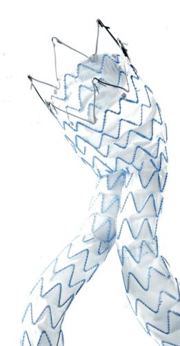
To stent or not to stent, that is the question


“Even the industry has recognised that no stent is a good stent for the leg,” said Gunnar Tepe, who was debating the topic ‘Balloon angioplasty is enough’. He was alluding to bioabsorbable stents, which are still in early development and conceded that these may be the future of stenting technology.


Speaking for the motion, Tepe told ilegx delegates in no uncertain terms to say “no” to stents as first line treatment.


“Stents are very expensive, for technical reasons only short spot stenting is feasible, there is no increased patency with stenting and if at all there is, it usually has no clinical relevance,” he said.


Speaking on balloon angioplasty as a sole treatment option, Tepe said that it showed “excellent” results. He also said that with stenting, there was often a new problem created, that of in-stent restenosis.


Overall, Tepe argued that it “much too early to say stents have to be used. In addition, bare metal stents vs. drug eluting stents have shown no significant difference.”


Thomas Zeller, Tepe’s opponent in the debate said, “Balloon angioplasty alone is not enough in below-the-knee revascularisation, especially in lesions smaller than 10cm.


For example, in a focal lesion across the knee, plain balloon angioplasty might be associated with insufficient primary success and high restenosis rate; atherectomy could be potentially associated with an increased risk of perforation and so a self-expanding low profile nitinol stent, might be the answer,” he said.


Zeller quoted Gunnar Tepe’s own paper “Self- expanding stents for treatment of infragenicular arteries following unsuccessfull balloon angioplasty,” which was published in European Radiology back at him.


He said data from the paper showed that when 24 stents were implanted in 20 such arteries, at six months follow-up of all patients, there was 100% technical success, 88% clinical imporvement and an 18% restenosis rate.


However, Tepe had pre-empted this argument for ilegx delegates by saying that this study had a small cohort, and had showed a lot of restenosis.


Endovascular therapy and surgery are complementary, not exclusive


In the debate between Dierk Scheinert, Leipzig, Germany, and Gerhard Rumenapf, Speyer, Germany, on the topic “Endovascular therapy is better than surgery,” ilegx delegates got to know first-hand how while results from historical studies and the idea that surgery and endovascular procedures were exclusive were one thing, reality on the ground is quite another.


Scheinert said that while “My opponent will say that surgery is the only viable and durable option for patients with long lesions and long total occlusions, as shown in some past studies, but in reality decisions are based on many factors. Comorbidities often need to be considered,” he said.


He put the results of past trials like BASIL which showed that outcomes and advantages of surgery were similar to endovascular therapy in terms of patients remaining asymptomatic in perspective. Scheinert told delegates that at the time of BASIL,”We had no appropriate stents and we had to use coronary stents off-label.” Nowadays we have a wide choice of self-expanding and balloon-expandable stents, but the restenosis rate is still too high, he admitted.


“Less is definitely more in the treatment of critical limb ischaemia,” said Scheinert referring to any less-invasive treatment options compared with the invasive nature of bypass. “New devices have helped to improve success rates of endovascular therapy and we are attempting to achieve what we must achieve, which is avoiding amputations,” he said.


On the opposing side of the debate, Rumenapf said “I think endovascular therapies and surgery should complement one another, not replace one another.”


“The Charing Cross Consensus, the International Diabetic Foot guidelines and the New England Journal of Medicine national guidelines all say the same thing. First, the treatment of these lesions should be interdisciplinary, second, if the short term and long term symptomatic improvement is expected to be equivalent, endovascular techniques should be used first. It is difficult to talk against this.”


He told ilegx delegates, “I cannot say anything against endovascular therapy if it is reasonably done. So before asking what remains for vascular surgeons or what is better, consider that the vascular surgeon can offer both techniques. The number of patients is rising but the number of bypass procedures remains constant,” he said.


After revascularisation, what next? Regular follow up!


“Regular follow-up and self surveillance are vital for critical limb ischaemia patients who have undergone revascularisation,” Ulrich Hoffman, Munich , Germany, told ilegx delegates .


He was talking on aspects of managing patients with critical limb ischaemia beyond revascularisation. Hoffman stated that such patients had high risk of restenosis . “These patients should be seen at least twice with physicians focussing on interval history, physical examination, ABI and imaging. We should also give clear instructions to patients on how to perform self-surveillance,“ he said.


Hoffman told ilegx delegates how important it was to revascularise critical limb ischaemia patients even if there were increased procedural risks, doubtful (long-term) success and high procedural costs. “This is because critical limb ischaemia is associated with high mortality, morbidity and costs,” he said.


Now angioplasty can treat femoral lesions once considered “impossible”


In an ilegx session that left interventionalists gasping at how far you could go with angioplasty, Lanfroi Graziani, succeeded in demonstrating how exactly to push the boundaries with this endovascular procedure.


How far can you really go with angioplasty? How do you go where no interventionalists usually go? These were the questions Lanfroi Graziani, Brescia, Italy addressed in a session titled “Angioplasty pushed to its limits.”


He told ilegx delegates, “Transluminal balloon angioplasty remains our best option in treating patients with critical limb ischaemia. Due to improvements in techniques and devices, we can now treat femoral lesions once considered impossible to treat – but specific training is required.


Graziani highlighted that the use of sophisticated instruments has enabled achieving good results in patients, even in the treatment of extreme lesions in dialysed subjects with critical peripheral ischaemia.


Angioplasty is a well-established technique in peripheral arterial disease and critical ischaemia, particularly in the case of the lower limb arteries and extremely calcified femoral popliteal segments,” he said.


Graziani said that in his institution, psoralen with long wavelength ultra violet radiation after balloon angioplasty is the first line treatment. “We believe in transluminal balloon angioplasty as it is a minimally-invasive, repeatable technique and a low-cost procedure,” he said.


In integrated extreme intervention, the lesion is usually approached from the femoral artery going down to the foot. “This kind of technique represents 90% of procedures used in critical limb ischaemia cases”, Graziani stated. “Stenting in the fem-pop segment and balloon angioplasty are combined together for the best revascularisation.”


Graziani has said that three tips to optimise the result of balloon angioplasty would be to ensure that 1 ) there is prolonged balloon inflation (>180 sec), 2) gradual high-pressure balloon dilatation and 3) dilatation using a correct balloon size.


The second annual ilegx symposium took place in Munich, Germany on 13-14 October, 2009 and was attended by around 250 delegates for the second year running.


Johannes Lammer wins CIRSE Gold Medal for 2009

0
Johannes Lammer wins CIRSE Gold Medal for 2009

Johannes Lammer was awarded the CIRSE gold medal at the opening ceremony on September 19, 2009, Lisbon, Portugal. “I am truly honoured to receive this medal from CIRSE,” said Lammer in his acceptance speech.

Lammer was born in Vienna, Austria and fittingly for a radiologist, the most image-based of all specialisations, originally wanted to be a filmmaker. He told Interventional News, “The decision to go into medical school came about because in the summer I finished high school, the Austrian Government decided that those who were going to medical school did not have to join the Army. I thought medical school was therefore, a good option.”


In the early Eighties, Lammer left for the US to learn about CT imaging.” It was the early times of CT, and it was said that the hospital of the University of Philadelphia had a very new CT scanner,” he recalled. However, when I arrived, this new CT machine was not there. So I was simply hanging around , observing barium studies. Then one day I met Ernest Ring, an interventional radiologist who saw that I was attracted to the specialty, and I learnt a lot from his training. So I came back from the US, not a CT expert, but an interventional radiologist,” he said.


Since 1992, Johannes Lammer has been heading the department of angiography, now Cardiovascular and Interventional Radiology in Vienna University Hospital and has been actively committed to furthering the cause of interventional radiology.


Both Anne Roberts and Anthony (“Tony”) A Nicholson were named as distinguished fellows of CIRSE at the opening ceremony.

Roberts was born in Boston Massachusetts, but grew up in Pasadena, California, near Los Angeles. After first studying history, education and doing a Master’s degree in the history of South Asia at UCLA, she decided to take up a career in medicine. A web profile of Roberts says that she believes that many procedures which used to require a surgical approach can now be done with “needles, wires and small plastic tubes” which are the basis of interventional radiology.


She is currently the Chief of Vascular and Interventional Radiology at the UCSD Thornton Hospital.


“I was absolutely thrilled to be named as a Distinguished Fellow of CIRSE. Especially as an American, and an American woman, to be so honoured was fantastic. Many of the previous fellows are heroes of mine, and to be in their company is truly a wonderful honour. I am very grateful to CIRSE for this tribute,” she told Interventional News.


Tony Nicholson was born in Liverpool. After studying biochemistry and microbiology, he did a Master ‘s degree in Cambridge. It was during this period that he decided on a career in medicine. In 1978 he entered Sheffield Medical School to study medicine from where he graduated MB ChB to obtain posts in medicine and surgery to registrar level at Sheffield Teaching Hospitals where he discovered interventional radiology.


He was much influenced here by the interventionalists of the time like David Cumberland, David Allison and Mike Collins and decided to become an interventional radiologist.


“I was very proud to receive this honour from a professional society like CIRSE whose membership I so respect”.


Nicholson is currently a consultant vascular and interventional radiologist at the Leeds Teaching Hospitals NHS trust.


The 2009 Gruentzig lecturer was Riccardo Lencioni, Pisa, Italy and the Roesch lecturer was Michael D Dake, California, USA.


Cook launch first SFA open-door registry

0
Cook launch first SFA open-door registry

Cook Medical is collaborating with physicians around the world to develop the SFA Open-Registry using OpenDoor, the first-ever transparent clinical data management suite.

Using an intuitive on-line web interface called OpenDoor, clinicians can track their patients, view the medical community’s trends in treating peripheral arterial disease and determine the most effective treatment options for their patients. Transparency will be a central objective of the SFA Open-Registry, with results for all treatment types available to participating clinicians. This level of transparency where all treatments types are included, as well as real-time access could lead to a reduction in bias in the presentation of clinical data, and allow participating clinicians access to real world patient results. It will also provide a forum for clinicians to communicate with each other about SFA treatments and outcomes.


The company says this observational registry will enable the exchange of real-world treatment results with other clinicians to find the most effective options for patients, track patient outcomes and view community trends. OpenDoor assists with data visualisation and analysis, while allowing clinicians to compare the outcomes of multiple treatments, identify patient subsets and more. In addition, it could help clinicians analyse with their practice-specific data to monitor patient outcomes.


Structure of the registry

The registry will collect clinical data including procedural and follow-up information (out to 5 years). The data will be accessible within 30 days of entry by the clinicians. It is intended to collect data on SFA cases including those treated with balloon angioplasty, bare metal stents, drug-eluting stents and surgical intervention.


Key features of OpenDoor:

Real-time data analysis

Data visualisation and analytics

Case portfolio management

Online forum for clinician collaboration

Resource library

Concierge service designed to assist clinicians with practice-specific analyses


Johannes Lammer wins CIRSE Gold Medal for 2009

0
Johannes Lammer wins CIRSE Gold Medal for 2009

Johannes Lammer was awarded the CIRSE gold medal at the opening ceremony on September 19, 2009, Lisbon, Portugal. “I am truly honoured to receive this medal from CIRSE,” said Lammer in his acceptance speech.

Lammer was born in Vienna, Austria and fittingly for a radiologist, the most image-based of all specialisations, originally wanted to be a filmmaker. He told Interventional News, “The decision to go into medical school came about because in the summer I finished high school, the Austrian Government decided that those who were going to medical school did not have to join the Army. I thought medical school was therefore, a good option.”


In the early Eighties, Lammer left for the US to learn about CT imaging.” It was the early times of CT, and it was said that the hospital of the University of Philadelphia had a very new CT scanner,” he recalled. However, when I arrived, this new CT machine was not there. So I was simply hanging around , observing barium studies. Then one day I met Ernest Ring, an interventional radiologist who saw that I was attracted to the specialty, and I learnt a lot from his training. So I came back from the US, not a CT expert, but an interventional radiologist,” he said.


Since 1992, Johannes Lammer has been heading the department of angiography, now Cardiovascular and Interventional Radiology in Vienna University Hospital and has been actively committed to furthering the cause of interventional radiology.


Both Anne Roberts and Anthony (“Tony”) A Nicholson were named as distinguished fellows of CIRSE at the opening ceremony.

Roberts was born in Boston Massachusetts, but grew up in Pasadena, California, near Los Angeles. After first studying history, education and doing a Master’s degree in the history of South Asia at UCLA, she decided to take up a career in medicine. A web profile of Roberts says that she believes that many procedures which used to require a surgical approach can now be done with “needles, wires and small plastic tubes” which are the basis of interventional radiology.


She is currently the Chief of Vascular and Interventional Radiology at the UCSD Thornton Hospital.


“I was absolutely thrilled to be named as a Distinguished Fellow of CIRSE. Especially as an American, and an American woman, to be so honoured was fantastic. Many of the previous fellows are heroes of mine, and to be in their company is truly a wonderful honour. I am very grateful to CIRSE for this tribute,” she told Interventional News.


Tony Nicholson was born in Liverpool. After studying biochemistry and microbiology, he did a Master ‘s degree in Cambridge. It was during this period that he decided on a career in medicine. In 1978 he entered Sheffield Medical School to study medicine from where he graduated MB ChB to obtain posts in medicine and surgery to registrar level at Sheffield Teaching Hospitals where he discovered interventional radiology.


He was much influenced here by the interventionalists of the time like David Cumberland, David Allison and Mike Collins and decided to become an interventional radiologist.


“I was very proud to receive this honour from a professional society like CIRSE whose membership I so respect”.


Nicholson is currently a consultant vascular and interventional radiologist at the Leeds Teaching Hospitals NHS trust.


The 2009 Gruentzig lecturer was Riccardo Lencioni, Pisa, Italy and the Roesch lecturer was Michael D Dake, California, USA.


Cryoplasty appears to have no place in diabetic patients

0
Cryoplasty appears to have no place in diabetic patients

“Cryoplasty is comparable to conventional balloon angioplasty in the femoropopliteal artery of the diabetic patients, and the technique is also associated with significantly more clinically driven repeat procedures”, said S Spiliopoulos, Patras, Greece.

He was presenting the results of a study titled “Cryoplasty versus conventional balloon angioplasty of the femoropopliteal artery in diabetic patients: long term results from a prospective randomised, controlled study” at CIRSE 2009, Lisbon, Portugal.


With regard to the results of the study, Spiliopoulos said, “the technical success rate between the two procedures was similar – 58% in the group randomised to PolarCath Cryoplasty (24 patients with 31 lesions, group A) vs. 64% in the group treated by conventional balloon angioplasty (26 patients, 34 lesions, group B) of the femoropopliteal artery.


There were no significant differences between the two groups with regard to overall mortality (12.5% for group A vs. 11.5% for group B, respectively) and limb salvage( 3.4% vs. 6.5% respectively)

While 24-month angiographic primary patency was not significantly different between the two groups (59% in group A vs. 55% in group B). On the other hand, significantly more re-interventions because of recurrent symptoms were required in the cryoplasty group up to 24 months (66% vs. 40% in the balloon angioplasty group; p<0.05 log-rank test), said Spiliopoulos.


Based on these results, he concluded that conventional cryoplasty was merely comparable to conventional balloon angioplasty, was associated with significantly more repeat procedures in diabetic patients.


Click here to visit ilegx.com, where you can register for the upcoming interdisciplinary leg summit, Oct 13-14, in Munich, Germany.


Setback for drug elution in the periphery as STRIDES follows SIROCCO

0
Setback for drug elution in the periphery as STRIDES follows SIROCCO

Johannes Lammer, Vienna, Austria, principal investigator of the STRIDES trial sponsored by Abbott Vascular, presented the six- and 12-month results at CIRSE 2009, September 19-23, Lisbon, Portugal.

“Dynalink E is a slow-eluting, self-expanding, drug-eluting stent releasing approximately 80% of the drug over three months. There was sustained clinical benefit with improvement in Rutherford-Becker clinical category in 80% of patients after 12 months and there were no observed stent fractures after 12 months.


“A retrospective comparison to the historical VIENNA Absolute trial suggested improved patency rate of DYNALINK-E vs. bare metal at six months. However, the improved patency rate was not sustained at 12 months,” he said.


This follows the results of the SIROCCO II study which showed that drug-eluting stents have delayed, but failed to conquer restenosis in the superficial femoral artery.


SIROlimus Coated Cordis SMART Nitinol Self-expandable stent for the Treatment of Obstructive Superficial Femoral Artery Disease (SIROCCO II) confirmed the short term efficacy of the slower release formulation identified in SIROCCO I. It found good outcomes in the bare SMART stent arm of the trial with an overall 6 month angiographic pooled restenosis rate of 11.6% (n=43) and an 18 month rate of 22.2% (n=45). However, slower eluting data pooled from SIROCCO I and II resulted in an early statistically significant difference in the primary endpoint (mean stent diameter), Showed that this advantage was lost by 18 months.


The STRIDES trial enroled 104 patients at 14 European sites, with a primary endpoint of in-stent restenosis (a measure of vessel re-narrowing) in the superficial femoral artery at six months. Secondary endpoints include angiographic (X-ray) measurements of the change in vessel lumen diameter between the time immediately following stent placement and at 12 months, restenosis at 12 months, as well as five-year clinical follow-up to track resolution of PAD symptoms, limb preservation and patient survival.


The STRIDES trial enroled complex patients, including 17% with critical limb ischaemia,45% with total limb occlusions, 39% with length greater than 10cm, 9.4% with restenosis and 78% with TASC C 2000 classification.


“This is a first-in-man evaluation of Dynalink-E in the superficial femoral arteries and the purpose of the first-in-human STRIDES trial was to evaluate the safety and efficacy of an everolimus-eluting nitinol stent for the treatment of superficial femoral and proximal popliteal arterial occlusive disease,” Lammer said.


“The superior patency rate of the drug-eluting Dynalink-E stent within the first six months may be beneficial to patients with critical limb ischaemia to improve early wound healing,” he said.


Experts find that while drug-eluting stents approved for use today were designed with the coronary arteries in mind, superficial femoral arteries in the leg present a different kind of anatomy that can be challenging to treat because re-obstruction of the vessel is a major concern. The STRIDES trial is evaluating the use of a self-expanding stent system specifically designed to withstand normal leg movement, combined with the anti-proliferative drug everolimus, as a longer-term treatment alternative for patients with superficial femoral artery disease.


Click here to visit ilegx.com, where you can register for the upcoming interdisciplinary leg summit, Oct 13-14, in Munich, Germany.


Cook launch Europe’s first drug-eluting stent for the SFA

0
Cook launch Europe’s first drug-eluting stent for the SFA

Against the backdrop of the disappointing STRIDES trial data, COOK Medical launched the first drug-eluting stent for use in the superficial femoral artery in Europe.

Michael Dake presented highly promising registry data out to two years, but this did not include patency as an endpoint which will be available once the results of the US randomised trial are presented next year. The Zilver PTX registry is the the largest registry of its kind ever conducted and results indicate that the Zilver PTX stent can effectively bridge the gap between the patient results achieved using open surgical bypass graft procedure.


“With the Zilver PTX stent, Cook has introduced a true landmark device that can reduce the number of PAD patients having to endure the trauma of leg amputations and bypass surgeries,” explained Rob Lyles, vice president and global leader of Cook Medical’s peripheral intervention unit. “Clinically proven, polymer-free and exceptionally durable, we expect the Zilver PTX stent to become the new standard of care for PAD in the SFA.”


Zilver PTX was introduced commercially to European physicians at the annual CIRSE meeting, held this year from 19-23 September in Lisbon. The launch included several important developments, including the introduction of a new website, http://www.cookmedical.com/zilverptx, with clinical and practical information on the device for patients and physicians in English and major European language languages, and a twitter page for live updates from CIRSE, http://www.twitter.com/ZilverPTX.


Cook also has initiated a first-of-its-kind open clinical database on SFA treatments. The SFA “Open Door” Registry provides physicians with an opportunity to exchange ongoing, real world treatment results. Utilising an intuitive web interface, clinicians may track their patients, view the medical community’s trends in treating peripheral arterial disease and determine the most effective treatment options. Transparency will be a central objective of the SFA Open-Registry, with results for all treatment types available to researchers, regulatory bodies and the public.


In an interview with Interventional News, Rob Lyles indicated that Cook had adopted a unique ‘affordable innovation’ strategy to overcome potential financial or reimbursement barriers in Europe.


Cook licenses the rights to use paclitaxel on peripheral stents and other noncoronary medical devices from Angiotech Pharmaceuticals of Vancouver, British Columbia, Canada. In the United States, the Zilver PTX Drug-Eluting Stent is an investigational device not available for sale at this time.


Click here to visit ilegx.com, where you can register for the upcoming interdisciplinary leg summit, Oct 13-14, in Munich, Germany.


Cryoplasty has no place in diabetic patients

0
Cryoplasty has no place in diabetic patients

Cryoplasty seems comparable to conventional balloon angioplasty in the femoropopliteal artery of diabetic patients, and the technique is also associated with significantly more clinically driven repeat procedures, said S Spiliopoulos, Patras, Greece.

He was presenting the results of a study titled “Cryoplasty versus conventional balloon angioplasty of the femoropopliteal artery in diabetic patients: long term results from a prospective randomised, controlled study” at CIRSE 2009, Lisbon, Portugal.


With regard to the results of the study, Spiliopoulos said, “the technical success rate between the two procedures was similar – 58% in the group randomised to PolarCath Cryoplasty (24 patients with 31 lesions, group A) vs. 64% in the group treated by conventional balloon angioplasty (26 patients, 34 lesions, group B) of the femoropopliteal artery.


There were no significant differences between the two groups with regard to overall mortality (12.5% for group A vs. 11.5% for group B, respectively) and limb salvage( 3.4% vs. 6.5% respectively).


While 24-month angiographic primary patency was not significantly different between the two groups (59% in group A vs. 55% in group B). On the other hand, significantly more re-interventions because of recurrent symptoms were required in the cryoplasty group up to 24 months (66% vs. 40% in the balloon angioplasty group; p<0.05 log-rank test), said Spiliopoulos.


Based on these results, he concluded that conventional cryoplasty was merely comparable to conventional balloon angioplasty, was associated with significantly more repeat procedures in diabetic patients.


Medtronic launches the KYPHON Express System as battle between vertebroplasty and kyphoplasty continues to rage

0
Medtronic launches the KYPHON Express System as battle between vertebroplasty and kyphoplasty continues to rage

Recent studies published in the New England Journal of Medicine suggest that the benefits patients derive from vertebroplasty is merely a placebo effect, although some spine interventionalists have criticised the design of these studies.

Against the backdrop of this raging debate, Medtronic has just launched the KYPHON Express System in Europe. This is a set of devices for the KYPHON balloon kyphoplasty procedure to treat vertebral compression fractures.


At the recent CIRSE 2009 conference, Lisbon, Portugal, some interventionalists asid they seem to prefer the kyphoplasty procedure which has the potential of providing added safety and a more consistent filling of the vertebral body with bone cement. Others have criticised the design of the studies in the NEJM in relation to their inclusion and exclusion criteria and population size, amongst other things.


Medtronic has said that newly introduced Kyphoplast Express System has tools, which are smaller than what is currently available for balloon kyphoplasty.


A company release says, according to a recently published study in The Lancet, patients whose VCFs are treated with balloon kyphoplasty experience significant improvement in quality of life, back function and back pain compared to patients receiving non-surgical management (including pain medication, bed rest, physiotherapy and back bracing) alone.


The Express System measures only 3.3mm in diameter, more than 20% smaller than devices currently available in Europe for the balloon kyphoplasty procedure. The smaller size may offer advantages in the treatment of smaller patients, such as petite women, and allows treatment of fractures in the upper thoracic area of the spine. Some 10-15 percent of VCFs are upper thoracic fractures2, which, until now, had some treatment limitations due to size availability of the balloon kyphoplasty devices.


Hendrik Fransen, from the Radiology Department of the AZ Sint Lucas hospital in Gent, Belgium said, “The KYPHON Express System will help us to make balloon kyphoplasty available to more patients. The KYPHON Express System was developed by Medtronic in direct response to physician requests for smaller-sized devices. This is good news, as we will now be able to perform this procedure more widely, which may improve the lives of more patients.”


Click here to visit ilegx.com, where you can register for the upcoming interdisciplinary leg summit, Oct 13-14, in Munich, Germany.


UEMS‰Ûödecision puts European IR in charge of its destiny

0
UEMS‰Ûödecision puts European IR in charge of its destiny

The recent vote by the European Union of Medical Specialists (UEMS) shows how interventional radiology has moved from a technical discipline to a true clinical discipline, which marks a real milestone – the official birth of a new medical specialty in Europe. CIRSE president, Jim A Reekers, said European standards for training in interventional radiology can now be implemented, which would result in the improvement of quality of care for patients

The last issue of Interventional News carried a report by CIRSE on how the European Union of Medical Specialists (UEMS) had voted positively on the establishment of an interventional radiology division in Europe. In this issue, Interventional News has gone deeper, and asked several prominent interventional radiologists in Europe what this significant decision means for the discipline.


CIRSE president, Jim A Reekers, The Netherlands, highlighted that this vote strengthens the perception of interventional radiology going from being a ‘professional hobby’ to a specialty. He said, “Interventional radiology was already recognised by the European Society of Radiology as a subspeciality of radiology for many years, but this is still recognition within your own group.


“For the outside world, a radiologist and an interventional radiologist were still more or less the same – an interventional radiologist being a radiologist who did some interventions on the side, like a professional hobby. Now, by the positive UEMS vote, interventional radiology is officially recognised in Europe as a true medical subspecialty, although still closely connected to diagnostic radiology. European standards for training in interventional radiology can now be implemented,” he said.


Andy Adam, UK, one of the editors-in-chief of Interventional News, emphasised that, “The recognition by UEMS is an important step in changing interventional radiology’s status from a technical discipline to a true clinical discipline, in charge of its own destiny.”


“Very simply, becoming a medical specialty means a lot,” said Marc Sapoval, France, CIRSE chairman of the scientific programme committee, “This decision is a very important step in the sense that it shows the way. From now on, no one can say ‘what is interventional radiology – who are these guys?’”


Riccardo Lencioni, Italy summed it up: “The UEMS decision is a milestone. It is the official birth of a new medical specialty in Europe. For several years interventional radiologists were facing the paradox of playing a key and well-defined role in patient care, but in the absence of a formal recognition as distinct medical specialists by several authorities. This decision marks the change in that.”


The UEMS decision also impacts other factors. Reekers says, “By being an official subspecialty in Europe, interventional radiology in all countries can now develop further to become a clinical specialty with direct patient referrals and clinical logistics. This is the next logical step.”


Training, skill certification and standardisation


Prominent interventional radiologists also feel that the decision will expedite the establishment of a training curriculum. It will enable a definite European skill certification and help in setting standards for quality.


Sapoval said, “This decision paves the way for a better definition of the training, capabilities, work organisation, and position among other specialties. Very likely it will help allow interventional radiologists to define themselves, set out what they need to develop, how to do it, at which pace and in which order.”


“The European skill certificate (interventional radiology examination) will be a fact in 2010. This examination will be based on the European interventional radiology training curriculum. And will be a huge step forward in standarisation of interventional radiology quality throughout Europe,” said Reekers.


Lencioni added, “CIRSE’s planned European Certificate of Interventional Radiology will represent a recognised common European qualification in interventional radiology. The UEMS decision has created a solid basis for this important and timely project, as well as for any other future initiative aimed at standardising training and expertise in interventional radiology across Europe.


Thomas Pfammatter, Swizerland, puts it in context for the country:


“The establishment of a European training curriculum is pivotal, particularly for a multilingual, small country such as ours, where many interventionalists are from abroad, and where several locally trained radiologists have undergone subspeciality training in Europe or the US. For a small national society the creation of our own training and quality control programmes would represent a huge effort. Therefore, the help of an international society, such as CIRSE, is definitely needed.”


Attracting young radiologists


“Subspecialty status will lead to designated interventional radiology posts in most hospitals and will ensure the future of this discipline. This will make interventional radiology more attractive and boost recruitment,” said Adam.


Lencioni said, “Recognition of interventional radiology as a distinct medical specialty will be instrumental in attracting young MDs. The cosmos of medicine is moving towards technology-assisted, minimally invasive approaches. UEMS’ decision makes it clear to the many young MDs who are interested in this exciting and ever-growing field of patient care, that there is one specialty for image-guided interventions, and this specialty is interventional radiology.”


Sapoval clarified some difficulties that young interventional radiologists have faced so far. “They have been in the fog for a long time. Now, they can see the way, what will be needed in terms of training, who will play for them, and who will guide and support them. As an example the SFICV (French Cardiovascular and Interventional Radiology Society) have been developing a specific training programme for young interventional radiologists that has proven to be very successful; more than 200 young interventional radiologists have entered the process in just four years.”


Pfammatter shares his views on some of the challenges facing recruitment. “Currently, a career as an interventional radiologist can be perceived as not an attractive one for young radiologists. This is because we are perceived as those constantly being exposed to turf battles, as those taking night calls until the end of their professional life and as those practicing in a field with an insecure job market. The recognition of a specialty status might help take care of a part of these problems,” he feels.


Different countries and scenarios


According to Lencioni, the UEMS decision will have a great impact on a national basis. “National authorities cannot disregard the UEMS decision and shall give national accreditation to interventional radiology. It will likely take a considerable amount of time to translate the decision into actual changes in the national regulations. But in the end, all European countries shall be aligned,” he says.


Referring to how the UEMS decision would affect the UK, Adam said, “The Royal College of Radiologists is seeking formal subspecialty status for interventional radiology in the UK. The UEMS decision will help in this effort.”


Sapoval adds, “The positive influence will be huge. In France, for instance, it will result in a strengthening of the Federation de Radiologie Interventionnelle, and influence the need for a specific training pathway managed by the “College des Enseignants de Radiologie de France (CERF).”


In Switzerland, discussions on the status of interventional radiology within radiology departments and on its current or future role in percutaneous imaging-guided therapies within the community of competing proceduralists has been going on over the last ten years or so. “Unfortunately, no consensus has been reached, and in 2009 interventional radiology is still practiced throughout the country according to local agreements and habits. There is no guarantee that it will survive. This decision might empower interventional radiology and represent an additional argument when it comes to defend or even enhance its status in Switzerland,” said Pfammatter.


Similarly, Jose Ignacio Bilbao, Spain, says “I´m not sure about the impact of this decision in Spain. People probably know that the administration here has decided that radiology is a four-year training specialty. We do not have any recognised programme of subspecialty fellowships and most of our specialised working places are not focusedon attaining this. It means that we, the spanish radiologists, have a lot of inner management to improve the situation. It is needless to say that any additional help from Europe will be very welcome.”

He believes that Spanish interventional radiologists and the Spanish Ssociety of IR (SERVEI), as well the Spanish Ssociety of Radiology (SERAM), will use this important decision to improve the situation.


The future


“The UEMS decision is not only important for interventional radiology but also for the accessibility of minimal invasive procedures for patients and through this for the improvement in the quality of care,” said Reekers.


So what lies ahead? According to Adam it is a clarion call to employers. “The next step is to ensure that employers recognise the importance of interventional radiology in modern medicine and the many advantages it offers for patient safety. They need to fund posts in IR, and provide the appropriate infrastructure for clinical practice.”


Johannes Lammer, Austria, adds, “Interventional radiology including vascular and non-vascular interventions as well as emergency treatments are an important part of state-of-the-art medical services today. Therefore it was important that interventional radiology is recognised as a medical subspecialty.


As a next step, it is important that in every European country interventional radiology is recognised as subspecialty as well.


Click here to visit ilegx.com, where you can register for the upcoming interdisciplinary leg summit, Oct 13-14, in Munich, Germany.


New Zilver PTX drug-eluting stent from Cook Medical could greatly reduce need for leg amputations and bypass surgery for European patients

0
New Zilver PTX drug-eluting stent from Cook Medical could greatly reduce need for leg amputations and bypass surgery for European patients

World’s first drug-eluting stent for a widespread form of peripheral arterial disease now available throughout the EU.

In a breakthrough development that could dramatically reduce the number of leg amputations and painful bypass graft surgeries performed annually on European patients, a first-of-its kind drug-eluting stent for a widespread form of peripheral arterial disease (PAD) is now available to physicians throughout the European Union.


Cook Medical’s new CE Marked Zilver PTX drug-eluting peripheral stent is widely expected to improve the standard of care for many patients with serious blockages in the superficial femoral artery (SFA) by creating a highly effective, completely new treatment option.


In the largest clinical trial of its kind ever conducted, the Zilver PTX stent was shown to effectively bridge the gap between the patient results achieved using open surgical bypass graft procedure — which is typically more painful and requires a longer hospital stay for the patient – and the less traumatic, but typically less effective, earlier minimally invasive treatment options for PAD such as balloon angioplasty and bare metal stenting, a press release issued by the company said.


“With the Zilver PTX stent, Cook has introduced a true landmark device that can reduce the number of PAD patients having to endure the trauma of leg amputations and bypass surgeries,” explained Rob Lyles, vice president and global leader of Cook Medical’s peripheral intervention unit. “Clinically proven, polymer-free and exceptionally durable, we expect the Zilver PTX stent to become the new standard of care for PAD in the SFA.”


Cook’s revolutionary device will be introduced commercially to European physicians at the annual CIRSE meeting, held this year from 19-23 September in Lisbon. The launch includes several important developments, including the introduction of a new website, http://www.cookmedical.com/zilverptx, with clinical and practical information on the device for patients and physicians in English and major European language languages, and a twitter page for live updates from CIRSE, http://www.twitter.com/ZilverPTX.


Cook also has initiated a first-of-its-kind open clinical database on SFA treatments. The SFA Open-Registry provides physicians with an opportunity to exchange ongoing, real world treatment results. Utilising an intuitive web interface, clinicians may track their patients, view the medical community’s trends in treating PAD and determine the most effective treatment options. Transparency will be a central objective of the SFA Open-Registry, with results for all treatment types available to researchers, regulatory bodies and the public.


Another key breakthrough is Cook’s commitment to a patient-focused strategy that should make the device available to virtually any PAD patient who needs it, Lyles explained. As part of its ongoing corporate mission to help reduce global health care delivery costs, Cook has adopted a unique ‘affordable innovation’ strategy aimed at reducing any potential financial or reimbursement barriers to its widespread adoption as the standard of care for PAD in the SFA.


The first drug-eluting stent approved for treating PAD in the superficial femoral artery (SFA), the difficult-to-treat, largest blood vessel in the leg, the Zilver PTX stent first expands and holds open the artery to restore blood flow. The device then delivers the drug paclitaxel to the cells in the vessel wall to reduce the risk of new blockages forming. In a major advance over previous drug-eluting technologies, the Zilver PTX achieves targeted drug delivery without using a polymer to adhere the drug to the stent body. This eliminates the potential patient risks associated with polymer-coated devices, including clot formation and inflammation.


Cook licenses the rights to use paclitaxel on peripheral stents and other noncoronary medical devices from Angiotech Pharmaceuticals of Vancouver, British Columbia, Canada. In the United States, the Zilver PTX Drug-Eluting Stent is an investigational device not available for sale at this time.


About PAD


Peripheral arterial disease (PAD), or peripheral vascular disease, is caused by atherosclerosis – the build up of fatty deposits (atheroma) within the lining of the arteries. The most common symptom of PAD is leg pain during exercise. Over time the arteries may narrow due to atherosclerosis, resulting in a reduction in blood flow. Severely reduced blood flow in the limbs is also known as critical limb ischaemia (CLI). It is characterised by leg pain at rest, non-healing wounds and gangrene and may lead to amputation of the limb.


Invatec launch the Maris Plus self-expanding peripheral stent system at CIRSE

0
Invatec launch the Maris Plus self-expanding peripheral stent system at CIRSE

Invatec has launched their latest self-expanding peripheral stent system, Maris Plus at CIRSE 2009. The Maris Plus stent has a “lesion-specific” design with larger sizes to meet the clinical requirements of the iliac region. The Maris Plus becomes the sixth stent platform now marketed by Invatec for the treatment of peripheral arterial disease.


“At Invatec, we pride ourselves on our ability to continually develop lesion specific solutions for cardiovascular arterial and venous disease. With the Maris Plus, we have created a novel, self-expanding stent platform specifically designed for the iliac artery,” said Stefan Widensohler and Andrea Venturelli, co-founders of Invatec. The Maris Plus is available in a 30 – 100mm length with 9.0 – 12.0mm diameter, meeting the variable needs of the iliac region. The distinct stent geometry allows for increased stability in the hip, yet it remains the most flexible stent on the market. Additionally, larger and fully integrated tantalum markers greatly enhance visibility for the physician.


Click here to visit ilegx.com, where you can register for the upcoming interdisciplinary leg summit, Oct 13-14, in Munich, Germany.


Invatec launch the Maris Plus self-expanding peripheral stent system at CIRSE

0
Invatec launch the Maris Plus self-expanding peripheral stent system at CIRSE

Invatec has launched their latest self-expanding peripheral stent system, Maris Plus at CIRSE 2009. The Maris Plus stent has a “lesion-specific” design with larger sizes to meet the clinical requirements of the iliac region. The Maris Plus becomes the sixth stent platform now marketed by Invatec for the treatment of peripheral arterial disease.


“At Invatec, we pride ourselves on our ability to continually develop lesion specific solutions for cardiovascular arterial and venous disease. With the Maris Plus, we have created a novel, self-expanding stent platform specifically designed for the iliac artery,” said Stefan Widensohler and Andrea Venturelli, co-founders of Invatec. The Maris Plus is available in a 30 – 100mm length with 9.0 – 12.0mm diameter, meeting the variable needs of the iliac region. The distinct stent geometry allows for increased stability in the hip, yet it remains the most flexible stent on the market. Additionally, larger and fully integrated tantalum markers greatly enhance visibility for the physician.


Click here to visit ilegx.com, where you can register for the upcoming interdisciplinary leg summit, Oct 13-14, in Munich, Germany.


Invatec announces worldwide launch of FiberNet, the smallest distal embolic protection system on the market at CIRSE 2009

0
Invatec announces worldwide launch of FiberNet, the smallest distal embolic protection system on the market at CIRSE 2009

System has lowest profile and highest level of performance, says a company release.

Invatec introduced FiberNet, a 3-dimensional distal embolic protection system (EPS) for carotid artery stenting with the lowest crossing profile (0.031’’) and highest performance capabilities on the market. Manufactured by Lumen Biomedical, FiberNet can provide superior deliverability and can be used in vessels ranging from 3.5 to 7mm.


With its unique design, FiberNet ensures excellent atraumatic wall apposition. It is also usable in the most complex vasculature formations, captures micro-emboli as small as 40µm via specially shaped PET-fibers and can aspirate these emboli from the body, allowing for safe and easy retrieval.


The FiberNet embolic protection system was evaluated in the EPIC Trial, a multicentre clinical study during carotid artery stenting of 237 high surgical risk patients with critical artery stenosis demonstrating a 30-day stroke rate of 2.1%, the lowest ever reported for a distal embolic protection device. The FiberNet EPS captures microemboli to prevent fragments traveling to the brain thus reducing the risk of ischaemic stroke. Andrea Venturelli and Stefan Widensohler, co-founders of Invatec said, “We are extremely excited about bringing this advancement in embolic protection technology to the worldwide market and believe it sets a new performance standard for distal embolic protection of carotid artery stenting, further reducing the risk to the patient. Our partnership with Lumen represents a tremendous opportunity to expand our position in the endovascular field.”


“The FiberNet EPS has several attributes including ease of use, low profile, and the ability to conform to an irregular surface in the vessel wall, all of which have contributed to the impressive results and safety profile that we observed in the EPIC trial,” said Subbarao Myla, medical director of cardiovascular research and endovascular intervention at Hoag Memorial Hospital in California and national principle investigator for the trial. “The device achieved the lowest stroke rate of any filter currently available, making FiberNet a top choice for physicians, and representing the next generation in embolic protection.”


Click here to visit ilegx.com, where you can register for the upcoming interdisciplinary leg summit, Oct 13-14, in Munich, Germany.


Invatec launch of REEF HP PTA balloon catheter in Europe at CIRSE

0
Invatec launch of REEF HP PTA balloon catheter in Europe at CIRSE

The REEF HP PTA ballloon catheter is specifically designed for AV shunts and hard-to-dilate peripheral lesions.


Invatec announced the launch of REEF HP, a PTA balloon catheter for use in all peripheral high pressure dilatation procedures. According to the company, the “lesion-specific” design of the balloon material is particularly useful in “hard-to-dilate” situations and the availability of variable shaft lengths mean that the Reef HP is equally suited to both arteriovenous (AV) shunts (50cm shaft),and peripheral applications (80/120cm shaft).


”The REEF HP is a robust and highly stable balloon, capable of counteracting even the most severe arterial lesions, with minimal slippage and a high pressure threshold,” said Stefan Widensohler and Andrea Venturelli, co-founders of Invatec. “It has a ‘lesion-specific’ design that is ideal for use in dialysis patients whose AV shunts often create short, fibrous, and hard-to-dilate lesions where the shunt is connected to the native vessel,” they added.


The REEF HP balloon is made from Invatec’s proprietary FLEXITEC XF, an extremely durable material with a large working pressure range of up to 22 bar, offering complete control during high pressure procedures. It is also a low compliant balloon, offering a uniform dilatation force and strong shape retention to dilate resistive lesions with greater success and extreme stability.


Click here to visit ilegx.com, where you can register for the upcoming interdisciplinary leg summit, Oct 13-14, in Munich, Germany.


New micro-bland embolization technique highlighted at CIRSE

0
New micro-bland embolization technique highlighted at CIRSE

CeloNova BioSciences has announced that Franco Orsi made two presentations at the Cardiovascular and Interventional Radiology Society of Europe (CIRSE 2009, Lisbon, Portugal) conference about “micro-bland embolization” which has demonstrated excellent clinical results in treating liver cancer.

Orsi and his colleagues at the European Institute of Oncology in Milan, Italy have developed a technique to cut off the blood supply inside liver tumours and liver metastases that leads to effective tumour control and reduced recurrence of tumours without the use of chemotherapy drugs.


“Embolic particles should be size-calibrated with a small bandwidth in diameter variations because during administration, larger particles within the same vial or syringe may occlude microvessels more proximally and prevent a deeper penetration of the smaller ones,” said Orsi, whose findings were presented at the CIRSE medical conference September 19-23 and published in the September edition of Vascular Disease Management.


“We are gratified that Orsi has developed this micro-bland embolization technique and demonstrated that the unique and very precise calibration of Embozene microspheres leads to superior clinical findings in the treatment of liver cancer,” said Thomas A Gordy, president and chief executive officer, CeloNova Biosciences. “Embozene microspheres, unlike other embolics, are precisely calibrated so that larger spheres do not prevent the deepest possible penetration of tumours. Patients with liver cancer treated in this new way can experience a longer life and a better quality of life.”


About Embozene microspheres


Embozene microspheres are the first and only microspheres to be colour-enhanced with a different colour for each size for increased procedural safety, efficiency and visibility. They are also available in a wider range of sizes than any other spherical embolic on the market. CeloNova’s Embozene microspheres consist of a hydrogel core and an exterior shell made from Polyzene -F, CeloNova’s proprietary polymer which is known to be anti-inflammatory and bacterial-resistant. Four design features distinguish Embozene microspheres from other spherical embolics: biocompatibility, precise calibration, stable suspension, and structural stability.


Opinion: A young interventional radiologist’s perspective

0
Opinion: A young interventional radiologist’s perspective

After years of painstaking lobbying and efforts from CIRSE, we are celebrating the official recognition of interventional radiology as a distinct medical subspecialty. Embracing the recent UEMS’ decision, Greece will also become one of the few European countries to officially offer 1-2 years of subspecialty training in interventional radiology.

Official European skill certification and training quality standardisation will further contribute to widespread legal coverage and adequate reimbursement of interventional radiologic procedures in the future.


However, being the devil’s advocate, I am wondering if that is enough. The UEMS’ decision is definitely paving the way towards an independent clinical specialty, but current interventional radiology practice trends are still far away from that point. The shortage of adequately trained clinically-oriented physicians and the unprecedented developments in medical technology render interventional radiology a victim of its own success.


Minimally invasive percutaneous and endovascular therapies have escaped the barriers of a small but ingenious group of physicians and have revolutionised the whole of modern medicine. Trying to withhold or restrict successful interventional radiology treatments within an elite professional group is at least short-sighted. Do not forget that we are not competing with each other, but with all the other specialties dreaming about sticking in a needle, a catheter or a stent.


Young interventional radiologists like me should forget about turf battles and have the motivation and the passion to become the best in what we do. Adequate knowledge of radiological imaging, technical expertise and wise clinical thinking built on years of everyday practice are just the minimal requirements.


Modern interventional radiology physicians have to choose from a wide variety of already established fields, like vascular interventional radiology, interventional oncology and musculoskeletal interventions. We have to continually advertise ourselves in our hospital and community, and above all, assume personal responsibility of the patients we treat while building on the level of scientific evidence of interventional radiology with high-quality scientific publications. We have to be aggressive and antagonistic, think “outside the box” and continue developing novel minimally invasive image-guided therapies, if we are to evolve to a new “medical species”. After all, only the best will survive.


Konstantinos Katsanos is an interventional radiologist from Greece


Angiotech pharmaceuticals announces commercial launch of the Option inferior vena cava filter in the United States

0
Angiotech pharmaceuticals announces commercial launch of the Option inferior vena cava filter in the United States

Angiotech Pharmaceuticals recently announced the commercial launch of the Option inferior vena cava (IVC) filter in the US, following FDA 510(k) clearance in June and a limited pre-launch in July.

Angiotech holds exclusive worldwide rights to market and distribute the Option IVC filter, which it obtained in a license agreement with privately held Rex Medical.


“The pre-launch of the Option IVC Filter last month was overwhelmingly successful, demonstrating its potential to be a market leading product in pulmonary embolism prevention,” said William Hunter, president and CEO of Angiotech. “We are excited to announce that the Option IVC filter is now available throughout the US through our dedicated interventional sales team.”


The Option IVC filter is used for the prevention of recurrent pulmonary embolism. The device is implanted, typically by interventional radiologists in a minimally invasive procedure, into the body’s inferior vena cava to prevent pulmonary embolism. The Option IVCfFilter is specifically designed for use as both a permanent or temporary implant (in temporary, or retrievable, indications, a physician may later perform a second surgical procedure to remove the Option IVC Filter if necessary or where mandated clinically).


NICE appraisal committee finds sorafenib ‰ÛÏnot recommended‰Û for advanced HCC

0
NICE appraisal committee finds sorafenib ‰ÛÏnot recommended‰Û for advanced HCC

The National Institute for Health and Clinical Excellence (NICE) has just issued a second appraisal consultation document on the first line use of sorafenib for advanced and metastatic hepatocellular carcinoma.

The UK health watchdog’s appraisal committee’s preliminary recommendations are:


• Sorafenib is not recommended for the treatment of advanced hepatocellular carcinoma (HCC) in patients for whom surgical or locoregional therapies have failed or are not suitable.


• People currently receiving sorafenib for the treatment of advanced HCC should have the option to continue treatment until they and their clinician consider it appropriate to stop.


In this context, a release from Bayer Schering Pharma finds that the National Institute for Health and Clinical Excellence (NICE) has, once again, proposed to deny Nexavar (sorafenib) for the treatment of advanced hepatocellular carcinoma (HCC). Nexavar is the only systemic treatment option that could potentially extend the survival of these patients.


Cases of liver cancer have almost tripled over the last three decades according to figures recently published by Cancer Research UK. In 1975, there were 865 cases of primary liver cancer and in 2006 that had risen to 3,108 in the UK. HCC accounts for 80-90% of these primary liver cancers.


Nexavar has demonstrated a 44% increase in survival for advanced HCC patients, compared to best supportive care alone. Nexavar is the first systemic drug for advanced HCC to show a significant survival benefit after 30 years of randomised, comparative trials.


Harpreet Wasan, medical oncologist, Hammersmith Hospital, Imperial College, London said: “Other than sorafenib, every systemic treatment that has been evaluated in advanced HCC, has failed to significantly extend survival. Today’s decision, unless reversed, puts us in a unique situation in cancer where we are left with nothing to offer advanced HCC patients apart from supportive and palliative care, thus denying them the life-preserving benefits of modern treatments. This is a devastating blow to patients and their families who will be robbed of precious time together. It is also painfully disheartening for British oncologists, many of whom were involved in the trials for this drug as they, effectively, will not be able to prescribe it.”


Graeme Poston, an eminent liver surgeon on behalf of the Hepatobiliary UK Group (HUG), president-elect of the Association of Upper Gastrointestinal Surgeons (AUGIS) and former president of the British Association of Surgical Oncologists (BASO) said: “I am naturally disappointed that NICE appears to have not taken into account the views of leading healthcare professionals in the field. The Hepatobiliary UK Group of doctors who specialise in treatment of HCC recently launched national guidelines for the UK which clearly state that sorafenib is the standard of care for patients with advanced HCC for whom no potential curative option is available. With this decision, physicians in the UK will be unable to provide the best possible care for their HCC patients, even though sorafenib treatment is readily available to patients in other parts of the world.’’


Alison Rogers, chief executive of the British Liver Trust said, “This is a very poor decision for patients with HCC in the UK. Liver cancer is a major concern for the Trust and the Trust calls for improvements throughout the liver cancer patient pathway, including better prevention, earlier diagnosis and access to curative and palliative treatments, together with the need for improved liver services as a whole”.


Nicole Farmer, Business Unit Head of Bayer Schering Pharma Oncology in the UK commented: “This proposal by NICE conflicts dramatically with the Government’s strategy to bring UK cancer outcomes in-line with the rest of Europe (where Nexavar is already widely available in countries such as France, Germany, Spain, Italy, Romania, and Greece) and reaffirms why the UK currently sits at 14-15 out of 18 EU countries with regard to cancer survival”.


About sorafenib for liver cancer


Sorafenib was licensed by the EMEA in October 2007. Sorafenib is licensed in the UK for the treatment of patients with hepatocellular carcinoma (HCC) the most common form of primary liver cancer who are unsuitable for loco-regional therapies. Sorafenib is the only licensed systemic therapy in the UK proven to significantly prolong survival for patients with advanced HCC versus best supportive care alone.


Sorafenib’s differentiated mechanism


Sorafenib targets both the tumour cell and tumour vasculature. In preclinical studies, sorafenib has been shown to target kinases known to be involved in both cell proliferation (growth) and angiogenesis (blood supply) – two important processes that enable cancer growth. These kinases included Raf kinase, VEGFR-2, VEGFR-3, PDGFR-B, c-KIT, FLT-3 and RET5. Preclinical models have also demonstrated that the Raf/MEK/ERK pathway has a role in HCC.


Interventional radiology resources cover peripheral arterial disease

0
Interventional radiology resources cover peripheral arterial disease

Society of Interventional Radiology offers publications, patient information brochures.

The Society of Interventional Radiology offers numerous resources just in time for September’s national peripheral arterial disease (or PAD) awareness month. As recent medical studies continue to highlight the seriousness of PAD, these resources are welcome additions to libraries for interventional radiologists and primary care physicians who want to learn more about minimally invasive treatments for this disease that affects an estimated 10 million people in the United States.


New for this year is “Aortic Diseases: Clinical Diagnostic Imaging Atlas”(Stuart J Hutchison, 2009, hardcover, 374 pages, includes DVD). This title in the brand-new Clinical Diagnostic Imaging Atlas Series offers authoritative guidance from a well-known cardiologist and imaging expert about when and how to perform the latest diagnostic imaging tests, interpret the results and effectively treat the emergency. Included are detailed discussions of hot topics, full-colour illustrations and a DVD of procedural videos, animation and downloadable image libraries. SIR member, $169; nonmember, $185.


Patient information brochures, which are useful for interventional radiologists, primary care physicians, other health care professionals and laypersons, give a better understanding of interventional radiology treatments. PAD-related titles include “What You Should Know About Peripheral Arterial Disease,” “Abdominal Aortic Aneurysm (AAA),” “Angiography” (English and Spanish versions), “Angioplasty,” “Stent Placement” and “What Is Interventional Radiology?” SIR member, $50 (100 brochures); nonmember, $100 (100 brochures).


Additional resources available at SIR Web site are listed below.


“Peripheral Vascular Interventions” (Krishna Kandarpa, 2007, hardbound, 720 pages, 1,146 illustrations): This comprehensive clinical reference describes the full range of endovascular interventions currently used for peripheral vascular problems. SIR member, $189; nonmember, $210.


“Image-guided Intervention: 2-Volume Set” (Matthew A. Mauro, Kieran Murphy, Kenneth Thomson, Anthony Venbrux and Christoph L. Zollikofer, 2008, hardcover, 1,928 pages): An international group of experts brings an exhaustive full-color, two-volume reference on every aspect of vascular and nonvascular interventions. Included are more than 1,600 examples of cutting-edge modalities such as MR, multislice CT, CT angiography and ultrasonography. SIR member, $339; nonmember, $370.


“Atlas of Vascular Anatomy: An Angiographic Approach” (Renan Uflacker, second edition, 2006, hardbound, 928 pages, 829 illustrations): This book, which presents the complete anatomy of the arteries, veins and lymphatic system by body region, includes full-colour drawings that are correlated with angiographic images to guide evaluation and management of vascular disease and performance of endovascular procedures. SIR member, $249; nonmember, $275.


“Cardiovascular Haemodynamics and Doppler Waveforms Explained” (Crispian Oates, 2008, paperback, 192 pages): This book provides the necessary understanding of the physical principles of blood flow in the body to produce clear and diagnostically secure scans. SIR member, $55; nonmember, $60.


“Vascular and Interventional Radiology” (Karim Valji, second edition, 2006, hardcover, 640 pages, 1,000 illustrations): This edition provides a thorough introduction to vascular and interventional radiology with a detailed review of the full spectrum of vascular and nonvascular diagnostic and interventional procedures. SIR member, $200; nonmember, $215.

“Vascular and Interventional Radiology: The Requisites” (John A. Kaufman and Michael J. Lee, 2004, hardcover, 510 pages): Part of the definitive Requisites series, this volume offers all the essentials necessary to pass exams in radiology and practice in the field. SIR member, $102; nonmember, $115.


“CT and MR Angiography: Comprehensive Vascular Assessment” (Geoffrey D. Rubin and Neil M. Rofsky, 2008, hardbound, 1,316 pages, 2,230 illustrations): This landmark work is the first comprehensive text on vascular imaging using CT and MR and provides a balanced view of the capabilities of these modalities and practical guidelines for obtaining and interpreting images. SIR member, $269; nonmember, $295.


“Handbook of Interventional Radiologic Procedures” (Krishna Kandarpa and John E. Aruny, third edition, 2001, softbound, 784 pages, 117 illustrations): This practical, take-along handbook features 20 new chapters and extensive updates throughout to keep pace with the rapid growth of interventional radiology and focuses on protocols and equipment. SIR member, $54.95; nonmember, $65. Also available is the “Handbook of Interventional Radiologic Procedures CD-ROM for PDA.” This version covers all current procedures from venous access techniques and embolizations to stent grafts and carbon dioxide angiography, providing instant access to information on protocols and equipment; indications, complications and clinical outcomes; drugs and dosages; and noninvasive studies and laboratory values. SIR member, $59.95; nonmember, $70.


Ernst-Peter Strecker

0
Ernst-Peter Strecker
Professor Ernst-Peter Strecker invented his first bare-metal stent in around 1985

Professor Ernst-Peter Strecker invented his first bare-metal stent in around 1985. He was also one of the first to implant a stent in a patient with a post-PTA restenosis of the right common iliac artery, in May 1987. Later, he invented the first nitinol stent which is mainly used for malignant gastrointestinal strictures. He told Interventional News about all his passions — stents, science, sport and now stars!

It is an exciting time to be in interventional radiology, which has just been recognised as a medical specialty…

Yes! interventional radiology (IR) is a very interesting and exciting medical field. Interventional radiologists have completely developed many new procedures that have had a great impact on the treatment of many different disease entities. Interventional radiology represents minimally invasive medicine which becomes more and more required by our patients. It is especially important for palliative care and geriatric patients.

Further, it is cost effective combined with a relatively short hospitalisation. This is wanted by all our patients and also in general by our health systems.

It is crucial to note that interventional radiology does not depend on big machines. You just need a good fluoroscopy unit or angiographic machine to perform most of the Interventional radiology procedures. This is available in nearly all hospitals.

As an interventional radiologist you just have to buy a new device system which is usually not a big investment. There is no need to ask your hospital administration when you want to start using a new catheter for a new type of procedure in your angio lab, so we have a fair amount of freedom in our decisions to establish new procedures in our department. I feel that many other medical specialties are envious of our potential. Some of the procedures developed by interventional radiology in the past are performed now by other physicians, eg. in the field of urology.

In this regard, it is a sign of the progress of our Society (Cardiovascular and Interventional Radiological Society of Europe), that we have been recognised as a medical specialty. This gives us more self-confidence and security. Also, I hope that this will encourage more young radiologists to join our field.

It should be emphasised that interventional radiology is a very broad field that is involved with many other (clinical) medical specialties. I think that one physician (in interventional radiology) cannot oversee and cover all interventional radiology procedures, it might be necessary to subdivide into further subspecialties. Tight cooperation and a good relationship with the colleagues of other clinical specialties is mandatory to understand the underlying disease of the patients to be treated.

Why did you choose medicine as a career, and how and why did you develop an interest in interventional radiology?

I think I was influenced as a child by my parents and their friends, who were physicians. I have always found my work exceedingly satisfying; helping sick people as a physician.

In the first year of my residency in radiology (1971), I had the chance to perform angiography, and I was fascinated by that work. Later, interventional radiology developed far beyond angiographic procedures. I enjoy working closely with patients, for me this is more satisfying than pure film or monitor reading, and this is possible with interventional radiology.

Interventional radiology has also allowed me to combine my interests in technique and physics while working with patients. This helped me to develop new medical instruments and devices.

Who have been your greatest influences?

In 1971/1972 at Johns Hopkins Hospital, EA James and Robert White motivated me most to perform experimental radiology and academic medicine. Back at Freiburg University Clinics, Germany, W Wenz and FJ Roth continued my education in angiography. At that time (1972–1978), we participated in the development of vascular interventional radiology with balloon percutaneous transluminal angioplasty and tumour embolization.

What innovations have shaped your career?

I think that the invention of stents and related developments have been the most important innovations for my career. After testing the first stent in animal experiments in Freiburg starting in 1985, together with my doctoral candidate, Beate Schneider, I implanted the first stent in a patient with a post- PTA restenosis of the right common iliac artery in May 1987.

In cooperation with Boston Scientific BSCI, we delivered the first bare-metal stent to the market in 1989. This balloon-expandable stent invented by me was knitted of a tantalum thread. Because of the material’s appropriate mechanical properties offering good stent flexibility, it was the leading vascular stent on the market for a long time. The corresponding self-expanding knitted stent design, also one of my inventions, was made of a nitinol wire, I think this was the first nitinol stent; it is mainly used for malignant gastrointestinal strictures. This stent is still highly appreciated and the leading stent for gastrointestinal and respiratory tract.

Stents opened a wide field for experimental research. So I performed the first experiments with drug-eluting stents for prevention of in-stent stenosis due to intimal hyperplasia. This was in cooperation with the Department of Pharmaceutical Technology of the University Heidelberg/Germany and the Department of Physiology of the University Tuebingen/Germany.

Together with D Liermann, I performed the first arterial intravascular brachytherapy to fight in-stent stenosis. Further, I designed stents as intravascular Can you share some of the proudest moments in your career?

I am proud of the contribution I have made to the field of interventional radiology and minimally invasive medicine.

I demonstrated that it is possible to design new medical instruments and implants with simple tools, which were principally available in the warehouse. I think it is very important to be creative in interventional radiology. Then it is possible to develop an idea and to follow it until it becomes a product.

Success does not necessarily depend on big institutions and companies, research can be performed in many places; it depends more likely on creativity, patience, and the capability to motivate other people to join your ideas and to support you.

Finally, I am proud to see that the stent idea has been accepted by so many physicians, and to see that I contributed with others to the improvement and the change of treatment procedures for vascular and gastrointestinal tract disease.

Which developing technologies or techniques in interventional radiology are you watching closely in the future?

I think the development of stent therapy is not yet finished. One of the main problems is in-stent stenosis caused by intimal hyperplasia in vascular disease, and by granulation tissue in the GI tract. I am now following new ideas to treat these entities with drug elution from stents and balloons, and the introduction of nanoparticles and stem cell therapy into this field. I am also interested in the new developments in interventional oncology.

What are the current problems/challenges facing interventional radiology?

Importantly, there is a shortage of young physicians going into interventional radiology; I hope this will change with our new status as a recognised medical specialty.

Then we have to be vigilant in the education of interventional radiologists. CIRSE has proposed three years of general radiology and two years for specialisation in interventions. Thus, within a period of five years a radiology resident could be a fully educated radiologist, which includes specialty in interventional radiology.

Also, interventional radiologists should maintain their position. Many procedures have been developed by radiologists, but people from other fields have taken over our work. I think that this is great insecurity and that it destroys a good atmosphere.

What are your current areas of research? What innovative projects are you working with?

I am trying to improve the platform of femoropopliteal stents. These should accommodate better to the anatomy and movement of the arteries. I am also developing stents for arterial bifurcations and trying to improve the systems with regard to more accurate deployment.

How do you keep up with this fast-developing field?

While it is true that IR is developing very fast, there are also many media that help to keep up with this fast development. Apart from the IR journals and IR meetings, there is the opportunity to use the internet. It is worthwhile to watch the TCT and PCR information because cardiologists have many problems with coronary arteries which are very similar to those we have with peripheral arteries. In addition, there are many basic research projects done by cardiologists from which we can transfer results to our projects.

It has become necessary to cooperate with other researchers from the field of biology, or with IT companies for further stent developments. In case you have an interesting project you will find support from other specialists.

Outside of medicine, what interests do you have?

I dedicate my time to my family. My interests include jogging, cycling and swimming. And as I am interested in science, I have started as a guest auditor to study astronomy at the University Heidelberg/Germany.

Fact File

Education and employment

1962–1968 Medical schools: Berlin, Wien, Heidelberg

Internships at the University Clinics of Lübeck, Berlin and Heidelberg/Germany

Medical degree and doctorate thesis at the University of Heidelberg/Germany in 1968

1970–1971 Training in radiology: Resident at Rhode Island Hospital, Providence/USA

1971–1972 Research Fellow at Johns Hopkins University, Baltimore/USA

1972–1978 Radiologist at the University Clinics Freiburg/Germany

1976 Residency in 1976 at the University of Freiburg.

1978–2007 Chief physician at the clinic for imaging,radiology, interventional radiology, and nuclear medicine at the Diakonissenkrankenhaus

Karlsruhe-Rueppurr Teaching Hospital of

the University of Freiburg/Germany.

Since 1982 Professor of radiology, University Freiburg/Germany

Since 2008 Consultant radiologist in Siloah Hospital Pforzheim, Germany.

Until 2009 Visiting professor in Asan Medical Centre, Department of interventional radiology, Seoul /Korea

Publications and awards

More than 300 publications in the literature, mostly in English, including chapters for books and peer-reviewed original scientific articles.

  • Boris-Rajewsky Medal, European Roentgen Society, 1980
  • Andreas Grüntzig Lecturer, Grüntzig Medal CIRSE, 2004
  • CVIR Editors Medal 2005, CIRSE

Special scientific and technical developments

  • Development of an experimental model for communicating hydrocephalus, in cooperation with AE James, MD
  • Development of a mathematical model for mesenteric perfusion (Mesentericography)
  • Development of an infusion pump for contrast media for small and large intestine examinations
  • Experiments with MRT, in cooperation with the Bruker Company Karlsruhe, 1980.
  • Development and invention of the Strecker stent 1986, first stent worldwide on the market, 1989
  • First experiments with drug eluting stents and first clinical studies, 1990/1991
  • Development of a percutaneously implantable catheter port system for regional arterial chemotherapy, 1990
  • Development of a stent releasing system with a string to be unravelled, used for gastrointestinal and respiratory stents and stent grafts
  • Development of a balloon stent graft for aortic aneurysms
  • Development of different braided and knitted stent designs
  • Development of a percutaneously implantable valve system
  • Development of a double coil stent design
  • Development of a Meander coil stent design
  • Development of a double coil-membrane design for aortic aneurysm exclusion

Current research interests:

Endovascular management of aneurysmal disease, in-stent restenosis and restenosis, femoro-popliteal artery disease.

Opinion: A review of current embolic agents

0
Opinion: A review of current embolic agents

Embolotherapy has become an increasingly popular procedure in the field of interventional radiology. The industry has been very active in developing sophisticated embolic agents including calibrated microspheres for bland embolization, drug-eluting microspheres and radioactive Y-90 microspheres to treat primary or secondary liver disease.

Conventional embolic agents are used mainly to treat intractable haemorrhage and to devascularise benign or malignant tumours. Since they are universally available as opposed to the novel embolization microspheres loaded with medications, this comment will focus on their characteristics. Embolization materials should be first classified according to their physical and biological characteristics.


Embolic particles are the most commonly used materials to perform uterine artery, hepatic artery or bronchial artery embolization. Regular non spherical polyvinyl alcohol (PVA) particles have been used for more than 25 years. Calibrated spherical particles made of tris-acryl have been developed to address some of the disadvantages associated with conventional PVA. More recently, three other types of microspheres, based on different polymers have been introduced on the market.


When choosing embolic particles, interventional radiologists should be aware that not all particles are equal. They differ in physical and biological properties including available sizes (or size range), uniformity (i.e. granulometric distribution of the particles compared to the advertised size), aggregation (mainly encountered with regular non spherical PVA particles), compressibility and elastic recovery (potentially leading to a more distal embolization than expected for soft microspheres with a slow elastic recovery) and visibility (coloured microspheres for better suspension and control at the time of injection).


What is the best currently available embolic particle? This frequently asked question remains a matter of debate provoking passionate discussions at scientific meetings and controversies in the literature.


Regular PVA particles do not completely occlude the lumen of the occluded arteries because of their irregular shape and heterogeneous calibration and occlusion is completed by thrombus formation. Non-spherical PVA particles cause moderate perivascular inflammatory change and recanalisation can occur after several months or years. The irregular shape of the material is associated with a larger granulometric range of the particles than advertised. Some of the particles are therefore smaller than expected and may cause distal or non-target embolization of normal tissue. Aggregation of PVA particles can lead to obstruction of the delivery microcatheter and potentially to an uncontrolled level of arterial occlusion. Finally, clumping of the embolic material may result in a false angiographic endpoint at the conclusion of the embolization.


Tris-acryl microspheres are made from precisely calibrated microporous cross-linked acrylic beads embedded with gelatin (Embosphere, Biosphere Medical, Roissy, France). Initially developed for high risk embolization in neuroradiology, tris-acryl microspheres are now considered the most common embolic particle used for uterine fibroid embolization.


­­The following size ranges are currently available: 40–120, 100­–300, 500–700, 700–900 and 900–1200µm. The diameter of occluded arteries correlates well with the microsphere size. The spheres are compressible, which allow easy passage through a microcatheter.


Angiographically, apparent clumping may occur with all types of microspheres and the embolic material redistributes depending on the infusion rate and the concentration. It is recommended to perform the post-embolization angiogram a few minutes (3–5 minutes) after injection of the embolic particle to obtain a true angiographic end point. For uterine fibroid embolization, a targeted embolization can be achieved using a limited uterine artery embolization technique (pruned-tree appearance). In the long-term, there is no chronic inflammatory reaction and no degradation of the polymer.


Two different types of PVA spheres have been recently introduced based on the successful use of tris-acryl microspheres and the long-term trackability of PVA as a polymer (Contour SE, Boston Scientific, Nattick, MA, US and Bead Block, Biocompatibles, Farnham, United Kingdom). PVA microspheres are easily visible because of their white (Contour SE) or blue (Bead Block) coloration. The following size ranges are currently available: 100–300, 500–700, 700–900 and 900–1200µm. PVA microspheres are easy to inject through microcatheters but are more compressible than tris-acryl microspheres. They tend to travel more distally than the irregular PVA particles or tris-acryl microspheres.


Calibrated microspheres consisting of a hydrogel core of polymethylmethacrylate with a thin coating of polyzene-F which may reduce inflammation for better biocompatibility were introduced on the market a few months ago (Embozene, Celonova, Newnan, US).


The following sizes are currently available: 40, 100, 250, 400, 500, 700, 900, 1,100 and 1,300µm. As opposed to the other types of microspheres, a tight size distribution has been chosen meaning that each syringe contains microspheres that are consistently the same size. Particles are colour-coded by particle size which may be useful to avoid inappropriate mixing, contamination of saline and contrast syringes and confirmation that optimal suspension is reached before embolization. The initial clinical experience for liver and uterine fibroid embolization has been associated with disappointing results probably due to inappropriate technique of use (size and angiographic end-point). Small sizes may travel very distally and cause non target embolization through intra-hepatic shunts or uterine artery-to-ovarian artery anastomosis.


All the particulate agents can be effective in clinical practice but there are some practical considerations to bear in mind. PVA microspheres always occlude at a more distal level than tris-acryl spheres because of different compressibility properties. In clinical practice, the interventional radiologist should consider upsizing particles when PVA or hydrogel polyzene-F microspheres are used instead of tris-acryl spheres. For uterine fibroid or bronchial artery embolization, the recommended diameter is 700–900µm (compared to 500–700µm tris-acryl microspheres) for PVA microspheres and 900 µm for hydrogel polyzene-F microspheres. Secondary redistribution seems to be more significant than with tris-acryl microspheres. Smaller particles may be selected to occlude more distal vessels and to induce tumor necrosis (liver embolization). For liver embolization, indicative size are 100–300µm or 300–500µm for tris-acryl or PVA microspheres. However, small microspheres should not be recommended in the presence of AV-shunting.


In summary, many permanent embolic particles are now available but they are not identical in their performance. Calibrated microspheres offer many potential advantages over regular PVA particles and have progressively become the first- line agent. However, among calibrated microspheres available, there are significant physical and mechanical differences that actually influence clinical outcomes. Interventional radiologists should bear in mind these differences and seek recommendations from colleagues or experts and not only from commercial brochures.


Jean-Pierre Pelage is Professor of Radiology at Hôpital Ambroise Paré, Université Paris Ouest, France. Audrey Fohlen and Vincent le Pennec are co-authors of this article. Both are from the Department of Radiology, Centre Hospitalier Universitaire de Caen, France.


Stenting has unexpected major role in venous reflux disease

0
Stenting has unexpected major role in venous reflux disease

Investigators say research presented at the 63rd Annual Meeting of the Society for Vascular Surgery, Denver, USA, is the basis for a new paradigm in the treatment of chronic venous insufficiency.

Seshadri Raju, principal investigator, and other researchers from the University of Mississippi and River Oaks Hospital, Jackson, USA, have found that stenting in iliac veins can significantly alleviate the symptoms of chronic venous insufficiency (CVI) by addressing the underlying obstructive deep vein lesions found in many of the patients with the condition.


“ Iliac vein obstructions should be considered and investigated in chronic venous disease, and our study demonstrates a new paradigm of treating venous disease replacing traditional open techniques with minimally invasive procedures,” says Raju.


Researchers said intravascular ultrasound has shown that obstructive deep vein lesions are found in more than 90% of CVI patients with severe symptoms. In about half of the cases, obstruction is caused by prior blood clots. In the other half, non-thrombotic mechanisms previously thought to be relatively rare, were the cause of blocking. Deep valve reflux is commonly present as well in both types of obstruction.


Combined obstruction/reflux is often present in CVI of post-thrombotic or primary aetiologies. Substantial symptom relief is obtained by percutaneous stenting alone. CVI responds to partial correction of combined pathology with stent and the residual reflux is well-tolerated, said Raju.


Raju and team presented on how patients with severe symptoms of CVI can be treated with newer, minimally invasive stent treatment technology alone. This outpatient procedure incorporates stent placement in iliac veins which are major venous drainage pathways from the lower limbs and are important sources of deep venous obstruction.


Investigators reviewed 513 intravascular iliac stenting procedures performed from 1997 to 2008, at their institution in CVI patients with associated deep vein reflux.

Using intravascular ultrasound, the researchers determined that patients limbs had either primary non-thrombotic (42%) or post-thrombotic (58%) reflux, with 58% of cases classified as severe (reflux segment = 3 and/or axial reflux).


Cumulative stent patency was 100% for non-thrombotic iliac vein lesions at six years, and 87% for post-thrombotic vein lesions. Complete pain relief occurred in 83% (cumulative) of the patients, with complete relief of swelling in 46% (cumulative). Also, complete healing of leg ulcers/freedom from recurrence was 63% (cumulative) at five years. Clinical outcomes were similar between non-thrombotic and post-thrombotic limbs, as well as severe and lesser reflux subgroups.


“Clinical results were impressive after stent placement alone and symptoms were so greatly relieved that further correction of the refluxing valves was not found to be necessary. Complete relief of pain was noticed in 83% of patients which was sustained long-term. Also, at six years, swelling disappeared in 46%t of patients and was improved significantly in an additional 25% of patients. A total of 63 percent of leg ulcers remained healed at five years after the procedure and quality of life measures also were improved.”


Raju explained that deep vein reflux is usually treated with complex open surgery, which is only available at a few specialty centers. On the other hand, stenting, which is a much simpler procedure, is more widely available.


“This research is a basis for a new paradigm in the treatment of CVI, he said. “Clinical results were impressive after stent placement alone and symptoms were so greatly relieved that further correction of the refluxing valves was not found to be necessary.”


Venous stenting has unexpected major role in reflux disease

0
Venous stenting has unexpected major role in reflux disease

Investigators say research presented at the 63rd Annual Meeting of the Society for Vascular Surgery, Denver, USA, is the basis for a new paradigm in the treatment of chronic venous insufficiency.

Seshadri Raju, principal investigator, and other researchers from the University of Mississippi and River Oaks Hospital, Jackson, USA, have found that stenting in iliac veins can significantly alleviate the symptoms of chronic venous insufficiency (CVI) by addressing the underlying obstructive deep vein lesions found in many of the patients with the condition.


“ Iliac vein obstructions should be considered and investigated in chronic venous disease, and our study demonstrates a new paradigm of treating venous disease replacing traditional open techniques with minimally invasive procedures,” says Raju.


Researchers said intravascular ultrasound has shown that obstructive deep vein lesions are found in more than 90% of CVI patients with severe symptoms. In about half of the cases, obstruction is caused by prior blood clots. In the other half, non-thrombotic mechanisms previously thought to be relatively rare, were the cause of blocking. Deep valve reflux is commonly present as well in both types of obstruction.


Combined obstruction/reflux is often present in CVI of post-thrombotic or primary aetiologies. Substantial symptom relief is obtained by percutaneous stenting alone. CVI responds to partial correction of combined pathology with stent and the residual reflux is well-tolerated, said Raju.


Raju and team presented on how patients with severe symptoms of CVI can be treated with newer, minimally invasive stent treatment technology alone. This outpatient procedure incorporates stent placement in iliac veins which are major venous drainage pathways from the lower limbs and are important sources of deep venous obstruction.


Investigators reviewed 513 intravascular iliac stenting procedures performed from 1997 to 2008, at their institution in CVI patients with associated deep vein reflux.


Using intravascular ultrasound, the researchers determined that patients limbs had either primary non-thrombotic (42%) or post-thrombotic (58%) reflux, with 58% of cases classified as severe (reflux segment = 3 and/or axial reflux).


Cumulative stent patency was 100% for non-thrombotic iliac vein lesions at six years, and 87% for post-thrombotic vein lesions. Complete pain relief occurred in 83% (cumulative) of the patients, with complete relief of swelling in 46% (cumulative). Also, complete healing of leg ulcers/freedom from recurrence was 63% (cumulative) at five years. Clinical outcomes were similar between non-thrombotic and post-thrombotic limbs, as well as severe and lesser reflux subgroups.


“Clinical results were impressive after stent placement alone and symptoms were so greatly relieved that further correction of the refluxing valves was not found to be necessary. Complete relief of pain was noticed in 83% of patients which was sustained long-term. Also, at six years, swelling disappeared in 46%t of patients and was improved significantly in an additional 25% of patients. A total of 63 percent of leg ulcers remained healed at five years after the procedure and quality of life measures also were improved.”


Raju explained that deep vein reflux is usually treated with complex open surgery, which is only available at a few specialty centers. On the other hand, stenting, which is a much simpler procedure, is more widely available.


“This research is a basis for a new paradigm in the treatment of CVI,” he said. “Clinical results were impressive after stent placement alone and symptoms were so greatly relieved that further correction of the refluxing valves was not found to be necessary.”


Interventional radiology is robust and active Down Under

0
Interventional radiology is robust and active Down Under

The 2009 Interventional Radiology Society of Australasia (IRSA) scientific meeting was held at Hamilton Island, Queensland from July 5–10. IRSA has over 230 members in Australia and New Zealand and is affiliated with the Royal Australian and New Zealand College of Radiologists.

“The IRSA meeting is unique, combining an excellent scientific content with a family friendly environment where delegates are encouraged to bring partners and children every second year. Alternate years are a more typical interventional radiology meeting, similar but much smaller than CIRSE or SIR,” said James Burnes, IRSA president.


He told Interventional News that interventional radiology in Australia and New Zealand is a subspeciality operating along similar grounds to that in Europe. “There are a number of full-time interventional radiologists working in the larger teaching and private hospitals in both countries, with many radiologists performing varying degrees of intervention in their day to day practice. Those performing full-time interventional radiology generally work along similar clinical lines to their European counterparts, with dedicated patient consulting and review sessions, however these would be the minority or interventional radiologists in Australia and New Zealand. There is a robust fellowship programme available in most capital cities of Australia and in Wellington, Christchurch and Auckland in New Zealand,” he said.


Burnes emphasised that “Like our European and American counterparts there is often keen competition from our non-radiology colleagues for procedures, particularly in the field of peripheral vascular intervention, however areas such as interventional oncology continue to grow at a rapid rate. Interventional radiologists are well accepted by our medical colleagues, but it would be fair to say that the general public lack much understanding of what we do. I am sure we are not alone in Australia in this regard,” he added.


Some key messages from the meeting came out in the area of IVC filter placement in trauma patients. “It clearly emerged that there is a need for a robust database of all patients to allow tracking and facilitate filter removal. There is a reduced incidence pulmonary embolism and death from pulmonary embolism in selected trauma patients receiving IVC filters and an increased risk of PE in trauma patients sustaining significant lower limb and/or pelvic fractures compared with those without significant lower limb or pelvic trauma,” said Dr Burnes. Two other areas that were a focus of the meeting were the increasing role for interventional radiology in treatment of malignancy and treatment of limb threatening ischaemia.


The scientific presentations included a number of lively debates comprising expert panelists, a film review session as well as scientific papers and reviews. There were also a number of sessions aimed at the basics of interventional radiology. The meeting benefitted from renowned international speakers Ziv J Haskal, professor of radiology and vascular and interventional radiology at the University of Maryland Medical Centre trials, H Bob Smouse, assistant professor of radiology and surgery at the University of Illinois College of Medicine at Peoria and David J Breen, clinical director of radiology at Southampton University Hospitals, UK, Thomas Jahnke, and SC Wang, from Sydney.


The 2010 IRSA scientific meeting will be held in Queenstown, New Zealand, from August 29- September 2.


Terumo Europe announces the initiation of e-MISAGO Registry and Misago stent range expansion

0
Terumo Europe announces the initiation of e-MISAGO Registry and Misago stent range expansion

Terumo Europe recently announced the initiation of e-MISAGO, one of the largest registries in the peripheral endovascular field. At the same time, the product range of Misago peripheral self-expanding stent has been expanded to cover wider indications.

e-MISAGO is a non-randomised, prospective, multicentre, observational registry with the main objective to validate the safety and efficacy of Misago Stent System in real-world patients. More than 100 European centres are expected to take part in e-MISAGO during the enrolment period scheduled until the end of September 2010, and targeting more than 2000 patients. The patients will be followed up at 30 days and 12 months.


The Misago stent has already been studied in the MISAGO 1 trial which enrolled 55 patients (treated with 83 Misago stents). At six months follow-up, the clinical outcomes were excellent, with repeat revascularisations needed in only 3.6% of the patients and a restenosis rate of 8.5%. The results of an ongoing the MISAGO 2 study which enrolled 770 patients in 79 centres will be presented at CIRSE 2009 and at TCT 2009.


In addition to the 6 and 8mm diameter Misago stents already available, 9 and 10 mm diameters, mainly dedicated to iliac arteries, have been added to the product range. The new items incorporate the same top class stent flexibility to fit tortuous or bent anatomy. The unique rapid exchange delivery system is expected to significantly enhance stent implantation also in iliac arteries, particularly in the crossover approach.


Journal of Vascular and Interventional Radiology increases impact and international reach

0
Journal of Vascular and Interventional Radiology increases impact  and international reach

Society of Interventional Radiology flagship publication ranks in top half of 90 imaging publications; Society offers journal-only access to professionals outside North America.

The Journal of Vascular and Interventional Radiology’s quality and influence has remained consistently high over the past five years, and the increase in its impact factor to 2.217 in 2008 ranks JVIR in the top half of 90 radiology, nuclear medicine and medical imaging journals, according to results from the Thomson Reuters Journal Citation Report.


As the journal continues to see a rise in its impact, the Society of Interventional Radiology is now offering an International Affiliate Program, online only access to JVIR to non-members outside North America.


“The Journal of Vascular and Interventional Radiology’s achievements extend well beyond the publication of papers with favourable citation statistics. Each month, JVIR delivers significant, scholarly, peer-reviewed scientific and clinical research,” said its editor, Albert A. Nemcek Jr.


“The Journal of Vascular and Interventional Radiology continues to attract a growing number of submissions from researchers at prestigious organisations throughout the United States and internationally,” added the interventional radiologist and professor of radiology and surgery at Northwestern Memorial Hospital in Chicago, US. “I applaud the efforts of our editorial board members who identify those manuscripts worthy of publication, assist authors in publishing clear and succinct papers and provide readers with essential articles on interventional radiology,” said Nemcek.


“The Society of Interventional Radiology must facilitate scientific collaboration and communication as new vascular and interventional radiology procedural solutions, devices and clinical management algorithms emerge to improve patient health care,” said SIR President Brian F. Stainken, who speaks for SIR’s 4,500 physician, scientist and allied health professional members. “As the global interventional radiology community matures and our innovative solutions are disseminated throughout the planet, we must redouble our efforts to share and learn from each other.


That is why SIR is initiating an International Affiliate Program, online-only access to its journal, as a pilot program for the next 12 months,” added the interventional radiologist who is an adjunct professor at Boston University and the chair of the diagnostic imaging department at Roger Williams Medical Center in Providence, US.


JVIR new manuscript submissions have increased significantly since the beginning of 2007, with a growing number coming from clinicians and researchers in more than 30 countries. Of the international manuscripts submitted over the past two years, the greatest numbers are from the following areas/countries: Europe, Japan, Korea, China and Canada. The number of JVIR international reviewers has grown as well in the past two years, with a number of reviewers coming from Europe, Canada, Korea, Brazil, Japan and China. In response to the growth in submissions, a new editorial board has been named, which includes 29 section editors in 11 subspecialty areas.


SIR President Stainken has said that several international societies have expressed interest in more formal collaboration or affiliations with SIR. The new International Affiliate Program is being offered to those who are not currently SIR members and who live outside North America at a special introductory rate for the next 12 months. “SIR remains eager and interested in assisting all national societies through sharing of standards, strategy, research and experience,” said Stainken.


JVIR’s impact factor is a direct measure of the number of times material in a particular journal is cited in relation to the total number of manuscripts a journal publishes. It is a way to estimate the frequency with which an average article in a journal is cited. Additionally, JVIR ranks in the top half of 56 journals in the field of peripheral arterial disease, and its five-year impact factor is 2.956, based on citations in 2008 to items published from 2003-07.


The Journal of Vascular and Interventional Radiology, which is celebrating its 20th anniversary, can be viewed online at www.jvir.org


Opinion: A transition in the treatment of renal cell carcinoma

0
Opinion: A transition in the treatment of renal cell carcinoma

Irrespective of the forthcoming 5-year data, percutaneous cryoablation has already carved a permanent place on the list of RCCa treatment options, writes Christos Georgiades, Assistant Professor of Radiology and Surgery, Johns Hopkins Hospital.

Every so often in medicine, a combination of seemingly unrelated events precipitates (or threatens, depending on your point of view) a change in the status quo. Albeit in the early stages, we are now witnessing such a transition in the treatment of renal cell carcinoma (RCCa). There are many factors contributing to this transition. In the United States there has been a steady and significant increase in the use of cross-sectional imaging studies as part of the patient’s primary diagnostic work up; so much so, that some in jest note that the physical exam has been outsourced to Radiology. To a lesser extent, the same trend is evident in Europe.


Mainly due to incidental detection (but also due to the increase in obesity rates, a known risk factor for RCCa), the incidence of RCCa has been steadily increasing on both sides of the Atlantic and in 2008 stood at 55,000 in the USA and 35,000 in Europe. Since there has been an increase in incidental detection, there has also been earlier detection of RCCa. This is why lesion size at diagnosis has decreased; 70% of detected lesions are smaller than 4cm. Another related factor is the evolution of cryoablation technology and the development of thin and more effective cryoprobes that allow for a percutaneous approach. The nature of RCCa itself is also important. It is always mass forming and not infiltrating and very slow growing (RCCa doubling time 603.1 +/- 510.1 days). The retroperitoneal location of the kidney mitigates bleeding complications and the fact that it is surrounded by fat allows for generous ablation margins.


All these factors have set the stage for the next step, which was to test cryoblation in clinical practice. During the last few years, a number of studies have been published showcasing the safety and efficacy of percutaneous cryoablation for RCCa. Although none of these was a prospective, randomized study, remarkably, they all arrived at the same conclusions: that for lesions <4cm, the efficacy of the procedure is approximately 95% and the rate of significant complications 6-8%.


These numbers compare very favourably with laparoscopic cryoablation and even nephron sparing surgical options. There is however, one catch – long term data are lacking. Precisely because of the slow growing nature of RCCa, 1- or 2-year data may not be adequate. Justifiably, referring physicians expect 5-year data before drawing any conclusions. Based on the time of publication of most of the 1- and 2-year data, the first 5-year efficacy results are expected to be released during the next 1-3 years.


Even so, the number of patients undergoing percutaneous cryoablation for RCCa will continue to increase. Given the preliminary but nevertheless very encouraging results, the group of patients that can benefit immediately are those who are, for whatever reason, unable to undergo surgery or general anaesthesia. Most such patients were previously simply being followed up, a not unreasonable option for older individuals given the expected RCCa growth rate. Others who might benefit could be a small percentage of patients, those with RCCa predisposing syndromes, multiple tumours or previous nephrectomy, who are likely to be steered towards ablation. There is yet another group of patients who will contribute to the increase in the use of percutaneous cryoablation. These are patients who simply do not want to have surgery, when given a reasonable alternative. Most members of this group usually independently research treatment options and stumble across ablation. The internet has been a catalyst for this group of patients, whose decision to forgo surgery and pursue ablation is further fuelled by the fact that a possible failure of cryoablation does not preclude surgery.


Irrespective of the forthcoming 5-year data, percutaneous cryoablation has already carved a permanent place on the list of RCCa treatment options. If the 5-year efficacy data confirm the earlier results, (going by unpublished data, this is likely) then percutaneous cryoablation may become the most important nephron-sparing treatment option for patients with small and percutaneously approachable RCCa. Not bad for an option many times less expensive than the current gold standard, one requiring only minimal sedation and one that can be performed mostly on an outpatient basis!


Opinion: A transition in the treatment of renal cell carcinoma

0
Opinion: A transition in the treatment of renal cell carcinoma

Irrespective of the forthcoming 5-year data, percutaneous cryoablation has already carved a permanent place on the list of RCCa treatment options, writes Christos Georgiades, Assistant Professor of Radiology and Surgery, Johns Hopkins Hospital.

Every so often in medicine, a combination of seemingly unrelated events precipitates (or threatens, depending on your point of view) a change in the status quo. Albeit in the early stages, we are now witnessing such a transition in the treatment of renal cell carcinoma (RCCa). There are many factors contributing to this transition. In the United States there has been a steady and significant increase in the use of cross-sectional imaging studies as part of the patient’s primary diagnostic work up; so much so, that some in jest note that the physical exam has been outsourced to Radiology. To a lesser extent, the same trend is evident in Europe.


Mainly due to incidental detection (but also due to the increase in obesity rates, a known risk factor for RCCa), the incidence of RCCa has been steadily increasing on both sides of the Atlantic and in 2008 stood at 55,000 in the USA and 35,000 in Europe. Since there has been an increase in incidental detection, there has also been earlier detection of RCCa. This is why lesion size at diagnosis has decreased; 70% of detected lesions are smaller than 4cm. Another related factor is the evolution of cryoablation technology and the development of thin and more effective cryoprobes that allow for a percutaneous approach. The nature of RCCa itself is also important. It is always mass forming and not infiltrating and very slow growing (RCCa doubling time 603.1 +/- 510.1 days). The retroperitoneal location of the kidney mitigates bleeding complications and the fact that it is surrounded by fat allows for generous ablation margins.


All these factors have set the stage for the next step, which was to test cryoblation in clinical practice. During the last few years, a number of studies have been published showcasing the safety and efficacy of percutaneous cryoablation for RCCa. Although none of these was a prospective, randomized study, remarkably, they all arrived at the same conclusions: that for lesions <4cm, the efficacy of the procedure is approximately 95% and the rate of significant complications 6-8%.


These numbers compare very favourably with laparoscopic cryoablation and even nephron sparing surgical options. There is however, one catch – long term data are lacking. Precisely because of the slow growing nature of RCCa, 1- or 2-year data may not be adequate. Justifiably, referring physicians expect 5-year data before drawing any conclusions. Based on the time of publication of most of the 1- and 2-year data, the first 5-year efficacy results are expected to be released during the next 1-3 years.


Even so, the number of patients undergoing percutaneous cryoablation for RCCa will continue to increase. Given the preliminary but nevertheless very encouraging results, the group of patients that can benefit immediately are those who are, for whatever reason, unable to undergo surgery or general anaesthesia. Most such patients were previously simply being followed up, a not unreasonable option for older individuals given the expected RCCa growth rate. Others who might benefit could be a small percentage of patients, those with RCCa predisposing syndromes, multiple tumours or previous nephrectomy, who are likely to be steered towards ablation. There is yet another group of patients who will contribute to the increase in the use of percutaneous cryoablation. These are patients who simply do not want to have surgery, when given a reasonable alternative. Most members of this group usually independently research treatment options and stumble across ablation. The internet has been a catalyst for this group of patients, whose decision to forgo surgery and pursue ablation is further fuelled by the fact that a possible failure of cryoablation does not preclude surgery.


Irrespective of the forthcoming 5-year data, percutaneous cryoablation has already carved a permanent place on the list of RCCa treatment options. If the 5-year efficacy data confirm the earlier results, (going by unpublished data, this is likely) then percutaneous cryoablation may become the most important nephron-sparing treatment option for patients with small and percutaneously approachable RCCa. Not bad for an option many times less expensive than the current gold standard, one requiring only minimal sedation and one that can be performed mostly on an outpatient basis!


Vascular Solutions launches the Trespass angiographic catheter

0
Vascular Solutions launches the Trespass angiographic catheter

Vascular Solutions has recently announced the availability of the new Trespass angiographic catheter, which is specifically designed for use by interventional radiologists and other physicians performing endovascular Abdominal Aortic Aneurysm (AAA) repair procedures. Its unique hybrid design offers simplicity and multiple functionality.

The Trespass angiographic catheter combines an angled tip design to facilitate guidewire delivery and direction with radiopaque markers for vessel sizing and sideports for high pressure injections. Because the Trespass is a flush catheter, angled tip catheter and vessel sizing catheter all in one, it replaces multiple catheters that are commonly used in AAA procedures with just one. The catheter comes in a 5F, 65cm configuration.


The Trespass catheter is intended for use for delivering radiopaque media to selected sites of the vascular system, and for pressure and anatomical measurements, and is currently available for sale in the United States.

RaySearch wins breakthrough order for proton treatment planning system

0
RaySearch wins breakthrough order for proton treatment planning system

RaySearch Laboratories AB has entered into a partnership and licensing agreement with Westdeutsches Protonentherapiezentrum Essen gGmbH (WPE). The partnership means that RaySearch will provide a comprehensive proton treatment planning system that will be used for planning patient treatments at the WPE when it becomes operational.

Since 2006 RaySearch has invested heavily to develop the most advanced proton system on the market. In 2008, it was used for the first time to treat a patient and this agreement represents the first commercial order for the system.


WPE is a proton therapy centre under construction at the University Hospital in Essen, Germany. Proton therapy is an advanced form of radiation therapy and is a highly effective method to selectively irradiate tumours and at the same time spare surrounding healthy organs.


The system will incorporate all the latest advanced tools and algorithms for optimisation and dose computation to take full advantage of the potential that proton therapy offers. It will also incorporate new software tools for adaptive therapy taking organ motion during and between treatment sessions into account. By adapting the treatment for changes occurring in the patient’s anatomy, the precision of the treatment can be improved even further. Adaptive therapy is a very promising area where RaySearch has conducted extensive research and built considerable expertise during several years and where no commercial solutions are available on the market today.


RaySearch will have full responsibility for the development and support of the software which will be based on RaySearch’s proprietary RayStation treatment planning platform. The system is scheduled to be fully operational during the summer of 2010.


Jonathan Farr, Head of Medical Physics, WPE, said: “With the advent of commercially available proton therapy pencil beam scanning systems (PBS) with superior dose deposition properties, PBS technology is evolving from traditional static tumour treatments also to those types that can include motion. With modern 4D CT-imaging, a patient’s anatomy and tumour motion can be monitored before dose delivery, representing an extensive and valuable data set for optimisation of the dose distribution and dose verification.”


“WPE is fortunate to have committed with their partner RaySearch, to build together an advanced treatment planning system to make full use of the available information from 4D-imaging for treatment guidance. WPE, together with RaySearch, are confident that this will bring clinical benefit to patients with large and mobile tumours whose clinical indications were not previously treated by the most highly conformal type of proton therapy, PBS,” Farr added.
“For RaySearch, this agreement is of very large strategic importance for several reasons. By winning our first proton order we have taken a big step towards becoming the leading player in the exciting field of proton radiation therapy,” says Johan Löf, CEO of RaySearch.


Brian Stainken

0
Brian Stainken

Interventional News’ Co-Editor-in-Chief, and new president of the Society of Interventional Radiology, Brian Stainken, on “organising the aisles” of the interventional superstore, and the “magic” of which interventional radiologists are too modest to boast.

Technological innovation in the field of interventional radiology is very exciting. Was this a major motivating factor for you as you moved into the field?

Like many, I found interventional radiology accidentally. I was a surgical intern in the early 80s at the Naval Hospital in San Diego, and I had a patient who suffered terribly from postoperative complications. I remember thinking that I was not sure I could ever look a patient in the eye and say that I thought major surgery was a good idea. That is a problem for an aspiring surgeon!

I was fortunate during that time to have seen some of the early giants of interventional radiology, people like Joe Bookstein and Skip van Sonnenberg. I read the first edition of Athanasoulis’ text book and I was hooked. This was something I could believe in. And I do.

For me, it is not so much about the technology, it is more about the elegance of the solutions, and the huge positive impact our approach has had on medicine. I can now look someone in the eye and say I can help.

What innovations have shaped your career?

I am not so sure any specific innovations have. Certainly, many people have: I was a fellow at the University of California, Los Angeles with Tom McNamara, Antoinette Gomes, Steve Rose, and Scott Goodwin. Even back then Tom was a visionary. Tom, Steve, and Scott taught me to do what I do. I draw on that experience every day. After LA, I joined the group at UCSD. I was lucky to be among the first to perform percutaneous cryoablation and I can count over a hundred percutaneous prostate and open liver cryoablation procedures to my credit, but what shaped my career more were the people: Anne Roberts, Karim Valji, Horacio D’Agostino, Giovanna Casola. You just cannot find any better. In the mid-90s, we moved “back east” to Albany, New York. Those were the home made endograft days. I had the opportunity to join forces there with a talented group of interventional radiologists and vascular surgeons who pushed my ‘endovascular’ skills to the limit. I built one heck of a home-made aortic endograft, or so I thought at the time! We also did a lot of work with mechanical thrombectomy, at that time also combining the tool with thrombolytic agents. I also recall returning from a meeting and suggesting to my partner, Gary Siskin, that he get involved with this new technique called fibroid embolization. I could go on for hours about innovations, but for me, it is really been about people.

What developing technologies or techniques are you watching closely for the future?

Interventional cancer therapies: There is a lot of excitement about the role of interventional radiology in cancer care. There is tremendous opportunity, not just in the tools, but in the approach. We are starting to think with a higher level of sophistication, looking at combinations, adjuvant approaches… these technologies are in their infancy. I believe that we need to push the horizon. Right now we are focused on firefighting non-resectable disease after it reaches our primitive thresholds for detection. As we are able to better stage or visualise the extent of disease, we will see interventional radiology solutions routinely being considered in the context of definitive treatment with curative intent. I believe we should watch developments in advanced visualisation (i.e. fused/molecular imaging), high resolution/microscopic targeting, and combined treatments. Having lost two parents to cancer in the past two years, I hope ultimately that there will be a better understanding of prevention, and that our children or grandchildren will regard our current use of burning lances and ice balls as …quaint and well intentioned.

Beyond cancer care, there is truly no disease state or organ system, and no specialty which has not enjoyed the benefits of interventional techniques. I used to say the only exception was psychiatry, but then we found out that carotid stents improve cognition. What could be better? I think I’ll sign up.

What innovative projects are you currently involved with?

I am a bit busy with the Society of Interventional Radiology these days. In that context, we are working on multiple fronts. We just enjoyed our most successful meeting ever in San Diego, pretty remarkable given the challenging economy. We are already busy planning for 2010 in Tampa, Florida. In addition, there are a host of ongoing projects, all focused on serving our members’ needs. We have an active research foundation, service line initiatives, advocacy (political, payor, and hospital), standards, publications and international relations all moving forward at a brisk pace. It is challenging and very exciting.
When I have time away from Society duties, I practice interventional radiology, in Providence, Rhode Island, while serving as Diagnostic Imaging Department Chair and General Nuisance. After 15 years in academic medicine, I have enjoyed many aspects of the transition to more of a small-hospital, private-practice environment, although I do miss training fellows.

When not busy otherwise, I have been blessed with an infinitely patient dermatologist wife of 25 years (Marsha), whom I met at medical school orientation, three children – two in college and one almost there. We even have a Portuguese Water Dog puppy named Gus (and yes, we did get ours before the Obamas!). We just finished building a house on the shore just south of Providence where we will someday retire. I enjoy boating, skiing, scuba, and chopping wood. I do not repair household appliances or cut the grass.

With exciting therapeutic developments hitting the headlines in the mainstream media, do high patient expectations of interventional techniques pose a challenge for interventionalists?

Not as long as the expectations are accurate. I practice interventional radiology full time and see patients in consultation every day. Most of my time with them is spent discussing expectations. It is so important to be on the same page, no one else can do that for you. The other part of the equation is to communicate the same to the other members of that patient’s healthcare team: Chief complaint, history of present illness, examination, planned procedure, intent, risks, benefit, and options; all typed and clear. Interventional radiologists need to use more letterheads and stamps. We are bad communicators.
I do believe that we have a tendency to oversimplify the skills required to do what we do. It is a bit like magic. If you do it well it seems effortless. But behind the scenes, it is much more complicated than it appears and it is okay to say so. Interventional radiologists are a bit too modest. You can do a lot of damage through a small hole. The word “minimal” applies to invasiveness, not requisite skill or training.

How do you keep up with this fast-developing field?

Well, of course I read every issue of Interventional News cover to cover!

I do read The Journal of Vascular and Interventional Radiology and other journals almost every month, and I try to attend meetings, although these days I find myself mostly in meetings at the meetings!

I do a fair amount of research on patients with complicated problems, mostly online. I know I will hear about this later, but I am an unabashed Google researcher. It is amazing what is out there and it is so easy to find.

As the new president of the Society of Interventional Radiology, how can the Society help professionals to keep up with developments and stay ahead of professional challenges?

Great question! We have been wrestling with organising the bounty of interventional radiology for a while. It is wonderful that there is such broad opportunity but, especially as each domain matures, it is a lot to stay abreast of.

It is analogous to the difference between a general store and a “superstore”. We have grown into the big box and its time for us to organise the aisles. For interventional radiology, what we see is a group of domains, or service lines, each focused on a disease state or organ system which comprises a major portion of our portfolio. Within each of these service lines we are developing educational programming, a research plan, business plans, and market analysis. Over the next few years, we see as many as a dozen domains under the society umbrella. We know that every practice is different. We will leave it to our members to decide what they want to buy. This concept translates well to our colleagues throughout the world, who we hope will partake of these materials and translate to their local marketplace as well.

We are extraordinarily fortunate to be where we are, when we are.

Factfile

Education
1975-1979 Bachelor of Science: Boston College, Massachusetts, USA
1979-1983 Doctor of Medicine: Georgetown University, Washington, DC, USA

Employment
1984-1986 Physician: Emergency Department, Naval, Hospital Guam, Agama Guam
2003-present President: Imaging Network of Rhode Island, Roger Williams Medical Center, Providence, Rhode Island, USA

Honours
1991 Navy Achievement Medal
2002 Fellow, Society of Interventional Radiology
2003 Fellow, Cardiovascular and Interventional Radiological Society of Europe
2008 Gold Medal, Association of Vascular and Interventional Radiographers
2003 Examiner, American Board of Radiology

Key publications
Stainken BF. Mechanical thrombectomy: basic principles, current devices, and future directions. Tech Vasc Interv Radiol 2003;6(1):2-5.
Siskin GP, Stainken BF, Dowling K, Meo P, Ahn J, Dolen EG. Outpatient uterine artery embolization for symptomatic uterine fibroids: experience in 49 patients. J Vasc Interv Radiol 2000;11(3):305-11.

Siskin GP, Stainken BF, Mandell VS, Darling RC, Dowling K, Herr A. Management of failing prosthetic bypass grafts with metallic stent placement. Cardiovasc Intervent Radiol 999;22(5):375-80.

Stainken BF, Sales J, Mandell V, Siskin G, Dowling K, Herr A. Mechanical thrombectomy for acute lower extremity ischemia: Experience in fifty patients. 25th Annual Scientific Meeting of the Society of Cardiovascular and Interventional Radiology: 2000 Mar 25-30; San Diego, USA. J Vasc Interv Radiol 2000;11(2):209.

Stainken B, Sansivero G, Chu A, et.al. Cooperative approach for the placement of peripherally inserted central catheters at a university hospital. Radiol 1995;197(p):521.

Stainken B, Casola G, Lim G, VanSonnenberg E, Schmidt J. Percutaneous transperineal cryoablation of the prostate as an adjunct or salvage procedure, Radiol 1995;197(p):217.

Stainken B. Clinical patient management. In: Siskin C, editor. Interventional radiology in women’s health. New York, USA: Thieme Medical Publishers; 2009.
Stainken B. Transcatheter arterial embolization in the management of splenic trauma. In: Baum SA, Pentecost MJ, editors. Abrams’ Angiography: Interventional Radiology. 2nd ed. Baltimore, USA: Lippincott, Williams and Wilkins; 2005. p. 1019-25.

Stainken B. Prostate Intervention, Clinical Urography ed 2, Pollack McClennan, 2000; chapter 131, pgs 3421-3428.

Opinion: Carotid stenting is far from dead

0
Opinion: Carotid stenting is far from dead

There are advantages and disadvantages of being responsible for a newspaper. My son Stephen and I began Vascular News in 1999 and Interventional News shortly afterwards with the support and encouragement of Andy Adam who has remained as an editor ever since alongside Brian Stainken who has just become the President of the Society of Interventional Radiology. It is most rewarding to have an immediate mode of communicating news but there is always a conflict in terms of editorial freedom and interference by the publisher.


Inevitably, I have my personal views as a clinician on some topics. But at the same time a journalist covers a meeting and comes away with a view and it is right that that journalist should write as he or she sees the story. When I go myself to a meeting, I cannot help taking a view on controversial topics. The visit to the recent “Controverses et Actualités en Chirurgie Vasculaire” meeting in Paris presented such an example. I took away from this meeting a view that a key presentation on carotid issues was given by Sumaira Macdonald. I was inclined at once to commission Sumaira, if she would agree to it, to write an article on her talk. It was so balanced and it seemed to me to be very fair. She duly wrote on “Carotid stenting: Does experience matter?”. When I read this well referenced article, I enjoyed it as I had her talk in Paris. It was then published in the Charing Cross edition of Vascular News. I now wish that it had appeared in Interventional News. I decided to write my personal views and offer it this time to Interventional News. It is a bit of a cheek for a vascular surgeon to write in that newspaper, but there is a good reason.


In the last edition of Interventional News, there is an article written by the journalist who covered the event under what I find is a regrettable headline which declared that “CACVS delegates say CAS is dead”. Whilst I accept the freedom of a journalist to write it as they see it, this does not mean that I personally agree with everything they write. Do I agree with this headline? No, I do not. The reason why I dislike this heading so much is because it is so biased without realising it! Controversies is essentially a vascular surgical meeting that is held in high esteem and run by my friend, Jean-Pierre Becquemin. The programme is very balanced and so it is sad that a biased article appeared in the newspaper, particularly as we had commissioned Sumaira’s article.


My view is that it is quite clear that proper training to deploy carotid stents is crucial. Sumaira emphasises that it is not just numbers of cases but the environment of learning and also the length of time being involved with the option of carotid stenting. It is apparent to me that it takes time to be sensitive to the type of anatomy which responds best to carotid stenting. This is all part of the learning process. I am less exercised by the background discipline and it is clear that interventional radiologists, vascular surgeons as well as interventional cardiologists are performing carotid stenting. The companies selling the stents do not shy away from selling to any discipline. Surely the key is that the patient must be safe and the operator well trained. It is also desirable that carotid bailout is always available. Thus it is a great advantage for there to be the availability of recovery of intracranial mishaps at once. Time delay is not good. Also required is the occasional need for carotid surgical bailout although this will be seldom required.


I am impressed by the revolution which has taken place in the management of varicose veins in an office based environment. From this I conclude that carotid stenting will sweep the board against carotid endarterectomy once the referring neurologists and doctors believe that stenting is as safe as the operation. At present, my view is that there is a background uncertainty but this can change at any time.This view is supported by data from the European Vascular and Endovascular Monitor (EVEM) that shows that there is a relentless change towards carotid stenting over time. However, there are clearly many referring doctors who are unsure and continue to refer for surgery. It goes without saying that, as far as I am concerned, an out-patient procedure will be preferred against a slit of the throat no matter how expertly performed. Nevertheless this will not happen until there is reliable evidence that there is at least parity. The results of the CREST trial in the United States and the European International Carotid Stenting Study (ICSS) are anxiously awaited. Such evidence as we have to this point leaves doubt but this is likely to be a temporary position. Therefore my view is that carotid stenting is far from dead. I hope that this makes clear my personal view and at the same time the dilemma that a publisher with knowledge of the field has at times like this.


Roger Greenhalgh, Imperial College, London, UK.


NeuroLogica gets Chinese FDA approval to provide CereTom portable CT scanner throughout China

0
NeuroLogica gets Chinese FDA approval to provide CereTom portable CT scanner throughout China

NeuroLogica Corporation, a provider of pioneering portable imaging equipment in CT and SPECT, recently announced its expansion into the Chinese medical device market with the approval of the company’s life saving CereTom portable CT Scanner by the Chinese State Food and Drug Administration (FDA).

The company also announced that it will establish a business liaison office in Beijing. CereTom is a portable eight slice CT scanner that can be used in the ICU, ER, OR, NICU, MICU, SICU, interventional suite, or any medical clinic. The CereTom is compact and lightweight; only 29in (74cm) deep, 5ft (153cm) tall and 4ft (134cm) wide and weighs approximately 750pounds (341kg). A company news release says it is being used by leading hospitals and medical centres around the world.


“We are extremely pleased with the cooperative effort among various Chinese State agencies, and especially the FDA, in granting approval of our Portable CT scanner for the Chinese market,” said Eric Bailey, President & CEO of NeuroLogica Corporation. “We believe the performance and convenience of our portable CT scanner will make a positive contribution to the quality of healthcare throughout China.”


Final approval of the CereTom Portable CT to the Chinese standard included several key components. The device was required to pass safety testing by an independent Chinese testing laboratory, including review of submitted safety data regarding the scanner’s history of performance. In addition, NeuroLogica Corporation’s design and manufacturing systems had to demonstrate compliance with the rigorous ISO 13485 International Quality System Requirements for Medical Devices and the U.S. Food & Drug Administration Quality System Regulations. These reviews were completed by the Chinese government agencies with no reservations regarding the systems’ capabilities.


Opinion: Limitations of the RECOVERY trial

0
Opinion: Limitations of the RECOVERY trial

By Lowell S Kabnick

The RECOVERY trial, presented at the 34th Annual Meeting of the Society of Interventional Radiology, addresses the highly debated topic of endovenous laser treatment outcomes compared to those of radiofrequency ablation. Specifically, the trial evaluated a 980nm endovenous laser (Biolitec, East Longmeadow, MA) using a 600 micron bare-tip fibre as opposed to the newly developed ClosureFAST®(RF) system (VNUS Medical, Sunnyvale, California) with a 7F catheter.


There is abundant clinical data comparing the original Closure procedure versus endovenous laser ablation with a bare-tip fibre; however, these studies were primarily focused on efficacy and safety.1,2 While the RECOVERY trial only assessed short-term results, several outcomes were evaluated, including pain, ecchymosis, tenderness, adverse sequelae, clinical severity score, and quality of life. The comprehensiveness of the study was further supported by the prospective, multi-centre, randomised protocol used to generate data for the 87 limbs.


Almeida et al3 reported 100% procedural success for both groups, with the RF group sustaining less postoperative pain, ecchymosis, tenderness and minor complications than the endovenous laser (EVL) group at 48 hours, one week. Additionally, the RF group demonstrated lower Venous Clinical Severity Scores (VCSS) and more optimal Quality of Life (QOL) scores than the EVL group through the initial two weeks.3 Conversely, no difference was noted in any of the endpoints between the two groups in the final postoperative evaluation at one month.3


The investigators of the RECOVERY trial,3 as well as several other thought leaders, support the theory that the initial bruising, tenderness, and pain experienced by patients after undergoing EVL can be attributed to perforation of the vein wall and extravasation of blood into surrounding tissue.4-6 Furthermore, Almeida et al3 assert that ClosureFAST produces less short-term side effects because the controlled and uniform heating of the 7cm electrode during the procedure precludes vein perforations.


Numerous variables of the EVL procedure have been evaluated for their impact on short-term side effects, including wavelength, linear endovenous energy density (LEED), and most recently, covered fibre tips.7-11 Studies, which have focused on differences between 810nm and 1470nm EVL wavelengths, have demonstrated that longer wavelengths produce slightly less short-term side effects than those associated with shorter wavelengths.8,10,11 This is attributed to the absorption characteristics of each wavelength; the shortest 810nm wavelength has a higher affinity for haemoglobin absorption; whereas, the longest 1470nm wavelength has greater propensity for water absorption.7,10,12 Because water absorption is more efficient than haemoglobin in absorbing energy, this may account for a decrease in vein perforations and a reduction in short-term pain and bruising.10,12 However, studies by Maulins and Pannier using the 1470nm diode do not support the decrease in pain.13,14


Linear endovenous energy density (LEED), which is the number of joules delivered per centimetre, has demonstrated influence on the efficacy of laser treatment. A higher LEED (> 80 J/cm) has shown greater success rates in EVL procedures.15,16 However, achieving a high LEED using a high power setting (>10W) with a bare-tip fibr has shown a marked increase in short-term pain and bruising.17 One of the downfalls of the RECOVERY trial is that 12W of power was used to achieve the target LEED. This high power setting likely contributed to the volume of short-term side effects observed with the bare-tip fibre in the study.


Another limitation of the RECOVERY trial was its use of bare-tipped fibres for the EVL procedures. As the RF technology has recently improved, so has the laser fibre technology with the advent of the jacket-tip fibre. This type of fibre features a “jacket” at the distal tip of the fibre, which covers the energy emanating portion, thus preventing the flat emitting face of the fibre from coming in contact with the vessel wall. Bare-tip fibre contact with the vein wall can lead to perforations, resulting in bruising and potential pain.3-6,18.

In a pilot study comparing the two fibre types, each produced a 100% success rate; however, the jacket-tip fibre generated significantly less postoperative bruising and pain (P< .005).17,18 The use of a jacket-tip demonstrated the ability to prevent vein wall perforations; hence, the difference in short-term side effects. These results also add supportive evidence that vein wall contact does not contribute to the mechanism of action of endovenous lasers.17 Alternatively, the thermal reaction created during the delivery of power appears to be the primary mechanism of lasers.17


With this data in mind, a prospective, randomised pilot trial was recently completed to evaluate a 980nm laser using a jacket-tip fibre (AngioDynamics, Queensbury, New York) versus ClosureFAST(VNUS, San Jose, California). In order to determine a difference in treatment outcomes, 85 patients were randomised to undergo either RF (n=50) using the ClosureFAST method or EVL (n=35) with a jacket-tip fibre, utilising a target LEED of 100J/cm at a power of 12W, and continuous pullback.17,19


Postoperatively, all patients wore 30-40mmHg compression hose and took ibuprofen.16 Duplex ultrasound was performed at 72 hours to confirm treatment success, which revealed a 100% closure rate for both groups.17,19

At one week, ecchymosis was graded (0-5) blindly by a nurse not involved in any of the procedures; additionally, analogue pain scores (0-10) were recorded each of the first seven days by every patient.17,19 The pain results were virtually identical for both groups, with average scores for RF reported at 0.804 and the EVL at 0.906.16,18 Similarly, bruising scores for each group were 1.34 for RF and 1.21 for EVL.17,19


This study provides compelling data for the new jacket-tip fibers, providing further evidence that short-term side effects of EVL are caused by perforations from bare-tip fibres. Even at an unfavourably high power setting of 12W, the jacket-tip fibre was able to produce efficacy and interim side effects equal to that of the ClosureFAST method. These results suggest that jacket-tip fibres generate a uniform thermal reaction similar to that generated by radiofrequency. Moreover, the performance of bare-tip fibers in the RECOVERY trial strengthens the argument for jacket-tip fibers, warranting the need for additional head-to-head trials of ClosureFAST compared to jacket-tip EVL with expanded endpoints.


In summary, while the RECOVERY trial provides formidable data for RF versus bare-tip fibers, the most current RF and EVL jacket-tip methods and devices are indistinguishable in efficacy and short-term side effects. With procedure time and tumescent anaesthesia also equivalent, these procedures present no genuinely significant difference to patients, making both radiofrequency and endovenous laser ablation exceptional options for the treatment of venous insufficiency.


References


1. Almeida JI, Raines JK. Radiofrequency ablation and laser ablation in the treatment of varicose veins. Ann Vasc Surg 2006; 20:547-552.


2. Morrison N. Saphenous ablation: what are the choices, laser or RF energy? Semin Vasc Surg 2005; 18:15-18.


3. Almeida JI, Kaufmann J, Gockeritz O, et al. Radiofrequency Endovenous ClosureFAST® versus Laser Ablation for the Treatment of Great Saphenous Reflux: A Multicenter, Single-blinded, Randomized Study (RECOVERY Study). J Vasc Interv Radiol 2009; DOI: 10.1016/j.jvir.2009.03.008.


4. Mundy L, Merlin TL, Fitridge RA, Hiller JE. Systematic review of endovenous laser treatment for varicose veins. Br J Surg 2005; 92: 1189-1194.


5. Proebstle TM, Gul D, Lehr HA, Kargl A, Knop J. Infrequent early recanalization of greater saphenous vein after endovenous laser treatment. J Vasc Surg 2003; 38: 511-516.


6. Min RJ, Zimmet SE, Isaacs MN, et al. Endovenous laser treatment of the incompetent greater saphenous vein. J Vasc Interv Radiol 2001; 12: 1167-1171.


7. Goldman MP, Mauricio M, et al. Intravascular 1320-nm laser closure of the great saphenous vein: a 6- to 12-month follow-up study. Dermatol Surg. 2004; 30:1380-1385.


8. Kabnick LS. Outcome of different endovenous laser wavelengths for great saphenous vein ablation. J Vasc Surg. 2006 Jan; 43(1):88-93.


9. Kabnick LS. Jacket-Tip Laser Fiber Vs. Bare-Tip Laser Fiber for Endothermal Venous Ablation of the Great Saphenous Vein: Are the Results the Same? Controversies in Vascular Surgery, Paris, 2008.


10. Proebstle TM, Moehler T, et al. Endovenous treatment of the great saphenous vein using a 1320 nm Nd: YAG laser causes fewer side effects than using a 940 nm diode laser. Dermatol Surg; 2005 Dec; 31(12): 1678-83.


11. Almeida JI, Mackay EG, et al. Saphenous laser ablation at 1470 targets the vein wall, not blood. 21st American Venous Forum, Phoenix, February 2009.


12. Goldman MP, Detwiler SP. Endovenous 1064-nm and 1320-nm laser treatment of the porcine greater saphenous vein. Cosmet Dermatol 2003; 31:257–62.


13. Maurins U, Rabe E, et al. Prospective Randomized Study of Endovenous Laser Ablation (EVLA) of Great Saphenous Vein with 1470nm Diode Comparing Bare with off –the –Wall Fiber and First Results with Radial Emitting Fiber. 22nd ACP congress, Marco Island, FL. November 6-9, 2008.


14. Pannier F, Rabe E, etal. Results After Endovenous Laser Ablation (EVLA) of Saphenous Veins with a New 1470nm Laser and Influence of Treatment Modifications. 22nd ACP congress, Marco Island, FL. November 6-9, 2008.


15. Timperman PE, Sichlau M, Ryu RK. Greater Energy Delivery Improves Treatment Success of Endovenous Laser Treatment of Incompetent Saphenous Veins. J Vasc Interv Radiol 2004; 15:1061-1063.


16. Timperman PE. Prospective Evaluation of Higher Energy Great Saphenous Vein Endovenous Laser Treatment. J Vasc Interv Radiol 2005; 16:791-794.


17. Kabnick LS. Venous Laser Updates: New Wavelength or New Fibers? 31st CX Vascular & Endovascular Controversies Update, London, April 2009.


18. Kabnick LS. Jacket-Tip Fiber vs. Bare-Tip Fiber for GSV Laser Ablation. Veith Symposium, New York, November 2008.


19. Kabnick LS. Covered Laser Fiber vs. Radiofrequency: Are The Results Similar? The International Symposium on Endovascular Therapy, Hollywood, FL, January 2009.

Opinion: Limitations of the RECOVERY trial

0
Opinion: Limitations of the RECOVERY trial

By Lowell S Kabnick

The RECOVERY trial, presented at the 34th Annual Meeting of the Society of Interventional Radiology, addresses the highly debated topic of endovenous laser treatment outcomes compared to those of radiofrequency ablation. Specifically, the trial evaluated a 980nm endovenous laser (Biolitec, East Longmeadow, MA) using a 600 micron bare-tip fibre as opposed to the newly developed ClosureFAST®(RF) system (VNUS Medical, Sunnyvale, California) with a 7F catheter.


There is abundant clinical data comparing the original Closure procedure versus endovenous laser ablation with a bare-tip fibre; however, these studies were primarily focused on efficacy and safety.1,2 While the RECOVERY trial only assessed short-term results, several outcomes were evaluated, including pain, ecchymosis, tenderness, adverse sequelae, clinical severity score, and quality of life. The comprehensiveness of the study was further supported by the prospective, multi-centre, randomised protocol used to generate data for the 87 limbs.


Almeida et al3 reported 100% procedural success for both groups, with the RF group sustaining less postoperative pain, ecchymosis, tenderness and minor complications than the endovenous laser (EVL) group at 48 hours, one week. Additionally, the RF group demonstrated lower Venous Clinical Severity Scores (VCSS) and more optimal Quality of Life (QOL) scores than the EVL group through the initial two weeks.3 Conversely, no difference was noted in any of the endpoints between the two groups in the final postoperative evaluation at one month.3


The investigators of the RECOVERY trial,3 as well as several other thought leaders, support the theory that the initial bruising, tenderness, and pain experienced by patients after undergoing EVL can be attributed to perforation of the vein wall and extravasation of blood into surrounding tissue.4-6 Furthermore, Almeida et al3 assert that ClosureFAST produces less short-term side effects because the controlled and uniform heating of the 7cm electrode during the procedure precludes vein perforations.


Numerous variables of the EVL procedure have been evaluated for their impact on short-term side effects, including wavelength, linear endovenous energy density (LEED), and most recently, covered fibre tips.7-11 Studies, which have focused on differences between 810nm and 1470nm EVL wavelengths, have demonstrated that longer wavelengths produce slightly less short-term side effects than those associated with shorter wavelengths.8,10,11 This is attributed to the absorption characteristics of each wavelength; the shortest 810nm wavelength has a higher affinity for haemoglobin absorption; whereas, the longest 1470nm wavelength has greater propensity for water absorption.7,10,12 Because water absorption is more efficient than haemoglobin in absorbing energy, this may account for a decrease in vein perforations and a reduction in short-term pain and bruising.10,12 However, studies by Maulins and Pannier using the 1470nm diode do not support the decrease in pain.13,14


Linear endovenous energy density (LEED), which is the number of joules delivered per centimetre, has demonstrated influence on the efficacy of laser treatment. A higher LEED (> 80 J/cm) has shown greater success rates in EVL procedures.15,16 However, achieving a high LEED using a high power setting (>10W) with a bare-tip fibr has shown a marked increase in short-term pain and bruising.17 One of the downfalls of the RECOVERY trial is that 12W of power was used to achieve the target LEED. This high power setting likely contributed to the volume of short-term side effects observed with the bare-tip fibre in the study.


Another limitation of the RECOVERY trial was its use of bare-tipped fibres for the EVL procedures. As the RF technology has recently improved, so has the laser fibre technology with the advent of the jacket-tip fibre. This type of fibre features a “jacket” at the distal tip of the fibre, which covers the energy emanating portion, thus preventing the flat emitting face of the fibre from coming in contact with the vessel wall. Bare-tip fibre contact with the vein wall can lead to perforations, resulting in bruising and potential pain.3-6,18.

In a pilot study comparing the two fibre types, each produced a 100% success rate; however, the jacket-tip fibre generated significantly less postoperative bruising and pain (P< .005).17,18 The use of a jacket-tip demonstrated the ability to prevent vein wall perforations; hence, the difference in short-term side effects. These results also add supportive evidence that vein wall contact does not contribute to the mechanism of action of endovenous lasers.17 Alternatively, the thermal reaction created during the delivery of power appears to be the primary mechanism of lasers.17


With this data in mind, a prospective, randomised pilot trial was recently completed to evaluate a 980nm laser using a jacket-tip fibre (AngioDynamics, Queensbury, New York) versus ClosureFAST(VNUS, San Jose, California). In order to determine a difference in treatment outcomes, 85 patients were randomised to undergo either RF (n=50) using the ClosureFAST method or EVL (n=35) with a jacket-tip fibre, utilising a target LEED of 100J/cm at a power of 12W, and continuous pullback.17,19


Postoperatively, all patients wore 30-40mmHg compression hose and took ibuprofen.16 Duplex ultrasound was performed at 72 hours to confirm treatment success, which revealed a 100% closure rate for both groups.17,19

At one week, ecchymosis was graded (0-5) blindly by a nurse not involved in any of the procedures; additionally, analogue pain scores (0-10) were recorded each of the first seven days by every patient.17,19 The pain results were virtually identical for both groups, with average scores for RF reported at 0.804 and the EVL at 0.906.16,18 Similarly, bruising scores for each group were 1.34 for RF and 1.21 for EVL.17,19


This study provides compelling data for the new jacket-tip fibers, providing further evidence that short-term side effects of EVL are caused by perforations from bare-tip fibres. Even at an unfavourably high power setting of 12W, the jacket-tip fibre was able to produce efficacy and interim side effects equal to that of the ClosureFAST method. These results suggest that jacket-tip fibres generate a uniform thermal reaction similar to that generated by radiofrequency. Moreover, the performance of bare-tip fibers in the RECOVERY trial strengthens the argument for jacket-tip fibers, warranting the need for additional head-to-head trials of ClosureFAST compared to jacket-tip EVL with expanded endpoints.


In summary, while the RECOVERY trial provides formidable data for RF versus bare-tip fibers, the most current RF and EVL jacket-tip methods and devices are indistinguishable in efficacy and short-term side effects. With procedure time and tumescent anaesthesia also equivalent, these procedures present no genuinely significant difference to patients, making both radiofrequency and endovenous laser ablation exceptional options for the treatment of venous insufficiency.


References


1. Almeida JI, Raines JK. Radiofrequency ablation and laser ablation in the treatment of varicose veins. Ann Vasc Surg 2006; 20:547-552.


2. Morrison N. Saphenous ablation: what are the choices, laser or RF energy? Semin Vasc Surg 2005; 18:15-18.


3. Almeida JI, Kaufmann J, Gockeritz O, et al. Radiofrequency Endovenous ClosureFAST® versus Laser Ablation for the Treatment of Great Saphenous Reflux: A Multicenter, Single-blinded, Randomized Study (RECOVERY Study). J Vasc Interv Radiol 2009; DOI: 10.1016/j.jvir.2009.03.008.


4. Mundy L, Merlin TL, Fitridge RA, Hiller JE. Systematic review of endovenous laser treatment for varicose veins. Br J Surg 2005; 92: 1189-1194.


5. Proebstle TM, Gul D, Lehr HA, Kargl A, Knop J. Infrequent early recanalization of greater saphenous vein after endovenous laser treatment. J Vasc Surg 2003; 38: 511-516.


6. Min RJ, Zimmet SE, Isaacs MN, et al. Endovenous laser treatment of the incompetent greater saphenous vein. J Vasc Interv Radiol 2001; 12: 1167-1171.


7. Goldman MP, Mauricio M, et al. Intravascular 1320-nm laser closure of the great saphenous vein: a 6- to 12-month follow-up study. Dermatol Surg. 2004; 30:1380-1385.


8. Kabnick LS. Outcome of different endovenous laser wavelengths for great saphenous vein ablation. J Vasc Surg. 2006 Jan; 43(1):88-93.


9. Kabnick LS. Jacket-Tip Laser Fiber Vs. Bare-Tip Laser Fiber for Endothermal Venous Ablation of the Great Saphenous Vein: Are the Results the Same? Controversies in Vascular Surgery, Paris, 2008.


10. Proebstle TM, Moehler T, et al. Endovenous treatment of the great saphenous vein using a 1320 nm Nd: YAG laser causes fewer side effects than using a 940 nm diode laser. Dermatol Surg; 2005 Dec; 31(12): 1678-83.


11. Almeida JI, Mackay EG, et al. Saphenous laser ablation at 1470 targets the vein wall, not blood. 21st American Venous Forum, Phoenix, February 2009.


12. Goldman MP, Detwiler SP. Endovenous 1064-nm and 1320-nm laser treatment of the porcine greater saphenous vein. Cosmet Dermatol 2003; 31:257–62.


13. Maurins U, Rabe E, et al. Prospective Randomized Study of Endovenous Laser Ablation (EVLA) of Great Saphenous Vein with 1470nm Diode Comparing Bare with off –the –Wall Fiber and First Results with Radial Emitting Fiber. 22nd ACP congress, Marco Island, FL. November 6-9, 2008.


14. Pannier F, Rabe E, etal. Results After Endovenous Laser Ablation (EVLA) of Saphenous Veins with a New 1470nm Laser and Influence of Treatment Modifications. 22nd ACP congress, Marco Island, FL. November 6-9, 2008.


15. Timperman PE, Sichlau M, Ryu RK. Greater Energy Delivery Improves Treatment Success of Endovenous Laser Treatment of Incompetent Saphenous Veins. J Vasc Interv Radiol 2004; 15:1061-1063.


16. Timperman PE. Prospective Evaluation of Higher Energy Great Saphenous Vein Endovenous Laser Treatment. J Vasc Interv Radiol 2005; 16:791-794.


17. Kabnick LS. Venous Laser Updates: New Wavelength or New Fibers? 31st CX Vascular & Endovascular Controversies Update, London, April 2009.


18. Kabnick LS. Jacket-Tip Fiber vs. Bare-Tip Fiber for GSV Laser Ablation. Veith Symposium, New York, November 2008.


19. Kabnick LS. Covered Laser Fiber vs. Radiofrequency: Are The Results Similar? The International Symposium on Endovascular Therapy, Hollywood, FL, January 2009.

Depuy Spine launches HealosFx injectable bone graft replacement – for minimally invasive spine procedures

0
Depuy Spine launches HealosFx injectable bone graft replacement – for minimally invasive spine procedures

DePuy Spine has recently announced the launch of Healos Fx injectable bone graft replacement (Healos Fx), the company’s first bone graft solution designed specifically for minimally invasive spine surgery (MIS).

Healos Fx is a mouldable, injectable version of healos bone graft replacement, which is the company’s osteoconductive and osteogenic (when combined with bone marrow aspirate) bone graft substitute which has been shown to achieve fusion rates equivalent to autograft in peer-reviewed, published human clinical studies.


Healos Fx is a ready-to-use fibrous material that can be moulded for open applications or injected via cannulas of differing lengths to reach difficult implantation sites in minimally invasive or small void surgical environments. The original form of Healos, which exists as pre-formed strips of varying sizes, has been available for more than seven years in America. and has been used in more than 65,000 procedures nationwide.


The new Healos Fx is a bone graft option for precise placement into difficult-to-reach surgical sites, particularly around pedicle screws and the interbody space, said Kornelis Poelstra, University of Maryland Shock Trauma, Baltimore. In addition, since the chemical composition of Healos and Healos Fx is identical, the compelling safety and performance records of the graft material have been well established.”


Healos Fx, which has a cohesive consistency when saturated with the patient’s bone marrow, provides both a continuous scaffold for bone formation and the osteoprogenitor cells needed to initiate new bone growth. The material is resorbed and remodeled into new bone as part of the healing process. Healos Fx was designed using proprietary DePuy Spine nanotechnology which promotes osteoprogenitor cell attachment and maturation. Healos Fx comes with a self-contained mixing and delivery device that allows for a smooth, simple mixing motion to create a uniform graft material in less than one minute.


Healos Fx is a proven technology platform that we are now able to offer in a new form to provide surgeons greater versatility, particularly when performing MIS procedures,” said Ian Burgess, Worldwide VP of Research and Development for DePuy Spine.


Healos Fx is compatible with DePuy Spine’s minimally invasive Viper2 Pedicle Screw Fixation System and can also be used with the LifeNet Health vertigraft portfolio of interbody spacers. HEALOS Fx Injectable Bone Graft Replacement, combined with autogenous bone marrow, is intended for use in filling bony voids or gaps of the skeletal system (i.e., the extremities, spine, and pelvis) that are not intrinsic to the stability of the bony structure. These defects may be surgically created osseous defects or osseous defects resulting from traumatic injury to the bone.


UK study validates technology for CT radiation dose reduction

0
UK study validates technology for CT radiation dose reduction

Swedish image enhancing software can contribute to cutting radiation induced cancers.

The UK population radiation dose arising from CT scans increased by 39% from 1997 to 2002, and contributes to 47% of the dose from medical exposure. A recent study from Glan Clwyd Hospital (North Wales Medical Physics, North Wales NHS Trust) shows that radiation during computed tomography (CT) scans can be significantly lowered, using new software from SharpView.


“We found that these filters can help reduce CT radiation dose by as much as 30 percent, even on systems which have already been optimised,” said Lynn Bateman, physicist at North Wales NHS Trust.


“The software preserves sharpness, reduces noise and most importantly, gives potential for reduction of CT radiation dose. We believe it is possible to decrease radiation dosage even further with optimisation of the software.”


Bateman is the primary investigator and will present the research at the annual UK Radiological Congress (UKRC) June 8-10 in Manchester. During the study radiologists were asked to rank images according to preference without being given information about doses or application of filters.


“Radiologists preferred filtered images to un-filtered in seven out of the nine data sets,” said Bateman. “This indicates that the diagnostic performance is significantly increased by using the filters.”


The software acts in a way which resembles the functions of human vision. It is based on mathematical algorithms that filter out noise, without affecting the diagnostic image quality, making imaging with lower radiation possible. CT Scans are often the best way to find anatomical changes in the body. This has lead to an increasing demand for CT examination and the usage continues to rise.


“During the past year, CT radiation dose has come under increasing scrutiny and is being debated in the UK,” said Magnus Aurell, President of SharpView. “The adoption of our technology will greatly benefit patients and make hundreds of thousands of CT scans every year much safer.”


Reference: HPA-RPD-001 Ionising Radiation Exposure of the UK Population 2005 Review. S J Watson, A L Jones, W B Oatway & J S Hughes.


Toshiba Medical Visualization Systems announces two major releases

0
Toshiba Medical Visualization Systems announces two major releases

Toshiba Medical Visualization Systems has announced two major releases in its Voxar 3D product line: Voxar 3D 6.3.2 Workstation and Voxar 3D 6.3.2 ActiveX. The products are the most widely-used advanced visualisation solutions for PACS, offering a rapid time-to-market with the most commonly-used advanced visualisation features. Voxar 3D Workstation is an application that offers the features through a best-in-class integration with PACS, and Voxar 3D ActiveX is a software component that offers them seamlessly embedded into PACS. Both systems are also designed to be easy to implement and support – operating on standard PACS platforms, and backed by a world-wide technical and clinical support organisation.


“Voxar 3D Workstation and ActiveX are ideal ways for a PACS vendor to add the power of advanced visualisation to their products,” said Calum Cunningham, Senior Vice President. “They provide outstanding functionality, and yet they are easy to integrate, deploy, and maintain.”


For ten years, Voxar 3D products have been designed by a dedicated team of clinical experts. The resulting advanced visualisation provides PACS customers with more productivity and greater insight, making PACS simply more competitive. “Our mature and robust technology opens up more sales opportunities, addresses the needs of a wider range of clinical specialties, and increases customer retention and loyalty,” continued Cunningham.


Voxar 3D Workstation has a full, enterprise-wide integration with PACS. It uses the PACS worklist, so users can access advanced visualisation with just one mouse click. What’s more, it can share data in memory with PACS, loading series at five times the speed of a traditional DICOM transfer. Once a PACS workstation has loaded a series, it is available to Voxar 3D Workstation immediately – there is no need to load it again from the workstation or over a network. And when captures are taken, they are stored in PACS as Live Images, which increases efficiency by allowing advanced visualisation to be saved and resumed throughout the workflow.


Voxar 3D ActiveX has all the integration benefits of Voxar 3D Workstation, but has the added value of deeply-embedded integration. With Voxar 3D ActiveX, Voxar 3D views are embedded and controlled within the PACS user interface. PACS customers no longer need to jump between applications to access advanced visualization, and they require less training because they use buttons that are familiar to them. In addition, the features look consistent with the rest of the system, maintaining the distinctive look and feel of the PACS.


Voxar 3D was the world’s first advanced visualisation product line to be integrated in PACS. Since then, it has always operated on COTS hardware and standard operating systems – with Voxar 3D 6.3.2 Workstation and ActiveX, it now works with Microsoft Vista. By operating on standard PACS platforms, the Voxar 3D product line allows PACS customers to use existing IT infrastructure and existing supplier arrangements. And by reducing the cost of sale, it allows PACS vendors to retain their margins.


“Toshiba Medical Visualization Systems has always provided PACS partners with advanced visualisation solutions that increase their revenues in low risk, low maintenance ways,” added Cunningham. “These latest releases offer our partners even more, and I am confident they will bring significant business opportunities.”


Misonix announces new HIFU distribution agreement for Portugal

0
Misonix announces new HIFU distribution agreement for Portugal

Sonablate is a state of the art High Intensity Focused Ultrasound (HIFU) instrument used for the trans-rectal ablation of tumours of the prostate gland.

Misonix, a developer of minimally invasive ultrasonic medical device technology, which is used in Europe for the ablation of tumours, and worldwide for other acute health conditions, has entered into a definitive, 5-year distribution agreement with Ekrior Lda, a division of Avanco Sistemas Medicos, based in Barcarena, Portugal. The agreement expands on and replaces a previous agreement with Avanco.


Under the terms of the agreement, Ekrior will market the Sonablate 500 High Intensity Focused Ultrasound (HIFU) System as a mobile, “fee for use” service to hospitals throughout Portugal. Misonix and Ekrior will share the “fee for use” revenue for the length of the agreement plus any extension periods. Procedure minimums for “fee per use” revenue are part of the agreement.


Ekrior management, drawing on Avanco’s long and successful history of introducing state of the art medical products and services, with an emphasis on urology and oncology, presents an excellent opportunity for rapid expansion in the Portuguese market.


Sonablate is a state of the art HIFU instrument used for the trans-rectal ablation of tumours of the prostate gland. Characteristics of HIFU, when used for this purpose, are clinical outcomes similar to other treatment modalities, but with low instances of urinary incontinence and sexual impotence.


BIBA Research‰Ûªs fourth quarter 2008 report confirms recovery of drug-eluting stent use

0
BIBA Research‰Ûªs fourth quarter 2008 report confirms recovery of drug-eluting stent use

The number of percutaneous coronary transluminal angioplasty (PTCA) procedures performed in Western Europe in the time frame 1 October to 31 December 2008 was measured at 195,611 – a 1.4% decrease since the third quarter and 2.8% lower than the fourth quarter one year earlier.

This is one of the main findings of the latest report on percutaneous coronary intervention trends in Western Europe from BIBA Research. The report collected and elaborated data from a sample of approximately 250 hospitals across Western Europe.


According to the report, procedures with a stent accounted for 88.8% of the total number of PCTAs. The stent market size has increased by .7% since the previous quarter, pushing the stent-per-procedure ratio in Western Europe up to the 1:1.29 level in Q4. The stent-per-procedure ratio had been measured at 1:1.26 in the same time frame one year earlier. The overall stent market size has decreased by .6% compared to the same period one year earlier.


The report states Germany remained the Western European market with the highest procedure-per-capita ratio, followed by Italy, Sweden, Switzerland and France. At the opposite end, Spain and the UK recorded two of the lowest rates.


Drug-eluting stent penetration rate
The drug-eluting stent penetration rate was measured at 51.6% in Western Europe: this represents the fifth consecutive increase since the third quarter 2007. Cardiologists have regained confidence in drug-eluting stent-based therapies since the late-thrombosis episodes two years ago. A drop in average price, product development and new clinical trial outcomes have been the three major drivers of the drug-eluting stent penetration recovery across Western Europe in the past 14 months, the report says.


Germany and France recorded the highest over-the-quarter drug-eluting stent penetration rate increase (+1.1%), followed by The Netherlands (+1%) and the UK (+0.9%). All the other European countries remained stable or grew at a conservative rate.


The drug-eluting stent market size was measured at approximately 130,400 units in Q4 2008, meaning a >US$202 million turnover. The annual turnover figure for the drug-eluting stent market was measured at 516,000 units or US$803 million.


Germany, Italy and France were the three largest European markets for the number of procedures, stent usage and turnover.


Devices pricing
The report highlights that drug-eluting and bare metal stent prices were stable over the Q4 2008 – although the exchange rate US Dollar/Euro and US Dollar/Pound Sterling had a considerable impact on stent pricing. Spain and the UK were the two countries with the highest and the lowest average bare metal stent price, respectively, with a brand price measured within the €1,200 range. UK and Germany were the two countries with the lowest prices in the drug-eluting stent segment, whereas Spain, France and Italy have recorded some of the highest device prices. The maximum price variation within brands across Western Europe in the fourth quarter was measured at approximately €1,800.


The overall market size for coronary balloons was measured at the 254,000-unit level in Q4, meaning just a 0.9% drop over the previous quarter. Guiding catheters usage was at approximately 343,000 units in the fourth quarter 2008. Guide wires usage was stable over the previous quarter at approximately 366,000 units.


First UK percutaneous mitral valve repair

0
First UK percutaneous mitral valve repair

A percutaneous approach for the treatment of clinically significant mitral regurgitation has demonstrated success in the first UK patients who have undergone the procedure. The first three cases of percutaneous mitral valve repair in the UK were performed in November 2008 in the Castle Hill Hospital, Hull. Recently, the percutaneous repair has also been performed in London.

Because the procedure does not require opening of the chest or a heart lung machine, it has the advantage of avoiding potential serious complications from open heart surgery, and typically requires only a short hospital stay. The procedure helps reduce symptoms of mitral regurgitation, improves heart function while preserving surgical options for the future in most patients.
The technology for the procedure, the MitraClip system (Evalve), received CE mark last year and is the only technology for percutaneous mitral repair commercially available in Europe for patients with functional or degenerative mitral regurgitation.


“This technology is cutting edge and may transform the way many of our patients receive heart surgery,” said Dr Farqad Alamgir, who led the team in Hull. “The MitraClip therapy reduces many of the risks and trauma currently associated with open heart surgery. Patients now have a second option which may not require them to have surgery. I believe the MitraClip system will not only play a key role for nonsurgical mitral regurgitation patients, but should also be considered as a therapy for select surgical candidates.” All three patients treated in Hull reported relief of symptoms at four months. The group was also the first in the world to perform angioplasty simultaneously with mitral valve repair on beating heart.


In London, one patient was treated in February with MitraClip by Drs Christopher Baker and Mike Bellamy at the Imperial College Healthcare NHS Trust. “The patient has done very well. The patient was someone with severe symptomatic mitral regurgitation and not thought to be fit to undergo a conventional procedure. The regurgitation was reduced from grade four, the most severe, to grade one, which is mild,” said Baker.


Approximately 500 patients have been treated with the MitraClip system worldwide. In Europe, 11 implanting hospital in five countries (Germany, Italy, UK, Switzerland, and The Netherlands) have treated 100 patients with the device.


The procedure
Percutaneous mitral valve repair is done under general anaesthetic, via the right femoral vein, with a 24 French device. The Evalve system consists of three subsystems: a steerable guide catheter, a clip delivery system, and the MitraClip device. The first part of the procedure consists of a transseptal puncture and the introduction of a wire across the septum into a pulmonary vein. The septal puncture is done under transesophageal echocardiogram control and is aimed to be very superior and posterior, explains Baker. “It is very high up and towards the back of the atrium, so it gives you enough room to move within it.” A super stiff wire is used to guide the guide catheter across the septum. The clip is introduced, guided by the echocardiogram. “You can choose precisely where on the valve you want to place the clip. It is possible to close the clip and look at the effect of that position on the degree of regurgitation using echo scans before releasing it. If it’s not optimal, you can release the valve leaflets and choose a better position. In our first case, our initial position was not as good as it needed to be, and we then tried several other positions before we were happy,” said Baker.


When the leak has been reduced adequately, the clip is deployed and the final result assessed. About 25% of the cases use two clips.


“The interesting thing is that you see the real time haemodynamic effect of reducing regurgitation. Our patient’s blood pressure, which was between 90mmHg and 100mmHg, as soon as we clipped the valve, would climb to 130mmHg and 140mmHg.”


According to Baker, a patient without major comorbidities would be able to go home on the following day after the procedure. “Our patient is being followed carefully,” he said. Follow-up will is being done at one, three, six and 12 months.


Baker said that the disadvantage is that not every patient or valve is suitable for the procedure.


The percutaneous repair was based on an open heart surgical technique introduced by Dr Ottavio Alfieri, in Milan, Italy, which involves fastening the leaflets together where the valve leaks using suture.

Simulation makes huge difference to malpractice claims

0
Simulation makes huge difference to malpractice claims

Simulation of image-guided surgical procedures can dramatically reduce malpractice claims, according to Steve Dawson, Massachusetts General Hospital, Boston, USA.

Speaking at the CX Symposium in April 2009, Dawson said: “My malpractice insurer has done a 15 year analysis of malpractice claims, and those physicians who have done simulation training, and those who haven’t. Simulation has made a huge difference in the losses suffered by my insurer, and as a result we now get a bonus back on our premium if we are simulation trained.”


Though the field of medical training simulation is just fifteen years old, he said, it has already proved that it works. But he warned that there are obstacles to be overcome to make further progress towards the huge training potential.


The first of these is disruption to the traditional mode of learning in medicine. “We’ve been doing animal surgery as a surrogate for learning for human surgery for a thousand years. And the master and apprentice tradition goes all the way back to the ancient Egyptians – 4,000 years. Simulation is a way to insert a computer between the master and apprentice, and let the apprentice learn and learn on their own.”


Other obstacles are technological, as much mathematical as they are medical. A computer simulation needs to respond to the operator as does the real world – the simulation needs physics. “It’s exceedingly hard to develop this,” said Dawson.


Once this has been satisfactorily achieved, the programmers can look to replicating the properties of tissues and devices.


“Now we get to the area where device companies might be interested in using the simulator to do bench-top testing. Instead of going through iterations and iterations with devices until they find the right one, if we have a fully-vetted, physics-based, algorithm-based simulator, you’ll be able to check that your device works correctly.”


Dawson predicted that simulation will become “the standard for procedural learning”.


Dawson told delegates that his claims for the benefits of simulator training were not tainted by conflicts of interest. “The Mentice system was invented by my group. I have no conflicts of interest, however, because my hospital didn’t think that it was anything and gave away the rights for nothing!”


Brazil sees UFE on wheels

0
Brazil sees UFE on wheels


Dr Nestor Kisilevzky from the Israeli Albert Einstein Memorial hospital, Sao Paulo, Brazil spoke on how a team of two interventional radiologists, one nurse, one assistant, and one driver took a mobile unit performing uterine fibroid embolizations (UFE) in female patients to Sao Paulo with low incomes.


Promising at the outset of his talk at the 2009 annual scientific meeting of the Society of Interventional Radiology, San Diego, to tell “a very different story”, Kisilevzky from the Israeli Albert Einstein Memorial hospital, explained how the “vast majority of interventional radiology facilities are located in the private hospitals.”


What really drew his attention in the last year was the vast number of patients who came seeking uterine fibroid embolizations within the public health system, only to be disappointed. These patients had neither insurance coverage nor the financial resources to afford the procedure privately.


Kisilevzky says, “This is what prompted us to think about how we can change this landscape. We realised that in our metropolitan areas, which are almost 300 square km in area, there are no UFE programmes available on a routine basis. This affects almost 1.6 million women with low incomes who need treatment for their fibroids.”


Since the team encountered difficulties in taking the low-income group patients to the private hospitals, they decided to take this technology to them. “That is how the concept of having a mobile interventional radiology unit was born,” says Kisilevzky.


The programme, sponsored by the Albert Einstein Social Responsibility Institute, aims to test the safety and efficacy of using a mobile unit on a hundred patients, who are selected by gynaecologists at local hospitals.


The team first obtained a small truck (called Angiomovel). Their slogan is “Providing medical technology with social responsibility” and the truck is loaded with all the equipment needed to perform uterine fibroid embolizations.


“We basically visit four selected public hospitals, with whom we have an agreement, once a week , carrying everything we need right from a modern mobile “C” arm, a radiological table, contrast medium, sheaths, catheters, microcatheters, embolic agent and protection aprons. We then ask for an empty room at the surgery which serves as our temporary angiosuite and perform several procedures in one day. When we finish the last procedure, we put all supplies back in the truck and leave,” says Kisilevzky.


He clarifies that supplies are partially donated by related companies like Philips, Bayer and Biosphere Med and that the interventional radiologists work on a completely philanthropic basis.


Quick facts on the procedures


59 UFE procedures in 12 weeks


Technical success: 100%


Mean procedure time: 41.5 minutes


Mean fluoroscopy time: 20.56 minutes


Mean amount of embolic used: 2.45 syringes, 2cc


Contrast used: 3.1 vials, 50 cc


Complications: None


Initial outcomes from the study


Patients discharged in 24 hrs – 58/59 -98.3%


Patients discharged at 48 hrs 2/59 – 3.4%


Readmissions at one week for pain management 3/59, 5.2%


Follow up @ 3 month (24 patients were seen at the office):


Improved: 21 (87,5%)


Unchanged: 2 (8,3%)


Worse: 1 (4,16%)

Osteoplasty has patients with metastatic bone disease walking again

0
Osteoplasty has patients with metastatic bone disease walking again

Miraculous results in some cases cause Turin radiologists to dub the treatment the “Lazarus effect”.

Osteoplasty—a highly effective minimally invasive procedure to treat the painful effects of metastatic bone disease by injecting bone cement to support weakened bones—provides immediate and substantial pain relief, often presenting individuals who are suffering terribly with the miraculous so-called “Lazarus effect,” said researchers at the Society of Interventional Radiology’s 34th Annual Scientific Meeting.

“The immediate good clinical results observed in our patients should encourage more widespread application of this palliative interventional radiology treatment,” said Giovanni C. Anselmetti, interventional radiologist at the Institute for Cancer Research and Treatment in Turin, Italy.


“Osteoplasty is not a first-line treatment. It is a highly effective minimally invasive procedure that provides pain relief for patients not responding to conventional pain medication treatments,” he said. Interventional radiologists can improve the quality of life for patients who have very large metastases and who are going to die because of their primary cancers,” added Anselmetti.


Metastatic bone disease is a painful condition that occurs when cancer cells at an original site metastasise to the bone. These metastases can become widespread throughout the skeletal system. Some bone metastases become painful due to ostelolysis. If left untreated, bone metastases can eventually cause the bone to fracture and seriously affect a patient’s quality of life.


Technique


Osteoplasty involves the injection of semi-liquid bone cement (typically, polymethyl-methacrylate or PMMA) into a bone lesion under constant and precise visual monitoring by CT or digital fluoroscopy imaging. The technique is similar to vertebroplasty, an interventional radiology treatment that has been used extensively in the spine to treat the pain of compression fractures.


Research


Data presented by Anselmetti at SIR found that the average pain intensity score for patients based on the 11-point visual analog scale dropped significantly from 8.8 +/1.4 to 1.8 +/2.1 within 24 hours of osteoplasty, said Anselmetti. “These patients experienced immediate and substantial pain relief. They did not require pain medication during the time of follow-up, and there were no clinically significant complications,” said Anselmetti. Of 81 patients (59 women, 12 men), 64 (79 percent) were able to stop taking narcotic drugs for their pain, and 43 (53 percent) could stop taking other pain medication. In this study, pelvic, femur, sacrum, ribs, humerus, scapula, tibia, pubis and knee bones were treated.


Case study


In one case, Anselmetti recounted, a 79-year-old Roman Catholic nun had severe pain and was bedridden because of cancerous osteolytic lesions in her pelvis which were previously treated unsuccessfully with chemotherapy and radiotherapy.


She underwent osteoplasty and experienced significant relief of pain and was able to walk two hours after the procedure. “This is the ‘Lazarus effect’,” said Anselmetti, referring to the term often used to connote an apparent restoration to life, with Lazarus being the subject of the miracle recounted in the New Testament in which Jesus raises him from the dead. The nun, like other patients, was able to be discharged from the hospital on the same day.


Pain relief


“I cannot emphasise enough how important it is to provide relief to patients who are dying from their cancers. In most cases we can provide pain relief, restore function for them to do daily activities and help them to stay ambulatory,” said Anselmetti. He noted that osteoplasty provided effective pain regression for individuals with both painful bone metastases and benign lytic lesions that did not respond to conventional analgesic treatment.


For the most part, the goal of treating bone tumors is not curative, but rather palliative by reducing pain, preventing additional bone destruction and improving function.

Toshiba Medical Systems introduces a new 160-detector row scanner

0
Toshiba Medical Systems introduces a new 160-detector row scanner

Aquilion/Premium is a scalable, upgradable platform designed to maintain the highest level of performance.

Toshiba Medical Systems Corporation introduces a new 160-detector row scanner; the Aquilion /Premium. The system generates 320 slices per 80 mm in a single rotation using the coneXact reconstruction algorithm. Its unique concept allows an easy upgrade path to 320-detector rows, with 160 mm coverage, and 640 slices per rotation, the same as the Aquilion ONE.


80-mm wide area detector
By virtue of 160 in-line “Quantum” detectors the Aquilion /Premium covers 80 mm in a single rotation. The very small detector aperture of just 0.5mm, the world’s smallest available in CT technology for patient care, provides true isotropic voxels of just 0.35mm, visualising the finest details for fast and accurate diagnosis.


coneXact
Aquilion /Premium generates 320 slices per rotation using the coneXact 3D volume reconstruction adopted in Toshiba’s flagship Aquilion ONE. By applying top end technology, reconstruction artefacts related to the wide cone angle are virtually neglected, providing highest quality images at any anatomical position.


Patient care
Pending the requested scan-range, up to 160 rows of data, or 80 mm, are scanned in 1 single rotation. Since the acquisition is completed in a shortest possible period of time, patient comfort is maximized. The unique “(i)-station” provides audio-visual instructions for the patients. During scanning the remaining breath-hold can be viewed by the patient. For children, special animated movies were developed easing the stress, and making examinations easy to follow. Precise anatomical positioning is warranted through the patient couch, which handles up to 300 kg.


Lowest dose
Through advanced dose reduction techniques, the Aquilion /Premium substantially lowers the patient dose by applying sophisticated algorithms on the raw-data. Working in 3 dimensions, “Quantum Denoising Software (QDS)”, a noise reduction algorithm, and Boost3D, an algorithm compensating for objects with increased absorption like the pelvis, lower the patient dose up to 50%, compared to usual levels.
In addition the Aquilion /Premium features “Active collimation”, minimizing unnecessary patient exposure caused by over-ranging when scanning in Helical mode, saving dose up to 20%.


Fastest scanning
The wide volume of 80 mm assure faster scan-times for all applications. A typical cardiac exam can be acquired in just 3 beats, therefore improving diagnostic accuracy. What’s more, when compared to a typical scan in Helical mode a substantial dose reduction is realised. Dynamic processes exceeding the 80 mm are assured by a volume shuttle mode. This wide volume acquisition technique provides extended coverage at the short scan-times, enabling excellent 4D viewing.


Green Power
The Aquilion /Premium uses a uniquely designed energy re-cycling system. When gantry rotation is ceased, released energy is used to power the computers, therefore reducing energy consumption.
Unsurpassed performance in low contrast detectability, 2mm at 0.3% and dose of just 22.5mGy, visualize small complex anatomical structures, such as coronary arteries.


Superb performance and maximised dose efficiency reduce the need for greater generators using more power. Resulting advantage is that the Aquilion /Premium offers better quality images at lower radiation levels, therefore lowering power consumption.


Image storage and transfer
Optimal patient management data is secured by large, on board, storage facilities. Up to 800.000 images can be stored for immediate access when needed. In addition the large raw-data storage facility keeps the acquired data for a prolonged period of time, when needed for additional specific reconstructions.


The user will be offered the latest DICOM standard. The newly developed, “Enhanced DICOM”, increases the transfer speed up to 10 times, compared to the usually applicable transfer rate. The transfer of 1200 images, which would normally take over 3 minutes, is completed in a mere 20 seconds.


Software at choice
Experience learned that selection of optional software packages can be difficult. What’s more, due to budget restrictions and a choice, which is sometimes overtaken by clinical reality, the users may be confronted with the need to have a different choice of software than originally selected. The Aquilion /Premium offers a half-year test period for a wide range of clinical software packages. Therefore the best choice for patient care is made with optimum use of the available budget.


TomoTherapy announces first Middle East installation of Hi‰Û¢Art cancer treatment system

0
TomoTherapy announces first Middle East installation of Hi‰Û¢Art cancer treatment system

Saudi Arabia’s King Faisal Specialist Hospital & Research Centre will become region’s first to treat cancer patients with next-generation radiation therapy solution.

TomoTherapy has recently announced that Saudi Arabia’s leading cancer centre, King Faisal Specialist Hospital & Research Centre (KFSH&RC), in Riyadh, will become the region’s first to treat patients with the Hi•Art treatment system, a next-generation radiation therapy solution designed to improve the precision of cancer care.


The company says the Hi•Art treatment system is unique among radiation therapy devices in its computed tomography (CT) scanner-based design. The system allows clinicians to efficiently acquire a true CT image of the patient immediately before daily treatment to help ensure accuracy. Then, using the same equipment as used in imaging, the treatment team can deliver highly-precise radiation in a continuous, 360-degree (helical) pattern.


KFSH&RC commemorated the installation of the Hi•Art treatment system with a ribbon-cutting ceremony during a scientific meeting held at the hospital March 9-12, 2009. The meeting, entitled “Innovative Approaches in Radiotherapy: Beyond Tomorrow”, attracted radiation oncology professionals from throughout the country and featured an impressive roster of speakers, including TomoTherapy co-founder and Chairman of the Board Thomas “Rock” Mackie.


“It was an honour for me to attend both the scientific meeting and Hi•Art system opening ceremony hosted by King Faisal Specialist Hospital & Research Centre,” said Dr. Mackie. “TomoTherapy is extremely pleased to partner with this world-renowned centre as it implements the only solution designed to deliver on the promises of image-guided, intensity-modulated radiation therapy (IG-IMRT).”


Post-market study announced for Talent thoracic stent graft

0
Post-market study announced for Talent thoracic stent graft

Medtronic Inc have announced the start of THRIVE, the company’s US post-market clinical study of its Talent thoracic stent graft for the endovascular repair of thoracic aortic aneurysms.

Addressing a condition of US Food and Drug Administration (FDA) approval, THRIVE will enroll a total of 451 patients at a minimum of 15 US sites. The study’s primary endpoint is freedom from aneurysm-related mortality – defined as death from aneurysm rupture or from any procedure intended to treat the segment targeted by the Talent system – at five years.


The study design incorporates the test group of 195 subjects from the earlier study that supported the device’s FDA approval in 2008, as well as an additional 256 new subjects to be prospectively enrolled. All subjects will be followed for five years.


“THRIVE will gather clinically-relevant data on the long-term safety and efficacy of the Talent thoracic stent graft in a real-world, US patient population,” said Karthikeshwar Kasirajan, the principal investigator of THRIVE, and leader of the team that performed the first study implant on 20 April at Emory University Hospital in Atlanta, USA.


“In my experience, the Talent system offers controlled deployment for placement accuracy, and enables more patients to be treated due to its broad range of diameters.”


With more than 10 years of clinical use worldwide, the Talent stent graft has been implanted in more than 20,000 patients. The device was approved by the FDA in June 2008.


Ozone treatment for herniated discs will soon become standard in the USA: Kieran Murphy

0
Ozone treatment for herniated discs will soon become standard in the USA: Kieran Murphy


Ongoing research in ozone treatment for herniated discs shows outcomes similar to surgery, and has researchers predicting that the minimally invasive treatment could soon become standard in the USA.

At the recent annual meeting of the Society of Interventional Radiology, held in San Diego, California 7-12 March, Dr Kieran Murphy, Vice Chair Deputy Chief Medical Imaging, University of Toronto, Canada, predicted that procedure will become standard in the United States within the next five years.


He said that much research in oxygen/ozone treatments has been done by interventional radiologists in Italy and as many as 14,000 individuals have received this treatment abroad over the past five years.


Back pain is the most common cause of job-related disability and a leading contributor to missed work. “Having a herniated disk can affect how you perform everyday activities and can cause severe pain that influences almost everything you do; however, you don’t have to undergo invasive surgery,” said Murphy who has worked on measuring the effectiveness and safety of ozone treatments for herniated lumbar discs as well as ozone’s mechanisms of action for relieving pain associated with herniated discs.


How is oxygen-ozone therapy performed?


Oxygen/ozone therapy involves injecting a gas mixture of oxygen and ozone into a herniated disc. The treatment can limit pain and inflammation by reducing the disc’s volume. Currently, open discectomy and microdiscectomy (both involving removal of disk material through an incision) are the standards in surgical

treatment for herniated disc.


“In oxygen/ozone treatment of herniated discs, interventional radiologists use imaging to guide a needle to inject oxygen/ozone into injured discs. The estimated improvement in pain and function is impressive when we looked at patients who ranged in age from 13 to 94 years with all types of disc herniations,” explained Murphy.


Mechanism of action


“Ozone shrinks disk volume; this is why it provides pain relief,” said Murphy. His team notes that the mechanism of action in relieving low back pain is complex; but the primary effect is a volume reduction of the proteoglycans in the nucleus pulposus.


Murphy’s research has uncovered that a simple incompressible fluid model predicted that reducing disk volume by 0.6% results in an intradiscal pressure reduction of 1 pounds per square inch. Thus a very small change in volume creates a large change in disc pressure, which reduces the applied pressure on the nerve and relieves pain. This model confirmed that a minimalistic alternative to a discectomy, such as oxygen/ozone treatment, is capable of relieving the pain caused by a herniated disk without causing irreparable damage.


Safety


Researchers are highlighting that oxygen-ozone therapy is an effective and extremely safe procedure. Also, while pain and function outcomes are similar to the outcomes for lumbar disks treated with surgical discectomy, the complication rate is much lower.


While highlighting the benefits over surgery, Murphy said, “Undergoing invasive surgical discectomy puts you on a path where you may be left with too little disc. Taking out a protruding disc may lose the shock absorption that naturally resides between them in the spine.”


Other advantages over surgery include significantly shorter recovery time for oxygen-ozone treatment over surgical discectomy and a lower complication rate (<0.1%).

Subspecialisation: resist evolution at your peril

0
Subspecialisation: resist evolution at your peril

Tony Nicholson says current attitudes towards subspecialisation leave interventional radiology stuck in the 20th century.

In the latter part of the 20th century there were tremendous advances in technology, materials, and therapeutics that demanded and enabled increasingly accurate diagnoses to be made, and ever more effective treatments to be given. The total knowledge base increased rapidly to a point where it became increasingly difficult for any one doctor to deliver top-class care in every medical or surgical specialty.


More recently, social and political demands to reduce the length of postgraduate medical training have exacerbated the difficulties of delivering training to a sufficiently high standard in all aspects of medicine and surgery. Whilst medical school education continues to provide, in four to six years, a good general understanding of basic medical sciences, outside of primary care, postgraduate medical education tries to deliver some general (core) skills/competencies, but increasingly concentrates on specialist skills.


Most responsible training bodies, recognising the trend, have slowly moved with these developments, though medicine has moved faster than surgery. In radiology, parallel developments have happened slowly and unofficially, though noted by the authorities that deliver radiology training. The perceived wisdom has been that a radiology department has to provide service for a multitude of specialties and subspecialties, as well as primary care, and that an impartial nonspecialist opinion from a radiologist with a broad spectrum of knowledge is of value to the subspecialist who may not be able to see the wood for the trees.


In addition, many radiologists consider that the lack of exposure to radiology during medical school years leaves new radiology trainees unable to make a decision as to whether they would like to specialise and in what area.


Such opinions and attitudes are part of 20th century thinking and in some countries are underpinned by the financial arrangements for healthcare. They leave 21st century radiology stuck in the 20th century where departments must deliver a complete general curriculum in which all aspects of radiology are covered. If specialist training is required, it has to occur outside the official training years.


However, in reality many radiology trainees and consultants recognise that the complexities of modern imaging require that the service they offer has to provide the specialist physician or surgeon with both a diagnostic and interventional service which complements the complexity of management. Whist the merits of thinking and seeing outside the specialty are undeniable, the recognition of what is normal and what isn’t has become more important to many radiologists than specific diagnosis outside their chosen field.


It is probable that there are now no large hospitals that do not have specialist groups of musculoskeletal radiologists, breast radiologists, neuroradiologsists, interventional radiologists, and cross-sectional imagers in organ-based subspecialties.


Interventional radiology resists such evolution at its peril. By recognising the move to subspecialisation, altering policy, and embracing the change now, it does so from a position of current strength. If it then grabs the opportunities and is unafraid of the perceived threats it can establish itself alongside other specialties and subspecialties.


By the time this is published, I hope UEMS (The European Union of Medical Specialists) will have approved a bid to make interventional radiology a subspecialty of radiology.

Look out for an expanded version of this article in the June 2009 issue of Interventional News. Don’t miss your copy: subscribe here.

Image-guided treatment avoids tubo-ovarian abscess surgery

0
Image-guided treatment avoids tubo-ovarian abscess surgery

Image-guided drainage of tubo-ovarian abscesses help women avoid surgery, according to a study performed at the Massachusetts General Hospital in Boston, USA.

The study included 54 tubo-ovarian abscesses in 46 patients. Results showed that “image guided tubo-ovarian abscess drainage avoided salpinto-oophorectomy (the surgical removal of a patient’s ovary and fallopian tube) in 95% of pelvic inflammatory disease cases, and in 72% of cases overall,” according to Robin Levenson, lead author of the study.


“Image-guided drainage should be considered as an alternative to salpino-oopherectomy for the treatment of tubo-ovarian abscesses. Successful treatment of the patient’s abscess and avoidance of surgery are important benefits of image-guided drainage,” said Levenson. “Additionally, salvation of a patient’s ovaries and fallopian tubes may preserve fertility as well as the endogenous hormones produced by the ovary.


“The procedure is quite safe. The complication rate in our series of image-guided drainage of tubo-ovarian abscesses has been extremely low. Only two out of 46 (4%) patients had minor complications. No major complications occurred.


“Awareness of the success of image-guided drainage in the treatment of tubo-ovarian abscesses will hopefully lead to fewer women requiring surgery and the resulting loss of their ovaries and fallopian tubes.”


This study will be presented at the 2009 American Roentgen Ray Society (ARRS) Annual Meeting in Boston, USA, on Tuesday 28 April.


Endovascular treatment for multiple sclerosis

0
Endovascular treatment for multiple sclerosis

Paolo Zamboni, University of Ferrara, Italy, presented the rationale and preliminary results of an endovascular treatment for multiple sclerosis at the CX Symposium Monday 6 April 2009.

Zamboni explained that, though multiple sclerosis is an inflammatory neurodegenerative disease of the central nervous system of unknown origin – widely considered to be autoimmune in nature – it is strongly associated with chronic cerebrospinal venous insufficiency.

This link was supported by Zamboni’s recent study of 65 patients affected by clinically defined multiple sclerosis, along with 235 “healthy” control subjects. Though this study left open the question as to whether venous stenoses are the cause or product of multiple sclerosis.

“I cannot answer this at the moment,” said Zamboni. “The interesting thing, though, is that when you can treat the stenosis, you have, in time, an improvement in those patients. Especially in the first phase.”

Zamboni’s current, ongoing study is exploring the effects of endovascular treatment for stenosed jugular and azygous veins in a cohort of 100 multiple sclerosis patients, with follow-up to one year.

“I think that this is really promising,” he said. “I have good cooperation with the neurologists in my country. And I think that this could be promising if neurologists and vascular people work back to back on this.”


US clearance extends below-the-knee balloon range to new lengths

0
US clearance extends below-the-knee balloon range to new lengths

Invatec has announced that it has received 510(k) clearance from the US Food and Drug Administration (FDA) to market its Amphirion Deep 150mm and tapered 210mm long percutaneous transluminal angioplasty balloons in the United States.

The Amphirion Deep long balloons join the pre-existing Amphirion Deep family of dedicated below-the-knee catheters, which are specifically designed to reach, access and treat arteries below the knee.

The 150mm and tapered 210mm long balloons are indicated to dilate stenoses in the femoral, popliteal and infrapopliteal arteries, while sharing the same unique design and quality features as the rest of the Amphirion Deep product line.


The unique tapered 210mm balloon is anatomically designed to provide more precise balloon-to-vessel conformability in the narrowed and tapered distal regions of the leg and foot.


Krishna Rocha-Singh, director of the Prairie Vascular Institute, Springfield, Illinois, USA, said: “Deep in the lower limb, below the ankle, the vessels narrow. The design of the tapered 210mm Amphirion allows the effective treatment of the most distal lesions.


“The tapered 210mm is an exceptional aid in the treatment of this very challenging patient population and greatly welcomed.”


“The availability of the Amphiron Deep long balloons underscores our commitment to offering a full range of peripheral solutions,” said Jack Springer, president of Invatec Inc.


“The 510(k) for the 150mm and unique tapered 210mm balloons has allowed us to offer more products to physicians, enabling the treatment of additional patients with below-the-knee disease. For many physicians, the Amphirion has become the product of choice and we are pleased to be able to extend the line to these new lengths.”


Endovascular repair safer for ruptured aneurysms

0
Endovascular repair safer for ruptured aneurysms

A study published in the April issue of the Journal of Vascular Surgery examines the national frequency, predictors, outcomes, and the effect of institutional volume metrics in cases where endovascular aortic repair was used to repair ruptured abdominal aortic aneurysms between 2001 and 2006.

Over the years, endovascular repair has gained wide acceptance for the elective treatment of abdominal aortic aneurysms. This success has led to increased interest in similar treatment of ruptured aneurysms, because most patients who suffer a ruptured aneurysm do not survive long enough to obtain medical care.


The mortality rate for patients who do survive and undergo traditional open surgical repair continues to exceed 40%.


In this study, an estimated 27,750 hospital discharges for ruptured aneurysm occurred, of which 11.55% were treated with endovascular repair. Data was secured through the Nationwide Inpatient Sample to evaluate operative outcomes.

“While the incidence of ruptured abdominal aortic aneurysm remained fairly constant, endovascular repair was used to treat ruptured aneurysm in an increasing proportion of patients – from 5.9% in 2001 to 18.9% in 2006,” said Andres Schanzer, assistant professor of surgery, University of Massachusetts Memorial Medical Center’s division of vascular and endovascular surgery, Worcester, USA.


Researchers found that endovascular repair was independently associated with a lower postoperative mortality risk than was open repair (31.7% vs. 40.7%).


“Elective surgery was the strongest predictor of the use of endovascular repair for ruptured aneurysm repairs,” added Schanzer. “The use of endovascular repair for ruptured aneurysm also increased in patients more than 80 years of age.


“Additionally, endovascular repair patients had a shorter length of stay (11.1 vs. 13.8 days for open repair); more discharges to home (65.1% vs. 53.9%); and lower hospital charges (US$108,672 vs. US$114,784).”


Procedure volume was determined for each institution where hospitals were categorised as low, medium or high volume. Researchers noted that even after adjustment for hospital surgical volume characteristics, teaching hospitals continued to show lower mortality risks following ruptured aneurysm repair than nonteaching hospitals.


Schanzer said that the study results support regionalisation of ruptured aneurysm repair to high volume centres whenever possible, and a wider adoption of endovascular repair nationwide. “Through such a system, appropriate patients could be rapidly transferred to institutions with endovascular capabilities, thus potentially decreasing the in-hospital mortality rate for this critically ill patient population,” he added.


Journal of Vascular Surgery

Radiofrequency ablation demonstrates superiority

0
Radiofrequency ablation demonstrates superiority

Radiofrequency ablation with the ClosureFAST (VNUS) catheter is superior to endovenous laser ablation, according to new research.

The RECOVERY trial compared 980nm endovenous laser therapy to the ClosureFAST radiofrequency thermal ablation device in the treatment of incompetent great saphenous veins.


Sixty nine patients were randomised to either laser or radiofrequency ablation. Where both legs required treatment, the patient received the same therapy in each. Eighty seven great saphenous veins were treated in all.


The study looked at short-term outcomes, including quality of life, venous clinical severity scores, and adverse events, during a one month period.


Apparatus were covered with a sheet and protective goggles were worn during each procedure to ensure that patients did not know which treatment they were receiving.


Follow-up was performed with duplex ultrasound at 24-72 hours and at one month, and clinical assessment and quality of life questionnaires at 24-72 hours, one and two weeks, and at one month.


Primary outcomes assessed were: Closure of treated vein within 3cm of saphenofemoral juntion; pain, as assessed by the patient on a ten-point scale; ecchymosis, measured by clinical staff on a five-point scale; and adverse sequelae.


Secondary outcomes included venous clinical severity score and quality of life as determined by the CIVIQ2 questionnaire.


There was 100% vein occlusion and elimination of reflux in both groups, but the ClosureFAST group of 46 patients reported only two adverse events (one case of hyperpigmentation and one of paresthesia), whereas the laser ablation group (41 patients) reported nine.


Patients also reported experiencing less post-procedural pain with radiofrequency treatment and were assessed to have higher venous clinical severity scores in the earlier stages of follow-up.


By one month, however, both groups demonstrated similar outcomes.


These results were presented to the 34th Annual Scientific Meeting of the Society of Interventional Radiology, San Diego, USA, in March 2009, by John Kaufmann of the Dotter Interventional Institute, Portland, USA.


Radiofrequency ablation demonstrates superiority

0
Radiofrequency ablation demonstrates superiority

Radiofrequency ablation with the ClosureFAST (VNUS) catheter is superior to endovenous laser ablation, according to new research.

The RECOVERY trial compared 980nm endovenous laser therapy to the ClosureFAST radiofrequency thermal ablation device in the treatment of incompetent great saphenous veins.


Sixty nine patients were randomised to either laser or radiofrequency ablation. Where both legs required treatment, the patient received the same therapy in each. Eighty seven great saphenous veins were treated in all.


The study looked at short-term outcomes, including quality of life, venous clinical severity scores, and adverse events, during a one month period.


Apparatus were covered with a sheet and protective goggles were worn during each procedure to ensure that patients did not know which treatment they were receiving.


Follow-up was performed with duplex ultrasound at 24-72 hours and at one month, and clinical assessment and quality of life questionnaires at 24-72 hours, one and two weeks, and at one month.


Primary outcomes assessed were: Closure of treated vein within 3cm of saphenofemoral juntion; pain, as assessed by the patient on a ten-point scale; ecchymosis, measured by clinical staff on a five-point scale; and adverse sequelae.


Secondary outcomes included venous clinical severity score and quality of life as determined by the CIVIQ2 questionnaire.


There was 100% vein occlusion and elimination of reflux in both groups, but the ClosureFAST group of 46 patients reported only two adverse events (one case of hyperpigmentation and one of paresthesia), whereas the laser ablation group (41 patients) reported nine.


Patients also reported experiencing less post-procedural pain with radiofrequency treatment and were assessed to have higher venous clinical severity scores in the earlier stages of follow-up.


By one month, however, both groups demonstrated similar outcomes.


These results were presented to the 34th Annual Scientific Meeting of the Society of Interventional Radiology, San Diego, USA, in March 2009, by John Kaufmann of the Dotter Interventional Institute, Portland, USA.


Kabnick warns on legal issues around sclerotherapy

0
Kabnick warns on legal issues around sclerotherapy

Lowell Kabnick, New York University Vein Center, USA, has warned US phlebologists to seek professional advice regarding the legal issues surrounding foam sclerotherapy and the use of compounded agents.


Lowell Kabnick, New York University Vein Center, USA, has warned US phlebologists to seek professional advice regarding the legal issues surrounding foam sclerotherapy and the use of compounded agents.

The US regulatory authority, the Food and Drug Administration (FDA), has not yet approved any foamed sclerotherapeutic product.


Sotradecol is, Kabnick said, the only commonly used FDA-approved sclerosing agent currently available in the USA. Because it is commercially available, it should not be compounded.

The FDA has historically avoided legal action against pharmacies practicing traditional compounding, according to Kabnick, as long as the compounders follow regulations.


At this time, polidocanol is not a known drug in the eyes of the FDA, and compounding of polidocanol is illegal.


Creating foam from an FDA-approved drug is known to alter its biological behavior, and is therefore classified as compounding. Once you change the biological behavior of a specific drug, a new application to the FDA for a “foamed product” is required. After the application has been filed, the drug in its new form must undergo rigorous testing to garner approval and become legally available for use.


Use of compounded or “altered drugs” could make the physician liable for criminal charges and significant fines, as well as invalidation of malpractice insurance.


Kabnick added that it is considered fraudulent to bill Medicare while using an unapproved drug.

Kabnick said that his presentation did not serve as legal advice, and that practitioners should speak to an attorney, the state board of medical examiners, or at least to their malpractice insurance carrier for validation and substantiation.

Kabnick issued the warning at the “It’s all about veins: Advances in venous therapy” event which was hosted by the Arizona Heart Institute and preceded the International Congress on Endovascular Interventions in February, 2009.


Don’t miss Lowell Kabnick at the CX Venous Day (7 April 2009). Register Now!


Stents must move with the arteries

0
Stents must move with the arteries

Stent grafts must be designed to move with the arteries through the cardiac cycle, as revealed by modern dynamic imaging techniques, according to Frans Moll, Universitair Medisch Centrum, Utrecht, The Netherlands.

Moll presented results of a study measuring arterial movement to delegates at the International Congress on Endovascular Interventions, Scottsdale, USA, in February 2009.


His team in Utrecht used electrocardiographically-triggered multislice computed tomography and electrocardiographically-gated magnetic resonance imaging to measure shape changes of the thoracic and abdominal aorta in healthy individuals and patients with aneurysm.


They found significant pulsatory changes of around 10%.


These changes were not distributed evenly along the arch, as he demonstrated with dynamic scans of pulsating aorta. “You tell me what size of diameter I should choose for my thoracic stent graft?” he asked the audience. “How much do I have to over-size my device?”


“With computed tomography scans you normally get you get somewhere in the middle – a kind of mean of the diameter.”


He showed results of a thoracic stenting – with a device no longer in use – in which, two years after implantation, the movement of the aorta had caused the stent to collapse. “We learned from these dynamics that we need more substantial support in the proximal end to prevent this kind of collapse in this dynamic environment.”


Further examples of stent damage caused by cardiac pulsation were presented from cases in the renal, iliac, and innominate arteries. Movements in three dimensions caused erosion leading to perforation of the graft tissue, and, in one case, simply broke the stent in two.


“These forces,” he warned, “are not to be underestimated.”


“If you want to adapt to this asymmetric pulsatility, you can probably can handle it with Z-shaped stents, or wave-form stents, and the amplitude should not be too high.


“However, if you use rings, it is different. It will work for the first couple of years – that has already been proven – but does it also work in the long term? It’s not very likely.”


Moll’s team wanted to see whether stents restricted the movement of the arteries, and found – in measurements of the renal arteries – a significant reduction in distensibility post-procedure.


He warned that movement in the renal arteries is caused not only by cardiac pulsation, but also by respiration; if the lungs go up and down, then the kidneys will also go up and down.


He urged that studies of artery movement are taken into account when designing new stent grafts, but admitted, “This talk probably will leave you with more problems than solutions.”


Don’t miss the CX Symposium (4-7 April 2009), inlcuding Ian Loftus, Hence Verhagen, Krassi Ivancev, and Florian Dick on imaging for endovascular aortic repair, and Frans Moll on the role of European biobanks. Register Now!

In peripheral artery disease, use the angiographic test that works for you, says Toomay

0
In peripheral artery disease, use the angiographic test that works for you, says Toomay


When comparing computed tomography angiography (CTA) and magnetic resonance angiography (MRA) for occlusive peripheral artery disease (PAD), Seth Toomay highlighted that interventions planned with CTA or MRA are both successful.

Speaking at the 34th Annual scientific meeting of the Society of Interventional Radiology, held in San Diego between 7-12 March in 2009, Dr Seth Toomay, Assistant Professor, University of Texas Southwestern found that CTA and MRA have similar sensitivities and specificities.


In a presentation titled, “CTA or MRA for occlusive peripheral artery disease: which test is better?” Toomay began by clarifying that lower limb peripheral arterial disease (PAD) is characterised by atheromatous narrowing or occlusion of one or more of the arteries of the leg. Symptoms include intermittent claudication, ischaemic rest pain, ulceration and gangrene and patients showed an ankle-brachial index < 0.90, he said.


The doctor from Texas, disclosing that his department used CTA, said that the advantages of the system were easy availability, rapid acquisition and lower cost. He also drew attention to the fact that the disadvantages in using CTA for PAD included radiation, the need for a nephrotoxic contrast agent and decreased sensitivity for patients showing extensive vascular calcifications, 3D reconstruction time.


On the other hand, he said, MRA had the advantages of not requiring a nephrotoxic contrast agent, it did not have the same sensitivity to calcium and significantly, there was no radiation from the procedure. But the disadvantages included patients developing nephrogenic systemic fibrosis associated with exposure to gadolinium, frequently used as a contrast substance for MR, and increased cost. Contraindications included patients who had pacemakers and claustrophobia and being aware that metal could cause MRI artifacts.


Referencing Ouwendijk et al and their 2005 publication in Radiology on the issue, Toomey said that when it came to CTA vs MRA, both provide equivalent diagnostic confidence.


In summary, CTA and MRA have similar sensitivities and specificities. “Interventions planned with CTA or MRA are both successful,” said Toomey. He pointed out that with both procedures six-month outcomes are equivalent, and recommended to colleagues to “use the test that works best for you.”


Hybrid aorta repair must be explored

0
Hybrid aorta repair must be explored

Joseph Coselli of the Baylor College of Medicine, Texas, USA, has called on the vascular field to explore hybrid endovascular techniques to repair diseased aorta with just one procedure.

“This business of staging the procedures has an ingrained risk in the interval between the two procedures,” said Coselli. “This can be anywhere from zero to 25%. We still have this to deal with. So the idea of treating everything at once is very appealing.


“The first early success using the arch replacement with debranching was published in 1998 in the Journal of Endovascular Surgery. where a failed arch repair was followed by debranching and stentgrafting.


“The concept became immediately and widely adopted. Potential benefits included a reduction in the use of cardiopulmonary bypass, eliminating hypothermic circulatory arrest and cardiac ischaemia, the prevention of late complications, and, being done in one procedure, avoiding the period of time between the two procedures.”


Coselli described techniques for treating the different zones of the aortic arch, the innovative technical variations that are being pursued, and the challenges inherent in each procedure.


He presented unpublished data from a series of 151 thoracic endovascular repairs, of which 61 were hybrid procedures. Within this hybrid group, 14 had chronic aortic dissection. Coselli reported a mortality rate from this group of 6.5%, and a paraplegia rate of 1.6%.


Coselli compared aortic arch repair to the Toyota Prius, the first mass-produced “hybrid” car – powered partially by petrol and partially by electricity – that, after a sceptical reception upon its launch in 1997, has come to dominate the new market for fuel-efficient vehicles.


The treatment options currently available demand creative use, he said. “Disease doesn’t follow these simple guidelines of 2cm above and 2cm below, which are the on-label use. Off-label uses are necessary for our armamentarium.


“There’s no question that when we get the really adaptable, off-the-shelf branch grafts we all would like to see that much of what we’re talking about will be rendered moot.”


Coselli was addressing delegates at the International Congress on Endovascular Interventions in Scottsdale, USA, in February 2009.

Opinion: Carotid artery stenting – Does experience matter?

0
Opinion: Carotid artery stenting – Does experience matter?

Sumaira MacDonald says that the EVA 3-S trial has demonstrated the world’s first reverse learning curve, and that carotid artery stenting can only get better

Evidence indicates a linear relationship between throughput and outcome for carotid endarterectomy.(1-3) This relationship is less clear for carotid artery stenting, however, it is likely to exist, as carotid artery stenting is technically complex.


The results of carotid stenting within the French national randomised trial (EVA 3-S) were significantly worse than the results for endarterectomy (4), and this trial has swayed public feeling, moving the (largely French) audience at the recent CACVS congress in Paris to vote in favour of the motion that “Carotid artery stenting is dead”!


Many argue that those performing the procedure within EVA 3-S were inexperienced. Indeed, 85% of all operators performing carotid stenting within this trial had performed ≤50 cases in total, but, despite this, the authors, concluded that “operator experience for carotid stenting was not a significant factor in the poor results”.


The EVA 3-S trial may well have demonstrated the world’s first reverse learning curve, with a nonsignificant increase in adverse event rate as operators “progressed” from their first to their fiftieth case.


The authors of the CAVATAS and SPACE trials, however, documented a more conventional relationship between volume or experience and outcome.(5, 6)


Our group performed a systematic review of the relationship between volume/experience and outcome for carotid stenting. Of over 700 publications yielded by the search strategy, only three randomised trials (RCTs), two post-marketing surveillance studies, two registries, and four large case series met the inclusion criteria.


The RCTs were clearly not designed to assess the effect of experience, and the two post-marketing surveillance studies (CASES-PMS and CAPTURE) seemed to suggest that prior experience was not relevant to outcome for carotid stenting.(7, 8) However, they both employed a structured training programme for operators at three different levels of experience and supported them with didactic and hands-on teaching, case reviews and simulator sessions.


Four case series notable for their size (totaling almost 3,000 patients) met the criteria for inclusion in our systematic review; Roubin et al, Boltuch (a series updated from Ahmadi et al), Verzini et al, and Setacci et al.(9-12) These series spanned more than a decade (1994-2006) and this particular period of time heralded conspicuous advances, such as improved pharmacology, dedicated stents, and cerebral protection.


There is evidence highlighting the influence of each of these innovations on outcome. A cynic might suggest that technical advances alone explain the improved results over time. However, meta-analysis of data from our systematic review showed statistically significant improvements with time (p=.0015 and p=.0001 respectively) both before and after the “tipping point” in technology (around 2000-2001).


Furthermore, registries like ProCAS (containing over 5,000 patients) clearly reveal that experience is an independent predictor of outcome, with a significant difference in performance for those who had performed 50 cases compared to 150 cases, and between those who had performed ≤150 and those who had performed ≥150 cases.(13)


While any large registry like this one collects cases over time, and is therefore subject to the major confounding variable of the influence of technical advances on outcomes, ProCAS also demonstrated that the positive temporal trend disappeared when adjustment was made for institutional experience, stressing the importance of experience rather than technical improvements.


To return to the question I was asked to address at the CACVS congress in January, the literature suggests that 50, 80, or 197 cases are required before the stroke and death rates for carotid stenting fall to a level acceptable to those performing endarterectomy.(11, 14, 15) Meta-analysis of these data reveal that it takes, on average, 1.82 years in centres with reasonable volume to get below an (arbitrary) event rate of 5%.


Is this the great Achilles’ heel of carotid stenting? The answer, I think, has many facets.


Endarterectomy is an “index procedure”, i.e. on the curriculum for vascular surgical trainees and is taught and examined formally. CAS is currently not.


Institutional experience is, arguably, as important as operator experience; the scrub nurses, radiographers, ward nurses, and possibly anaesthetists, all have a role to play. Institutional learning extends to decision making, i.e. the whole multidisciplinary team learns appropriate case selection, and this is as vital as pure technical skill for any procedure, not least carotid stenting.


The importance of case selection will be highlighted during the CX Symposium, at which I will present a scoring system developed to aid novices (i.e. those with experience of ≤50 cases) in selecting patients for carotid stenting according to agreed anatomic criteria.


Finally, should we worry about that vote in Paris? Probably not: EVA 3-S did indeed demonstrate that carotid stenting performed by novices in unselected patients is less safe than endarterectomy performed by experts.


An important message for us all, I think.


References
1. Cowan JA Jr, et al. J Am Coll Surg 2002;195:814-21.
2. Holt PJ, et al. Eur J Vasc Endovasc Surg 2007;34:646-54.
3. Nazarian SM, et al. J Vasc Surg 2008;48:343-50.
4.Mas JL, et al. N Engl J Med 2006;355:1660-71.
5. Brown MM, et al. Lancet 2001;358(9297):1998-9.
6. Fiehler J, et al. Neuroradiol 2008;50:1049-53.
7. Katzen BT, et al. Catheter Cardiovasc Interv 2007;70:316-23.
8. Gray WA, et al. Catheter Cardiovasc Interv 2007;69:341-8.
9. Roubin GS, et al. Circulation 2001;103:532-7
10. Boltuch J, et al. J Endovasc Ther 2005;12(5):538-47.
11. Verzini F, et al. J Vasc Surg 2006;44:1205-11.
12. Setacci C, et al. Eur J Vasc Endovasc Surg 2007;34:655-62.
13. Thiess W, et al. Stroke 2008;39:2325-30.
14. Lin PH, et al. Am J Surg 2005;190:850-857.
15. Ahmadi R, et al. J Endovasc Ther 2001;8:539-46.


Don’t miss Sumaira MacDonald at the CX Symposium (6 April 2009). Register Now!

Trial results for retrievable filter

0
Trial results for retrievable filter

Matt Johnson, Indiana University School of Medicine, USA, presented results of a clinical trial of the Option inferior vena cava filter (Angiotech) to the 34th Annual Scientific Meeting of the Society of Interventional Radiologists, held in San Diego, USA, March 2009.

The study set out to assess the safety and effectiveness of the device in terms of the protection it offers against pulmonary emboli as both a permanent and retrievable device.


According to Johnson, “The potential for retrieval increases the population in which inferior vena cava filters could be appropriately implanted.


“It also, arguably, improves the safety profile of the filters,” he added.


As well as being made of non-thrombogenic material, the manufacturers say that the Option filter offers unique filter apex and retention anchors, and is suitable for insertion through either the femoral or jugular route.


The multicentre, single-arm trial implanted the Option filter in a cohort of 100 patients at risk of pulmonary embolism in whom anticoagulation was either contraindicated or had failed. Thirty nine patients had filters removed within 175 days of implantation, and were then followed up for a period of 30 days. Other patients were considered to have permanent implants and were followed up to 180 days post implantation.


Five suspected cases of pulmonary embolism were reported in the study cohort, two cases of cabal thrombosis and four instances of filter tile of greater than 15 degrees. One case of recurrent pulmonary embolism and one of caval thrombosis were held to be related to the device. Of 15 deaths within the group, and 14 cases of deep vein thrombosis, none were found to be related to the filter.


The study reported an 88% clinical success rate, defined as successful filter placement without subsequent pulmonary embolism.

Thirty six patients had their filters successfully removed after a mean period of 67 days.


Johnson concluded that the Option filter offers a similar safety profile to other inferior vena cava filter devices.


This evidence will support the manufacturer’s application to the Food and Drug Administration in order to receive clearance to market and sell the Option vena cava filter in the USA.


Don’t miss Raman Uberoi, Oxford Radcliffe Hospital, UK, on the latest evidence around the use of inferior vena cava filters at the CX Venous Day (7 April 2009). Register Now!


Name change for UK health technology research programme

0
Name change for UK health technology research programme

From April 2009 the UK’s National Institute for Health Research (NIHR) Coordinating Centre for Health Technology Assessment (NCCHTA) will be undergoing a name change to NETSCC, HTA, as part of its integration into the growing NIHR Evaluation, Trials and Studies Coordinating Centre (NETSCC).

This name change will not affect the running of the Health Technology Assessment programme.


The renamed NETSCC, HTA will continue to oversee this, ensuring that it funds independent research that is important to the NHS.


The website, contact details and other working arrangements will also remain unchanged.


NETSCC has been created to manage evaluation research on behalf of the NIHR and facilitate a more coordinated approach to the handling of research applications, ensuring publicly funded research is carried out in the most efficient way.


As part of this, NETSCC is applying the successful NCCHTA model of research management to the new NIHR research programmes it manages.


For more information on other programmes managed at NETSCC, visit the NETSCC website.


For further details on this and the HTA programme please visit the HTA website.

AV fistulas remain dialysis gold standard at any age

0
AV fistulas remain dialysis gold standard at any age

A new study shows that for those individuals with chronic kidney disease, it doesn’t matter if you’re young or old: arteriovenous fistulas remain the gold standard for maintaining access to one’s circulatory system to provide life-sustaining dialysis.

Interventional radiologists found no difference between the two age groups when it comes to “patency” or the openness of fistulas or accesses needed for dialysis. Their results were presented at the Society of Interventional Radiology’s 34th Annual Scientific Meeting.


“Elderly patients’ arteriovenous fistulas – vascular accesses needed for dialysis treatment – responded just as well as those in younger patients in length of time the access stayed open and in moving blood flow efficiently. An arteriovenous fistula is the preferred access at any age,” said Andrew Forauer, an interventional radiologist at Dartmouth-Hitchcock Medical Center in Lebanon, USA


“One of the greatest challenges facing patients and their doctors is keeping an individual’s vascular access graft open for dialysis. Arteriovenous fistulas remain the gold standard of access for kidney dialysis patients. They last longer, need less rework and are associated with lower rates of infections, hospitalisation and death than other types of access,” explained Forauer.


A significant number of patients with chronic kidney failure receive dialysis using synthetic bridge grafts that tend to clot or malfunction, decreasing reliable access for life-sustaining dialysis and causing considerable morbidity, discomfort and inconvenience for dialysis patients, noted Forauer.


“Arteriovenous fistulas are underutilised in the United States yet they are best for keeping blood vessels open for access so individuals can continue to get their life-saving dialysis,” he said.


Society of Interventional Radiology

US market approval for ReeKross

0
US market approval for ReeKross

ClearStream Technologies have announced that it has received approval from the US Food and Drug Administration (FDA) for the sale in the United States of its ground-breaking ReeKross family of catheters.

ClearStream is an innovative designer and manufacturer of specialist medical devices including catheters and drug-eluting stents used in interventional procedures.


The ReeKross is used in the treatment of critical limb ischemia. It was developed under ClearStream’s research and development programme which is supported by Enterprise Ireland.


Andrew Jones, Chief Executive of ClearSteam Technologies, said: “This FDA approval is potentially one of the most exciting developments for ClearStream in that it enables us to supply the largest market in the world for peripheral catheters.


“It gives significant additional impetus to our drive to build and develop ClearStream as one of the world’s leading suppliers of innovative devices for the treatment of critical limb ischemia and diabetic foot.


“US healthcare systems are well developed and organised for interventional treatment of these conditions, and we now have approval to supply some of the most effective products available on the market.”


ClearStream Technologies Group plc

Varicose vein associated with right-left shunt

0
Varicose vein associated with right-left shunt

An association between patent foramen ovale and varicose veins must be explored, according to David Wright of BTG PLC, London, UK, who addressed delegates at the American Venous Forum, Arizona, USA, in February.

Foam sclerotherapy, an increasingly popular endovenous treatment for varicose veins, introduces bubble emboli in the venous circulatory system.


“Obviously, only patients with a right-to-left shunt have the possibility that bubbles will cross from the venous circulation into the arterial circulation,” said Wright.


“Once there, they’re going to be distributed to all organs, most importantly, to the brain.”


Wright says that neurological events have been reported as occuring in about one or two per cent of patients treated with foam sclerotherapy. These events range from mild onset migraine to stroke.


Of the right-to-left shunt causes, patent foramen ovale is by far the most common and is a contraindication of endovenous ablation treatment.


Wright’s study took a cohort of 217 varicose vein patients, aged between 18 and 60 years, and tested them for right-to-left shunt using transcranial Doppler middle cerebral artery blood-flow monitoring. One hundred and twenty eight patients (59%) tested positive, either at rest or after Valsalva manoeuvre.


This, said Wright, is significantly higher than the 26-27% prevalence of patent foramen ovale reported to be present in the general population.


“There is an association between varicose veins and right-to-left shunt,” Wright concluded, “and almost certainly with patent foramen ovale.


“Unfortunately, these data really don’t give us any clue as to what the nature of this association is.”


For in-depth exploration of the issues around foam sclerotherapy, be sure to attend the CX Symposium Venous Day (7 April 2009), featuring presenations from high-profile phlebologists, including Nick Morrison, Phoenix, USA.

Vascular imaging is ‰ÛÏpoor man‰Ûªs surrogate‰Û

0
Vascular imaging is ‰ÛÏpoor man‰Ûªs surrogate‰Û

Three dimensional and functional imaging is set to revolutionise the vascular field, Joachim Wildberger, University Hospital of Aachen, Germany, told delegates at the European Vascular Course, Maastricht, in February.

Wildberger said that vascular imaging presently relies on the “poor man’s surrogate”: “We check the patient for the length of the lesion and the diameter – usually, we place a ruler on the level of the lesion, in order to get some kind of idea,” he said.


He also described how the field acquires “pseudo-3D” images through rotational angiograms, and derives additional “functional” information from the delivery of contrast media.


A poll of the audience showed that European vascular surgeons have high expectations for functional imaging, but that, for lower limb procedures, they mostly still rely on conventional catheter angiography (48.1%), and for carotid procedures they have a preference for ultrasound (44.7%).


Half of the audience felt that computed tomography and magnetic resonance angiography modalities are presently of limited use.


Wildberger responded: “From my perspective the wrong answer is that computed tomography and magnetic resonance angiography are not robust enough to serve as a basis for individual decision making.


“Nonetheless, it’s quite true that there is still a need for a lot of post-processing, which can limit their use unless you have a dedicated technician available.”


However, he said that, as they increase their capacity for gathering functional data, these modalities could be of great value to the vascular surgeon.


Wildberger described the increasing utility of 3D images: “If you can make this kind of assessment, then you do not just rely on an estimate, but can really take a measurement on the level of the disease.”


“The trends in imaging are that we are leaving anatomy, because most of the questions regarding anatomy are already covered,” he added, “and we are going more and more into the area of biology.


“Or, we can put it another way; we are leaving the morphology and are interested in more functional data.”


New CAD-based lymph node analysis application will be launched by Definiens

0
New CAD-based lymph node analysis application will be launched by Definiens

Definiens is all set to launch their first CAD application, LymphExpert, at the European Congress of Radiology in Vienna which will be held from 6-10 March.

The company, which was founded by Gerd Binnig, the 1986 Nobel Laureate in Physics, says LymphExpert is capable of accurately analysing, screening, and sorting high volumes of images, allowing radiologists to operate more efficiently and make more-informed diagnoses. It also helps radiologists to identify, analyse and measure the size of lymph nodes over time, making it easier to detect the metastatic spread of cancer.


A press release issued by Definiens says is the first and only such application capable of automatically segmenting images of lymph nodes. It says manual segmentation and analysis of lymph nodes, even by experienced radiologists, is extremely challenging and the results are often inconsistent.


The widely used RECIST and WHO criteria are two-dimensional respectively: manually measured they do not necessarily give a true picture of the size of a lymph node. Volume is a far better measure but, until now, it has not been possible for radiologists to assess the volume of individual lymph nodes.


Consistent, reproducible analysis


Definiens LymphExpert facilitates the manual detection of lymph nodes in a CT image. Once a radiologist identifies a lymph node, the application automatically segments the node and analyses its properties. It quantifies the lymph node according to RECIST and WHO guidelines and visualises its volume. Definiens LymphExpert results are accurate, fast and reproducible, supporting physicians in deciding the most effective course of therapy.


Definiens LymphExpert analyses lymph nodes in 3D and visualises their volume. With a precise, three-dimensional picture of a lymph node’s growth or diminution over time, the cancer’s progression or remission can be more accurately measured.


Aneurysm deaths fall as EVAR increases

0
Aneurysm deaths fall as EVAR increases

Elective repair for abdominal aortic aneurysms is on the rise in the USA, yet total related deaths continue to decline since the introduction of endovascular repair (EVAR), according to an ongoing, long-term research report.

The study evaluates the overall annual number of aneurysm repairs, abdominal aortic aneurysm-related deaths, and mortality rates for both elective and rupture repair, rupture diagnoses without repair, as well as the effect of endovascular repair on the annual volume of aneurysm repair and its impact on rupture occurrence.


The updated research is published in the March 2009 issue of the Journal of Vascular Surgery.


“We have found that use of EVAR, which was approved by the Food and Drug Administration in 1999, has increased steadily,” said senior author Marc Schermerhorn, assistant professor of surgery, Harvard Medical School and section chief of endovascular surgery at the Beth Israel Deaconess Medical Center department of vascular surgery.


“In 2005 [endovascular repair] accounted for 56% of repairs, yet only 27% of the deaths for intact repairs.”


The overall number of related deaths (intact repair, ruptured repair, unrepaired ruptures) from 1993 to 2005 was 79,955, and the number of annual deaths decreased by 38%.


The updated study showed that by 2005, the mean annual number of intact repairs increased from 36,122 in the pre-endovascular era (1993-1998) to 38,901 in the period 2001-2005.


Despite the increase in repairs, the mean annual number of deaths related to intact abdominal aortic aneurysms repair decreased from 1,693 pre-endovascular to 1,207.


Mortality for all intact aneurysm repair had decreased from 4% to 3.1%, yet open repair mortality remained unchanged.


Journal of Vascular Surgery

NICE publish EVAR appraisal

0
NICE publish EVAR appraisal

The UK National Institute for Health and Clinical Excellence (NICE) published final guidance this February on the use of endovascular stent grafts for the treatment of infrarenal abdominal aortic aneurysms.

The guidance recommends:


• Endovascular stent grafts are recommended as a treatment option for patients with unruptured infrarenal abdominal aortic aneurysms, for whom surgical intervention (open surgical repair or endovascular aneurysm repair) is considered appropriate.


• The decision on whether endovascular aneurysm repair is preferred over open surgical repair should be made jointly by the patient and their clinician after assessment of a number of factors including:


1. Aneurysm size and structure/shape


2. Patient age, general life expectancy and fitness for open surgery


3. The short- and long-term benefits and risks of the procedures including aneurysm-related mortality and operative mortality


• Endovascular aneurysm repair should only be performed in specialist centres by clinical teams experienced in the management of abdominal aortic aneurysms


• Endovascular aortic stent grafts are not recommended for patients with ruptured aneurysms except in the context of research.


Dr Gillian Leng, NICE deputy chief executive, said: “The overall incidence of abdominal aortic aneurysms has increased in recent years and is likely to increase further with the ageing of the general population.


“The independent Committee carefully considered the evidence and concluded that, where appropriate, endovascular stent grafts offered a good use of NHS resources for unruptured infrarenal abdominal aortic aneurysms.”


NICE


Biasi calls time on carotid stenting

0
Biasi calls time on carotid stenting

A standard time limit for carotid artery stenting procedures was suggested by Giorgio Biasi of the University of Milano-Bicocca, Italy, speaking at the International Congress on Endovascular Interventions XXII in Scottsdale, Arizona, USA, this month.

Biasi presented the final results of the RISC study (Registro Italiano per lo Stenting Carotideo), a study of 1,350 cases, which found that the minimally-invasive procedure has good long-term results, both in terms of neurological complications and restenosis.


The study aimed to collect data on carotid artery stenting in a “real world” setting.


The data was compiled by professionals from across disciplines concerned with preventing stroke due to carotid plaques. “The interesting point is,” said Biasi, “that the major components – vascular surgeons, radiologists and cardiologists – were roughly equally represented.”


Each of the study centres was free to use different techniques and devices.


Patient follow-up took place at one, six, 12 and 24 months with computed tomography or magnetic resonance imaging, always in the presence of an independent neurologist. The primary endpoints of the study were 30-day combined any stroke and death rate, restenosis at 24 months, and stroke and death rate at 24 months.


The study found a 30-day stroke and death rate of 2.4%. Biasi highlighted a peak in complication rates in patients aged between 70 and 80 years, which fell in those over 80 years of age.


A difference between experienced and inexperienced centres (those that had performed more or less than 30 procedures respectively) was observed, but this, he said, was not found to be significant (p=.09). He also showed that the data strongly supported statin administration and the use of embolic protection devices.


“The total procedure time was very important. When the time was over 60 minutes, there was a great difference from those taking less than 30 minutes,” he said.


“The presence of calcified plaques is also very important [increasing the chance of stroke and death], and the least important factor is the percentage of stenosis.”


The study found no statistically significant difference between open and closed cell stent designs.


Beyond 30 days, there were 23 deaths up to two years, 15 of which were from non-neurological causes. There were 13 strokes recorded in this period and a rate of new restenosis of 4.4%.

Bard’s Lifestent receives FDA approval

0
Bard’s Lifestent receives FDA approval

C  R Bard Inc has announced that it has received pre-market approval from the United States Food and Drug Administration to market the LifeStent FlexStar and FlexStar XL vascular stent systems. The devices are approved for the treatment of occlusive disease in native superficial femoral arteries (SFA) and proximal popliteal arteries, and are marketed by the Bard Peripheral Vascular Division, located in Tempe, Arizona.

Two-year clinical data on the LifeStent vascular stent demonstrated a freedom from target lesion revascularization rate of 78% vs. 42% (p<.0001) for percutaneous transluminal angioplasty alone.


In addition, the device exhibited a low fracture rate of 3.8% at 18 months. Dr Barry Katzen, founder and medical director of Baptist Cardiac & Vascular Institute and co-principal investigator of the LifeStent RESILIENT trial, said: “The long-term data recently presented are important because they demonstrate the sustained performance of the LifeStent vascular stent.


“Of particular significance to clinicians and patients is the approval of the 170mm stent, the longest commercially available in the United States.  Henceforth, long and diffuse lesions presenting in the SFA can be effectively treated with a single stent, which should help improve both clinical outcomes and procedural costs.” 

Timothy Ring, chairman and chief executive officer, said: “The approval of the LifeStent vascular stent positions Bard as the only company offering a stent indicated for the treatment of SFA and proximal popliteal disease in the USA.


“It’s also the third pre-market approval we have received for a peripheral vascular stent or stentgraft in the last five months. This demonstrates our ability to execute rigorous clinical trials and our commitment to introducing innovative products to address the needs of clinicians and their patients.”


Bard’s Lifestent receives FDA approval

0
Bard’s Lifestent receives FDA approval

C  R Bard Inc has announced that it has received pre-market approval from the United States Food and Drug Administration to market the LifeStent FlexStar and FlexStar XL vascular stent systems. The devices are approved for the treatment of occlusive disease in native superficial femoral arteries (SFA) and proximal popliteal arteries, and are marketed by the Bard Peripheral Vascular Division, located in Tempe, Arizona.

Two-year clinical data on the LifeStent vascular stent demonstrated a freedom from target lesion revascularization rate of 78% vs. 42% (p<.0001) for percutaneous transluminal angioplasty alone.


In addition, the device exhibited a low fracture rate of 3.8% at 18 months. Dr Barry Katzen, founder and medical director of Baptist Cardiac & Vascular Institute and co-principal investigator of the LifeStent RESILIENT trial, said: “The long-term data recently presented are important because they demonstrate the sustained performance of the LifeStent vascular stent.


“Of particular significance to clinicians and patients is the approval of the 170mm stent, the longest commercially available in the United States.  Henceforth, long and diffuse lesions presenting in the SFA can be effectively treated with a single stent, which should help improve both clinical outcomes and procedural costs.” 

Timothy Ring, chairman and chief executive officer, said: “The approval of the LifeStent vascular stent positions Bard as the only company offering a stent indicated for the treatment of SFA and proximal popliteal disease in the USA.


“It’s also the third pre-market approval we have received for a peripheral vascular stent or stentgraft in the last five months. This demonstrates our ability to execute rigorous clinical trials and our commitment to introducing innovative products to address the needs of clinicians and their patients.”


SENTIS trial shows reduced mortality with NeuroFlo

0
SENTIS trial shows reduced mortality with NeuroFlo

Interim safety/outcome analysis through 94 enrolled patients showed reduced mortality with the use of NeuroFlo (CoAxia) catheter for the treatment of ischaemic strokes. The mortality rate in the group treated with the double balloon catheter in the abdominal aorta was 6.4% vs. 14.9% in the control group.

The purpose of the SENTIS (Safety and efficacy of NeuroFlo technology in ischaemic stroke) trial is to assess the safety and efficacy of the device, intended to increase blood flow to the brain and potentially reduce the damage caused by stroke.


SENTIS is a prospective randomised, single blind, multi-centre trial designed to demonstrate the safety and efficacy of the NeuroFlo treatment relative to medical management alone in improving neurologic outcome. Principal investigator is Dr Ashfaq Shuaib, University of Alberta, US. Edmonton One hundred patients enrolled at 45 sites have been enrolled. Baseline NIHSS scores range from five to 18 and time from symptoms onset averages six hours at baseline. Treatment involves placement of a double balloon catheter in the abdominal aorta, with sequential inflation of the balloons in the infra- and supra-renal positions, with 70% luminal occlusion for 45 minutes.


Progression on NIHSS stroke index was encountered in 12.5% of the patients in the treated group and 37% in the patients in the control group. No serious cardiac, renal or aortic adverse events have been noted to date. Interim safety and outcome analysis of the 94 patients supports continuation of the trial and hints for reduced mortality in the treated group. Enrollment will continue through 2009.

Scrub caps for chefs‰Ûª hats at the ISET

0
Scrub caps for chefs‰Ûª hats at the ISET

Adding a big of comic relief to the International Symposium on Endovascular Therapy 2009, in January, (left to right) Timothy Clark, New York University Lan Gone Medical Center, Matthew Johnston, Indiana University, and Ziv Haskal, University of Maryland, change scrub caps for chefs’ hats as they demonstrate radiofrequency ablation technology platforms on bovine liver during the Symposium on Clinical Interventional Oncology.


Drug-eluting balloons data show safety and efficacy

0
Drug-eluting balloons data show safety and efficacy

Drug-eluting balloons were a much-discussed topic at this year’s TCT conference, in October 2008, Washington DC. Two sets of results showed that the area is making headway, and the usefulness of this type of device.

0
National differences in abdominal aortic aneurysms treatment reported

A new report that documents the demographics, treatment modes and outcomes of more than 84,000 patients from 386 hospitals, in ten countries who underwent treatment for abdominal aortic aneurysms (AAAs) or carotid reconstruction, will be released at this year’s European Society for Vascular Surgery (ESVS) meeting in Nice, France.

0
BASIL trial bypass benefit debated

At the ESVS meeting in September 2008, Professor Andrew Bradbury, Birmingham, UK, presented an update on the UK-based, HTA-funded Bypass versus Angioplasty in Severe Limb Ischaemia of the Leg (BASIL) trial.

The BASIL trial was designed to investigate whether, in patients with severe limb ischaemia (rest pain, tissue loss) due to infra-inguinal disease, bypass surgery or balloon angioplasty are associated with a better outcome in terms of amputation-free survival, all cause mortality, a range of secondary clinical endpoints, health-related quality of life and hospital costs.

Since the interim results were published in the Lancet in 2005 patients have been followed for a further 2.5 years; 54% of patients have now completed more than five years follow-up. Although the latest analysis has shown no significant difference in amputation-free survival between the two groups, Bradbury explained that "in patients who survive two years after intervention, surgery was associated with a significant 7.3 month improvement in subsequent all cause mortality at an additional non-significant hospital cost of circa £3,500 over the first three years."

Commenting on the results, Bradbury asked ‘Why might surgery be better in the longer term?’ One explanation, he said, is that revascularisation with surgery appears to be more complete and durable than with angioplasty. In addition, patients who had surgery were less likely to require further intervention, so avoiding the dangers of being back in hospital. A third reason may be aftercare. "Most of the patients who underwent bypass surgery were put under some kind of graft surveillance programme, and also there is a suggestion that these patients have better medical aftercare," Bradbury said.

In conclusion, the BASIL trial strongly suggests that angioplasty should be considered first line treatment for high-risk patients (specifically those with a predicted survival of less than two years) and for patients with no usable veins (the BASIL trial data relates largely to vein [75%] rather than prosthetic grafts). This is because, in the short term (up to one or two years), angioplasty is less morbid and less expensive and such patients will not live to enjoy the longer-term benefits of surgery. However, for all other patients (about 75% of the BASIL cohort) Bradbury recommends surgery as first line treatment. "What we really need is a predictive tool to try to determine which patients will live to two years," he said, adding that such a tool is currently being developed from the BASIL trial cohort to aid future decision-making.


Revascularisation goes genetic in chronic limb ischaemia

0
Revascularisation goes genetic in chronic limb ischaemia

By Florian Dick

The current prognosis of critical limb ischaemia is still very poor and amputation rates continue to increase across Europe despite the fact that arterial reconstruction and, in particular, endovascular options for below-the-knee recanalisation have evolved at an incredible pace in recent years.

0
Drug-eluting balloons data show safety and efficacy

Drug-eluting balloons were a much-discussed topic at this year’s TCT conference, in October 2008, Washington DC. Two sets of results showed that the area is making headway, and the usefulness of this type of device.

0
National differences in abdominal aortic aneurysms treatment reported

A new report that documents the demographics, treatment modes and outcomes of more than 84,000 patients from 386 hospitals, in ten countries who underwent treatment for abdominal aortic aneurysms (AAAs) or carotid reconstruction, will be released at this year’s European Society for Vascular Surgery (ESVS) meeting in Nice, France.

0
ilegx: an initiative to address a rising major amputation rate in Europe

By Roger Greenhalgh

An inaugural meeting of the ilegx initiative was launched at Imperial College at the weekend of 25–26 October 2008. The initiative took place under the organisation of BIBA Conferences, which runs the Charing Cross Symposium series. However, it was very much the creation of three interdisciplinary programme directors, Dr Michael Edmonds, diabetologist, Kings College Hospital, London; Professor Gunnar Tepe, interventional radiologist, Tuebingen, Germany; and Dr Dieter Mayer, vascular surgeon and wound care expert, Zurich, Switzerland. The initiative was led in BIBA by Antje Kiewell, who has a background of “blue chip” marketing at Procter & Gamble. Those skills have been tested to the maximum on this initiative.

From the medical point of view, I became supportive of this concept when, to my horror, I realised one day that I had led a Regional Vascular Service in West London for some 25 years and the amputation rate is rising alarmingly. It is really upsetting to have to accept that the population is less well served now than when I was trained. And I thought I was saving legs all the time! I went to sleep at night pleased to have served my patients and with various reconstructions, kept their legs on. Were the vascular surgeons not crowing that even if we do not prolong life, our patients die “with their boots on.” It seems not. I was shocked and needed to investigate why and if this really is the case.
Antje organised by teleconference with the busy programme directors a programme to throw light on this matter. It soon emerged that the initiative would be pan-European to see if the problem is widespread and to see if the cures for the problem in each country are similar or contrasting. There was to be a diagnostic day on the first day and a management day after that .It was agreed that the programme directors would act as chairmen and also summarise the findings at the end of each day.
The speakers were carefully briefed and stuck to the systematic approach. In particular the audience appreciated that the speakers went logically through the sequence of causes of leg and foot ulceration .The diagnostic “wheel” was frequently quoted and acted as a spur to logical coverage of diagnosis in this sequence.

100 legs lost per week in the UK

Michael Edmonds addressed almost 300 delegates from 24 different countries and from many disciplines including vascular surgeons, diabetologists, endocrinologists, dermatologists, neurologists, podiatrists, orthopaedic and plastic surgeons, and wound care experts, as well as vascular scientists and nurses. He opened the ilegx Consensus Summit Meeting with these words: “In the UK, 100 legs per week are lost in diabetic patients,” quoting Dr Douglas Smallwood, Chief Executive of Diabetes UK. “Conservative estimates are that 50% of these are preventable. This is not acceptable.” It soon emerged that the rise in major amputations is by an increase in diabetic patients. Such patients have an increased chance of having arterial insufficiency and once an ulcer occurs, it can extend to amputation very fast indeed. The sugar laden tissues are a good culture medium for infection and it is vital to make the diagnosis rapidly. The faculty explained that diagnosis is achieved in the age-old way of history, physical examination and special investigations. There is a majority chance that an ulcerated foot has a vascular, arterial, venous or lymphatic cause. Vasculitis was discussed, and the need to involve a dermatologist for this diagnosis emerged and with it the need for biopsy.

Urgent referral needed

The second day began with prevention and I had a serious shock as a member of the audience. It simply appears too much to expect that a diabetic patient should be diagnosed earlier and the course of the disease altered. I had thought this would be the way. No. It seems the best we hope for is for early referral once an ulcer has occurred. This was big news for me and raised the issue of why these patients are not referred sooner, if this is focally important!
Apparently about two ulcerated feet per year are likely to be seen by a general practitioner and the family doctor does not pay enough attention to something that occupies so little of his time. What is needed is urgent referral. There is apathy and this is not confined to one country but is widespread. On top of this, the prevalence of diabetes is increasing along with rising obesity in many Western countries.
There is even talk of rewarding the doctor who does not refer to secondary care! In Switzerland we heard that there is endless resource available for amputation but no reimbursement for preventative podiatry and sensible footwear. That is not all of the problem. There is an inbuilt obstruction to cross referral as this costs money and so interdisciplinary working is not encouraged. It is surmised that the rising amputation rate is because of such issues, late referral of diabetic ulceration of the foot and lack of referral to an open access interdisciplinary approach. Dr Edmonds champions this approach but I question if it is as easy now to set this up as it was when he started it. The National Health Service (NHS) has changed in the UK. At about the same time as Michael Edmonds was setting up his diabetic open access clinic, Charles McCollum, then at Charing Cross with Christine Moffatt, set up the Riverside Venous Ulcer Service. This had a similar open access approach and encouraged nurses to refer to nurses from the community to the specialist centre, in this instance, the Regional Vascular Service at Charing Cross.
So, at the inaugural ilegx meeting, we learned that ulcers should be referred early and we learned that upon patient arrival in hospital, it is imperative that the circulation should be assessed and that it should be ascertained if the patient is diabetic and especially if there is infection in the foot or leg and if the blood supply is adequate. The action step is to control infection that moment not to delay it. Debridement should be adequate and this can imply minor amputation such as ray amputation. Above all, the infection must be controlled at once and the wound cleaned and washed regularly. Antibiotics also play a role. If blood flow is compromised it should be corrected, as debridement is done not in stages. It would be helpful if there were data to show that early referral is beneficial and it would be likewise good for proof that interdisciplinary working does benefit the patient. Much is anecdotal but we must start somewhere.

The interdisciplinary wheel

The audience appreciated that Antje Kiewell had summarised the disciplines that could be involved in various pathologies based upon what the programme directors had fed to her before the meeting. This was in the form of a moving wheel, which was appreciated. This enabled the group at least to have a system to add to and update. It is a type of algorithm that enables the specialist to be reminded of which disciplines might have an interest given a particular diagnosis.
This was just a start. At least it has been discussed and I feel better that there are others who now share my disappointment over the rising amputation rate. It is Europe wide. It is mainly from late treated diabetes. We should be able to save 50% more legs than we do. It would not just be better for the patient. It would be cheaper. Is that the key? I hope not! We should stop this trend for people and mankind not for bankers. These poor souls are not part of the “credit crunch.” They could be yours and my loved ones.
I will end with an example that is vivid in my memory. My son, Stephen, called his grandfather, the father of my wife, Karin, “Biba.” This “Biba” had a stroke and lost sensation in the paralysed leg. I am not sure he had diabetes at the end but he definitely had to take too much dead weight pressure on his heel. I saw him in his Alpine village hospital bed whilst I was on holiday and reminded the nurses to keep pressure off the heel and cushion the foot. Debridement was delayed and he came to major amputation and died soon after. That was correctable. It should not have happened. Stephen called his company after “Biba.” There are thousands of instances of “Bibas” and we need to stop it. An ulcer on the foot can lead to amputation. ilegx says that interdisciplinary skills should be deployed and this should save 50% of the amputations at least.


BASIL trial bypass benefit debated

0
BASIL trial bypass benefit debated

At the ESVS meeting in September 2008, Professor Andrew Bradbury, Birmingham, UK, presented an update on the UK-based, HTA-funded Bypass versus Angioplasty in Severe Limb Ischaemia of the Leg (BASIL) trial.

The BASIL trial was designed to investigate whether, in patients with severe limb ischaemia (rest pain, tissue loss) due to infra-inguinal disease, bypass surgery or balloon angioplasty are associated with a better outcome in terms of amputation-free survival, all cause mortality, a range of secondary clinical endpoints, health-related quality of life and hospital costs.

Since the interim results were published in the Lancet in 2005 patients have been followed for a further 2.5 years; 54% of patients have now completed more than five years follow-up. Although the latest analysis has shown no significant difference in amputation-free survival between the two groups, Bradbury explained that "in patients who survive two years after intervention, surgery was associated with a significant 7.3 month improvement in subsequent all cause mortality at an additional non-significant hospital cost of circa £3,500 over the first three years."

Commenting on the results, Bradbury asked ‘Why might surgery be better in the longer term?’ One explanation, he said, is that revascularisation with surgery appears to be more complete and durable than with angioplasty. In addition, patients who had surgery were less likely to require further intervention, so avoiding the dangers of being back in hospital. A third reason may be aftercare. "Most of the patients who underwent bypass surgery were put under some kind of graft surveillance programme, and also there is a suggestion that these patients have better medical aftercare," Bradbury said.

In conclusion, the BASIL trial strongly suggests that angioplasty should be considered first line treatment for high-risk patients (specifically those with a predicted survival of less than two years) and for patients with no usable veins (the BASIL trial data relates largely to vein [75%] rather than prosthetic grafts). This is because, in the short term (up to one or two years), angioplasty is less morbid and less expensive and such patients will not live to enjoy the longer-term benefits of surgery. However, for all other patients (about 75% of the BASIL cohort) Bradbury recommends surgery as first line treatment. "What we really need is a predictive tool to try to determine which patients will live to two years," he said, adding that such a tool is currently being developed from the BASIL trial cohort to aid future decision-making.


Revascularisation goes genetic in chronic limb ischaemia

0
Revascularisation goes genetic in chronic limb ischaemia

By Florian Dick

The current prognosis of critical limb ischaemia is still very poor and amputation rates continue to increase across Europe despite the fact that arterial reconstruction and, in particular, endovascular options for below-the-knee recanalisation have evolved at an incredible pace in recent years.

0
Afshin Gangi
Professor Afshin Gangi is a well-respected interventional radiologist

Professor Afshin Gangi is a well-respected interventional radiologist from the University Hospital of Strasbourg, France. He recently spoke to Interventional News about his career and passion for medicine, and his love of fast cars.

Did you always want a career in medicine?

Yes, even as a child I knew I wanted to be a doctor. I think it was because of my uncle – he was a physician. When I was about five or six I decided that I wanted to follow in his footsteps. So basically ever since I can remember, I wanted a career in medicine.

When did you decide to specialise and become an interventional radiologist?

When you become a physician in France you have to choose a speciality. In the beginning, I did not want to be a radiologist because there was no patient contact and I’ve always wanted to be able to physically examine and treat patients. I did one semester in internal medicine and then I did pneumology. A friend of mine told me to do radiology as it was really interesting and exciting and so I thought I would give it a try. After six months in radiology I thought I had found my calling. At that time, radiology was the new kid on the block, on the verge of the technology revolution with many new techniques and computers – a man’s dream. But there was a problem – no patient contact. It’s like looking at the fish in an aquarium; I don’t just want to look at the fish I want to touch them as well!
So I spoke to my boss, Professor Jean-Louis Dietemann, and explained to him that I found radiology very exciting but missed the doctor-patient relationship. He advised me – “why not try interventional radiology?”
After three of four terms in diagnostic radiology, he sent me to Luxemburg to Dr R F Dondelinger – the big man of IR. I worked there for a year and then returned to Strasbourg. I soon discovered that everyone was doing vascular interventional radiology, but no one was doing non-vascular intervention. After consultation with Professor Dietemann, I started with non-vascular interventions and from that point on became interested in bone and spine IR.

Who have been the greatest influences in your career?

I think one of the greatest influences would have to be my boss at Strasbourg, Professor Jean-Louis Dietemann. He was the first person who pushed me to do interventional radiology and supported me when I started my career.
Andy Adam has also been a great influence. I have worked a lot with him and he has the one of the sharpest minds I know in interventional radiology. He is a very good clinical radiologist and has excellent decision-making skills. And finally, Peter Muller, from Boston.

You have obviously had many great moments in your career, what have been the proudest moments?

I would think receiving my PhD in Laser Physics. It did involve a lot of sacrifice from my family. I also cherish receiving my first award at RSNA.
What brings me the greatest joy today, is when a patient comes to me the day after treatment and tells me that the pain they have been suffering from has disappeared and that life is normal again. There is nothing compared to that.

You have taught many workshops and seminars, do you think that young surgeons and medical students should gain experience in interventional procedures and techniques as well as surgery?

I think the future will involve mixing radiology and surgery. I don’t think there will be any choice. Today I am working with surgeons who accept us as part of their team with a two-way flow of ideas so that patients receive the best possible care that modern medicine can provide.
I did surgery while I was a medical student and this training has stood me in good stead as an interventional radiologist. Today’s interventional radiology training should include some semesters in surgery, some in interventional radiology, with a very strong base in diagnostic radiology. If you cannot diagnose, you cannot treat and come up with appropriate interventional ideas and solutions.
In Strasbourg, we perform both diagnostic and interventional radiology so when we report scans we also offer any further radiological management if possible. However, now 90% of our activity is interventional.
Kyphoplasty has been described as a ‘first cousin’ to vertebroplasty. Would you say that kyphoplasty is an essential successor to vertebroplasty or is it a more expensive and time-consuming procedure that doesn’t have any significant advantages over vertebroplasty?
This is a very delicate subject because there is a lot of power and money involved. The truth is vertebroplasty is an established technique which is easy to perform under local anaesthesia after appropriate training.

Kyphoplasty, on the other hand, is a more complex and expensive procedure but has some specific indications. In Strasbourg, we use kyphoplasty especially for stable, traumatic fractures in young patients within the first week of the traumatic injury. For tumour cases, I would never use kyphoplasty.

One of the objectives of kyphoplasty is the reduction of complications, mainly cement leakage. Experts trained in vertebroplasty know that the number of cement leakages is low and 95% of leakages are asymptomatic with no clinical consequences. You cannot replace vertebroplasty with kyphoplasty. It does not make economic sense to replace the relatively inexpensive vertebroplasty by kyphoplasty which costs 2000 Euros per level.

What are your current areas of research?

There are many research areas which I am excited about.
I am really interested in pain management. I am looking at tumour decompression, not ablation. Tumour decompression helps reduce pressure in the tumour, which is especially important in the spine where you can have cord compression.
We are also working with robotics. We have engineers from the Physics School of Strasbourg working with us. We are working on utilising robotic technology in the performance of image-guided interventions. This is a big subject as technology has to be extremely precise. This is the future; I don’t think it will be available tomorrow but perhaps in a few years.
We have obtained with Professor Michel de Mathelin, a professor of robotics, a dedicated interventional MR system which will be available in our department at the end of this year.
We are also working on new types of cement for vertebroplasty. Another very exciting clinical research concern cryoablation. For 15 months we have been working on tumour ablation with cryoablation. The technique is very promising in many organs particularly bone and soft tissue and renal tumours.
Soon on the market, there will be focused ultrasound machines which we hope to use for bone tumours and in pain management.
So there are plenty of interests!

What do you think are the current challenges facing interventional radiology, with particular reference to the spine?

Spine and bone intervention is becoming minimally invasive and is performed under image guidance. We are the creative guys innovating the new techniques. We should be more involved with patient care. Today the greatest risk to interventional radiology is the absence of clinical accountability. I think that not only should we be doing the procedure but also be involved in the clinics, the aftercare and follow-up of patients.
Furthermore, I think we should have our own ward like everyone else, with our own beds. In Strasbourg, the university hospital is building us a new interventional department with three interventional theatres including all modalities CT, MR, US and fluoroscopy.

Do you prefer to teach interactively?

I like human contact and to me the most rewarding aspect of a university job is teaching. When you are teaching in a workshop, you are communicating directly with the individual. You can see their enthusiasm and know they are learning and understanding the procedure you are demonstrating. The hands-on experience gives the individual a sense of accomplishment. At the end of the workshop, you can see the excitement in their eyes and this is very satisfying and rewarding. Workshop teaching is a lot more time consuming, but the outcomes are more productive.
I think that when you are teaching you should not be too serious. It’s good to minimise stress by having fun and sharing jokes and being more relaxed. Learning is laughing and having fun.

Where do you think the future of spinal treatment lies?

The future is multi-modality imaging-guided treatment. In our interventional suite, we utilise CT, ultrasound, fluoroscopy and 3D imaging, and at the end of this year, we will have interventional MRI and robotics.

Outside of medicine, what other interests do you have?

I like anything that moves fast. My big, big problem is fast cars! But I am getting over that now as the love of fast cars has been replaced by my Persian greyhound. So recently I have changed my sports car to something that the dog can fit in.
I love reading. My favourite hobby is to read and collect antique books on Persian, Greek and Roman history. I also appreciate impressionist art such as Monet, and when I’m travelling I always make it a point to visit the museums.

Fact File

Born  
7 December 1962, Teheran, Iran

Present position  
Professor of Radiology, University Hospital of Strasbourg, France

University and hospital titles
1987         Residency at the Medical school of the University Hospital of Strasbourg (France)
1990–91    Residency in Interventional Radiology (Prof R.F. Dondelinger), Luxembourg
1991         Medical degree of the Medical school of Strasbourg (France)
1991         Board of diagnostic radiology
1991–97    Fellowship in radiology at the University Hospital of Strasbourg (France)
1994         Master of science in medical biology, option medical imaging at the University Claude-Bernard of Lyon (France)
1997         PhD in laser physics – CNRS Strasbourg (France)
1997         Associate professor in diagnostic and interventional radiology at the University Hospital of Strasbourg (France)
2000         Full professor of radiology
2003         Associate Professor – King’s College of London

Society membership (selected)

  • RSNA (Radiological Society of North America)
  • CIRSE (Cardiovascular and Interventional Radiological Society of Europe)
  • Goupe de Travail Pluridisciplinaire sur le Rachis (Rachis 50)
  • Subcommittee Member ECR 2004

Editorial boards (selected)

  • Assistant Editorial Board of Investigative Radiology
  • Member of the Editorial Board of Radiologie Journal du CEPUR
  • Member of the Editorial Board of the Journal de Radiologie (Société Française de Radiologie)

Awards (selected)
1993         Certificate of Merit RSNA 93 Scientific exhibit
1999         Prize of CEPUR (Collège d’Enseignement post-Universitaire de Radiologie)
1999         Magna Cum Laude RSNA
1999         CUM Laude RSNA
2000         CUM Laude RSNA
2002         Certificate of Merit, European Congress of Radiology, Vienna
2002         Magna Cum Laude, European Congress of Radiology, Vienna
2003         Medal of the Royal College of Radiologists, London

Peter Gaines

0
Peter Gaines
Interventional News talks to Peter Gaines

Interventional News talks to Peter Gaines, current President of the British Society of International Radiology (BSIR), about the important issues facing IR today, how the speciality has evolved, and his love of fishing and rugby.

When did you decide you wanted a career in medicine?

My parents told me that medicine would be rewarding and make me rich. They were only half right.

Why did you decide to specialise in interventional radiology?

Mammography was dull and barium enemas messy.

Who have been your greatest influences?

  1. My mother for her determination. She was from a mining family in South Yorkshire and won a scholarship to the local grammar school.
  2. My father for his great ability to relax.
  3. My wife for calm in the face of three daft kids and a career radiologist.
  4. My three children for being such fun.
  5. I stand in awe of two radiologists. Hugh Saxton was head of Radiology when I first started training at Guy’s Hospital. His attention to detail and regard for the importance of planning and good technique were the ground rules of good interventional radiology. I moved from Guy’s Hospital, London, UK, to Sheffield to work under David Cumberland. He was the complete interventionist. Incredibly bright, the best pair of hands I have witnessed, and a really nice guy. He inspired a breed of interventional radiologists to push the limits in a controlled way.
  6. Joy Division for being the greatest band ever.

What have been your proudest moments?

  1. Proving my parents half right.
  2. A talented group of teenagers playing rugby union took me along as coach so that they could win the Yorkshire Plate in 2003.
  3. In 1994 I was part of a team with Jonathan Beard and Richard Wood who developed the Sheffield Vascular Institute. That revolutionary step placed interventional radiologists alongside surgeons as a single clinical team outside radiology and general surgery. The result was a group of clinicians working together cohesively to a high standard of clinical care. I now have the privilege of working with a group of interventionists who are now my friends and who will further push the boundaries of what we are able to achieve.

As BSIR President, what have you achieved so far and what do you hope to achieve over the next year?

Interventional radiology in the UK is on a very sound footing having been steered over the last few years by some of the great visionaries of our Society. In my time Mike Dean, Tony Nicholson, Tony Watkinson and Andy Adam have all worked tirelessly to guide our speciality to a bright future. As practising diagnosticians and interventionists we have an obligation to accurately document our outcomes. Over the last year, we have worked hard to rationalise our national registries and develop a sound basis for future data collection. In addition, we have radically changed our website and within that, we will be delivering tools to enhance the practise of our members. Specifically, we are producing treatment pathways developed by experts within certain fields, easily accessible patient information leaflets, and comparative outcomes of index procedures delivered at the click of a button. Enhanced funding has been provided to attract young radiologists into the field and fund education and research.
Interventional radiology, in the future, needs to be recognised and sustainable. To achieve both requires a shift in the way that our government perceives the speciality, the way that our services are reimbursed, the way that our Royal College of Radiologists recognises our skills, and the way that patients are referred. We are about to embark upon a long path that will eventually take our speciality to that more certain future.

What do you think are the important issues facing interventional radiology today?

It is important that interventional radiologists have a defined identity. Part of that is related to the issues of recognition detailed above. However, we need recognition not only from the medical profession and government but also from the public.
Other specialities wish to acquire the skills developed in interventional radiology. There may be good reasons for this but the heady standards that we have achieved should not be given up lightly simply because other specialities may be in charge of the patient. Were I to start doing open aneurysm repairs or carotid endarterectomies without training, or after attending a short fellowship without curriculum or assessment, I should be held up for ridicule. Similarly, clinicians without formal structured training in IR skills really should not be allowed near patients.
Interventional radiology now plays a major part in many patients treatment. So that patients are offered the most appropriate treatments based upon informed decision, interventional radiologists should be involved all along the patient pathway. This requires that the interventional radiologist leaves the comfort of their own environment, move to the wards and out-patients, and be willing to make clinical judgements before and after their treatment episode. Clearly such practise is best undertaken as part of a team environment and it will have the benefit of enhancing the credibility of the individuals, their profession, and the clinical team as a whole.

How has interventional radiology evolved since you began your career?

Our practise was restricted by the equipment we used. The miniaturisation of devices through exquisite engineering and the combination of those devices with drugs has relaxed those constraints. The limits are now those of our imagination and intellect.

In which areas have you seen the most change?

Wonderful devices and the refinement of the technique have revolutionised the management occlusive and aneurysmal disease, and we are about to see great developments in interventional oncology. More significantly, and to my great delight, data are now being generated that justify the use of such therapies. Eventually, open surgery will be a faded memory.
Perhaps just as important are the failed lessons of history. How often do we need to see new atherectomy catheters fail before finance stops investing in projects only designed to make quick money for the inventors? How often do we need to see lasers fail as recanalisation devices before medicine remembers the past?

What are your current areas of research?

We are fortunate at the Sheffield Vascular Institute in having a cohesive group of surgeons and radiologists. The group is large enough for us to be able to indulge in specific interests. We all have to do our bit of the mundane work, but my specific areas of clinical and research interests include thoracic aortic disease, carotid disease and vascular anomalies. I am fortunate enough in developing a collaboration with Imperial College to research virtual reality in endovascular therapy.

Outside of medicine, what interests do you have?

  1. Good food and wine.
  2. By coaching junior rugby I can enjoy a standard of sport that I never achieved.
  3. Nature provided Sheffield with some of the finest fly-fishing in the world. I have fished with my father since I was five and the old man is still better than me.
  4. Man provided Sheffield with great golf. I play enthusiastically but badly.
  5. Our house is full of music. My children play, shamefully I don’t.


FACT FILE
Medical education:

1973–1978     Manchester Medical School
1978             MB ChB
1981             MRCP
1987             FRCR
2002             FRCP

Career
1978–1979    House Officer Medicine, North Manchester General Hospital
1979–1979    House Officer Surgery, Park Hospital, Manchester
1979–1980    SHO, Cardiothoracic Unit, Wythenshawe Hospital, Manchester
1980–1981    SHO/Registrar, General Ipswich Hospital, Ipswich
1981–1982    Registrar, Greenwich District Hospital
1982–1986    Registrar Guy’s Hospital
1986–1990    Senior Registrar, Diagnostic Radiology, Sheffield Hospitals
1989–1990    Visiting Lecturer, Prince of Wales Hospital, Chinese University, Hong Kong
1990–1992    Senior Lecturer, Sheffield University
1992–1995    Vascular Consultant, Central Sheffield University Hospitals Trust
1995–Now     Consultant Vascular, Sheffield Vascular Institute
Radiologist, Northern General Hospital Trust
2006–Now     Honorary Professor, Sheffield Hallam University

Selected awards
1996             Elected Fellow of CIRSE
2002             Elected as a Fellow of the Royal College of Physicians, London
2002             Oliphant Professor to Flinders University, Adelaide, Australia
2002             The Andreas Gruntzig Memorial Lecture, CIRSE, Lucerne
2005             Watty Fletcher memorial Lecture, BSIR Annual Scientific Congress
2006             Honorary Professor to Sheffield Hallam University
2007             First Prize: British Society of Interventional Radiology

Current committee and society memberships:

  • President of the British Society of Interventional Radiology (BSIR)
  • Member of the Payment by Results Clinical Advisory Panel
  • Board of the Faculty of Clinical Radiology, Royal College of Radiologists.
  • Programme Organiser for the Charing Cross International Vascular Symposium
  • Advisor to the Belgian Government regarding the introduction of new endovascular techniques
  • Member of the CIRSE Interventional Radiology Certification Task Force
  • Executive Committee member of the TACIT (Trans-Atlantic Carotid Intervention Trial) Group
  • Executive Committee member of the International Carotid Stent Study (ICSS)

Journal editorships:

  • European Journal of Vascular and Endovascular Surgery (1997–2002)
  • Vascular Medicine
  • Cardiovascular Interventions Online
  • Interventional Radiology Monitor
  • Carotid and Neurovascular Intervention
  • Cardiovascular and Interventional Radiology

Opinion: Analysis on early treatment with PTFE-TIPS

0
Opinion: Analysis on early treatment with PTFE-TIPS

Professor Ziv J Haskal MD, Vice Chair and Chief of Vascular and Interventional Radiology at the University of Maryland, Baltimore, US


This early data from this study appear to bolster the already substantial controlled literature comparing TIPS to endoscopic therapy of oesophageal varices. There is undisputed Class I, Level A evidence that proves that TIPS, i.e. portosystemic shunt therapy, yields markedly lower esophageal variceal rebleeding rates compared to endoscopic band ligation or sclerotherapy. Notably, most prior trials, and meta-analyses, were comprised of patients whose shunts were created with bare metal stents. Treated populations varied amongst studies, from ones including Child C patients, to ones specifically excluding them in favour of healthier elective patients. Most, like this one, showed significantly higher crossover from endoscopic therapy to TIPS rescue.

It’s clear that modern TIPS means PTFE stent grafts. The patencies achieved, out-of-the-gate, vastly exceed those of bare stents. For this commentator, a decade of hundreds of bare stent TIPS revisions has essentially disappeared. It stands to reason that the reduced need for TIPS revisions (a need as high as 38% in some controlled trials) will correlate with lower recurrent symptoms, perhaps further magnifying the disparity between endoscopic therapies and TIPS. Several controlled trials comparing stent graft TIPS to medical therapies are underway. This is one such study.

The data in this study is still in early stages, and the details available to us in the abstract and presentation are necessarily thin. While the current conclusion seems eminently plausible, we must wait for more data. Some of the questions to be answered include: why is this a “high risk cirrhotic patient” population per the authors’ title? High risk for mortality – by virtue of their bleeding, amount of transfusion, liver function, metabolic derangement, intubation status, aspiration pneumonia, etc? Indeed, their exclusion of Child-Pugh score patients >13 generally defines the population as ‘healthier’ cirrhotics, but for their bleeding episode. If there is a mixture of oesophageal and gastric varices, then the cohorts will need to be separated, as therapies and endpoints vary in both medical and TIPS groups. Better characterisation of the acuity and metabolic abnormalities that define high risk, i.e. early mortality will be needed, perhaps using APACHE scores and, secondarily, MELD – this is appears to be one of this study’s niches. Indeed, one point that distinguishes most prior randomised trials was time to randomisation. As delays lengthen, from one day to one week, the selection bias toward ‘healthier’ surviving patients grows. Further, details describing graded rates of encephalopathy, the natural downside of TIPS, will be needed. All this data should undoubtedly follow, in the ultimate manuscript.

Finally, we can consider the basic premise: that a PTFE stent graft might provide better outcome than a bare stent when compared to endoscopic therapy. In reality, I would expect the early results of these stents to be relatively similar. In truly acutely bleeding patients, survival is first judged at 30 day intervals, not at one year. The relative upshot of a PTFE stent graft over a bare stent, in this acute window, is likely real, but small. The stent graft, if properly employed, would prevent the acute thromboses attributed to biliary-TIPS fistulae. But again, I’d expect this early effect is small.

RESILIENT: 2-year results support primary stenting of the SFA

0
RESILIENT: 2-year results support primary stenting of the SFA

After the Vienna ABSOLUTE trial RESILIENT is the only randomised controlled trial to report long-term results comparing primary stenting of the superficial femoral artery (SFA) and angioplasty.


As the optimal endovascular approach to SFA occlusive disease has been discussed very controversially for many years, these results are an important step towards establishing a missing basis of evidence and were received accordingly with great enthusiasm at two recent international vascular meetings: the British Society of Inventional Radiologists (BSIR) in Manchester in November 2008, and the VEITH meeting in New York a couple of weeks later. As in ABSOLUTE, primary femoropopliteal stenting was also in RESILIENT superior to angioplasty for symptomatic SFA occlusive disease and advantages were sustained at two years.


RESILIENT is an ongoing transatlantic multicentre trial that is conducted at 24 sites throughout the United States and Europe and involved 206 patients with life-style limiting claudication due to femoropopliteal lesions with a maximum length of 15 cm (mean lesion length 66 mm). The patients were randomised in a 1:2 ratio to either angioplasty alone (n=72) or angioplasty plus primary stenting (n=137) using C.R. Bards’ selfexpanding nitinol LifeStent (R). Primary endpoint was the clinically driven need for repeated revascularisations and secondary endpoints were initial procedural success, long-term vessel patency as assessed by duplex and sustained clinical improvement.

Dr. Barry Katzen, Founder of the Baptist Cardiac and Vascular Institute in Miami, Florida, and primary investigator of RESILIENT gave a detailed and comparative subgroup analysis of the European and US experience at BSIR in Manchester and an provisional interim analysis at 24 month follow-up at VEITH, both of which had not been published yet. “All patients have reached their 24 month follow-up by now”, Katzen said in New York, “however, some analyses are still being performed and are not available yet”. Essentially, the 12 month results showed a primary patency and freedom from repeated revascularisation in 80% and 87% of patients after primary stenting, respectively, whereas corresponding rates after angioplasty alone were 38% and 46% (P


Two-year results in favour of primary SFA stenting

“Ultrasound data were not recorded beyond 12 months”, Katzen explained long-term follow-up,”however, the Kaplan-Meier analysis of clinically driven revascularisations demonstrates an impressive advantage of primary stenting:  at 24 months only 20% of stented patients had needed a repeated revascularisation, whereas it was 62% after angioplasty alone.” The rate of major adverse clinical events was similar in both arms at two years (20%) and showed, as at 12 months, a similar safety of primary stenting as compared to angioplasty alone. The overall stent-fracture rate at 18 months remained low at 3.8% (3.1% at 12 months) and fractures occurred almost exclusively in the middle segment of the SFA and often when 2 or more stents had been used.

These results were very impressive and illustrated the enormous progress in recent stent technology as compared to earlier generations of balloon-expandable stainless steel stents. “It is important to remember that RESILIENT compared primary stenting to angioplasty alone and not to the common clinical strategy of angioplasty with optional stenting in cases of dissatisfactory morphological results”, Katzen pointed out. Hence bail-out stenting after angioplasty was analysed as failure of angioplasty according to the intention to treat and, indeed, bail-out stenting was needed in about 40% of patients in the angioplasty arm. However, primary stenting has been tested before against a clinical strategy of angioplasty with optional stenting in the Vienna ABSOLUTE and ASTRON trials, respectively, and even in these trials, patients benefitted from primary stenting. “Although use of drug-eluting balloons is promising to improve the long-term durability of angioplasty in the SFA, their use is unlikely to change the need for initial bail-out stenting drastically. However, they may justify a policy of angioplasty with selective SFA stenting in the future, but randomised trials have to show that first”, Katzen expanded on the clear demonstration of limitations of stand-alone angioplasty of the SFA in RESILIENT. As bailout stenting was significantly more often necessary for longer or more calcified lesions, the results of RESILIENT are in line with results of the other randomised trials on the subject (FAST and the Vienna trials) indicating that primary stenting is the superior strategy for long and complex SFA lesions. “We may not have solved the ‘SFA conundrum’ yet, but the emerging evidence points into a clear direction”, Katzen concluded.


Results at European and US sites similar

An interesting subanalysis was given at BSIR comparing the European experience at Leipzig, Germany, and Vienna, Austria (n=55) with their US counterparts. The demographic and disease specific characteristics of the patients were comparable across the atlantic. While bail-out stenting was less often needed at the European sites (20% vs 40% overall) and, thus, the results in the angioplasty arm were somewhat better, the advantages of primary stenting were still of similar magnitude in the European experience as in the US at 12 months follow-up (Figure).

Thus the long-standing controversy over the right endovascular approach to SFA occlusive disease is shifting currently from anecdotal to high-level evidence. Importantly, the available evidence is fairly consistent across the trials so far, and emerging long-term results confer a comfortable degree of certainty. Finally, meaningful debate becomes possible and indeed one of the ‘great debates’ at the upcoming 31st Charing X meeting next April in London is dedicated to this important topic: it is certainly promising to become one of the exciting and entertaining highlights of the symposium.

PRECISION V shows DC Bead is safer and effective for treatment of HCC

0
PRECISION V shows DC Bead is safer and effective for treatment of HCC

At the CIRSE meeting 2008, held in Copenhagen, Denmark, Professors Johannes Lammer, Anthony Watkinson, and Riccardo Lencioni presented new data supporting the use of Biocompatibles’ PRECISION TACE with DC Bead (DC Bead loaded with doxorubicin) for the treatment of hepatocellular carcinoma (HCC). The multi-centre study compared drug-eluting beads (DEB) with conventional transarterial chemoembolization (cTACE).


Trial overview

Professor Johannes Lammer, Medical University of Vienna, Austria, presented an overview of the PRECISION V trial, first discussing the epidemiology of HCC. He stated that the disease is globally the fifth most common cancer, and over 600,000 new cases are diagnosed worldwide each year. HCC particularly affects the Eastern Asian population (370,000 cases each year), followed by Japan, Europe and the US populations, with 40,000, 32,000 and 19,000 new cases identified each year, respectively. Lammer added that HCC is the third leading cause of cancer-related mortality, and is the leading cause of death in cirrhotic patients.

The BCLC (Barcelona-Clinic Liver Cancer) Staging and Treatment Schedule for HCC categorises three main stages for treating the disease, explained Lammer. Stage 0 (early stage) normally results in resection, although, liver transplantation or PEI/RF (Percutaneous ethanol injection/radiofrequency) may be indicated. Stage A-C (intermediate stage) typically results in chemoembolization, PEI/RF or the administration of new agents, such as sorafenib. For terminal stage cancer, Stage D, symptomatic treatment only is administered (Figure 1).

The aim of the PRECISION V randomised phase II study was to investigate the safety and efficacy of chemoembolisation with DC Bead loaded with doxorubicin (PRECISION TACE with DC Bead) in an international, multi-centre trial. Roughly 200 patients were recruited in 23 European centres, with 100 patients randomised to each arm. Patients assigned to the control arm of the study were administered cTACE using doxorubicin mixed with lipiodol followed by a bland embolic. Patients received up to three treatments occurring at baseline, two and four months, and follow-up was for six months.

The primary endpoint was six-month tumour response rate measured by magnetic resonance imaging (MRI) and response criteria according to the EASL (European Association for the Study of the Liver). Secondary endpoints were safety (toxicity according to the South West Oncology Group and focussing on doxorubicin related events), time to progression, tumour response according to RECIST (Response Evaluation Criteria In Solid Tumors), local tumour response (EASL), time to discharge, cardiotoxicity, quality of life (QoL) and healthcare resource use.


Inclusion and exclusion criteria

According to Lammer, patients included in the study had HCC not suitable for curative treatments, multinodular HCC without vascular invasion or extrahepatic spread, recurrence following resection or percutaneous ablation, preserved liver function (Child Pugh A and B), or were on the transplant list but may not receive a transplant within six months.

Excluded patients were those with another primary tumour, previously treated with chemo- or radiotherapy, advanced liver disease, advanced tumoural disease, any contraindication for doxorubicin administration, and any contraindication for hepatic embolization procedures.

Lammer reported that after three treatments, technical success for the DC Bead group was 97% compared with 99% in the cTACE group (Figure 2). In terms of dose, the mean dose of doxorubicin for treatment 1 was 142.1mg vs. 102.9mg for the DC Bead and cTACE groups, respectively. For the second treatment, the dose was 115.3mg vs. 91.6mg, respectively, and for the third treatment it was 95.8mg vs. 85.4mg, respectively.


Efficacy outcomes – the primary endpoint

Professor Lammer, who was the principal investigator for the study, also presented tumour response data, which demonstrated greater tumour response in patients treated with PRECISION TACE with DC Bead.

DC Bead demonstrated an advantage in complete response (27 vs. 22%), objective response (52 vs. 44%) and disease control (63 vs. 52%). Furthermore, Professor Lammer showed that in patients with more advanced disease (those with Child Pugh B, ECOG 1, Bilobar or Recurrent Disease) DC Bead was significantly more effective (p<.05).


Safety outcomes

Professor Anthony Watkinson, the Royal Devon and Exeter Hospital, Peninsular Medical School, Exeter, UK, presented the safety outcomes of the PRECISION V trial, which was measured by the rate of adverse events and serious adverse events (SAEs), treatment-related events, and doxorubicin-related events.

He reported that the total number of adverse events was higher in the cTACE group (497) compared with the DC Bead group (423). SAE rates were equal for both groups (22) and related SAEs were slightly more common in the cTACE group (7) compared with the DC Bead group (6). In the majority of cases, it was observed that the number of SAEs by organ system was higher in the cTACE group than DC Bead (Figure 3).

In patients with more advanced disease, such as Child Pugh B, ECOG 1, bilobar disease, and recurrent disease, SAEs were more common in the cTACE group within 30 days of treatment, explained Watkinson. Death due to disease progression was also higher in the cTACE group than the DC Bead group (3 vs. 1, respectively), however, the total number of deaths due to all causes were equal (8).

In terms of doxorubicin-related side effects, such as alopecia, mucositis, marrow suppression and skin discolouration, the occurrence of each was very significantly higher in the cTACE group (Figure 4).


Patient impact

In discussing the safety outcomes and patient impact, Watkinson said the trial showed that patients treated with PRECISION TACE with DC Bead experienced fewer treatment-related SAEs, Grade 3 and 4 adverse events, and adverse events overall. Compared with the DC Bead group, 100 patients treated with cTACE experienced an additional two more related SAEs, eight more related Grade 3 and 4 adverse events, 24 more doxorubicin-related events, and 46 more adverse events related to the treatment.

He concluded by stating that PRECISION TACE with DC Bead is safe, shows equivalent overall numbers of events when compared to cTACE, but there is a significant (p=.0001) benefit from the DC Bead in reducing the effects of systemic doxorubicin. He also added that there is a near complete absence of alopecia and a marked reduction in serious liver toxicity in DC Bead patients, and the safety of the DC Bead therapy is maintained in advanced patients.


PRECISION V – Summary of results

To summarise the data and interpret the results, Professor Riccardo Lencioni, Pisa University Hospital of Medicine, Pisa, Italy, reported that overall, DC Bead has a greater rate of objective response (p=.11) and lower treatment-related adverse events and SAEs. Furthermore, DC Bead has a significant (p<.05) advantage in objective response in more advanced patients (p=.038) and disease control in more advanced patients (p=.026). Lencioni added that DC Bead demonstrates a highly significant (p<.01) advantage in the reduction of doxorubicin-related side effects (p=.0001) in all patients.

PRECISION TACE with DC Bead is “safe, efficacious and reproducible”, he said. Adding, “There is a significant advantage of using DC Bead in patients with more advanced disease – those with more compromised liver function, poorer performance status, bilobar tumour and recurrent tumour – greater response, greater disease control and improved safety.”. He observed that DC Bead offered a rare example of a new cancer treatment with greater efficacy and also reduced toxicity.

Currently, the AASLD (American Association for the Study of Liver Diseases) guidelines do not recommend chemoembolization for Child B and ECOG 1 patients, but given the results from the PRECISION V trial, the data shows that these patients can now be safely treated with PRECISION TACE with DC Bead, Lencioni concluded.

Most read

Features

Profiles

Alex Barnacle

Sarah B White

Sarah B White

Muneeb Ahmed