This advertorial is sponsored by Terumo Interventional Systems EMEA.

In this article, Flavio Andresciani (Santa Maria Goretti Hospital, Latina, Italy) and Mireia Teixidor (Park Taulí University Hospital, Barcelona, Spain) discuss transradial access (TRA) when performing emergency vascular interventions, each sharing a case report demonstrating its use.
Endovascular interventions are increasingly used in the management of acute haemorrhagic and ischaemic vascular emergencies. While transfemoral access remains the most commonly used approach, TRA is gaining adoption in visceral interventions due to its favourable safety profile and potential anatomical advantages.1,2 However, experience with TRA in emergency visceral interventions remains more limited, making real-world examples particularly relevant.
In recent years, endovascular therapy—specifically percutaneous mechanical thrombectomy (thromboaspiration)—has emerged as a minimally invasive alternative to open surgery, allowing for prompt restoration of mesenteric blood flow in selected patients.
Historically, transfemoral access (TFA) has been the most commonly used approach for visceral interventions due to its versatility, operator familiarity and broad applicability across a wide range of procedures.3 However, TRA is increasingly being adopted as an alternative access strategy in selected visceral interventions due to its ability to address specific anatomical, clinical and procedural challenges.
The potential advantages of TRA may vary depending on the vascular territory being treated, the patient’s anatomy and the procedural objective. In some circumstances, TRA may facilitate catheterisation through a more favourable vessel trajectory. For example, in interventions involving a highly acute, downward-oriented superior mesenteric artery (SMA), TRA may provide improved coaxial alignment and facilitate cannulation. In other situations, TRA may be advantageous when femoral access is limited by trauma, anatomical constraints, prior interventions, patient positioning requirements or other procedural considerations.
TRA may also offer access-site advantages while allowing for early patient mobilisation.1 Feasibility of this approach remains dependent on appropriate patient selection and preprocedural assessment, including evaluation of radial artery patency, vessel diameter and adequate collateral circulation, particularly when larger-profile devices are anticipated. Consequently, TRA should not be viewed as a replacement for femoral access, but rather as a complementary strategy that may expand procedural options in appropriately selected patients. Potential limitations include radial artery size constraints, device shaft-length restrictions, vessel spasm, and the inability to use certain large-bore devices, reinforcing the importance of careful patient and procedure selection.
The two cases presented in this article illustrate how different clinical and anatomical challenges may favour TRA for different reasons—from improved coaxiality in challenging mesenteric anatomy to situations where femoral access is limited by trauma and pelvic stabilisation measures. As experience with radial techniques continues to grow, developing familiarity with radial-specific devices, procedural planning and patient selection may help expand treatment opportunities in selected complex or emergency situations where femoral access is difficult, suboptimal or unavailable.
Case report: SMA thrombosis
First presented at the Terumo Interventional Systems Radial to Visceral user meeting at the 2025 Cardiovascular and Interventional Radiological Society of Europe (CIRSE) congress (13–17 September, Barcelona, Spain), Andresciani describes his case which involved treatment of acute mesenteric ischaemia (AMI) secondary to SMA thrombosis requiring rapid revascularisation to prevent irreversible bowel necrosis.
Clinical presentation
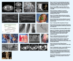
A 75-year-old female presented to the emergency department with acute onset abdominal pain. Laboratory evaluation revealed leucocytosis (white blood cell count: 15,000/µL) and elevated serum lactate levels (3.8mmol/L). An abdominal computed tomography (CT) scan was performed, demonstrating early signs of small bowel ischaemia (Figure 1) secondary to acute occlusion of the middle third of the SMA (Figure 2).
The patient was scheduled for a percutaneous mechanical thrombectomy (thromboaspiration) of the SMA.
 Anatomical and access challenges
Preliminary evaluation of the patient’s vascular anatomy revealed a very acute origin angle of the SMA (Figure 3), along with tortuosity of the iliac axis (Figure 4); both features represented unfavourable factors for a femoral approach.
Rationale for TRA
In this selected case, TRA was chosen because it offered a more favourable coaxial approach from above. Preliminary ultrasound evaluation of the right radial artery demonstrated an adequate calibre (2.8mm), with proper collateral flow from the ulnar artery confirmed by a normal collateral circulation assessment, including Allen test and ultrasound evaluation.
Procedure
The right radial artery was punctured under ultrasound guidance (Figure 5), and a 6Fr Radifocus™ (Terumo) radial sheath was placed (Figure 6).
Although a sheath-to-artery ratio greater than one is a known predictor of transient radial artery spasm and vascular wall stress, interventional cardiology experience—particularly with 8Fr sheathless guiding catheters—has demonstrated feasibility in selected patients when appropriate vessel assessment, sheathless techniques and anticoagulation strategies are employed.4
In this case, a weight-adjusted dose of 5,000IU of unfractionated heparin was administered immediately after TRA was secured to prevent thromboembolic complications and catheter-induced thrombosis. Notably, owing to the preparation of the vessel with the positioning of the 6Fr radial introducer, the seamless profile of the sheathless system and meticulous catheter manipulation, no spasmolytic agents (such as nitroglycerin or verapamil) were required in this case, and the procedure was completed without any clinical or angiographic evidence of radial artery spasm.
To allow the placement of a long sheath into the radial artery, a highly supportive 0.035-inch Radifocus™ Glidewire Advantage™ hydrophilic coated guidewire (Terumo; Figure 7) was deployed, enabling the exchange for a 100cm Neuron MAX 088 long sheath (Penumbra).
Catheterisation of the descending thoracic aorta was then performed using a 5Fr Berenstein catheter (Merit) and the Radifocus™ Glidewire Advantage™ 0.035-inch guidewire, positioning the guiding catheter at the proximal segment of the SMA (Figure 8).
Preliminary mesenteric angiography confirmed the occlusion (Figure 9), demonstrating poor collateral compensation.
Multiple sessions of SMA thromboaspiration were performed using a RED 72 reperfusion catheter (Penumbra; Figure 10), removing abundant thrombotic material (Figure 11).
Superselective aspiration sessions of the jejunal branches were also carried out over a 0.014-inch Radifocus™ Glidewire Advantage™ guidewire (Figure 12), in an attempt to reopen the branches that were still occluded.
Outcome
The final angiographic control documented successful restoration of patency in the mesenteric artery, with a persistent sub-occlusive defect at a jejunal branchpoint (Figure 12), which showed good distal collateralisation. The catheters were therefore removed, and haemostasis of the TRA site was achieved using a TR Band™ compression device (Terumo; Figure 13).
The patient underwent a follow-up CT scan 10 days after the procedure, which showed proper intestinal reperfusion without signs of ischaemia (Figure 14–15) and complete restoration of SMA patency (Figure 16). During hospitalisation, the identification of new-onset atrial fibrillation reinforced the embolic origin of the SMA occlusion and informed subsequent medical management.
Key learning points
In this case, the combination of a supportive 0.035-inch guidewire and vessel preparation with a radial sheath facilitated transradial deployment of an 8Fr guiding catheter. This tailored endovascular approach may provide a valuable alternative to femoral access in selected anatomical and clinical scenarios, enabling successful performance of complex visceral interventions while minimising access-site complications.
Case report: Hepatic bleeding
Teixidor demonstrates the importance of performing elective cases that require TRA, helping to build experience that may save valuable time when femoral or groin access is not obtainable in an emergency setting.
Clinical presentation
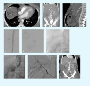
A 35-year-old man presented following a motorcycle accident with a Glasgow Coma Scale (GCS) score of 13 (Figure 1). Orotracheal intubation was performed and, upon arrival at the hospital, it was determined that the patient had an unstable pelvic fracture. A subsequent X-ray determined pubic symphysis diastasis associated with a fracture-dislocation of the left sacroiliac joint, for which a pelvic binder was inserted.
The patient was identified with a 500mL hemoperitoneum and hepatic laceration, which was treated with liver and preperitoneal packing. Sigmoid perforation was also identified, requiring sigmoid resection.
Although bleeding in the liver was brought under control, the patient remained unstable and so underwent a subsequent CT scan. This showed critical laceration of the hepatic parenchyma (Figure 2) and rupture of the common hepatic artery.
 Anatomical and access challenges
Due to the severe pelvic fracture, the presence of a pelvic binder, and limited access to the groin, femoral access was not feasible. In addition, the angle between the coeliac trunk and the aorta was very sharp, making it difficult to approach from below through the femoral artery.
Rationale for TRA
At this point, the patient was brought to the interventional radiology (IR) suite. Given the anatomical challenges (Figure 3) and limited groin access, TRA was employed.
Procedure
Ultrasound-guided puncture of the radial artery was performed and a 6Fr long sheath was inserted. Advancing then to the proximal abdominal aorta, an arteriogram was performed, which confirmed the source of the bleeding (Figure 4).
The coeliac trunk was then catheterised, as well as the hepatic artery, and the arterial rupture was crossed (Figure 5). Once crossed, a 6mm Viabahn VBX covered stent (Gore) was preferred in order to rapidly exclude the arterial rupture while maintaining hepatic arterial perfusion (Figure 6–7).
Outcome
The final arteriogram showed that the artery was patent, bleeding had resolved and that the patient was haemodynamically stable (Figure 8). The sheath was then removed, and manual compression of the radial artery puncture site was performed.
The patient underwent a follow-up CT scan one day later—as per protocol—which confirmed the patency of the stent and that no complications were associated with the procedure (Figure 9).
Key learning points
In this case, there are two particularly important learning points: the first is that, when you have an unstable patient and a focused assessment with sonography in trauma (FAST) ultrasound shows free blood inside the abdomen, the correct approach is to take the patient to surgery. However, if the surgeon cannot identify the cause of the instability, or if they identify it but cannot completely resolve it, then the next step is to take the patient to the IR suite.
Secondly, when clinically feasible, CT imaging can help accurately identify the source of bleeding and facilitate procedural planning.
TRA was particularly useful in this scenario because the angle between the coeliac trunk and the aorta was very sharp, making it difficult to approach from below through the femoral artery. Additionally, because of the pelvic trauma, the patient had a pelvic binder in place, making establishment of an optimal sterile femoral access field more challenging; when implanting a covered stent, a completely sterile area is essential.
To conclude, performing radial artery access in daily practice is useful for later application during emergency procedures. This is particularly true in patients with unfavourable arterial ostia (such as celiac trunk or SMA), or in patients who require a pelvic binder—which is common in any patient who has suffered blunt trauma.
Conclusion
Radial and femoral approaches are both safe and effective for trauma patients, with femoral access remaining an important complementary option, as not all emergency visceral interventions are suitable for TRA. Procedure selection should consider operator experience, radial artery calibre, device length and compatibility, as well as adequate collateral flow. The two cases presented here illustrate how TRA may provide distinct advantages in selected emergency scenarios, whether through improved anatomical alignment or when femoral access is limited. Importantly, routine exposure to radial techniques in elective cases may help operators develop familiarity that can be valuable when TRA is required in selected emergency scenarios.2,5
References