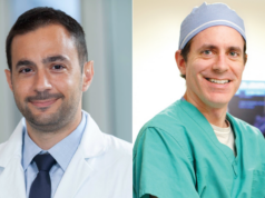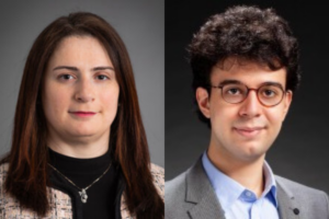
Artificial intelligence (AI) in radiology has moved past the era of experimental novelty into a phase of structural necessity. In interventional oncology (IO), AI applications span the entire pre-procedural, intra-procedural, and post-procedural continuum. Here, Dania Daye and Mustafa Ege Seker (both University of Wisconsin, Madison, USA) survey the most exciting clinician uses of AI, which, as implementation develops, positions clinicians as orchestrators rather than operators of the technology.
In the pre-procedural phase, AI enables departments to create highly efficient schedules, potentially reducing patient wait times and increasing procedure room utilisation; patient triaging and expedition of urgent/emergent cases, minimising patient wait times and improving access to care.1–4 Furthermore, AI models can rapidly and accurately segment anatomical structures, pathologies and critical vessels from cross-sectional imaging modalities and register between modalities.5–7
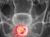
AI is revolutionising planning with tools like voxel-based personalised dosimetry for Y-90 radioembolization, which calculates toxicity thresholds to optimise tumoricidal doses while sparing healthy tissue.8,9 Needle-based procedures, benefit from improved safety and accuracy by AI, with real-time guidance and compensation for patient movement intraoperatively.10 We are witnessing the transition from qualitative estimation to quantitative precision. For thermal ablation, for instance, the recent COVER-ALL trial has demonstrated that software-based margin assessment is superior to our traditional visual checks, offering a real-time safety net that verifies technical success before the patient leaves the table.11
Similarly, AI-driven treatment response and risk stratification is becoming increasingly vital. AI models showed promising results for prediction of treatment response and overall survival with transarterial chemoembolization (TACE) in patients with hepatocellular carcinoma (HCC).12,13 Similarly, computed tomography (CT) texture analysis combined with machine learning has shown promise in improving post-ablation prognostication for patients with adrenal metastases.14
Despite these recent advances, the transition from experimental novelty to clinical necessity is hindered by significant structural barriers. Clinical implementation faces challenges regarding workflow integration and the ‘black box’ nature of AI models, which limits clinician trust without robust explainability.15 Generalisability remains a major hurdle due to the scarcity of diverse, well-annotated datasets that may lead to algorithmic bias across different patient populations.16 Post-deployment monitoring is essential yet difficult to implement, requiring continuous oversight to ensure algorithms do not drift in performance over time.17 Furthermore, the rise of foundation models introduces the risk of generative hallucinations, where AI creates plausible but factually incorrect medical details, necessitating strict human verification.18 We must be vigilant against automation bias and adhere to emerging reporting standards to ensure these tools function as supportive clinical partners.
Looking ahead, the true revolution lies in the ‘consultation room’ rather than the operating theater. Generative AI has emerged as a powerful bridge for health literacy. We are now able to utilise AI tools to translate dense, jargon-filled procedural reports into narratives readable by all, demystifying care for our patients.19 It allows us to bridge language barriers and anxiety gaps that human providers, constrained by time, often struggle to span.
Beyond text-based communication, immersive technologies such as extended reality (XR) are being integrated with AI to provide embodied, visual explanations of anatomy and surgical options. These platforms also allow patients to visualise their tumour and the planned intervention in a 3D space, which has been shown to lower anxiety levels and reduce decisional conflict. This visual literacy complements verbal communication, providing a more holistic understanding of the patient journey.20,21
As we integrate these new tools into clinical workflows, our role evolves from operator to orchestrator. We are moving toward a ‘human-on-the-loop’ model where we leverage computational power to see the invisible and predict the unpredictable. The question is no longer if AI will change our practice, but how we will govern that change to ensure it serves our patients first. The future of IO is not automated; IO with AI will lead to augmented and better patient care.
References
- Ranschaert, E., Topff, L. and Pianykh, O. (2021) ‘Optimization of radiology workflow with artificial intelligence’, Radiologic Clinics of North America, 59(6). https://doi.org/10.1016/j.rcl.2021.06.006
- Gaddum, O. and Chapiro, J. (2024) ‘An interventional radiologist’s primer of critical appraisal of artificial intelligence research’, Journal of Vascular and Interventional Radiology, 35(1), pp. 7–14. https://doi. org/10.1016/j.jvir.2023.09.020
- Petry, M., Lansky, C., Chodakiewitz, Y., Maya, M. and Pressman, B. (2022) ‘Decreased hospital length of stay for ICH and PE after adoption of an artificial intelligence-augmented radiological worklist triage system’, Radiology Research and Practice, 2022, 2141839. https://doi.org/10.1155/2022/2141839
- Batra, K., Xi, Y., Bhagwat, S., Espino, A. et al. (2023) ‘Radiologist worklist reprioritization using artificial intelligence: Impact on report turnaround times for CTPA examinations positive for acute pulmonary embolism’, American Journal of Roentgenology, 221(3), pp. 324–333. https://doi.org/10.2214/ AJR.22.28949
- Wasserthal, J., Breit, H.C., Meyer, M.T. et al. (2023) ‘TotalSegmentator: Robust segmentation of 104 anatomic structures in CT images’, Radiology: Artificial Intelligence. Published online 5 July. https:// doi.org/10.1148/ryai.230024
- Xu, J., Dong, A., Yang, Y. et al. (2025) ‘VSNet: Vessel structure-aware network for hepatic and portal vein segmentation’, Medical Image Analysis, 101, 103458. https://doi.org/10.1016/j.media.2025.103458
- Fang, X., Xu, S., Wood, B.J. et al. (2020) ‘Deep learning-based liver segmentation for fusion-guided intervention’, International Journal of Computer Assisted Radiology and Surgery, 15(6), pp. 963–972. https://doi.org/10.1007/s11548-020-02147-6
- Garin, E., Tselikas, L., Guiu, B. et al. (2024) ‘Long-term overall survival after selective internal radiation therapy for locally advanced hepatocellular carcinomas: Updated analysis of DOSISPHERE-01 trial’, Journal of Nuclear Medicine. Published online 11 January. https://doi.org/10.2967/jnumed.123.266211
- Jia, Y., Li, Z., Akhavanallaf, A., Fessler, J.A. et al. (2023) ‘90Y SPECT scatter estimation and voxel dosimetry in radioembolization using a unified deep learning framework’, EJNMMI Physics, 10, 82. https:// doi.org/10.1186/s40658-023-00598-9
- Matsui, Y., Ueda, D., Fujita, S. et al. (2025) ‘Applications of artificial intelligence in interventional oncology: An up-to-date review of the literature’, Japanese Journal of Radiology, 43(2), pp. 164–176. https://doi.org/10.1007/s11604-024-01668-3
- Odisio, B.C., Albuquerque, J., Lin, Y.M. et al. (2025) ‘Software-based versus visual assessment of the minimal ablative margin in patients with liver tumours undergoing percutaneous thermal ablation (COVER-ALL): A randomised phase 2 trial’, The Lancet Gastroenterology & Hepatology, 10(5), pp. 442–451. https://doi.org/10.1016/S2468-1253(25)00024-X
- Morshid, A., Elsayes, K.M., Khalaf, A.M. et al. (2019) ‘A machine learning model to predict hepatocellular carcinoma response to transcatheter arterial chemoembolization’, Radiology: Artificial Intelligence, 1(5), e180021. https://doi.org/10.1148/ ryai.2019180021
- Kim, J., Choi, S.J., Lee, S.H., et al. (2018) ‘Predicting survival using pretreatment CT for patients with hepatocellular carcinoma treated with transarterial chemoembolization: Comparison of models using radiomics’, American Journal of Roentgenology, 211(5), pp. 1026–1034. https://doi.org/10.2214/ AJR.18.19507
- Daye, D., Staziaki, P.V., Furtado, V.F. et al. (2019) ‘CT texture analysis and machine learning improve post-ablation prognostication in patients with adrenal metastases: A proof of concept’, Cardiovascular and Interventional Radiology, 42(12), pp. 1771–1776. https://doi.org/10.1007/s00270-019-02336-0
- Saw, S.N., Yan, Y.Y. and Ng, K.H. (2025) ‘Current status and future directions of explainable artificial intelligence in medical imaging’, European Journal of Radiology, 183, 111884. https://doi.org/10.1016/j. ejrad.2024.111884
- Simon, B.D., Ozyoruk, K.B., Gelikman, D.G., et al. (2025) ‘The future of multimodal artificial intelligence models for integrating imaging and clinical metadata: A narrative review’, Diagnostic and Interventional Radiology, 31(4), pp. 303–312. https://doi. org/10.4274/dir.2024.242631
- Jiang, S., Bukhari, S.M.A., Krishnan, A. et al. (2025) ‘Deployment of artificial intelligence in radiology: Strategies for success’, American Journal of Roentgenology, 224(2), e2431898. https://doi. org/10.2214/AJR.24.31898
- Das, A.B., Sakib, S.K. and Ahmed, S. (2025) ‘Trustworthy medical imaging with large language models: A study of hallucinations across modalities’, in Proceedings of the IEEE/CVF International Conference on Computer Vision Workshops (ICCVW), pp. 1265–1272. Available at: https://openaccess. thecvf.com/content/ICCV2025W/CVAMD/html/ Das_Trustworthy_Medical_Imaging_with_Large_ Language_Models_A_Study_of_ICCVW_2025_paper. html
- Tripathi, S., Mutter, L., Muppuri, M. et al. (2025) ‘PRECISE framework: Enhanced radiology reporting with GPT for improved readability, reliability, and patient-centered care’, European Journal of Radiology, 187, 112124. https://doi.org/10.1016/j. ejrad.2025.112124
- Saccenti, L., Huth, H., Varble, N. et al. (2025) ‘Augmented interventional radiology via augmented reality’, Journal of Vascular and Interventional Radiology, 36(12), pp. 1937–1944.e2. https://doi. org/10.1016/j.jvir.2025.09.010
- Evans, T., Turna, A., Stringfellow, T.D. and Jones, G.G. (2025) ‘Uses of augmented reality in surgical consent and patient education – A systematic review’, PLOS Digital Health, 4(4), e0000777. https:// doi.org/10.1371/journal.pdig.0000777
Dania Daye is an associate professor of radiology at the University of Wisconsin School of Medicine and Public Health, Madison, USA and Mustafa Ege Seker is a research fellow at the University of Wisconsin-Madison, Madison, USA.