Rotarex™S: A 25-year legacy continues

This advertorial, sponsored by BD, is only available in selected countries and geographies. [geoip-content country=”AT, BE, BG, HR, CY, CZ, DK, EE, FI, DE, GR, HU, IE, IT, LV, LT, LU, MT, NL, PL, PT, RO, SK, SI, CH, ES, SE, GB”]

This year marks a significant milestone as BD celebrates 25 years of its RotarexS rotational atherothrombectomy system, a journey that started in the Swiss mountains with Immanuel Straub, and has today touched the lives of over 250,000 patients. In this article, Vascular News outlines the heritage and history of the device and presents a case report highlighting its continued real-world application and outcomes.

The story of RotarexS begins in the early 1990s, when Immanuel Straub, a Swiss engineer and founder of Straub Medical, teamed up with a medical doctor to create a tool to treat peripheral arterial disease (PAD). Straub combined knowledge from Swiss watchmakers (specialists in micro engineering) and Swiss mountain tunnellers (with their ability to break through the toughest materials) and along with his own expertise in creating high-quality and high-durability springs for the car industry, they came up with a concept for the treatment of arterial blockages. This concept became RotarexS.

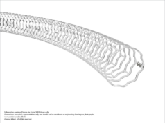
The RotarexS rotational atherothrombectomy system is designed to remove both thrombus and plaque in acute to chronic arterial occlusions. It is indicated for use in peripheral arteries for native blood vessels or vessels fitted with stents, stent grafts or native or artificial bypasses. With four functions, the device is designed to detach occluding material from the vessel, aspirate detached material into the catheter head, fragment the aspirated material and transport it out of the patient’s body. The device is available in three size options: 6F, 8F and 10F.

Case report

Mark Portou

Mark Portou (Royal Free London NHS Foundation Trust, London, UK) has extensive clinical experience with the RotarexS and here shares with Vascular News a case report demonstrating the effective use of the device. Specifically, the consultant vascular surgeon, along with vascular specialty registrar Katherine Hurndall, outline utilisation of the RotarexS rotational atherothrombectomy catheter in an occluded deep-vein reconstructed aorto-bifemoral bypass graft.

Procedural overview

A 41-year-old female patient presented with a fourday history of sudden onset severe right leg pain, worsening paralysis, and progressive paraesthesia. She had undergone an autologous deep venous aortoiliac reconstruction 18 months prior, following the explantation of an infected dacron aorto-bifemoral bypass. This was performed for symptomatic occlusive disease. In addition, an emergency right common femoral (CFA) to superficial femoral artery (SFA) long saphenous vein (LSV) jump graft and fasciotomies were performed in the days following the explant and deep vein reconstruction, following an acute graft occlusion. She had continued to smoke.

She presented tachycardic with fixed mottling below the knee. There was no motor or sensory function and no pulses throughout the right leg. All pulses were palpable on the left side. A computed tomography (CT) angiogram demonstrated an occlusion from the origin of the right neo-iliac venous conduit at the anastomosis with the native aorta, throughout the length of the graft, in the LSV jump graft conduit, the native SFA throughout its length, with contrast reconstituted in the right profunda femoris (PFA), just distal to the origin (Figures 1, 2 and 3).

Figure 1: Preoperative CT angiogram at the level
of the origin of the bilateral deep venous grafts.
Figure 2: Preoperative CT angiogram
demonstrating occlusion of the right limb of the
deep vein aorto-iliac bypass graft.
Figure 3: Preoperative CT angiogram highlighting
contrast in the right native profunda femoris.
Surgical clips are visible on the occluded
autologous LSV jump graft.

The patient was counselled regarding the likely unsalvageable nature of the right lower leg, and concerns regarding the ability of the remaining limb to heal post-amputation were discussed. Open surgical options to restore inflow and enable a successful amputation were considered, but avoided due to extensive scarring, previous infection, lack of covering tissue and the presence of an underlying vein jump graft.

An open left brachial cut down for 6Fr-sheath access was performed, along with an ultrasoundguided percutaneous retrograde right popliteal puncture using a micropuncture sheath. Through-andthrough access was obtained following wire passage from the antegrade left brachial sheath (Figure 4) through the soft thrombotic occlusion and the LSV conduit, with rendezvous in the proximal native SFA. A through-and-through approach was taken in case of perforation of either vein graft or disruption of the surgical anastomosis, to enable balloon occlusion or rapid covered stent repair (Figure 5).

Figure 4: DSA image from 6F brachial access sheath. Short stump of right limb of deep venous conduit visible. Patent left side as seen on the CTA.
Figure 5: Antegrade-retrograde through-and-through access obtained via a left brachial approach and retrograde right popliteal access.

The V18 wire (Boston Scientific) was exchanged for the 0.018 Rotarex wire through a micro catheter. The 6F RotarexS rotational atherothrombectomy catheter was deployed from the antegrade left brachial sheath with a pressurised heparinised saline flush. A full mechanical thrombectomy of the neo-aorta iliac graft, the CFA jump graft, the PFA and SFA was performed (Figure 6). Bolus doses of intra-arterial catheter directed thrombolysis were targeted down the PFA and SFA.

Figure 6: 6F RotarexS catheter passed antegrade from long brachial sheath into the occluded right graft limb over the externalised 0.018 RotarexS wire.

Conclusion

Flow into the PFA and SFA was achieved, with restoration of the right femoral pulse. Patency of the right deep vein and LSV jump grafts was confirmed on angiography (Figure 7). She subsequently underwent a successful amputation, which healed without complication and after rehabilitation is now able to walk up and down stairs independently with a prosthetic limb.

Figure 7: Catheter-directed DSA demonstrating
complete clearance of the thrombotic occlusion
in the deep vein aorto-iliac graft and restoration
of flow through the profunda femoris and
superficial femoral artery through the LSV
jump graft.
Figure 8: Follow-up CT angiogram demonstrating
ongoing patency in both aorto-iliac graft limbs.

Visit the BD website to find out more about the evolution of the RotarexS system and register to get exclusive access to RotarexS workshops, designed to deepen your understanding and skills with rotational atherothrombectomy technology.

Disclaimer: The opinions and clinical experiences presented herein are for informational purposes only. The results from this Advertorial may not be predictive for all patients. Individual results may vary depending on a variety of patient specific attributes. The clinician has been compensated by Becton, Dickinson and Company to participate in this advertorial. BD Switzerland Sarl, Terre Bonne Park – A4, Route De Crassier, 17, 1262 Eysins, Vaud. Switzerland, Tel: +41 21 556 30 00. Fax: +41 44 722 5370. bd.com. BD, the BD logo and Rotarex are the property of Becton, Dickinson and Company or its affiliates. © 2024 BD. All rights reserved. bd.com. Please consult product labels and instructions for use for all indications, contraindications, hazards, warnings and precautions. BD-117405


[/geoip-content]


LEAVE A REPLY

Please enter your comment!
Please enter your name here