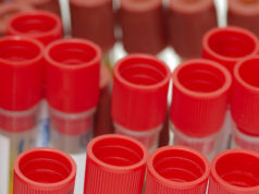
Where is liquid biopsy today? Bruno Damascelli, Vladimira Tichà, Giancarlo Beltramo, Alberto Gramaglia and Gianluigi Patelli write on the complexities and challenges of the procedure as it gains ground among select interventional radiologists.
Liquid biopsy is gaining momentum on its path toward clinical utility, but the current role of this important advance in oncology is difficult to pinpoint. While it is easy to obtain a blood sample, molecular analysis of tumour biomarkers is still remarkably complex. The aim is to identify the genetic mutations of cancer in the biological markers it releases into blood. The most widely used of these is circulating tumour DNA (ctDNA) which is released into circulation primarily through apoptosis and cell necrosis. In the blood, ctDNA is mixed with cell-free DNA (cfDNA) which is produced in larger quantities because of cell turnover in normal tissues. This means ctDNA must be separated by an extraction process, which is followed by genetic sequencing—known as next generation sequencing (NGS)—covering a variable number of genes chosen according to their frequency in tumours. The cases presented here used Guardant360 (Guardant Health) comprehensive genetic profiling which reports the amount of ctDNA as a percentage of cfDNA.
The greatest difficulty in molecular analysis is the scarcity of ctDNA detectable in the general circulation, which has given rise to various methods of obtaining a significant amount of this biomarker. The simplest method, investigated by us and confirmed by another research group, is to draw venous blood in proximity to the tumour site. Percutaneous venous catheterisation is part of the armamentarium of interventional radiologists and relies on their familiarity with navigating the vascular system under fluoroscopic guidance. Sampling sites can be chosen as dictated by the natural history of the disease, which includes both primary and metastatic sites.
Unlike tissue molecular diagnosis, which is inevitably restricted to a few sections, liquid biopsy provides a wider reflection of the tumour’s epigenetic heterogeneity. Other advantages of selective sampling compared with conventional peripheral sampling are that it avoids the ctDNA dilution that occurs in the general circulation, and it exploits the topographical contribution of lymphatic drainage, a leading route of cancer spread. Lymph from the left hemithorax, the entire abdomen and the lower limbs drains into the thoracic duct, which empties into the left venous angle. Sampling from this site can provide information about intra- and extra-peritoneal cancers. Lymph from the right hemithorax and part of the head and neck drains into lymphatic trunks that empty into the right venous angle, where venous sampling provides more effective molecular characterisation than peripheral sampling.
Liquid biopsy is beginning to be considered for use in diagnosis of lung nodules (Case one) for which no convenient solution is offered by imaging methods or percutaneous biopsy, which can prove inconclusive or unfeasible because of the associated risks.

Radiosurgery is increasingly used in cancer treatment, sometimes as the only option, with a consensus for its use in the absence of histological examination. Liquid biopsy can confirm the diagnosis and assess treatment response or resistance during follow-up. A liquid biopsy showing a mutation consistent with glial neoplasm (Case two) is decisive not only when histological diagnosis is not possible but also for monitoring treatment in the event of recurrence or progression. Molecular diagnosis has a higher success rate on cerebrospinal fluid than on peripheral blood but repeated lumbar puncture is not without risk for the patient. Retrograde percutaneous catheterisation of the jugular veins is an outpatient procedure known since its use in the diagnosis of pituitary tumours.

Another application of selective venous sampling is in ocular cancers. Uveal melanoma (Case three) can express mutations predicting disease evolution. Follow-up relies on abdominal ultrasound to detect hepatic metastases, which are invariably detected too late for effective treatment. Sampling from the jugular vein on the melanoma side allows molecular characterisation of the tumour, predicting its behaviour and guiding the use of new drugs with recent, promising results. Until now, this tumour has been treated with radiotherapy based on a diagnosis made with physical and radiological means applied to the intact eye.

Prostate adenocarcinoma (Case four) affects 16% of the male population. Early diagnosis of this cancer has had a positive effect on survival but metastatic spread leads to a long-lasting deterioration in quality of life. Peripheral sampling or selective sampling from the hypogastric veins can provide timely detection of the somatic mutations involved in resistance to radiotherapy or androgen suppression, guiding appropriate personalised treatments.

Liquid biopsy is not yet recognised for its diagnostic accuracy, clinical utility and patient benefit, and few molecular tests are approved by regulatory bodies. While it is unrealistic to imagine that liquid biopsy could replace histological examination, it is nonetheless true that in monitoring the evolution of a tumour and its sensitivity or resistance to treatment, blood sampling is always possible, whereas tissue biopsy often is not.
References:
Tichà V., Patelli G., Basso G. et al, Case Report: Potential role of selective venous sampling for liquid biopsy in complex clinical settings: Three case presentations, Frontiers, https://doi:10.3389/fgene.2023.1065537.
Tamrazi A., Sundaresan S., Gulati A. et al, Endovascular image-guided sampling of tumour-draining veins provides an enriched source of oncological biomarkers, Frontiers, https://doi:10.3389/fonc.2023.916196.
Saife N. Lone, Sabah Nisar, Masoodi T. et al., Liquid biopsy: a step closer to transform diagnosis, prognosis and future of cancer treatments, Molecular Cancer, https://doi:10.1186/s12943-022-01543-7.
Bruno Damascelli is an interventional radiologist at Fondazione Falciani ONLUS, Milan, Italy.
Vladimira Tichà is an interventional radiologist at Fondazione Falciani ONLUS, Milan, Italy
Giancarlo Beltramo is head of radiosurgery at Centro Diagnostico Italiano, Milan, Italy.
Alberto Gramaglia is head of radiotherapy at Monza General Hospital, Monza, Italy.
Gianluigi Patelli is head of radiology at Casa di Cura San Francesco, Bergamo, Italy.


