
 In an exciting session at the Charing Cross Symposium (CX; 24-27 April, London, UK), the audience witnessed the demonstration of a novel treatment strategy with intravascular lithotripsy for calcified arterial stenosis. It will now become a CX tradition to feature live cases to showcase what has been discussed in the presentations, so that the audience can observe the evidence given in practice.
Giving some background to the concept of intravascular lithotripsy, Andrew Holden, Auckland, New Zealand, who performed the first-in-human intravascular lithotripsy procedure, explained, “the basic concept is to create apposition of the angioplasty balloon to the vessel wall at very low pressure to perform lithotripsy, to create microfragmentation of the calcium in both the medial and intimal layers, change the compliance of the artery and then allow the balloon to dilate out to nominal diameter at low pressure and hopefully, with a low instance of residual stenosis or dissection.”
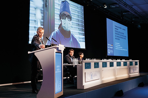
The live case was broadcast from Bad Krozingen, Germany, with operator Thomas Zeller.
The patient was an 84-year-old female with Rutherford stage 3 claudication, with a history of coronary vessel disease and internal carotid artery stenosis. Previously, the patient had an occlusion of the right superficial femoral artery, extending into the popliteal artery, with significant calcification that was treated with lithotripsy and drug-coated balloon angioplasty, after which she had been pain free on her right leg.
In the procedure broadcast at CX, Zeller used lithotripsy to prepare the vessel in the patient’s left leg for drug-coated balloon angioplasty using the protocol from the DISRUPT III randomised controlled trial which is comparing lithotripsy plus drug-coated balloon to drug-coated balloon as standalone therapy.
Providing some tips based on his experience with the lithotripsy device, Zeller placed a lot of importance on the preparation of the catheter. “In every balloon there is still some air trapped and it is important to evacuate the air from the folded balloon before you inflate it and before you try to apply the sonic pressure waves because if there is still air trapped in the balloon, the pressure waves will not be transmitted to the vessel wall,” he said.
Further, pulling from lessons learned in the DISRUPT I and DISRUPT II trials, Zeller advised that the balloon should be slightly oversized in order to improve acute outcomes. “Undersizing leads to inferior outcomes acutely and also in the longer term. Similarly, increasing the dose, i.e. doing more lithotripsy cycles in the same position, does improve the overall outcome of the lithotripsy procedure.”
Describing how the device is used by the operator during the procedure, he explained, “the device has seven markers in total. The markers at the edges represent the proximal and distal balloon shoulders and the five markers in-between are the emitters. It is important to position the emitters in the lesion area and the balloon marker can be outside.”
Watching the case, Holden commented that the lithotripsy is a dose that is delivered to refractory stenoses to try to improve the acute outcome—Zeller then added, “the specific feature of lithotripsy is that you disrupt medial calcification.”
As for his strategy following intravascular lithotripsy, Zeller reported that at his centre, it is followed up with a drug-coated balloon. “That is our general strategy, because, based on what we learned from the pilot study, lithotripsy is a very good tool for achieving good acute outcomes, but in the long-term we still see restenosis development. This leads me to believe the combination of vessel preparation with lithotripsy followed by drug-coated balloon angioplasty is a logical approach to treat these lesions. DISRUPT III will prove whether it is true or not,” Zeller stated.
Polling following the live case showed that 50% of the CX 2018 audience believes that calcified tibial vessels are best managed with a drug-coated balloon following vessel preparation during endovascular procedures. Further, 69% of the CX 2018 audience would not use a drug-coated balloon for a heavily calcified lesion without vessel preparation.
A focus on calcification
Broadening the discussion on calcification, Arne Schwindt, Münster, Germany, weighed in with his personal experience, adding, “lesions that are unresponsive to stenting and high pressure ballooning could be treated. We have seen acceptable clinical and angiographic outcome in rock-like calcified common femoral arteries. Early experience shows promising results of intravascular lithotripsy in highly calcified small mesenteric and celiac branches, and has the potential to make EVAR/TEVAR/TAVI feasible in patients with poor iliac access.”
In her assessment, Renu Virmani, Gaithersburg, USA, called for a “dedicated device for calcified lesions that either cracks or removes calcified areas in order to achieve adequate vessel expansion. Probably, such lesions need drug-eluting stents rather than drug-coated balloons.”
In the end, Fabrizio Fanelli, Florence, Italy, concluded that calcium significantly increases procedural complications and that most studies have excluded severely calcified lesions from endovascular treatments. He said that acute and long-term endovascular outcomes are inferior when severe calcium is present, and that it represents a great limitation of superficial femoral artery procedures due to vessel recoil after balloon dilatation, flow-limiting dissection related to high pressure use and a higher stent implantation rate. “Multiple technologies are available nowadays but ‘the ideal’ is not available yet. Atherectomy devices increase the luminal gain and may also improve drug uptake. Further, vessel preparation with plaque scoring and lithotripsy do not debulk calcium, but are able to increase vessel permeability, drug absorption, and patency rate.”


