

A prototype wearable device, tested in animal models, can continuously collect live cancer cells directly from a patient’s blood. Proposed as an alternative to biopsy, the system allows for the analysis of larger blood volumes, leading its developers to write in Nature Communications that “this technology could potentially be used to analyse larger number[s] of circulating tumour cells to facilitate translation of analytical information into future clinical decisions”.
Developed by a team of engineers and doctors at the University of Michigan (Ann Arbor, USA), it could help doctors diagnose and treat cancer more effectively.
“Nobody wants to have a biopsy. If we could get enough cancer cells from the blood, we could use them to learn about the tumour biology and direct care for the patients. That is the excitement of why we are doing this,” says Daniel F Hayes, the Stuart B Padnos professor of breast cancer research at the University of Michigan Rogel Cancer Center and senior author of the Nature Communications paper.
Cancerous tumours can release more than 1,000 circulating tumour cells (CTCs) into the bloodstream in a single minute. These have become an established biomarker for prognosis in patients with various carcinomas, with elevated levels of CTC isolated from a single blood draw being indicative of metastatic breast, colorectal, prostate, or lung cancers. Current methods of capturing CTCs from blood rely on samples from the patient—usually no more than a tablespoon taken in a single draw. Some blood draws come back with no cancer cells, even in patients with advanced cancer; an average 7.5mL sample from a patient with metastatic breast cancer contains no more than 10 cancer cells, identified within the context of billions of erythrocytes and millions of leukocytes.
Over a couple of hours in the hospital, the new device—a portable aphaeretic system—could continuously capture CTCs directly from the vein, screening much larger volumes of a patient’s blood. In animal tests, the cell-grabbing chip in the wearable device trapped 3.5 times as many cancer cells per millilitre of blood as it did running samples collected by blood draw.
“It is the difference between having a security camera that takes a snapshot of a door every five minutes or takes a video. If an intruder enters between the snapshots, you would not know about it,” explains Sunitha Nagrath, associate professor of chemical engineering at the University of Michigan, who led the development of the device.
Research shows that most cancer cells cannot survive in the bloodstream, but those that do are more likely to start a new tumour. Typically, it is these metastases that are deadly, rather than the original tumour. This means CTCs captured from blood could provide better information for planning treatments than those from a conventional biopsy. Indeed, the investigators of the present proof-of-principle study hypothesised that “CTC evaluation might be used for early detection of malignancy, if an assay with sufficient sensitivity and specificity could be developed.”
The team tested the device in dogs at the Colorado State University’s Flint Animal Cancer Center (Fort Collins, USA) in collaboration with Douglas Thamm, a professor of veterinary oncology and director of clinical research there. They injected healthy adult animals with human cancer cells, which are eliminated by the dogs’ immune systems over the course of a few hours with no lasting effects.
For the first two hours’ post-injection, the dogs were given a mild sedative and connected to the device, which screened between 1–2% of their blood. At the same time, the dogs had blood drawn every 20 minutes, and the cancer cells in these samples were collected by a chip of the same design.
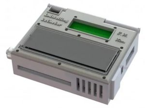
The device shrinks a machine that is typically the size of an oven down to something that could be worn on the wrist and connected to a vein in the arm. For help with the design, the engineering team turned to Laura Cooling, a professor of clinical pathology at the University of Michigan and associate director of the blood bank, where she manages the full-size systems.
“The most challenging parts were integrating all of the components into a single device and then ensuring that the blood would not clot, that the cells would not clog up the chip, and that the entire device is completely sterile,” comments Tae Hyun Kim, who earned his doctorate in electrical engineering in the Nagrath Lab and is now a postdoctoral scholar at the California Institute of Technology (Pasadena, USA).
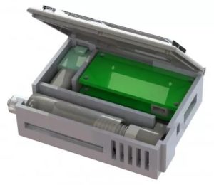
Each component of the device is integrated into a compact 3D printed, portable structure, and is controlled through a custom built mobile application via wireless communication. The system consists of four major parts: a micro-controller, peristaltic pump, heparin injector, and a CTC capture module that contains a microfluidic CTC capture chip.
The investigators developed protocols for mixing the blood with heparin, to prevent clotting, and sterilisation methods that killed bacteria without harming the antibodies on the chip. Kim also packaged some of the smallest medical-grade pumps in a 3D-printed box with the electronics and the cancer-cell-capturing chip.
The chip itself is a new twist on one of the highest-capture-rate devices from Nagrath’s lab. It uses the nanomaterial graphene oxide to create dense forests of antibody-tipped molecular chains, enabling it to trap more than 80% of the cancer cells in whole blood that flows across it. The chip can also be used to grow the captured cancer cells, producing larger samples for further analysis.
In the next steps for the device, the team hopes to increase the blood processing rate. Then, led by Thamm, they will use the optimised system to capture cancer cells from pet dogs that come to the cancer centre as patients. Chips targeting proteins on the surfaces of canine breast cancer cells are under development in the Nagrath lab now.
Hayes estimates the device could begin human trials in three to five years. It would be used to help to optimise treatments for human cancers by enabling doctors to see if the cancer cells are making the molecules that serve as targets for many newer cancer drugs.
“This is the epitome of precision medicine, which is so exciting in the field of oncology right now,” says Hayes.


")


