

Kevin Stadtlander outlines how his institution is benefitting from the “creativity” of interventional radiologists during the coronavirus pandemic, here providing a step-by-step account of a bedside inferior vena cava (IVC) filter placement for a COVID-19-positive patient in intensive care.
A 38-year-old male with past medical history of hypertension, admitted for complications of COVID-19, required mechanical ventilation and extracorporeal membrane oxygenation (ECMO). The patient developed an intracranial parenchymal haemorrhage felt to be related to ECMO, with fluctuating haemodynamics, while on intravenous (IV) heparin protocol. After removal of the ECMO venous cannula from the right groin, the patient was also found to have right iliac vein and femoral deep venous thrombosis (DVT), as well as below-knee DVT. The patient was referred to interventional radiology (IR) for inferior vena cava (IVC) filter placement, with a contraindication to systemic anticoagulation due to his intracranial bleed.
Given the ongoing COVID-19 pandemic, we had already implemented a plan that included having a designated IR procedure room in the department. Our IR plan included the directive to try to do as much as possible at beside in COVID-positive patients. For this IVC filter placement, we quickly mapped out both options, and decided to take the care directly to the patient room.
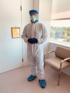
We located a portable C-arm with digital subtraction angiography (DSA) capability and did a trial in an empty intensive care unit (ICU) room to ensure that the C-arm would fit under the bed, and that images would be clearly visible through the ICU bed. These goals were both accomplished, but it did necessitate maximum ICU bed height to fit the C-arm. The COVID patient room was negative pressure and separated from the hallway (considered dirty) by sliding glass doors. Since no true anteroom was available, we used the empty ICU room as a staging room across the hall. The entire procedure plan was first reviewed in a huddle with all team members. We chose to have one IR physician and two radiology technologists in the room for the procedure. We were also accompanied by IR nurses (outside the COVID room), who helped with staging and acted as buddies for donning and doffing. The sterile procedure tray for the IVC filter was made up in a second empty ICU room and covered. Informed consent was obtained via telephone from a family member.
The IR physician and two technologists donned their personal protective equipment (PPE) and lead aprons in the staging room with the help of the nursing buddies. The C-arm, the C-arm monitor, and the ultrasound unit were covered with a plastic barrier, and then taken into the COVID-positive patient’s room by the “dirty” technologist (who was wearing full PPE, excluding sterile surgical gown and gloves). The second technologist (wearing full PPE and designated as procedure scrub technologist) then wheeled the procedure tray into the room, put on her surgical gown and gloves, and began prepping the patient’s left groin. We chose the left groin due to a thrombus in the right iliac vein, and in order to stay away from the head of the bed. The IR physician then entered the room, put on a sterile gown and gloves over full PPE, and helped to complete patient preparation. Importantly, a step stool (covered in plastic) was used by the physician during the procedure, as bed height necessary for C-arm made this a requirement. Sign in/time out was performed, IVC filter placement was completed in 10 minutes, and sign out was performed. Throughout the procedure, the patient’s ICU nurse monitored the patient and controlled his IV sedation via drips that were outside the room as per COVID protocol.
The surgical scrub gowns and top layer of gloves were removed in the normal fashion in the room and discarded. The procedure tray breakdown was quickly performed, and all items were disposed of in the room trash. Plastic wraps were removed from equipment and discarded in the room as each unit was moved from room to hallway. Each unit was terminally cleaned with X-ray machine bleach wipes. Each of the three caregivers then performed careful doffing of PPE (with our nurse buddies using a checklist) at the door threshold, ultimately leaving only N95 mask and bottom layer eye goggles on as they exited back to the staging room. Lead aprons were removed and wiped down with a lead X-ray apron cleaner disinfectant. N95 mask and goggles were then removed, and caregivers were free to head to the surgical locker room for scrub change and shower. A debrief huddle with all team members was then performed.
This case illustrates how IR creativity allows for procedures to be performed “portably” at the bedside in a manner safe for both caregivers and patients, while having the added benefit of preserving PPE. By way of follow-up, this patient ultimately came off the ventilator, became COVID-negative, and a CT brain has since demonstrated resolution of the intracranial bleed. The patient has been discharged from hospital.
Kevin Stadtlander, MD is an interventional radiologist at the Cleveland Clinic Florida, Weston, USA.


