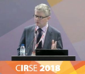
Researchers report the use of ultrasound to detect embolization in real time for patients undergoing treatment for benign prostatic hyperplasia (BPH). Richard Owen (University of Alberta, Edmonton, Canada) presented these results at the Cardiovascular and Interventional Radiological Society of Europe (CIRSE) annual meeting (22–25 September, Lisbon, Portugal), demonstrating the clear visibility of the embolic agent Occlusin 500 Embolization Microsphere (OCL 503) within 24 hours of the prostatic artery embolization (PAE) procedure.
The study investigators set out to evaluate the safety and effectiveness of the embolic product OCL 503 in PAE for the treatment of men with BPH. The trial was small; 10 patients with moderate to severe lower urinary tract symptoms secondary to BPH were treated in this open label, single centre pilot study. A standard embolization technique in PAE was used: bilateral embolization using a 2.8F microcatheter. “A 2.8F microcatheter was required, because the embolic product is not compressible, and does not pass through a smaller microcatheter,” Owen said of this choice. The catheter position was confirmed with a cone beam CT at the time of the procedure.
Patients were imaged using a Philips Model IU22 ultrasound imaging system with a transabdominal approach before the procedure, within 24 hours of PAE, and at three months’ follow up.
Owen commented: “These particles [the embolic agents] are clearly visible in all of these patients, in the prostatic tissue, within 24 hours of embolization. A blinded observation of the ultrasound images was able to identify patients that had a bilateral or unilateral embolization by the presence of these particles. Qualitative assessment of the signal intensity within 24 hours of PAE correlated with the number of microspheres delivered to the prostatic tissue. As part of this study, we knew how many microspheres we delivered, and we knew in which patients ,of course. Within three months, none of the particles were visible.
“I am not sure what the clinical role will be, but it [the embolic agent] is clearly visible in the embolized prostate, and it is in fact visible in other tissues,” Owen continued. “I am not at liberty to discuss this here, but it is visible in other embolized tissues, as you would expect. That relates to the density; the density is higher than that of normal soft tissue, and that is why you get such a profound reflection from ultrasound.”
According to the study investigators, the echogenicity relates to the amount of embolic delivered. Owen explained at CIRSE: “It [ultrasound imaging] may well provide a useful tool at the time of embolization to confirm the entirety of embolization. We used to use some other measures post-procedure, CT, for example, in post-procedure imaging, to see where the emboli are, but we could do a real time ultrasound here to see if we have reached an adequate endpoint—but it would require more work.”
Owen concluded his talk by expressing his gratitude to his mentor: “I did visit Portugal several years ago, and I learnt this technique from professor Martin Pisco (Hospital Saint Louis, Lisbon, Portugal), so thanks to him for starting the ball rolling in much of the world.”
A CIRSE attendee asked Owen if he had any experience with contrast enhanced ultrasound, which the questioner claimed to find “extremely useful” in the immediate monitoring of embolization. Owen replied: “You can do that, of course, although it requires the addition of a different agent; the intravenous injection of a contrast. However, there could be other factors, for example spasm in the vessel, that might affect that endpoint [immediate imaging of emboli]. The use of OCL 503 and ultrasound imaging is not affected by those endpoints; it would reflect the actual embolic material in the tissue.”
The embolic agent
OCL 503 is a biodegradable microsphere, comprised largely of poly (lactic-co-glycolic acid) or PLGA, a bioresorbable suture material. It is a Class II medical device approved by the US FDA for use in hypervascular tumours, and is currently pending CE mark approval, as well as clearance for use in Canada. Owen described the microsphere: “It comes as a dry powder, similar to other embolics, and it is reconstituted with contrast—Isobuoyant with Omnipaque 240 in this case.” OCL 503 is eliminated from the body in three to six months, as has been evidenced in a previous animal study published in Cardiovascular and Interventional Radiology. The microspheres degrade to carbon dioxide and water, and have a density of 1.1g/ml (in comparison to the 1.03g/ml of liver tissue, and 1g/ml of water).
This research forms part of an ongoing trial investigating the OCL 500 series in PAE and uterine artery embolization, sponsored by IMBiotechnologies. Owen is also a recipient of the Alberta Innovative Grant, and is a part-time consultant for Cook Medical.


