
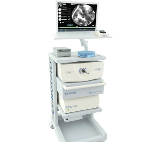
 What if physicians could perform an optical biopsy (defined by Thomas D Wang, associate professor, Gastroenterology, University of Michigan, USA, as one that uses the properties of light to enable the operator to make an instant diagnosis at endoscopy)? Further, what if during a cryoablation procedure, an interventional radiologist can actually see what happens during the procedure at a cellular level in real time? A new tool, Cellvizio (Mauna Kea) does just that.
A study presented at the European Conference on Interventional Oncology (ECIO, 17–20 April, Dublin, Ireland) reported preliminary data on the safety and feasibility of performing endomicroscopy during percutaneous image-guided procedures such as biopsy or ablation of tumours in the liver or kidney. The study was titled confocal laser endomicroscopy for microscopic characterisation of kidney and liver tumours during in vivo percutaneous biopsies and ex vivo cryoblation. Julien Garnon, Strasbourg, France, told delegates that the team are also creating an atlas of images using confocal laser endomicroscopy and that they are correlating these with histology.
As reported at ECIO, the study protocol involved CT positioning of a 17g coaxial needle in the lesion; insertion of the endoscopy probe; injection of 2.5ml of fluorescein; video recording of the tissue using confocal laser endomicroscopy; obtaining histological samples using an 18g semi-automatic needle; video recording of the sample using endomicroscopy; and analysis of endomicroscopy and samples by a pathologist in order to correlate them.
Garnon stated that in the ongoing study, there were no adverse events related to the endomicroscopy procedure. “There was a satisfactory or good image quality obtained in 7/10 cases (2/10 were not evaluated at the time of presentation). The mean endomiscroscopy recording time per procedure was 11.3 minutes and the mean number of endomicroscopy sequences per procedure was 13. There was one technical failure,” he said. He further clarified that there was no histological diagnosis done using confocal laser endomicroscopy in the study, but noted that this is possible.
Garnon concluded: “In vivo confocal laser endomicroscopy is feasible and safe. However, we are radiologists and not pathologist and there is a learning curve that is associated with this procedure. It has several potential applications: for instance in needle biopsy, the technology could help achieve better target biopsies and improve the diagnostic yield by enabling real-time microscopic tissue characterisation. For ablation procedures, it can ensure better control and completeness of ablation, thus reducing recurrence rates while preserving functional tissue. In chemoembolization procedures the use of confocal laser endomicroscopy could ensure that the chemical agent reaches the centre of the tumour and can help to select the appropriate particle size.”
Afshin Gangi, Oncological Interventional Radiology lead, King’s College London, London, UK, head of the department of Interventional Radiology, Université de Strasbourg, Strasbourg, France and chairman of Radiology and Nuclear Medicine, CHU Strasbbourg, told Interventional News: “The tool was developed for endoscopists, but has now become available to us. It allows us to visualise tissue in grey scale. We can actually see individual cells (but not their nuclei) and the architecture of the tissue. We are able to see the blood vessels and circulation. As a scientist and researcher, it enables me to place my props or instrument anywhere I want and watch what is happening. While carrying out a biopsy, I can be directly connected to the pathologists in our institution and they can see exactly what we do, and can guide us to the right position where there are viable cells, which greatly improves the position for the biopsy (In fact, pathologists love this tool as they are seeing living tissue for the first time). Further, I can use this to see the environment that I will be working in as an interventional radiologist. My colleague, Pramod P Rao, has done a lot of work on this study.”
Gangi added: “Prior to commencing the treatment, I can see whether the environment is hypovascular and see what the cells look like. For instance, with ablation, I can clearly see the biological effect of treatment. This allows you to histologically monitor the ablation. Is this absolutely necessary? I do not know. The fact is that I have the physical ability to see this and it is amazing to do so. However, you can only see one layer of cells, not any more deeply than that.”
Gangi clarified that this tool can add to the cost of the procedure as it uses extremely specific fibres that are difficult to manufacture, and can add up to 200 Euros for each biopsy.
“It is exciting to see the use of confocal laser endomicroscopy in vascular treatments such as chemoembolization. We placed the needle adjacent to the feeding artery and then advanced it to the centre of the lesion from the periphery to see the distribution of the drug. It was helpful to see the changes occurring in the tumour after embolization. These included the changes in the intercellular matrix, the fluid movement and the changes in the microvasculature. In the cryoablation procedure we could observe the pattern of crystal formation. Every stage of cryoablation could be appreciated in real time from intra and extracellular ice formation to the swelling of cells and cell rupture with complete loss of tumour matrix and pattern. During chemoembolization, this enables us to immediately decide whether the beads are too big and to administer smaller beads, if necessary. It also works with lipiodol chemoembolization, in which you can actually see the drug leaving the lipiodol,” he explained.
“All of this means that you can adapt your treatment intracellularly and really tailor the treatment to the patient. This is not macroscopic visualisation on imaging but microscopic visualisation inside the tissue, or simply in vivo imaging. It opens up a new field of research and imagination,” he concluded.


