
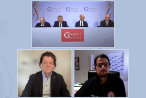
The second day of the CX Aortic Vienna 2022 Digital Edition (24–26 October) saw presentations on dynamic computed tomography angiography (CTA), a device with the potential to bring hologram guidance to vascular procedures and an edited case highlighting the role of intravascular ultrasound (IVUS) intraoperatively in thoracoabdominal branched endovascular aneurysm repair (BEVAR). The multidisciplinary moderating panel, which included a cardiac viewpoint from Heinz Jakob (Essen, Germany), questioned these technological developments for imaging in the post-presentation discussions—in terms of the limited access to them for cost reasons, and whether the reduction in radiation exposure the technologies offer can justify hospitals assuming the financial burden.
The first presentation came from Alan Lumsden (Houston, USA), who demonstrated why dynamic CT imaging might be considered the “new gold standard” for diagnosis and interventional planning. “It is obviously important to choose the right diagnostic tool at the right phase of the treatment modality,” the presenter began, declaring CT as a “very flexible” modality. This means that it can lend itself to dynamic imaging, Lumsden explained—static imaging and the single “view” it offers is sometimes akin to “no view” at all.
Lumsden then detailed how and why time-resolved CTA, developed for cardiac imaging, can be advantageous for use in endovascular aneurysm repair (EVAR) follow-up. The approach involves “moving the patient in and out the scanner, taking snapshots”, which can then be “put together” to form a three-dimensional composite image over time, Lumsden relayed, likening it to a “movie”. This allows the surgeon to see the contrast dye’s flow pattern and how long it takes to “arrive”, meaning “you can work out what type of endoleak you have.”
Furthermore, the presenter asserted that there is no radiation exposure increase as compared to triphasic CTA. His concluding remarks were that dynamic CTA can be applied to “guide subsequent interventions” and can be “widely performed on conventional scanners”.
It was whether this imaging approach can, in fact, be “widely performed” that featured in the discussion that followed. Moderator Markus Steinbauer (Regensburg, Germany) acknowledged its potential as a “valuable tool”, but that he does not currently have access to it in his practice. Anchor Roger Greenhalgh (London, UK) concurred—“we encourage the concept, but it needs to be more widespread”.
Another point the discussion raised was whether the reduced radiation exposure facilitated by dynamic CTA warrants shouldering the financial cost of implementing the requisite technology. For this reason, moderator Tilo Kölbel (Hamburg, Germany) posited that it would “take a while until this is standard practice”.
The result from the interactive audience poll suggested that dynamic CTA could indeed become the gold standard for diagnosis and interventional planning, with 88% of respondents voting in the affirmative.
The edited case presented by Michele Antonello (Padua, Italy), which shed light on intraoperative intravascular ultrasound (IVUS) in thoracoabdominal branched endovascular aneurysm repair (BEVAR), also yielded noteworthy discussion. Fellow presenter Jan Heyligers (Tilburg, The Netherlands) again posed a question on radiation exposure to Antonello—“is it mainly a reduction of radiation that you are aiming at?”—to which the response was that IVUS can “reduce contrast [use] by 30%”. Kölbel queried the impact IVUS has on procedure time, highlighting that this is also a factor when evaluating the cost-effectiveness of the approach as compared to the standard of care.
Heyligers’ own presentation demonstrated the scope for holographic augmented superposition of patient-specific imaging. The device in question, Heyligers suggested, could, with further development, solve the “problem” in vascular surgery of having to interpret three-dimensional information from a two-dimensional display. With smart glasses projecting directly onto the exact location on the patient, Heyligers shared with delegates that thus far, the technology has proved promising in static applications such as in a foot or wrist. Future iterations will be “smarter”, and capable of assisting in a “moving environment”, such as the vasculature, the presenter ventured.
Once more, the hologram technology elicited lively discussion, with Kölbel keen to hear how this could work in endovascular procedures. Heyligers underlined that other technologies could be harnessed “once inside vessels”, reiterating that the holographic capability to “recognise patient-specific surroundings” can help fulfil the “ambition” to have “faster, more precise” procedures, with “less radiation”.
Steinbauer pointed to the potential for applying the technology in educational settings, such as to project guidelines onto a patient to show where and where not to operate. This was an idea with which Heyligers agreed— a “remote experience” is possible, he added, thanks to the device’s internet connectivity, which could allow a seasoned surgeon “to help younger colleagues out” even if they are not in the room.
The thread running through this session on thoracic imaging was that practising at the forefront technological and procedural advancements in vascular surgery does not come without significant financial cost, ultimately constituting a barrier to entry for many centres. The moderating panel emphasised that radiation reduction is a priority when considering whether or not to implement a new procedure and shoulder the cost of the technology required to perform it.


