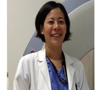
While most radiologists probably do not view a biopsy as being the most exciting or technically challenging procedure within our repertoire, it may be time to give the image-guided biopsy a second look, writes Alda Tam.
The goal of personalised medicine is to deliver “the right drug, for the right patient, at the right dose, at the right time.” This healthcare model is feasible only if an individual’s genetic information can be accessed for decision making. In cancer care, the image-guided biopsy, where tumour tissue is extracted for analysis, has become the mainstay by which patient genetic data is acquired, playing a vital role both in determining standard-of-care therapy options and enrolment onto clinical trials. As the focus for the treatment of oncology patients has shifted towards engaging upon molecular subsets within a cancer type, there has been a corresponding shift in regards to the purpose and utility of image-guided biopsies. It is clear to the oncological community that tissue acquired from a biopsy that is adequate only for histological diagnosis is no longer sufficient. A similar understanding is required of the radiology community as we are responsible for performing the majority of image-guided biopsies.
If personalised cancer care is to become a reality, the current state in which 10–15% of patients do not have biopsy samples sufficient for mutational characterisation by next generation sequencing technologies must change. While most radiologists probably do not view a biopsy as being the most exciting or technically challenging procedure within our repertoire, it may be time to give the image-guided biopsy a second look as it actually can provide tremendous opportunities for collaboration and innovation. Similar to how pathologists assess for specimen adequacy on a fine needle aspiration or evaluate for resection margins intra-operatively, a radiologist can play an active role in medical decision making by selecting a suitable lesion for biopsy. We are the most qualified physician on the healthcare team to interpret the diagnostic images, choose the lesion that is most likely to yield sufficient and biologically relevant tissue for molecular testing, and weigh the risks vs. the benefits of the biopsy procedure for the patient. Communications with the oncology and pathology teams are essential to driving quality improvement for biopsy yield.
In terms of innovation, the fundamental technique of biopsy has changed negligibly over time. This presents a noticeable opportunity for radiologists to change the way biopsies are performed either through device design, or imaging guidance and navigation during tissue acquisition.
Advances in molecular imaging beyond the standard fluorodeoxyglucose positron emission tomography (FDG PET/CT), the emergence of molecular tracers, the maturation of the field of radiogenomics, and the incorporation of optical imaging techniques into the procedure itself for sample verification are some technological developments that have the potential to improve biopsy yield.
The field of oncology today is energised by the multiplicity of novel agents under development and the potential for therapy that is predicated on biomarkers. In the face of this rapidly and constantly moving environment, the tumour tissue acquired from image-guided biopsies will be expected to fulfil multiple and varied analytical objectives. Therefore, it is essential for the radiologist performing image-guided biopsies, whether in an academic or community hospital setting, to realise that every biopsy needs sufficient tissue for molecular markers and that practice changes may be required—if your biopsy routine is just to sample the tumour once or twice, recognise that it may not provide satisfactory material for biomarker testing. The question of biological relevance and clonal evolution, which places the most recently acquired tissue at the forefront of directing next line therapy, is also important and implies that radiologists should expect to see patients undergoing image-guided biopsies at multiple time points throughout the course of their disease. Lastly, we should not be surprised if in the near future there is a revolution in the tools and processes used to perform image-guided biopsies to allow for a tissue sample to be representative of the complete mutational signature of a tumour.
Alda Tam is associate professor, Department of Interventional Radiology, MD Anderson Cancer Center, Houston, USA. Tam is a medical monitor to Galil Medical and receives research support from AngioDynamics


