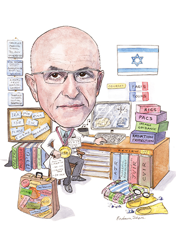
“In the early days, I used to have to cut a 7F plastic tube, make side holes, send it off for sterilisation and shape it using steam just before the procedure. We used to basically manufacture our own disposable equipment,” Gabriel Bartal, director, Department Medical Imaging and Interventional Radiology, Meir Medical Center, Kfar Saba, Israel tells Interventional News. Bartal is also president of the Israeli Society of Interventional Radiology (ILSIR) and alludes to the start-up culture in the country.
What drew you to medicine and interventional radiology?
The short answer is probably my Jewish mother and my older brother. The longer answer involves my interest in human biology and its many applications. My main motivation was looking into new frontiers to facilitate change and help those who need it most.
After medical school, I served in the Israel Defense Forces as a physician. At first, it was very clear to me that I would become a surgeon or a cardiologist. Later, I started to become uncertain about my path in medicine. I shared my doubts with one of my senior colleagues, who told me to read about interventional radiology. I went to the medical library early one morning and started to read about the fascinating field of interventional radiology. I read papers published by the founding fathers and about the evolving field of interventional radiology. By the time I was asked by the librarian to leave late in the evening, I knew I would do a residency in radiology, and the eventual destination was crystal clear—interventional radiology.
What do you remember of the early days in interventional radiology?
Diagnostic vascular interventions like digital subtraction angiography were very challenging because we had to inject intravenously to image the arteries. Later there was an amazing shift to the intra-arterial imaging that we use today.
In the early days, I used to have to cut a 7F plastic tube, make side holes, send it off for sterilisation and shape it using steam just before the procedure. We used to basically manufacture our own disposable equipment. Now, invasive diagnostic angiography has nearly been replaced by noninvasive imaging like Doppler ultrasound, CTA and MRA, and interventional radiology has become a therapeutic specialty in modern medicine. Advanced image guidance techniques eliminate most of the limits of transcatheter therapies and this is just the beginning.
Who were your mentors in the field?
There have been several remarkable radiologists who guided me from the very beginning. Starting from my residency in Tel Aviv Medical Centre, with professor Albert Solomon, who made sure that I would become a “well-balanced radiologist” and not one who merely carries out procedures. Dr Josef Papo taught me the very basics of interventional radiology, dedication, the ability to think out of the box and most important of all, procedural safety.
I was lucky to be accepted for a fellowship in interventional radiology at the Hammersmith Hospital in London, UK. There I was mentored by professor David Allison and professor Andy Adam. The former made sure that I was trained in the various fields of interventional radiology and I used to make a 400-mile round trip around the UK, learning from the leading lights about angioplasty and peripheral interventions. In the USA, professors Frederick Keller and Barry Katzen have been very supportive in shaping my career.
As the current president of the Israeli Society of Interventional Radiology (ILSIR) could you provide an overview of interventional radiology in Israel today?
The most significant recent event that took place was the “CIRSE meets Israel session: Innovations in IR” at CIRSE 2014. This was an initiative of past CIRSE presidents, professor Michael Lee and professor Anna Maria Belli, and was supported by the society’s executive committee. As a start-up country, it was very natural to talk about innovations. ILSIR was established over 10 years ago and has 55 active members who practice in all the fields of interventional radiology. We have 21 recognised interventional radiology units throughout Israel. About a third of ILSIR members are fellowship-trained in the USA, Canada or the UK.
In order to raise the level of interventional radiology and to increase the number of certified interventional radiology fellows in Israel, we have structured a fellowship programme in accordance with the recently ratified SIR Fellowship programme, with the active help of professor John Kaufman. Our goal is to make sure that interventional radiology will dominate in all fields of image-guided interventions. We constantly seek ways to attract more young, gifted doctors into our specialty and the foundation of the fellowship program is one of the milestones in this goal.
Israel is emerging as a key destination for many start-ups and medical device companies…
There is a start-up culture that pervades Israel. We currently have over 5,000 start-up companies, with about 15% annual growth. There are over 1,000 Israeli companies based in healthcare and life-science products, including 700 specialising in medical devices. Approximately half are already generating revenue.
One can wonder how it is that Israel, a country with a population of less than eight million people, only 65 years old, in a constant state of war and with no natural resources is able to produce more start-up companies than large, peaceful, and stable nations such as Japan, South Korea, Canada and the UK?
Many interventional radiology devices including variations of angioplasty balloons, specially designed stents and aortic stent-grafts have been invented, designed and evaluated in Israel. They are manufactured and marketed by major international companies.
You have long been an advocate of protection from radiation. What are the key developments in this area?
Radiation protection and safety is something that everybody speaks about, but not many people really understand. Fluoroscopy is one of the cornerstones of modern imaging and is vital for interventional radiology. Most of the vendors of angiography systems provide dose reduction options to reduce over 70% of patient effective dose, with near-identical image quality. Use of rotational angiography with cone beam CT options will expand and allow safe access and accurate performance.
Normally we use dosimetry badges that are “read” periodically and we know about exposure only in retrospect. In addition to the traditional badges, real-time occupational dose information found in the DoseAware system (Philips) may have a real impact on our practice and lead to necessary behavioural changes.
What essential steps need to be followed to keep exposure to ionising radiation to a minimum?
Interventionalists need a basic understanding of radiation exposure mechanisms. Acceptance and implementation of the radiation safety routine in daily practice is key to safe interventional radiology practice.
Radiation protection measures should be part of the planning of the procedure. For example, perform CT or MR angiography prior to the endovascular procedure and prepare the necessary disposable equipment based on the pre-acquired imaging.
- Use available patient dose reduction technologies (low-fluoroscopy-dose-rate settings, low frame-rate pulsed fluoroscopy, spectral beam filtrationc).
- Keep the X-ray tube as far as possible from the patient and image receptor as close as possible to the patient.
- Adjust collimator blades tightly to the area of interest.
- Use protective shielding and position yourself in a low-scatter area.
- Use all available information to plan the interventional procedure.
- Routinely use power injectors for contrast material injections when feasible.
- Step out of the procedure room during digital subtraction angiography acquisitions.
- Obtain appropriate training in radiation protection and dose management.
Why is simulation important, and what is the latest evidence supporting its adoption in training?
Complex cases treated by interventional radiology requires doctors with profound clinical backgrounds and a high level of technical skills, which have to be constantly re-evaluated and updated—very often on a case-to-case basis. The only reliable, scientifically validated way of developing and improving these skills is through medical simulation. Advanced simulation systems with clearly defined features, can create a feeling of reality and allow the best and most efficient training of the staff. Endovascular training should use simulation that has been shown to be a valid representation of the training objective and provide a realistic replication at an appropriate level of fidelity.
Endovascular simulation training should be tailored to the trainees and become an integral part of their residency and or fellowship. Virtual reality training runs on a computer, which provides an opportunity to collect information on trainee performance. User data can also be used to create critiques and to generate an individual’s learning curve over time as well as compare an individual to a group of colleagues. This gives a clear advantage over non-computer based models. Simulation should comprise both radiation safety and dose management training and become an integral part of the residency and or fellowship.
In your view what are the most important developments in interventional radiology?
We already live in a world that uses volumetric imaging. I believe that the use of fusion imaging with real-time overlay integrated with live fluoroscopy in the angiosuite will become a basic tool in the future.
ν A head-mounted stereo display of the 3D image will allow direct access to any vessel (blood, bile or other) without the need to rotate the C-arm or “fishing” for the vessel’s true lumen.
ν Use of endovascular robotics will evolve and replace the need to stand in the radiation danger zone next to the patient. This advancement will reduce patient dose and improve the accuracy of vessel catheterisation and device deployment because a robot never gets tired thus allowing procedure performance with high precision.
ν Use of tissue or pathology specific nanoparticles will allow targeted drug delivery with minimal if any “collateral damage”.
Can you describe a memorable case that you treated?
I was back from my fellowship to my hospital in Tel Aviv, Israel, and it was in the era when self-expandable metal stents (Wallstent, Boston Scientific) had just been introduced into the UK, following a successful clinical evaluation.
A very famous actor had pancreatic cancer with biliary obstruction and jaundice. At that time only external drainage and plastic stents were available. Consulting this patient I mentioned the possibility of internal drainage using a metal stent without the need for external drainage. The hospital administration did not like the idea of using an expensive stent instead of a simple and cheap tube, but the exemption was made for this famous person and we performed a successful procedure. Several days later the actor was back on the stage and I was invited to a play in a prime seat. He announced to the audience that thanks to our treatment he was able to be back on stage very quickly. Unfortunately, I could not attend the performance as I had to embolize a trauma victim on the same night. The Wallstent then became a routine tool in biliary and later in endovascular interventions.
My fields of interest outside medicine are reading, mostly poetry in languages such as Russian, Hebrew and English. I also enjoy arts, music and sports. I used to enjoy contact sports most of all, but it has recently been mostly swimming, and trekking in nature.
Fact File
Current academic and hospital appointments
2005–present Director, Department Medical Imaging and Interventional Radiology, Meir Medical Center, Kfar Saba, Israel
1992–present Lecturer of Tel-Aviv University postgraduate medical school in interventional radiology
Education
1985–1989 Radiology, Computers in Medicine, Sackler School of Medical Education, Tel-Aviv Univeristy, Israel
1975–1978 Graduated faculty of Medicine, Tel-Aviv University, Tel Aviv, Israel
Postgraduate education (Selected)
1990–1991 Lewis Foundation Scholarship, Royal Postgraduate Medical School, Hammersmith Hospital and Royal Free Hospital, both London, Sheffield General Hospital, Sheffield, UK
1992 Gastroenterology Course for Radiologists, University of Leeds, Leeds, UK
Society memberships:
President, Israeli Society of Interventional Radiology (ILSIR)
Member, Executive Committee Israel Radiological Association
Member, Israel Medical Association
Member, Radiology Society of North America (RSNA)
Member, European Society of Radiology (ESR)
Fellow, North American Society of Interventional Radiology (SIR)
Fellow, Cardiovascular and Interventional Radiology Society of Europe (CIRSE)
Committees
Member, European Working Group for Management in Radiology (EWGMR)
CIRSE representative in Medical Radiation Protection and Training (MEDRAPET)
Member, CIRSE Radiation protection Subcommittee
Member, Israeli National Professional Commission on Radiation Control (INPCRC)


