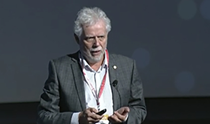
Jim Reekers, Amsterdam, The Netherlands, delivering the Josef Roesch honorary lecture at CIRSE 2015 (26–30 September, Lisbon, Portugal), urged delegates to look at new definitions for critical limb ischaemia and pointed to the potential of perfusion angiography in predicting the outcome of endovascular intervention. “Look past the re-opening of vessels alone,” he said.
Reekers’ lecture was titled “Critical limb ischaemia treatment: Beyond pipe fitting” and he at once set out that the current definition of critical limb ischaemia (defined as rest pain for more than two weeks and/or ulceration and gangrene, and ankle systolic pressure below 50mmHg and/or toe systolic pressure of below 30mmHg in the Second European Consensus Document in 1992) was often inadequate.
“There are problems with this definition as it does not provide clear parameters for the severity of the disease, and cannot be used to identify the patients that need treatment and separate them from those who do not. Also, it cannot be used as a parameter for outcome. Additionally, in diabetic arterial disease, pressure measurements are often unreliable due to severely calcified arteries,” Reekers, professor of Radiology and Interventional Radiology at the University of Amsterdam, noted.
There are other problems with the current definition of critical limb ischaemia; as-yet unpublished meta-analysis data suggest that around 50% of patients classified as having critical limb ischaemia who do not undergo revascularisation will not have an amputation. Further, more than 50% of graft occlusions do not lead to amputation, and 15 to 20% of critical limb ischaemia patients will go on to have amputations despite optimal revascularisation (either after a surgical bypass, or endovascular treatment, for reason not entirely understood, he explained.
“We need a holistic definition of critical limb ischaemia to aid diagnosis but also predict an outcome,” he noted adding that one such definition that holds true might be that if you have critical limb ischaemia, without revascularisation, you will lose your leg.
Reekers also stated that research in critical limb ischaemia tended to focus on proxy endpoints such as target lesion revascularisation rather than endpoints that are important to patients, such as limb salvage.
Current treatment numbers
“Where do we stand in the treatment of critical limb ischaemia today? In studies published in the last decade, [in which either bypass or endovascular treatment were carried out], the limb salvage rates at 12 months are constantly reported to be in the region of 70 to 80%,” he said. Reekers made the point that the “80%” number comprised the nearly 50% who do not lose their leg anyway, irrespective of intervention, and the 30–35% who benefit from revascularisation. The remaining 15% or so went on to lose their leg, despite revascularisation. “Medicine is all about finding the right patients to treat,” he noted.
Reekers then went on to note that people with critical limb ischaemia felt local pain, stiffness of the muscles with reduced movement and nerve paralysis due to the lactate that is formed when there is a shortage of oxygen.
So, critical limb ischaemia can also be defined as “shortage of oxygen in the tissue of the foot,” he said, adding that decreased inflow of blood was a macrovascular parameter while mismanagement of blood in the foot was microcirculation.
“In the microcirculation, oxygen moves through the capillaries by diffusion, and therefore the pressure of blood will be of less consequence than the true volume, or amount of blood. Therefore, oxygen concentration in the tissue is mainly dependent on volume flow through the capillaries, and so critical limb ischaemia is dependent on volume flow through the capillaries,” Reekers said. So, if you measure the volume flow through the capillaries, you can glean information about the amount of oxygen and about subsequent critical limb ischaemia, he noted.
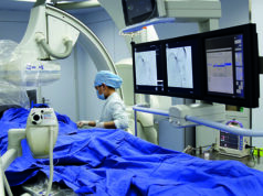
Perfusion angiography, a new imaging modality which is developed by Reekers in cooperation with Philips Medical, measures volume flow in the whole foot, both macro and microcirculation over time.
“Remember the foot is less than 10% macrocirculation and more than 90% microcirculation, and with the technique it is important to measure information from the whole foot, not only the location of the ulcer. Contrast, like oxygen, moves through the capillaries by diffusion and this allows the imaging of flow at the level of the capillaries. Perfusion angiography is post processing software that analyses standard digital subtraction angiograms that are taken as a regular part of the procedure,” he said making the point that no extra contrast or radiation is needed.
Reekers further explained that as the foot was the end organ of peripheral arterial disease, it was important not to concentrate on the ulcer. “The entire foot is diseased and the ulcer is only an expression of that disease. The whole microcirculation of the foot is diseased, not just the angiosome,” he exhorted.
Using perfusion angiography in daily practice
Perfusion angiography might be used to determine a non-angiographic endpoint for endovascular revascularisation by measuring the increase in volume flow through the foot pre- and post- intervention, said Reekers.
“We are currently carrying out a trial to see how much increase in flow is necessary to correlate to a good outcome [for endovascular intervention],” he said in reference to the PALI-study, a multicentre study being carried out in six European centres with 120 patients.
Another use of perfusion angiography in critical limb ischaemia is to test the functionality of the microcirculation of the foot in critical limb ischaemia.
Reekers clarified that this was “absolutely breath-taking” because it can possibly be used to predict outcome and to select patients. “Selective vasodilation of the capillaries will lead to increased microcirculation flow by shunting at the microcirculation level. Quantification of this response to local stimulation is a parameter for the functionality of the microcirculation. This is calculated as the capillary resistance index (CRI), he said.
Describing findings from very preliminary data that Reekers emphasised was not “good science”, but very interesting nevertheless, he reported a retrospective, consecutive subgroup analysis of 21 patients. Comparing the outcomes of 10 patients who underwent revascularisation (two by bypass) to 11 who had no treatment and with their being an equal number of diabetics in both arms, Reekers stated that there were seven early amputations (around 30%) seen in the group. When all the 21 patients were analysed in terms of capillary resistance index, it was seen that six had a CRI of more than 0.9 and 15 had a CRI of less than 0.9. Interestingly, six of the seven early amputations were from the group with CRI>0.9, while the remaining one was from the group with CRI<0.9, showing that CRI was a strong predictor of outcome. “This could bridge the gap between patency and clinical improvement in critical limb ischaemia,” Reekers pointed out.
“So, perfusion angiography could help to determine a non-angiographical endpoint for revascularisation and could preselect patients, with functional imaging who will benefit from revascularisation at the end of a revascularisation procedure,” Reekers concluded, while again cautioning that these are very early data and stating that prospective data are needed to further support these findings.
He emphasised that accurate standardisation of perfusion acquisition parameters is absolutely “essential” for the technique to work. “Good research is mandatory to prevent uncontrolled opportunism and premature clinical introduction,” he added.


