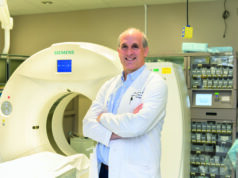
With a focus on magnetic resonance imaging (MRI)-guided procedures and cancer signalling pathways which promote cancer resistance to ablation, David Woodrum (Mayo Clinic, Rochester, USA) discusses how advancements in MRI have solidified its place in the diagnosis and treatment of prostate cancer.
Focal prostate ablation continues to challenge the standard paradigm for prostate cancer treatment. Prostate cancer was the most common cancer in men in 2024, with 299,010 new cases and 35,250 deaths in the USA alone.1 Standard treatments include surgery or radiation therapy, but no matter how expertly done, these therapies carry significant morbidity and risk to the patient’s quality of life, with an impact on sexual, urinary, and bowel function.2 Furthermore, screening programmes using prostatic-specific antigen (PSA) level and transrectal ultrasound-guided systematic biopsy have identified increasing numbers of low risk, low grade “localised” prostate cancer.
With this, prostate focal therapies with ultrasound and MRI guidance have become available, fuelling the debate on the suitability of focal or regional therapy for patients with low or intermediate risk prostate cancer. Some of the largest unresolved issues include prostate cancer multifocality, suboptimal staging with accepted imaging modalities, limitations of current biopsy strategies, and, most important, whether focal therapies can be safely and effectively used for prostate cancer.3
Advancements in MRI have made it the platform of choice for identifying prostate cancer, whether focal or multifocal, and in disease staging.4 Targeted biopsy of suspected cancer lesions detected with MRI is associated with increased detection of high-risk prostate cancer and decreased detection of low-risk prostate cancer, particularly with the aid of MRI/ultrasound fusion and direct in-bore biopsy platforms.5
Focal prostate ablation is often considered for patients with low to intermediate risk prostate cancer who may not require radical whole gland treatments like surgery or radiation therapy. Many focal ablation methods can be guided and monitored with ultrasound or MRI. Ultrasound is the most common imaging modality for guiding and monitoring focal ablation, but it cannot reliably depict the prostate cancer target and has limited capability to real-time monitor during the ablation. Although MRI requires more specialised hardware and training, it provides the most robust targeting and monitoring imaging guidance platform. Use of real-time MRI enables temperature mapping, allows for real-time visualisation of tissue heating, as well as real-time precise imaging of the ice ball as it forms in the case of cryoablation. It is important to note that long-term outcomes of MRI-guided focal prostate cancer ablation compared to traditional treatments are still areas of ongoing research and evaluation.
References
- Siegel RL, Miller KD, Wagle NS, et al. Cancer statistics, 2023. CA: a cancer journal for clinicians. 2023;73(1):17-48.
- Potosky AL, Davis WW, Hoffman RM, et al. Five-year outcomes after prostatectomy or radiotherapy for prostate cancer: the prostate cancer outcomes study. J Natl Cancer Inst. 2004;96(18):1358-67.
- Onik G, Vaughan D, Lotenfoe R, et al. “Male lumpectomy”: focal therapy for prostate cancer using cryoablation. Urology. 2007;70(6 Suppl):16-21.
- Spektor M, Mathur M, Weinreb JC, et al. Standards for MRI reporting-the evolution to PI-RADS v 2.0. Transl Androl Urol. 2017;6(3):355-67.
- Siddiqui MM, Rais-Bahrami S, Turkbey B, et al. Comparison of MR/ultrasound fusion-guided biopsy with ultrasound-guided biopsy for the diagnosis of prostate cancer. JAMA. 2015;313(4):390-7.
David Woodrum is a professor of interventional radiology at the Mayo Clinic in Rochester, USA.
Disclosures: The author declared no relevant disclosures.


