
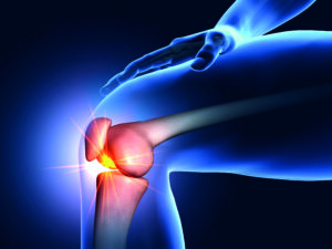
 Musculoskeletal (MSK) embolization has steadily gained ground as a minimally invasive option for chronic joint and tendon pain in selected patients. Once primarily associated with interventional oncology (IO) and haemorrhage control, embolization has been applied to conditions such as osteoarthritis (OA), tendinopathies and adhesive capsulitis, with early studies suggesting meaningful pain reduction.
In a session held at the recent Interventionell Radiologisches Olbert Symposium (IROS; 14–17 January, Salzburg, Austria), several speakers described the growing biological rationale underpinning the technique and reviewed available data, emphasising its potential to “break the cycle of pain” in patients with chronic MSK conditions.
Opening the session, Clayton Kraft (Helios Hospital Krefeld, Krefeld, Germany) provided an orthopaedic viewpoint, stating that pain is often wrongly considered a “minor” issue.
“If you look at everyday clinical practice in an orthopaedic setting, you see a very large number of tendinopathies and joint problems—not infections or tumours, but degenerative joint disease. Lower back pain is one of the biggest groups, but many patients present with joint pain or tendon insertion pain.”
“Mechanically driven joint pain often begins with malalignment or trauma,” Kraft continued. “This leads to irritation or synovitis, meaning joint inflammation, although we don’t always know exactly what ‘inflammation’ means at the microscopic level. Over time, cartilage damage progresses, leading to bone-on-bone contact, and eventually OA and pain.”
In his description of tendinopathy, Alexander Loizides (Medical University Innsbruck, Innsbruck, Austria) characterises the pain cycle as initiated by the “activation of macrophages” and the subsequent release of inflammatory mediators. This process leads to the development of neoangiogenic factors and the ingrowth of new vessels into the tendons and tendon sheaths. Loizides further notes that this progression involves a “neural component, or a neural cascade,” which significantly contributes to pain generation.
Within this context, the primary objective of embolization is to disrupt the abnormal microvascular supply in the affected tissues. As Loizides explains, the goal is to “interrupt this vicious cycle, breaking the cascade.”
Positioning MSK in the treatment algorithm
Presenters across the session were clear that embolization is not considered first-line therapy, instead positioning it within a “stepwise treatment algorithm” between conservative management and surgical intervention, as Kraft stated during his presentation.
Kraft described treatment for OA and tendinopathy as following a “pyramid model”, beginning with physiotherapy, medication and activity modification, and progressing to injections and other minimally invasive therapies, reserving surgery as the “ultima ratio”.
He continued, defining transarterial embolization in this context as a “minimally invasive, joint-preserving option” for patients who have persistent symptoms despite prior conservative treatment. Kraft added that typical candidates are those experiencing ongoing pain for at least six months with significant impact on quality of life.
Equally, speakers across the IROS 2026 session emphasised that careful patient selection is critical, particularly in younger patients or those who are still candidates for corrective surgery. “Mechanical problems, for example a loose body or a meniscal tear requiring repair, must be treated mechanically,” said Kraft, adding that infections are also a “key contraindication” which is particularly relevant in prosthetic joints.
“Without a control arm, interpretation is difficult, particularly when endpoints are patient-reported outcomes such as pain or function scores” – Clayton Kraft
Upper extremity embolization: Non-linear improvement
While early clinical experience focused largely on knee OA, presenters highlighted that embolization is now being explored in other regions, including adhesive capsulitis of the shoulder, symptomatic rotator cuff disease, Achilles tendinopathy, plantar fasciitis and selected cases of persistent pain following joint replacement.
Speaking on shoulder applications, Christoph Binkert (Medizinisch Radiologisches Institut, Zürich, Switzerland) observed that “night pain is usually dominant at baseline” and often improves relatively quickly after embolization, whereas improvements in range of motion may occur more gradually. Binkert made clear that, particularly in cases of frozen shoulder, patients must be counselled on expectations regarding pain relief and functional recovery, as he stated that “improvement occurs, but can take time”.
Binkert noted that pain reduction in this area is often “substantial” and can provide long-term relief, but “effects are not always linear”. Additionally, he added that knowledge of the vascular anatomy in upper extremity cases is “crucial” for safe embolization, as there are multiple small branches at risk requiring careful technique to avoid non-target embolization.
Larger studies needed
Speakers reported that available studies—largely single-arm and observational—suggest that embolization can lead to substantial reductions in pain scores and improvements in quality-of-life measures. Binkert conveyed that approximately 70% of patients show meaningful clinical response in several series and meta-analyses involving the upper extremities.
As Kraft noted during his presentation, the main issue is lack of control groups in these investigations, stating that “without a control arm, interpretation is difficult, particularly when endpoints are patient-reported outcomes such as pain or function scores”.
Throughout the IROS session, speakers called for larger, multicentre, randomised studies to strengthen the available evidence base, to more clearly define the magnitude and durability of treatment effect in chronic MSK conditions.
Federico Collettini (Charité Universitätsmedizin Berlin, Berlin, Germany), who presented on the current evidence for knee joint embolization, stated that a significant area in need of larger datasets is persistent pain after joint replacement.
“This is an important topic that is close to my heart,” he stated, describing how a significant number of patients continue to experience pain following treatment. “We have developed an algorithm: first exclude all other causes: infection, loosening, malalignment, mechanical problems. If everything is excluded and pain persists, embolization of hypervascularised synovium can be considered,” Collettini said.
To date, there are prospective, single-centre studies showing “significant symptom improvement” with this treatment, but more data are needed to confirm this, he said. Roughly 70% of patients achieve clinically meaningful improvement of symptoms after this treatment, Collettini detailed, which is often defined as a ≥50% pain reduction. Although follow-up data show pain reduction persisting at one and two years in many cases, he emphasised that no therapy works in 100% of patients.
A “shift” toward resorbable particles
Later in the session, Loizides outlined the clinical shift toward using resorbable particles for MSK embolization rather than permanent embolic agents. He highlighted that studies—most notably those by Yuji Okuno et al—have demonstrated favourable outcomes, showing significant pain reduction in the treatment of tendinopathy.
“For tendinopathies, we use imipenem almost exclusively,” Loizides explained, noting its ability to form irregular microparticles that effectively provide occlusion of pathological vessels. A defining characteristic of imipenem is its rapid resorption, with near-complete dissolution typically within two hours.
Loizides further emphasised that meticulous selective catheterisation is paramount, particularly in anatomical regions like the Achilles tendon, which often features a dual arterial supply.
“The core technique involves selective angiography to pinpoint specific areas of hyperaemia within the Achilles tendon,” he noted. “For instance, while a posterior tibial artery injection may show no clear hyperaemia, selective catheterisation of the fibular artery can reveal marked hypervascularity. Following selective embolization, the completion angiogram typically demonstrates near-complete devascularisation.”
MRI: Necessary or not?
Each of the speakers highlighted the importance of imaging and its central role in identifying target areas, yet, in Kraft’s view, imaging findings are not always a clear indicator of the severity of the patient’s condition. He explained that imaging can show severe radiologic OA, for example, with little pain reported by the patient, whereas mild imaging findings can be accompanied by severe pain.
Binkert underlined that contrast-enhanced magnetic resonance imaging (MRI) and MR angiography can help to identify hypervascularised synovial or capsular tissue, and when hyperaemia is present, he added that embolization can be “helpful”. Collettini noted that although contrast-enhanced MRI can help differentiate OA phenotypes and potentially refine patient selection, routine implementation in clinical practice remains challenging in many settings due to cost and limited access.
More to come
Speakers highlighted MSK embolization as a promising but developing technique, emphasising the heterogeneous duration of treatment effect and pain reduction from patient to patient. “We should not expand the armamentarium blindly”, said Kraft, contending that embolization is a useful tool to introduce slowly.
While early clinical results suggest encouraging pain relief in selected patients, speakers stressed the need for rigorous trials to confirm its efficacy. In closing, Collettini stated that therapy decisions in this patient population are “highly individual”, requiring cautious consideration of symptoms and patient quality of life reports.


