By John Kaufman
On 16 January 1964, Charles Dotter (who was then chairman of Radiology at the University of Oregon Medical School) performed the first recorded angioplasty in the world when he used progressively larger catheters to dilate a distal superficial femoral artery stenosis [Fig 1A and Fig 1B]. The patient was an elderly woman with rest pain and gangrenous toes who had only been offered amputation of her foot.
Her rest pain abated, her toes auto-amputated, and she kept her foot until her death two years later from a myocardial infarction. Dotter had suggested that angiographic catheters could be used as surgical instruments the previous year during a lecture in the then Czechoslovakia. Why he chose this patient to be his first, and what her told her before and after the procedure is lost to us now. However, when he dilated that stenosis, he initiated an era of innovation and invention in image-guided intervention that has changed the patient experience, altered how medicine is practised, led to the creation of new industries, and improved our understanding of many diseases. An estimated 60 million patients have undergone vascular angioplasty or stent placement (another one of Dotter’s original ideas) over the past 50 years. Many tens of millions more have undergone other image-guided interventions such as embolizations, drainages, and ablations.
The world in which Dotter worked was one in which image-guided interventions required ingenuity, creativity, and great bravery. Catheters were initially handmade from plastic tubing for each patient using blow-torches, steam, hole punches, and scalpels. There were no standardised shapes or dimensions, such that each new anatomy encountered would generate a new catheter shape. The catheters were difficult to see fluoroscopically, and challenging to control. Guidewires were crudely constructed, lacked coatings or safety wires, and stiff. There were no haemostatic sheaths, balloons, microcatheters, coils, or any of the tools we now take for granted. Even the most basic interventions were dramatic, sometimes risky procedures. Nevertheless, doctors and patients together accepted uncertainty, and image-guided interventions flourished.
Early partnership with industry was absolutely essential to the development of image-guided interventions. Without the support of the medical device industry, image-guided interventions could never have progressed. The personal relationships of early interventionalists with individuals such as William Cook, John Abele, and Peter Nicholas were key, as these visionaries shared an enthusiasm for and belief in this developing approach to treatment. In the very different regulatory environment of that time, a new device could be described by a physician, fabricated by a company, sterilised, and used in a patient within weeks [Fig 2]. With few dedicated tools and devices, the pace of innovation and invention was intense. Procedures and devices were created as each new clinical problem was encountered.
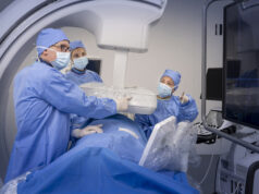
One of the most remarkable aspects of the early interventionalists was their ability to do so much with such crude imaging equipment. This required a close partnership between interventionalist, technologists, and other support staff [Fig. 3]. Techniques that are considered the standard of care today, such as digital subtraction angiography, ultrasound, and CT, were decades away. All images were recorded on film that needed to be developed after each acquisition, enforcing a slower pace to procedures. Positioning patients, setting technique, placing filters and grids, and injection rates were all art forms. Reliable film changers that allowed rapid exposure of a series of films over the course of an injection of contrast were major advances. The contrast agents were ionic and high osmolar, which was painful to patients and resulted in more frequent contrast reactions. Procedures were performed with minimal physiologic monitoring, often without nurses.
Initially, most procedures were performed by radiologists, as they had the best access to equipment. Few non-radiologists had an interest in image-guided interventions, as the techniques were mysterious, the procedures relatively rare compared to conventional surgical methods, and their true roles in medicine undefined.
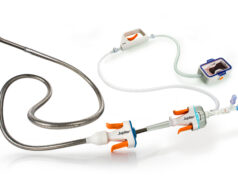
Fifty years later, the landscape of image-guided intervention is almost unrecognisable compared to 1964. Devices are incredibly sophisticated, and now routinely combine drug or energy delivery with the device.
They are readily available and much easier to use, with wide choices in everything from access needles to closure devices. Many different specialties routinely utilise image-guided interventions for diagnosis and treatment. Imaging equipment is fast, almost automated, entirely digital, and able to merge multiple modalities. The range of diseases and patients treated has expanded far beyond the vascular space, and now includes almost every organ but the skin. Whole classes of once standard surgeries have been replaced by image-guided procedures, such as the transjugular intrahepatic portosystemic shunts (TIPS), originated by another Oregon pioneer in intervention, Josef Rösch [click here to read a personal memoir of Charles Dotter by the 87-year-old legend Josef Rösch]. This made surgical decompression of the portal venous system in cirrhosis a rare operation.
Medicine has also changed dramatically during this time. The level of data required to support adoption of a new device or image-guided procedure extends far beyond one person’s report of successful outcomes. To introduce a disruptive procedure such as angioplasty today would require extensive pre-clinical bench-top and then animal testing, tiered safety and efficacy trials in ultimately large numbers of probably randomised patients, cost-effectiveness analyses, quality of life assessments, and determination of long-term outcomes. Several different regulatory agencies would be required to approve not only the devices used for the procedure, but payment. Once introduced, post-market surveillance would be likely. Dotter’s report of the first angioplasty triggered a chain of events that are impossible today.
Although we live in a world that is vastly different to that of the Dotter and the early pioneers in image-guided interventions, this area of medicine remains one of intense excitement, innovation, invention, and creation of new knowledge. Where we will end up in the next 50 years with image-guided interventions will probably be just as different to us as today would be to Dotter.
To acknowledge the 50th anniversary of the first angioplasty, a unique symposium will be held in Portland, Oregon, on 23 and 24 July 2014 [click here to find out more on the Image-guided Intervention: The next 50 years (IGI 50) symposium]. The goal of this meeting will be to look at the future of image-guided interventions from all perspectives. The speakers and attendees will not only be multispecialty and international, but from all disciplines and professions that are involved in image-guided interventions. Leaders from medicine, industry, government, payors, healthcare administration, education, innovation, public health, and academics will engage in moderated discussions in which the audience will be able to participate using an adaptation of social media. The interactive format will push all present to think outside of current parameters and comfort zones, whether discussing training, new technologies and procedures, measurement of outcomes, payment, unmet needs of patients and diseases, implementation of image-guided interventions in developing health care systems, and barriers and opportunities in innovation.
There are great challenges in image-guided interventions today, but also great potential. Growth will continue to be explosive, but it will be different than that of the last 50 years. Great advances will come from the interdisciplinary application of imaging, technology, and pharmacology to deliver image-guided interventions that improve healthcare and lead to better understanding of diseases. In this manner the durable creativity and energy that surrounds image-guided interventions portends even greater successes in the future.
John Kaufman is director, Dotter Interventional Institute and Frederick S Keller Professor of Interventional Radiology, Oregon Health & Science University Hospital, Portland, USA
All the images are courtesy of The Dotter Interventional Institute, Portland, USA


