
Early research on a simple method to produce radiopaque drug-eluting microspheres that can be incorporated into the current clinical transcatheter arterial chemoembolization workflow has also been reported.
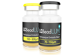
BTG has announced that it has received 510(k) clearance from the US Food and Drug Administration for LC Bead Lumi, the first commercially available radiopaque embolic bead, for the embolization of hypervascular tumours and arteriovenous malformations.
A press release from BTG states that LC Bead Lumi is a next generation development of LC Bead (known as DC Bead in Europe) that enables real-time visible confirmation of bead location during embolization. “This has the potential to provide interventional radiologists increased control, enabling real-time adjustments to optimise patient treatment. The lasting radiopacity of LC Bead Lumi means they will also be visible in follow-up scans, allowing precise evaluation of the completeness of tumour treatment,” the release adds.
Louise Makin, BTG’s CEO, comments, “LC Bead Lumi reinforces our leadership in embolization technology and our focus on bringing to market innovative products that better serve specialist physicians and their patients. We look forward to securing regulatory clearances for additional radiopaque bead products in other markets.”
Keigo Osuga, Diagnostic and Interventional Radiology, Osaka, Japan, commenting on the future direction of embolic agents at a special session at the Cardiovascular and Interventional Radiological Society of Europe (CIRSE) annual meeting in 2015 in Seville Spain, notes, “There is growing interest to improve the visualisation of microspheres to monitor the behaviour of microspheres during injection and to evaluate the intralesional deposit of microspheres after the procedure. Investigation of radiopaque or MR-detectable microspheres is underway.”
A study, published online ahead on print in November 2015 in the Journal of Vascular and Interventional Radiology (JVIR), has concluded that it is possible to develop a simple method to produce radiopaque drug-eluting microspheres that can be incorporated into the current clinical transcatheter arterial chemoembolization workflow. The researchers further showed that these radiopaque drug-eluting beads were discernible enough to be visualised with X-ray imaging techniques.
Carmen Gacchina Johnson, The National Institutes of Health, Bethesda, USA, and colleagues added an ethiodised oil (Lipiodol, Guerbet) and ethanol solution to a lyophilised 100–300µm bead before loading with doxorubicin. These radiopaque drug-eluting beads were evaluated in vitro for x-ray attenuation, composition, size, drug loading and elution, and correlation between attenuation and doxorubicin concentration. The investigators evaluated in vivo conspicuity in a VX2 tumour model.
The investigators report in JVIR that Lipiodol was loaded into lyophilsed beads using two glass syringes and a three-way stopcock. Maximum bead attenuation was achieved within 30 minutes. “Doxorubicin loading efficiency and total amount eluted were similar to DC Bead (Biocompatibles). However, the elution rate was slower for radiopaque drug-eluting beads (p<0.05),” they write. They also report that radiopaque drug-eluting beads were seen in tumour feeding arteries after administration by fluoroscopy, computed tomography, and microcomputed tomography, and that their location was confirmed by histology.
In other research reported at CIRSE 2015, K Ashrafi, Innovation group, BTG International, Camberley, UK, and colleagues, presented on the characterisation of a novel drug-eluting radiopaque bead embolic, based on the DC Bead that has been developed to be visible in vivo under clinical X-ray imaging.
The team noted in their abstract: “The radiopaque bead embolic was developed to maintain equivalent performance to DC Bead, whilst providing clinicians with intra- and post-procedural visualisation for TACE procedures. This study focused on the in vitro characterisation of the radiopaque bead and showed that the drug-loading times were equivalent to DC Bead, and no significant change in bead diameter was measured between preloaded, loaded and eluted conditions.” Further results from the study showed that good handling and deliverability of radiopaque bead was achieved using standard materials and techniques.


