 Showcasing cutting-edge applications of robotics in interventional radiology (IR), presenters at the Society of Interventional Radiology (SIR) annual scientific meeting (23–28 March, Salt Lake City, USA) delivered results from a range of pioneering studies.
Flexible robotic microcatheter “reduces procedural complications”
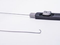
First to the podium was Christopher Bailey (Johns Hopkins University School of Medicine, Baltimore, USA) whose team demonstrated “breakthrough capabilities” in fabricating soft robotic microcatheters capable of steering and navigating through “small, complex, tortuous and/or delicate” vasculature.
Bailey and his team went about creating a strategy for fabricating microfluidic multilumen tubing to serve as the microcatheter tubing, employing two-photon direct laser writing (DLW) for three-dimensional (3D) printing of soft microrobotic actuators with submicron-scale resolutions.
Subsequently, they produced multilumen tubing with an outer diameter of roughly 2.1Fr and inner microfluidic channels enabling actuation of 0.21Fr in diameter. The 3D microprinted soft robotic unidirectional microcatheter tip is fluidically sealed to a microfluidic chip and is capable of achieving deflection angles of greater than 115 degrees and burst pressures larger than 600kPa, Bailey explained.
Transarterial interventions are routine in IR, the speaker noted; however, in geometrically complex anatomy, interventionists can face an inability to effectively manoeuvre microcatheters/guidewires, causing a “substantial everyday challenge”.
“By uniting these strategies,” Bailey said, “a new class of microcatheter can be created that overcomes manoeuvrability deficits, which can cause unsuccessful catheterisation” and can minimise risk of complications in arterial interventions.
Bailey indicated that their microcatheter shows the “unique additive micromanufacturing techniques” that can serve as the “basis for future multidirectional designs and innovations”. Ultimately, their team hope to expand catheter-based procedures and improve procedural success rates.
“High technical success” with robotic percutaneous bone biopsy
Next, Agnieszka Witkowska (Rhode Island Medical Imaging, Providence, USA) presented high technical success and diagnostic yield, with reduced complications, for percutaneous computed tomography-guided bone biopsy using a patient-mounted robot.
Witkowska explained that their study sought to evaluate the feasibility of a robotic system with steering capabilities in patients with cancer. Including 40 consecutive biopsies in 39 outpatients, their retrospective observational study looked at biopsies that were performed in the pelvis, spine, ribs, shoulder, femur, and sternum. The median size of lesions was 26mm and lesion characteristics biopsied included 14 lytic (35%), 16 mixed (40%), and 10 sclerotic (25%). Witkowska outlined that, for mixed and sclerotic lesions, needles were manually exchanged over a Kirschner wire prior to drilling for lesion access.
Delivering their results, Witkowska stated that they achieved 100% technical success, with a mean trajectory length of 55.5mm. She noted that intermediary checkpoints were utilised in eight biopsies, but overall, procedure time was kept “low”.
Demonstrating this, she detailed that median time to needle insertion from skin to target was 19 seconds, time from first to final scan was 21 minutes, procedure time was 30 minutes and dose length product and effective dose were 536.6mGycm and 7.1 millisievert, respectively. Continuing, she noted that the diagnostic yield was 72.5% for cancer in this cohort.


