Facebook
Linkedin
Mail
Twitter
Youtube
Home
Latest News
Features
Interventional Oncology
Profiles
Media
Videos
Podcasts
Events
Supplements
Past Issues
Subscribe
Search
Facebook
Linkedin
Mail
Twitter
Youtube
Interventional News
Home
Latest News
Features
Interventional Oncology
Profiles
Media
Videos
Podcasts
Events
Supplements
Past Issues
Subscribe
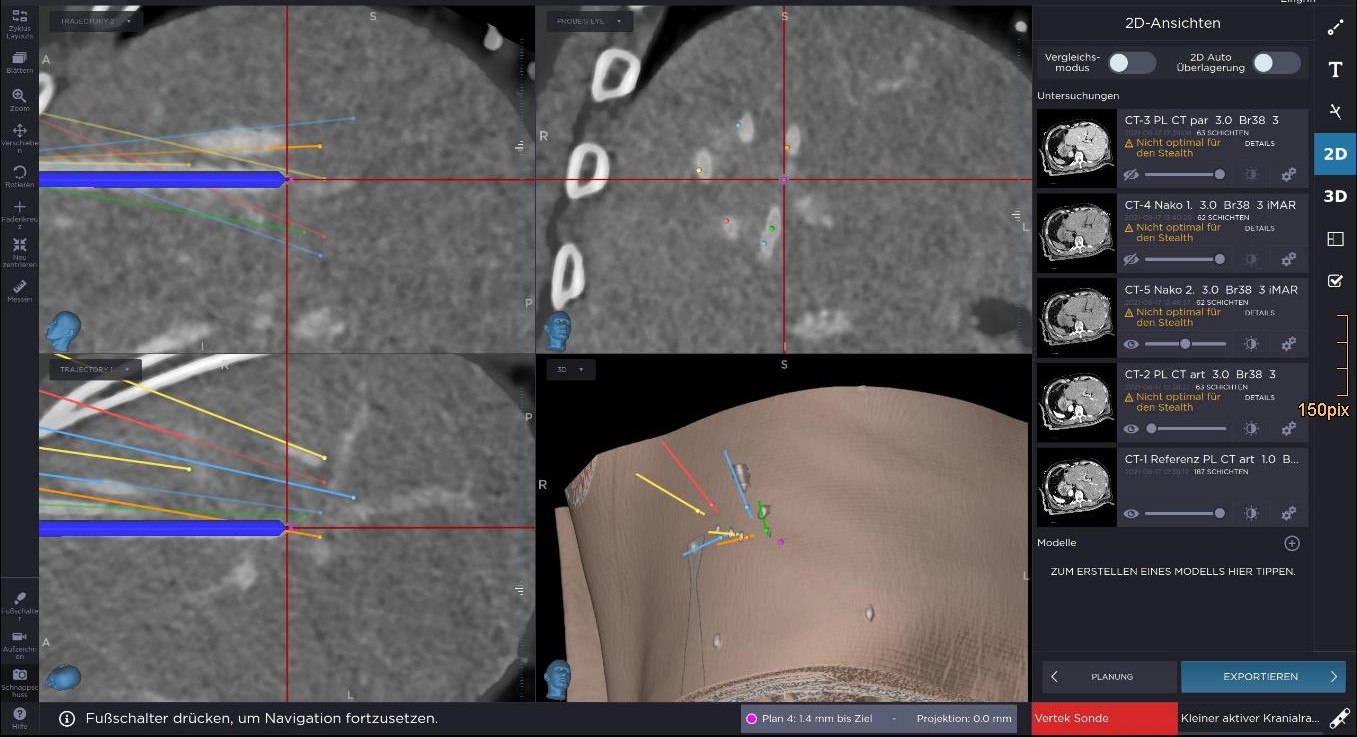
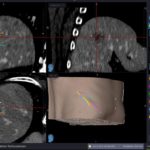
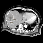
Fig 1c Verification of precise coaxial needle placement by image fusion.